Article

Selenium Nanoparticles Suppressed Oxidative Stress and Promoted Tenocyte Marker Expression in Tendon-Derived Stem/Progenitor Cells

by
Pauline Po Yee Lui
1,2,*,
Caihao Huang
3,4 and
Xing Zhang
3
1
Department of Orthopaedics and Traumatology, The Chinese University of Hong Kong, Shatin, New Territories, Hong Kong SAR, China
2
Center for Neuromusculoskeletal Restorative Medicine Ltd., Hong Kong Science Park, Shatin, New Territories, Hong Kong SAR, China
3
Institute of Metal Research, Chinese Academy of Sciences, Shenyang 110000, China
4
School of Materials Science and Engineering, Dalian University of Technology, Dalian 116000, China
*
Author to whom correspondence should be addressed.
Antioxidants 2024, 13(12), 1536; https://doi.org/10.3390/antiox13121536
Submission received: 1 December 2024 / Revised: 12 December 2024 / Accepted: 12 December 2024 / Published: 15 December 2024
(This article belongs to the Section Natural and Synthetic Antioxidants)

Abstract

:
Traumatic tendon injuries generate reactive oxygen species and inflammation, which may account for slow or poor healing outcomes. Selenium is an essential trace element presented in selenoproteins, many of which are strong antioxidant enzymes. Selenium nanoparticles (SeNPs) have been reported to promote tissue repair due to their anti-oxidative, anti-inflammatory, anti-apoptotic, and differentiation-modulating properties. However, its effects on the functions of tendon-derived stem/progenitor cells (TDSCs) and tendon healing have not been reported. This study examined the effects of SeNPs on the functions of hydroperoxide (H2O2)-stimulated TDSCs. Rat patellar TDSCs were treated with H2O2 with or without SeNPs. The viability, marker of proliferation, oxidative stress, inflammation, apoptosis, and tenocyte marker expressions of H2O2-stimulated TDSCs after SeNPs treatment were assessed. Our results showed that SeNPs increased the viability and expression of the marker of proliferation of TDSCs exposed to H2O2, while concurrently reducing oxidative stress, inflammation, and apoptosis. Additionally, the expressions of tenocyte markers were significantly elevated in H2O2-treated TDSCs after treatment with SeNPs. Furthermore, the expressions of Sirt1 and Nrf2 also increased after SeNPs treatment in H2O2-stimulated TDSCs. In conclusion, SeNPs mitigated oxidative stress, inflammation, and apoptosis while enhancing the survival and expression of the marker of proliferation of TDSCs in an oxidative stress environment. Additionally, it promoted the fate of TDSCs towards the tenocyte lineage in the presence of such oxidative stress. The increased expressions of Sirt1 and Nrf2 likely mediated the anti-oxidative and anti-inflammatory effects of SeNPs. SeNPs hold promise as a novel intervention for promoting tendon healing.

1. Introduction

Tendons and ligaments endure substantial tensile stresses and are prone to rupture due to overuse or trauma, resulting in significant pain and functional impairment. The regenerative capacity of tendons and ligaments following an injury is generally poor, which represents a significant burden to the healthcare system. Both conservative treatments and surgical interventions for tendon injuries often yield unsatisfactory outcomes characterized by extended healing time, formation of scar tissue, adhesions, occasional bone formation, and a higher likelihood of re-injury. The accumulation of microinjuries during loading as a result of ineffective tendon regeneration after acute injuries is a predisposing factor for the development of chronic tendinopathy, presented with persistent pain and functional disability over the long term.
Following exercise and traumatic injury, tendons constantly produce reactive oxygen species (ROS) [1,2]. Excessive ROS exceeding the tendon’s antioxidant capacity can trigger inflammation, causing tendon injury. The accumulation of microinjuries may lead to tissue fibrosis and impaired healing [3]. Notably, elite professional soccer players exhibiting ultrasonographic (US) evidence of tendon damage showed significantly higher levels of oxidative stress compared to those with normal US findings [4]. The level of ROS was also elevated in the injured tendon compared to the normal tendon in a patellar partial transection rat model [5]. Previous studies have shown that the tenogenic properties of tendon-derived stem/progenitor cells (TDSCs) were compromised, with increased osteo-chondrogenic differentiation potential in an inflammatory environment [6,7,8]. Therefore, more regenerative healing is expected through reducing oxidative stress and inflammation.
Selenium (Se) is an essential trace element in human health and diseases including immune response, neurodegeneration, cardiovascular diseases, and cancer [9,10,11,12]. The protective effect of selenium on mammalian cells is mainly carried out by a series of selenoproteins in which selenium is incorporated as selenocysteine. There are 25 selenoproteins in the human body and most of them are strong antioxidant enzymes [13]. Pretreatment of human dermal fibroblasts with selenium nanoparticles (SeNPs) reduced cell death induced by heat shock [14]. In addition, SeNPs also inhibited prostaglandin E2 (PGE2), which is an activator of pro-inflammatory cytokines like interleukin-6 (IL-6) and tissue necrosis factor alpha (TNF-α) [15]. In addition, SeNPs reduced apoptosis and intracellular ROS levels as well as accelerated myogenic differentiation of C2C12 in the presence of H2O2 [16]. The incorporation of SeNPs in the wound dressing made of bacterial cellulose/gelatin enhanced the anti-inflammatory, anti-oxidative, and anti-bacterial effects of the hydrogel [17]. However, there has been no study about the effects of SeNPs on TDSCs and tendon healing.
This study aimed to examine the effects of SeNPs on the functions of H2O2-stimulated TDSCs. We hypothesized that SeNPs would increase the viability, proliferation, and tenogenesis while abrogating oxidative stress, inflammation and apoptosis of TDSCs under oxidative stress. The effects of SeNPs on the expressions of Sirt1, FoxO1, and Nrf2, key regulators of oxidative stress and inflammation, were also investigated.

2. Materials and Methods

2.1. Study Design

The study was approved by both the animal research ethics committee (Ref. 23-229-NSF, approved on 5 January 2024) and the University Laboratory Safety Office. Adherence to the ARRIVE guidelines was ensured throughout the research process.
TDSCs were seeded in a culture plate in complete culture medium (α-Modified Eagle’s Medium (α-MEM) containing 10% fetal bovine serum (FBS), 100 U/mL penicillin, 100 μg/mL streptomycin; all from Invitrogen Corporation, Waltham, MA, USA) overnight. The cells were washed with phosphate-buffered saline (PBS) and treated with different concentrations of SeNPs (0–2 µg/mL) and H2O2 (200 µM) in a serum-free medium. At various times after treatment, the viability, marker of proliferation, oxidative stress, inflammation, apoptosis, expressions of tenocyte markers, and transcription factors regulating oxidative defense mechanisms and inflammation of TDSCs were assessed. Hence, H2O2 was present during the whole experiment. The viability and expression of the marker of proliferation of TDSCs were assessed by AlamarBlue assay and immunofluorescence (IF) staining of Ki67, respectively. The oxidative stress of TDSCs was assessed by CM-H2DCF-DA assay kit, mRNA expressions of glutathione peroxidase 1 (Gpx1), glutathione peroxidase 3 (Gpx3), glutathione peroxidase 4 (Gpx4), thioredoxin reductases 2 (Txnrd2), selenoprotein M (Selenom), superoxidase dismutase (Sod1), catalase (Cat), NADPH oxidase 1 (Nox1), heme oxygenase 1 (Hmox1), IF staining of nitrotyrosine and malonaldehyde (MDA), and catalase activity. The catalase activity of TDSCs was measured using an assay kit. The mRNA (Il6, Il1b, Cox2) and protein (IL-6, Cox-2) expressions of inflammatory cytokines were measured by qRT-PCR and IF, respectively. Apoptosis of TDSCs was examined by mRNA expressions of pro-apoptotic (Bax, Bad, Bid, Casp3) and anti-apoptotic markers (Bcl2l1, Bcl2), and IF staining of Bax. The mRNA expressions of tenocyte markers (Col1a1, Col3a1, Eln, Tnc, Dcn) and transcription factors regulating oxidative defense mechanisms and inflammation (Sirt1, FoxO1, Nrf2) of TDSCs were examined by qRT-PCR.

2.2. Preparation of SeNPs

SeNPs were prepared in our laboratory and the quality including the crystallinity, shape, purity, hydrated particle size, and zeta potential were examined. Selenous acid (13 mM) was dissolved in 5 mg/mL of polyvinyl alcohol (PVA) and then mixed with ascorbic acid (33 mM) with vigorous stirring. The selenium particle size was controlled by adjusting the concentration of reactants and reaction times in the reduction reaction of ascorbic acid. The crystallographic structure of the selenium particles was examined by X-ray diffraction analysis (XRD, Rigaku D/max 2400 diffractometer, Rigaku Corporation, Tokyo, Japan) using a monochromated Cu Kα radiation source with an accelerating voltage of 40 kV and a current of 250 mA. The elemental composition of the selenium particles was analyzed by energy dispersive X-ray spectrometer (EDX, QUANTA 450, FEI, Hillsboro, OR, USA) coupled with transmission electron microscopy (TEM, Tecnai F30, FEI, Hillsboro, OR, USA). The zeta potential and hydrated particle size of the selenium particles were measured by a zeta potential analyzer and laser particle size analyzer (ZEN3690, Malvern Panalytical, Malvern, UK), respectively. SeNPs were dissolved in deionized water at 6 mg/mL and sonicated for 30 min. The solution was further diluted with a cell culture medium before use.

2.3. TDSC Isolation

Patellar TDSCs were isolated from male Sprague Dawley rats (6–8 weeks, 150–220 g). The procedures for TDSC isolation are well-established [18]. Briefly, the tendon tissue was minced, and enzymatically digested using type I collagenase (Cat no.: C0130, Sigma Aldrich, St. Louis, MO, USA) to produce a single cell suspension, seeded at an appropriate low cell density to isolate the stem cells, and then cultured the cells to facilitate colony formation. The expressions of stem cell markers, clonogenicity, and multi-lineage differentiation potential of the isolated cells were determined according to our established protocols [18]. Only TDSCs at passages fewer than 5 were used for experiments.

2.4. Alamar Blue Assay

TDSCs at 4000 cells were seeded in a 96-well plate and treated with or without different concentrations of SeNPs (0–2 µg/mL) and H2O2 (200 µM) (Cat no.: 23E124006, VWR, Philadelphia, PA, USA) in serum-free medium. At 1 h and 4 h after treatment, the AlamarBlue solution (Cat no.: DAL1100, Thermo Fisher Scientific, Waltham, MA, USA) was added for 2 h. The fluorescence intensity was measured at the excitation wavelength of 560 nm and the emission wavelength of 590 nm. Cell viability relative to the untreated group was calculated and compared among different groups. There were 5 samples/group.

2.5. Immunofluorescence Staining of Ki67, IL-6, Cyclooxygenase-2 (Cox-2), Bax, Nitrotyrosine, and Malonaldehyde (MDA)

TDSCs were seeded on slides and treated with 0.5 µg/mL of SeNPs and H2O2 (200 µM) in a serum-free medium. At 4 h after H2O2 exposure, the cells were washed, fixed with 4% paraformaldehyde (Cat no.: A11313.36, Thermo Fisher Scientific, Waltham, MA, USA), and permeabilized with 0.3% Triton-X100 (Cat no.: 17-1315-01, PlusoneTM, GE Healthcare Bio-Sciences KK, Tokyo, Japan). The primary antibodies against Ki67 (Cat no.: 11-5698-82, Thermo Fisher Scientific, Waltham, MA, USA, 1:1000 dilution), IL-6 (Cat no.: ab290735, Abcam, Cambridge, UK, 1:1000 dilution), Cox-2 (Cat no.: A1253, Abclonal Technology, Woburn, MA, USA, 1:1000 dilution), Bax (Cat no.: #MA5-32031, Thermo Fisher Scientific, Waltham, MA, USA, 1:1000 dilution), nitrotyrosine (Cat no.: 9691S, Cell Signal Technology, Danvers, MA, USA, 1:1000 dilution), or MDA (Cat no.: ab27642, Abcam, Cambridge, UK, 1:1000 dilution) were added for 1 h at room temperature. After washing, the cells were treated with goat anti-rabbit IgG (H + L) secondary antibodies conjugated with Alexa FluorTM 488 (Cat no.: A-11008, Thermo Fisher Scientific, Waltham, MA, USA, 1:500 dilution) for 45 min at room temperature. The slides were counter stained with 4′,6-diamidino-2-phenylindole (DAPI) (Cat no.: P36935, Invitrogen Corporation, Waltham, MA, USA). The fluorescence signals were viewed using a fluorescent microscope (Leica DM5500 B, Leica Microsystems, Wetzlar, Germany). The percentages of Ki67-positive cells (3 samples/group), IL-6-positive cells (n = 4/group), Cox-2-positive cells (n = 4/group), Bax-positive cells (n = 4/group), nitrotyrosine-positive cells (3–4 samples/group), and MDA-positive cells (n = 4/group) were counted.

2.6. CM-H2DCF-DA Assay

TDSCs were seeded at 40,000 cells in a 12-well plate and treated with 0.5 µg/mL of SeNPs and H2O2 (200 µM) in a serum-free medium. At 4 h after H2O2 exposure, the cells were washed and incubated with 1–10 µM of CM-H2DCF (Cat no.: C6827, Thermo Fisher Scientific, Waltham, MA, USA) for 30 min at 37 °C. The cells were rinsed, fixed with 4% paraformaldehyde, and counter-stained with DAPI. The fluorescence signals were viewed using a fluorescent microscope (Leica DM5500 B). The percentage of ROS-positive cells was counted. There were 3 samples/group.

2.7. Catalase Activity Assay

TDSCs were seeded at 40,000 cells in a 12-well plate in serum-free medium and the total protein was extracted. The collected protein was treated with 0.5 µg/mL of SeNPs and H2O2 (200 µM). At 2 h after H2O2 exposure, the catalase activity was measured using an assay kit (Cat no.: S0051, Beyotime Institute of Biotechnology, Suzhou, China). The optical density at 520 nm was measured. The activity level was normalized by the total protein content measured by the bicinchoninic acid (BCA) protein assay (Cat no.: #23227, Thermo Fisher Scientific, Waltham, MA, USA). There were 4 samples/group.

2.8. qRT-PCR

TDSCs were seeded at 40,000 cells in a 12-well plate and treated with 0.5 µg/mL of SeNPs and H2O2 (200 µM) in a serum-free medium for 4 h. qRT-PCR was performed using the QS7-Pro 384 and StepOne Plus 96 qPCR system (both from Applied Biosystems, Waltham, MA, USA). The relative gene expression was calculated using the 2−ΔCT formula. We tested both Gapdh and Actb in the analysis of some markers and similar results were observed. Therefore, we used Actb as the house-keeping gene. The primer sequences are shown in Table S1.

2.9. Statistical Analysis

Quantitative data are presented in boxplots. The sample sizes for the experiments in this study ranged from 3 to 5, which were considered small. In addition, the SeNPs without an H2O2 group were not included in this biological investigation because biologically, treatment with SeNPs is needed only when there is an increased production of ROS stimulated by H2O2 supplementation in this study, we therefore used the Kruskal–Wallis test to compare the difference in more than 2 groups. If there was a significant difference in the Kruskal–Wallis test, the Mann–Whitney U test was subsequently used to identify the differences. The effect size, r, was reported. A value of r at 0.1, 0.3, and 0.5 were regarded as small, medium, and large effects, respectively. All the data analyses were performed using the SPSS analysis software (SPSS Inc., Chicago, IL, USA, version 26.0). A value of p < 0.05 was regarded as statistically significant.

3. Results

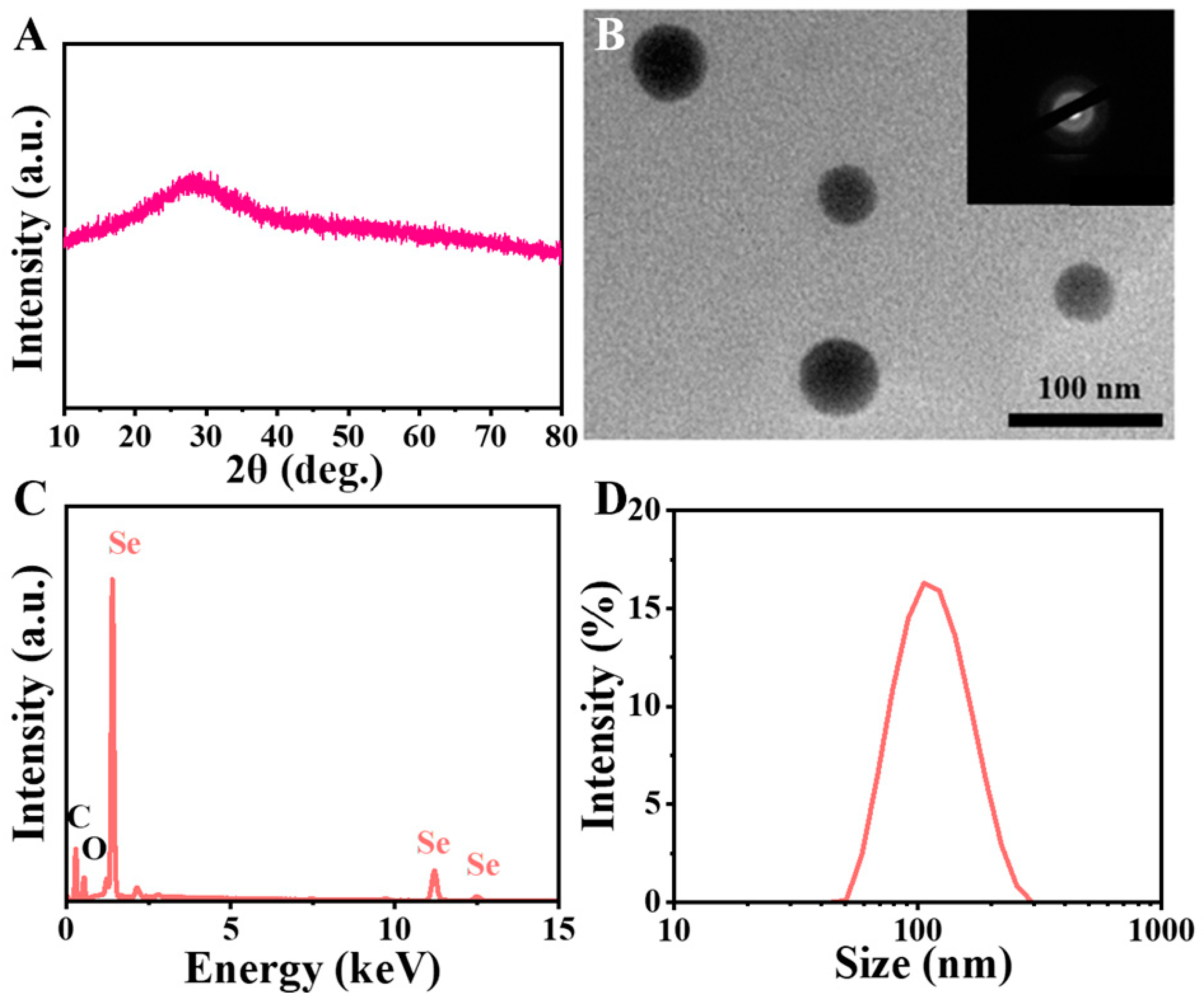
3.1. Characterization of SeNPs

SeNPs used in this study were synthesized in our laboratory and the quality of the resulting particles was analyzed. Our results showed that the color of the reaction solution changed from colorless to red when selenous acid was added to PVA and ascorbic acid, indicating the synthesis of SeNPs from selenite oxyanion. XRD analysis showed a broad peak, suggesting that SeNPs were amorphous and lacked a good crystalline structure (Figure 1A). The transmission electronic microscopy (TEM) micrograph of dried SeNPs (Figure 1B) showed that the particle was spherical, and the selected area electron diffraction pattern (SAED) further demonstrated its amorphous nature. Energy dispersive X-ray analysis (EDX) was used to analyze the elemental composition of the selenium particles (Figure 1C). Selenium (Se), carbon (C), and oxygen (O) atoms were observed in the sample. The occurrence of carbon and oxygen atoms was due to the presence of residual polyvinyl alcohol (PVA) in the sample. The mean hydrated particle size and zeta potential of SeNPs were 91.3 nm (Figure 1D), and −7.6 mV, respectively, suggesting that SeNPs were in the nanometer scale. These SeNPs were used in the following experiments to examine the effects of selenium on the functions of H2O2-treated TDSCs.

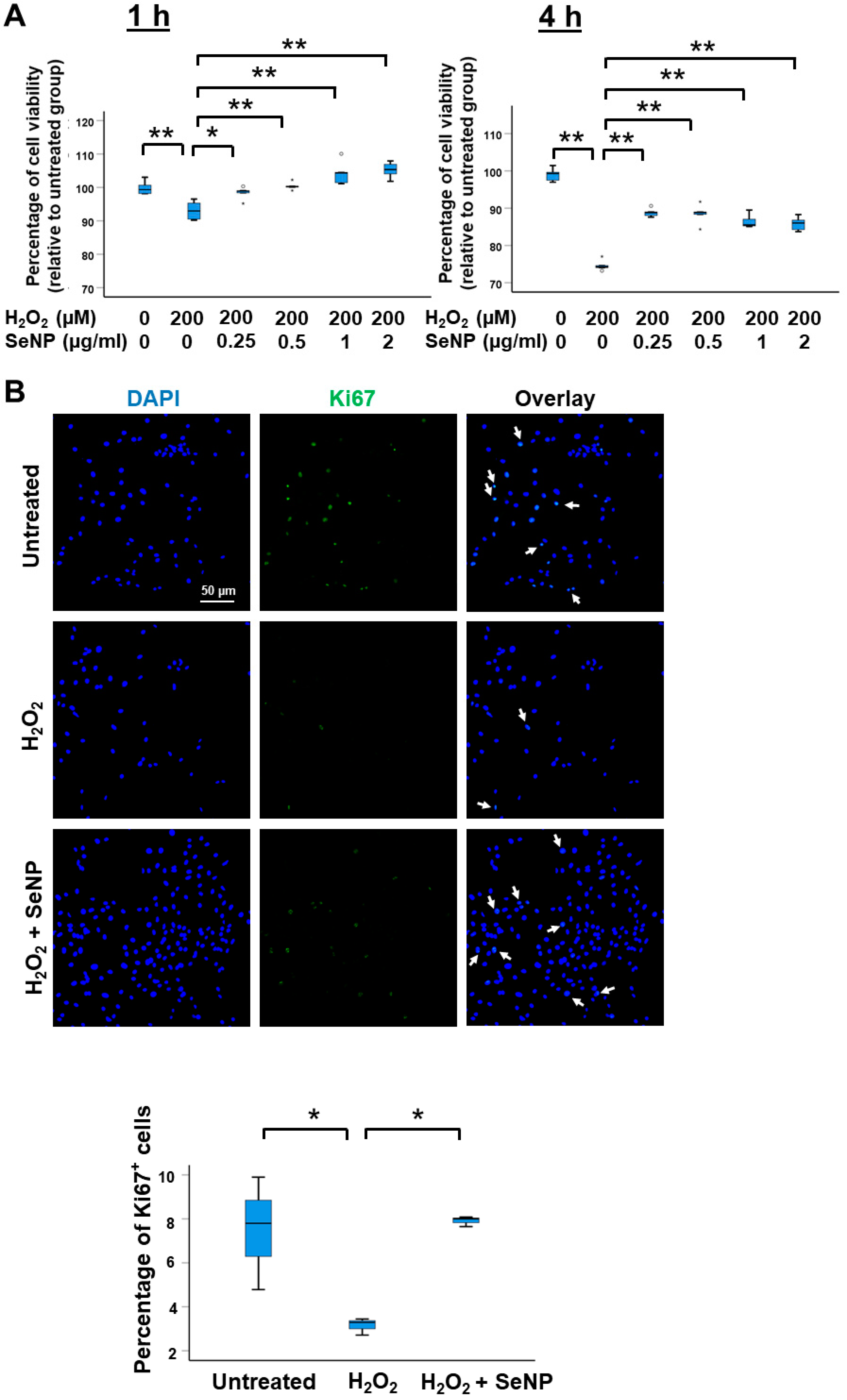
3.2. Viability and Expression of Marker of Proliferation

H2O2 reduced the viability of TDSCs at 1 h and 4 h after treatment (both p < 0.01; r = 0.83) (Figure 2A). The addition of SeNPs significantly increased the viability of the H2O2-treated TDSCs (p < 0.05, r = 0.69 for SeNPs (0.25 µg/mL) treatment for 1 h; others (p < 0.01, r = 0.83) (Figure 2A). H2O2 reduced the percentage of Ki67+ cells (p < 0.05; r = 0.80) (Figure 2B), and the effect was reversed by co-treatment with SeNPs (p < 0.05; r = 0.80) (Figure 2B).

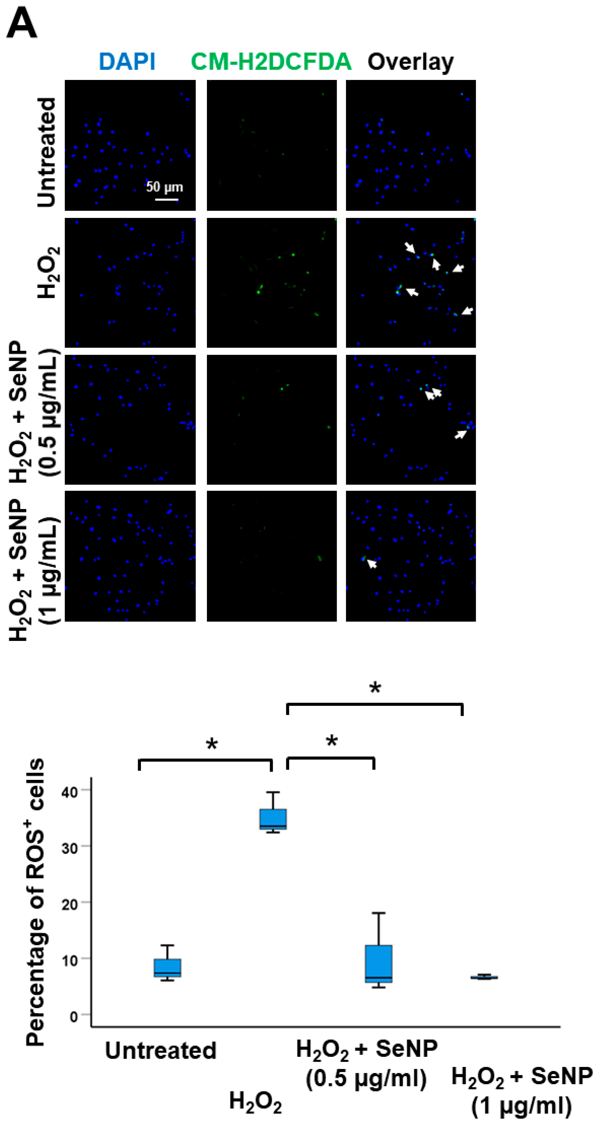
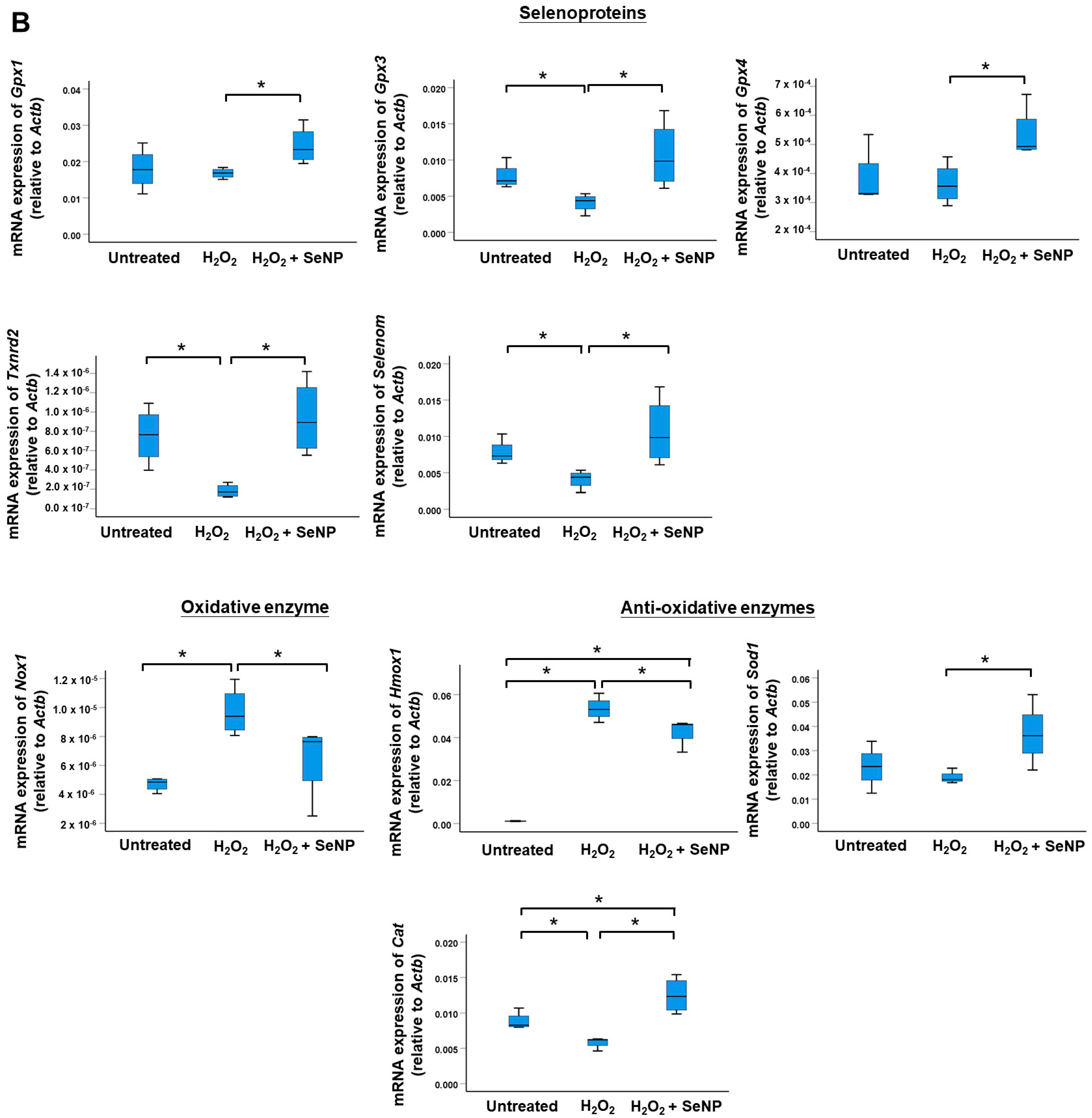
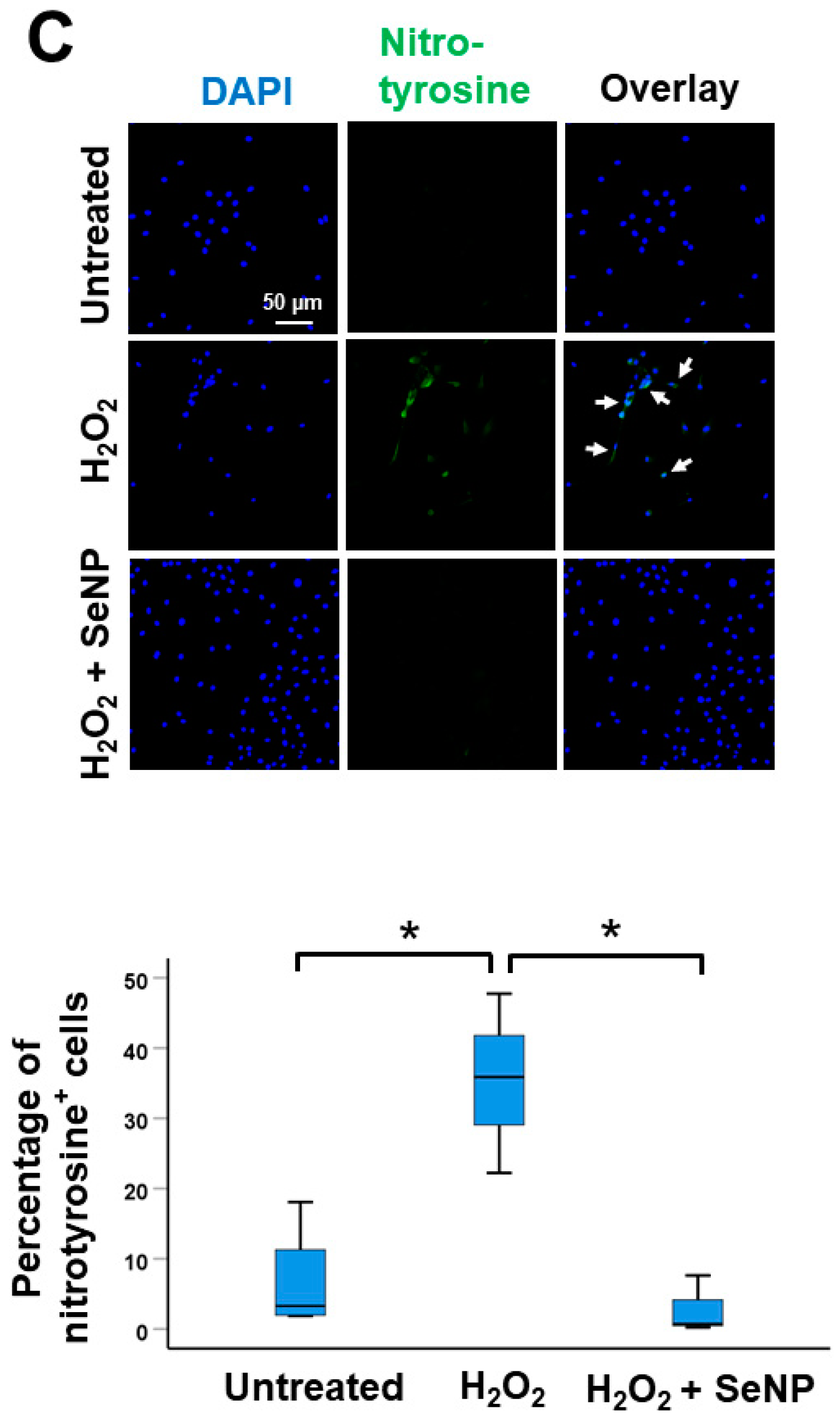
3.3. Oxidative Stress

H2O2 increased the percentage of ROS+ cells (p < 0.05; r = 0.80) and the addition of SeNPs at 0.5 µg/mL and 1 µg/mL returned the percentage of ROS+ cells to normal (both p < 0.05; r = 0.80) (Figure 3A). Similar results were observed in the mRNA expressions of selenoproteins (Gpx1, Gpx3, Gpx4, Txnrd2, Selenom), and enzymes implicated in oxidation (Nox1) and anti-oxidation (Hmox1, Sod1, Cat) (Figure 3B). H2O2 suppressed the expression of Gpx3, Txnrd2, and Selenom in TDSCs (all p < 0.05; r = 0.82) but had no effects on the expression of Gpx1 and Gpx4 at the concentration and time tested (both p > 0.05; r = 0.2 for Gpx1 and r = 0.1 for Gpx4). SeNP supplementation significantly increased the expression of these five markers (all p < 0.05; r = 0.82) (Figure 3B). H2O2 elevated the expressions of oxidative enzyme Nox1 (p < 0.05; r = 0.82) and anti-oxidative enzyme Hmox1 (p < 0.05; r = 0.82), reduced the expression of Cat (p < 0.05; r = 0.82) but has no significant effect on the expression of Sod1 (p > 0.05; r = 0.41) (Figure 3B). The addition of SeNPs significantly reversed the effects of H2O2 on the expression of Nox1, Hmox1, and Cat (all p < 0.05; r = 0.82) as well as increased the expression of Sod1 (p < 0.05; r = 0.71) (Figure 3B). Similarly, SeNPs reduced the expressions of both protein oxidation products (nitrotyrosine) (p < 0.05; r = 0.8). (Figure 3C) and lipid peroxidation product (MDA) (p < 0.05; r = 0.82) (Figure 3D) in H2O2-treated cells. In addition, the catalase activity of H2O2-treated TDSCs increased after treatment with SeNPs (p < 0.05; r = 0.82) (Figure 3E).

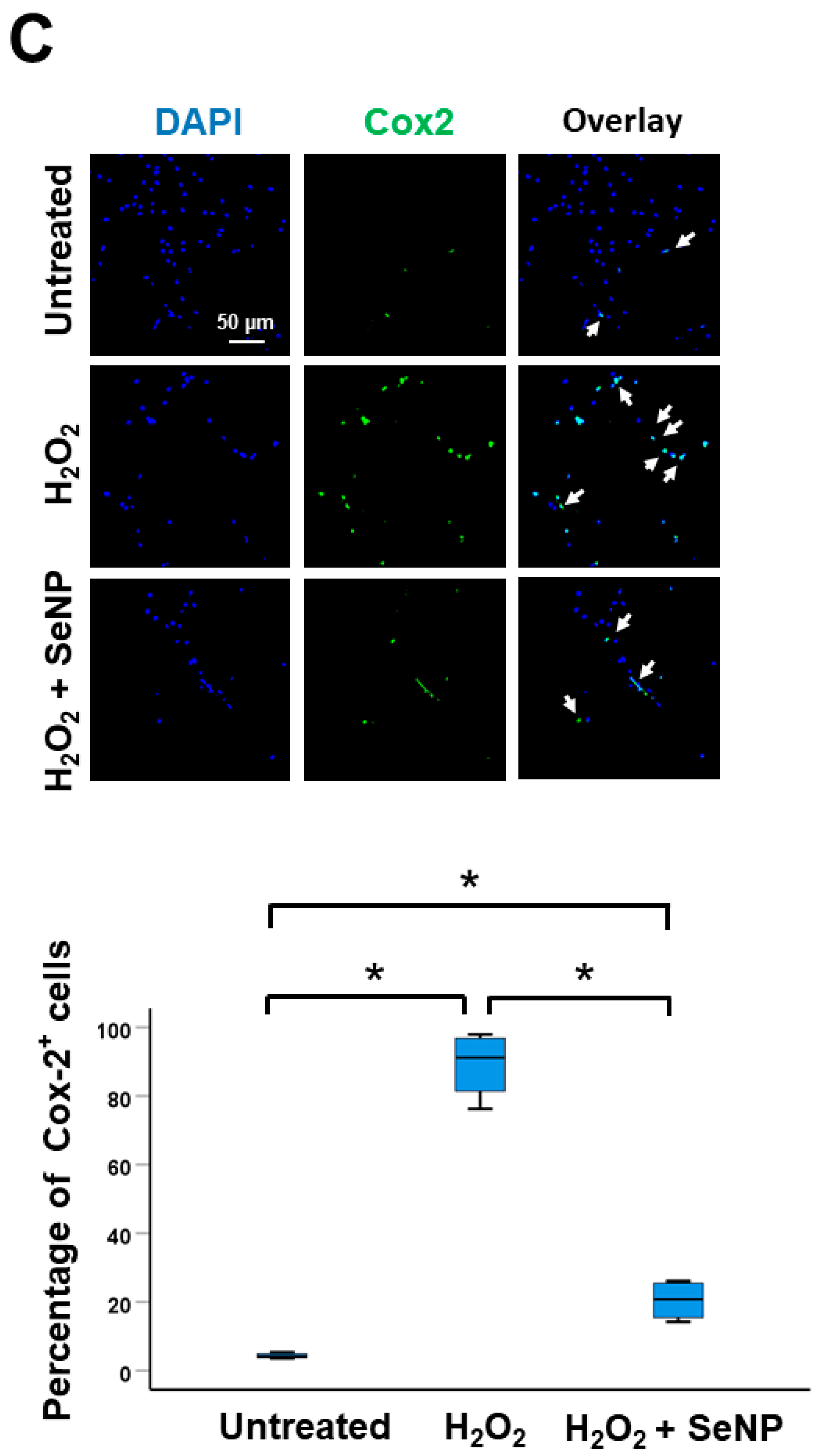
3.4. Expression of Inflammatory Cytokines
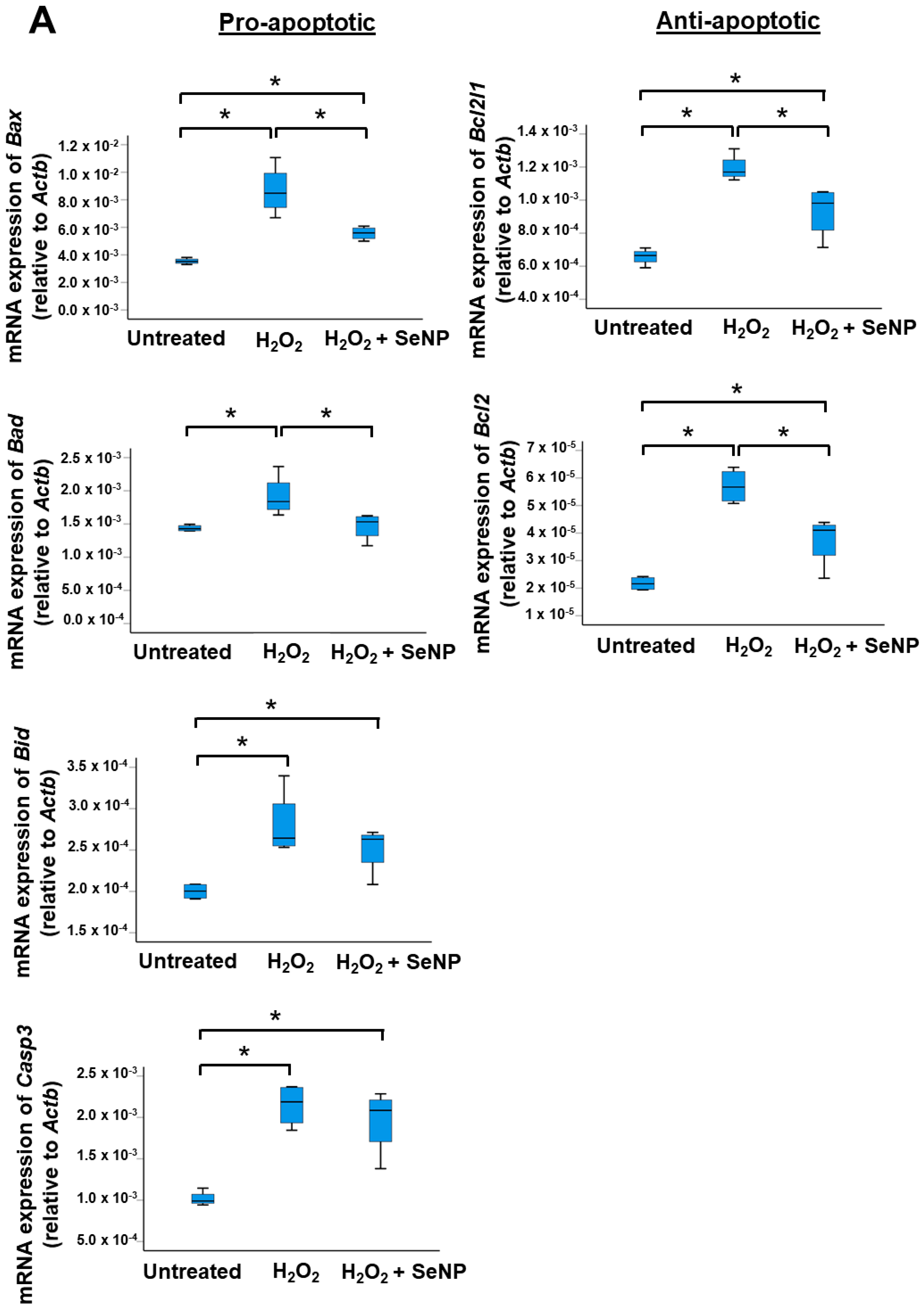
H2O2 significantly upregulated the mRNA expressions of Il6, Cox2, and Il1b (all p < 0.05; r = 0.82 for Il6 and Cox2; r = 0.87 for Il1b) (Figure 4A) as well as the percentages of IL-6+ cells (Figure 4B) and Cox-2+ cells (Figure 4C) (all p < 0.05; r = 0.82). SeNPs decreased the expressions of these inflammatory cytokines in TDSCs stimulated by H2O2 (all p < 0.05; r = 0.82) (Figure 4A–C).

3.5. Apoptosis

The exposure of TDSCs to H2O2 significantly elevated the mRNA expressions of pro-apoptotic and anti-apoptotic markers (all p < 0.05; r = 0.82) (Figure 5A) as well as the percentage of Bax+ cells (p < 0.05; r = 0.82) (Figure 5B) in TDSCs, as compared to the untreated group. Conversely, the addition of SeNPs significantly reduced the mRNA expressions of Bax, Bad, Bcl2l1, and Bcl2 (all p < 0.05; r = 0.82) (Figure 5A) and the percentage of Bax+ cells (all p < 0.05; r = 0.82) (Figure 5B) triggered by H2O2. The mRNA expressions of Bid and Casp3 also decreased after the addition of SeNPs but did not reach statistical significance (p > 0.05; r = 0.2) (Figure 5A).

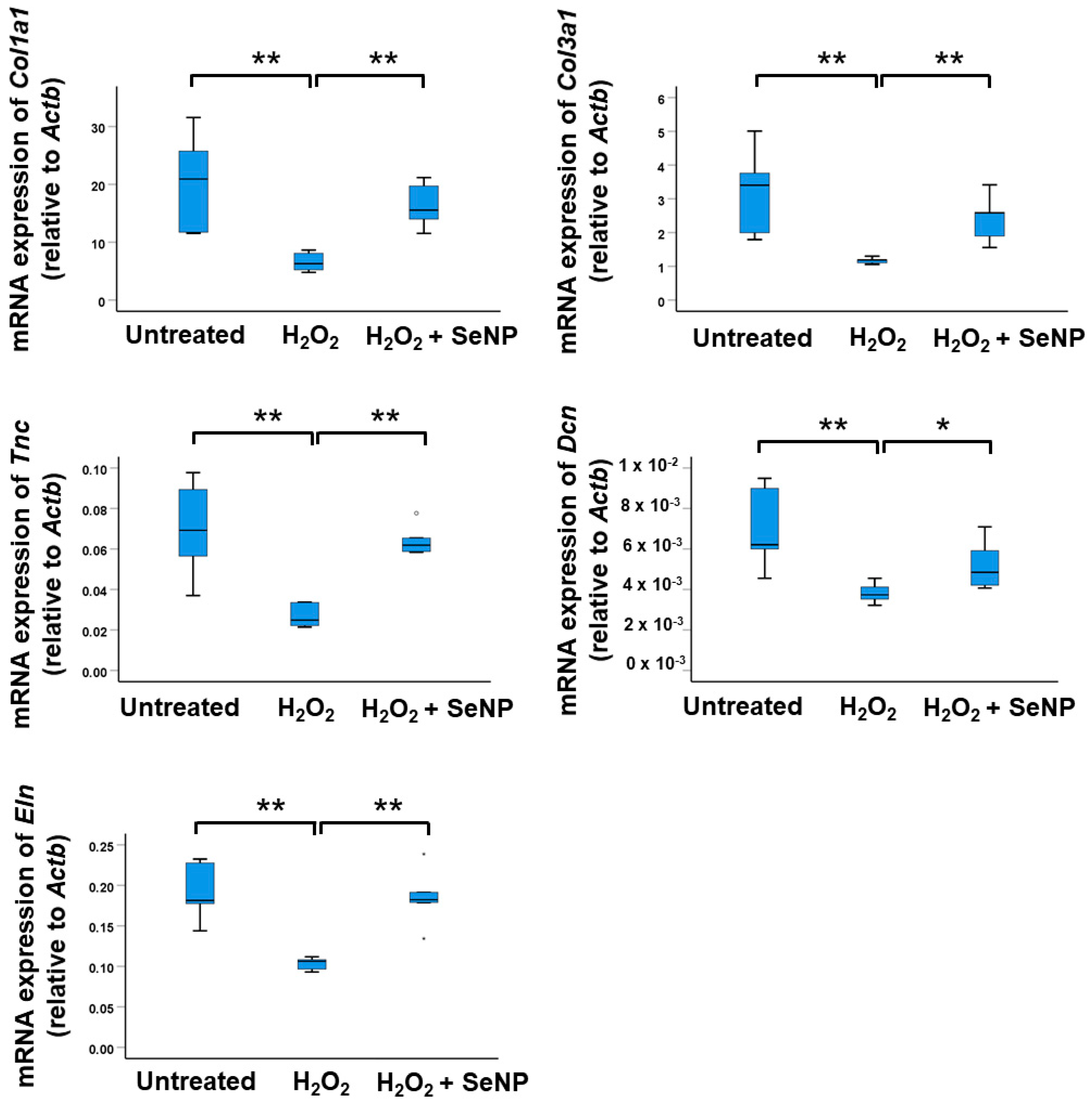
3.6. Expression of Tenocyte Markers

The addition of H2O2 resulted in a significant decrease in the mRNA expressions of tenocyte markers (Col1a1, Col3a1, Eln, Tnc and Dcn) in TDSCs (all p < 0.01; r = 0.83) (Figure 6). The addition of SeNPs significantly increased the mRNA expressions of the tenocyte markers compared to the H2O2-stimulated group (Dcn: p < 0.05; r = 0.63; others: p < 0.01; r = 0.83) (Figure 6).

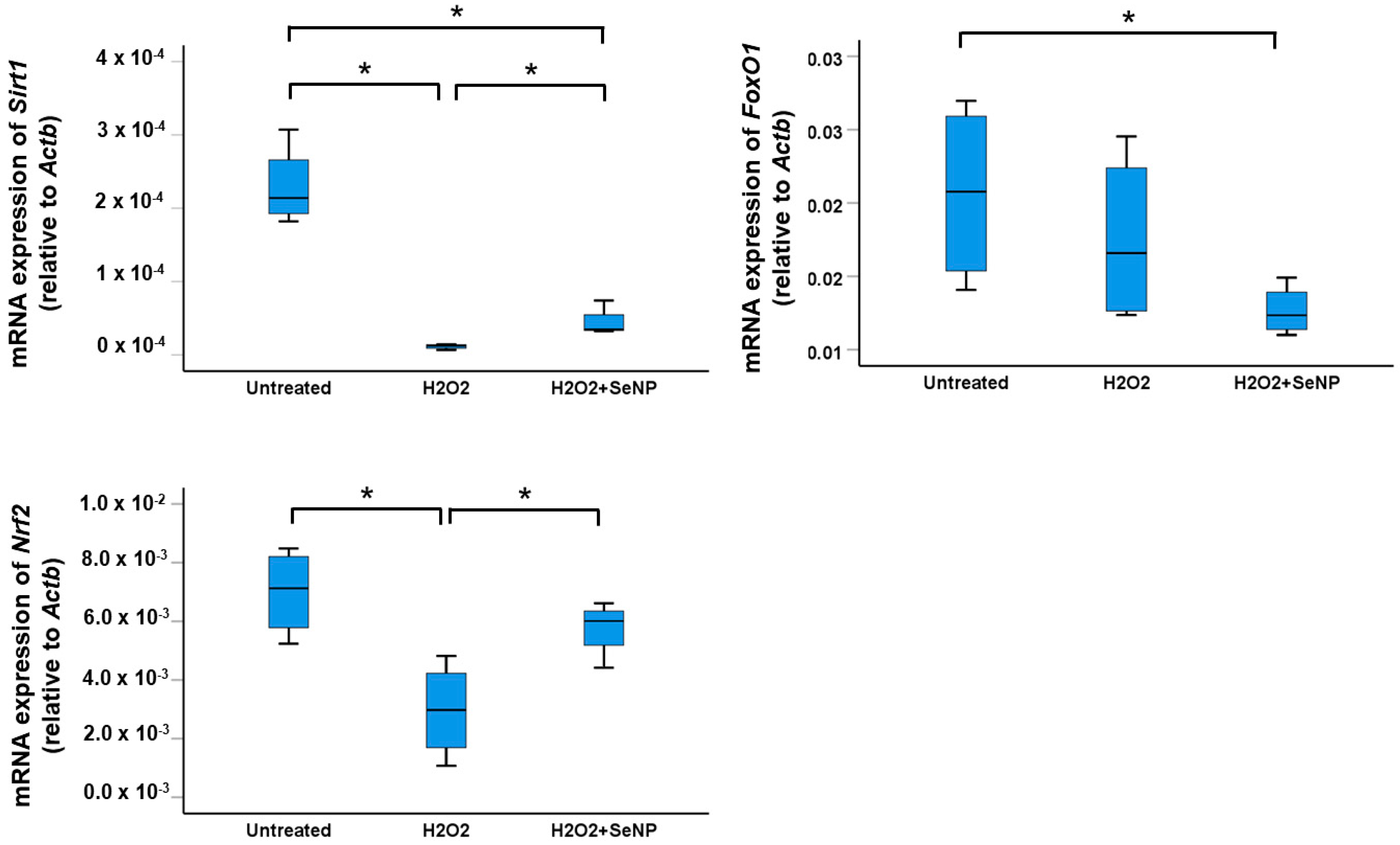
3.7. Expression of Transcription Factors Sirt1, FoxO1, and Nrf2

Treatment of TDSCs with H2O2 for 8 h significantly reduced the expressions of Sirt1 and Nrf2 (both p < 0.05; r = 0.82) but not FoxO1 (p > 0.05; r = 0.41) (Figure 7). Supplementation of SeNPs significantly increased the expressions of Sirt1 and Nrf2 in TDSCs compared to the H2O2-treated group (both p < 0.05; r = 0.82 and 0.71 for Sirt1 and Nrf2, respectively) (Figure 7).

4. Discussion

Our results showed that SeNPs increased the viability and expression of the marker of proliferation, as well as reduced the oxidative stress, inflammation, and apoptosis of H2O2-treated TDSCs. In addition, the expressions of tenocyte markers, Sirt1 and Nrf2 were significantly elevated in H2O2-treated TDSCs after SeNPs supplementation.
H2O2 is an important oxidative stress inducer. Overproduction of H2O2 causes oxidative damage, activation of endoplasmic reticulum stress, and apoptotic death of tenocytes [19]. In addition, H2O2 was also reported to impair colony formation, stemness marker expression, and differentiation capacity, as well as suppress cell migration, cell viability, apoptosis, and proliferation of TDSCs [20,21,22], similar to the results reported in this study. Strategies that can scavenge excessive ROS in the tendon may be useful for the promotion of tendon healing.
Previous studies have shown that inflammation promoted non-tenocyte differentiation of TDSCs, with higher expressions of osteogenic, chondrogenic, and adipogenic markers as well as reduced expression of tenocyte makers [6,7,8]. The pool of TDSCs available for tenogenic differentiation and regenerative repair was reduced, contributing to slow and poor tendon healing and occasionally ectopic bone formation at the late stage of tendon repair [23,24]. TDSCs are a major cell type involved in tendon repair after injury. The reduction in inflammation, oxidative stress, and apoptosis, and improvement in the survival and tenogenic activity of these cells after treatment with SeNPs in this study suggested that the delivery of SeNPs in a biodegradable biomaterial that supports its slow release might be used to promote tendon repair. This requires further research and evaluation of histology, and biomechanical properties of tendon after transplantation of SeNPs in a tendon injury animal model. SeNPs-loaded in biodegradable biomaterial can be prepared as an off-the-shelf, ready-to-use product for tendon repair. The quality control, storage conditions, and manufacturing costs are expected to be lower compared to the use of other biologics such as stem cell-based therapies. There is no standard treatment for reducing ROS production in tendons. Steroid and non-steroidal anti-inflammatory drugs (NSAIDs) are commonly used to reduce pain and inflammation after tendon injury. The inclusion of these drugs for comparison may provide more information about the relative efficacy of SeNPs in the promotion of tendon healing compared to the current practice, although there is no evidence of their long-term efficacy.
While there has been no study about the effects of SeNPs on TDSCs and tendon healing, our results were consistent with the effects of SeNPs reported in other tissues. Previous studies have shown that SeNPs alleviated homocysteine-mediated oxidation and endothelial dysfunction both in vitro and in vivo [25]. In addition to its anti-oxidative effect, SeNPs also exhibit anti-inflammatory and anti-apoptotic activities as well as modulate stromal cell differentiation. SeNPs inhibited the phosphorylation of nuclear factor of kappa light polypeptide gene enhancer in B-cells inhibitor alpha (IkB-α) and hence, prevented the release of nuclear factor kappa B (NF-κB), which is a central regulator of the synthesis of Cox-2, inducible nitric oxide synthase (iNOS), and many pro-inflammatory cytokines [26]. In addition, SeNPs also inhibited PGE2, which is an activator of pro-inflammatory cytokines like IL-6 and TNF-α [15]. In addition, selenium is also a critical regulator of macrophage activation. The addition of Se increased M1 to M2 polarization of interleukin-4 (IL-4)-treated macrophages [27]. Supplementation of SeNPs coated Ulva Lactuca polysaccharide (ULP-SeNPs) suppressed the activation and infiltration of macrophages in colon tissue by inhibiting the nuclear translocation of NF-κB [28]. In addition, SeNPs also increased the viability and osteogenic differentiation of human embryonic stem cell-derived mesenchymal stromal cells (MSCs) by modulating oxidative stress and preventing excessive ROS accumulation [29]. In addition, SeNPs reduced apoptosis and intracellular ROS levels as well as accelerated myogenic differentiation of C2C12 in the presence of H2O2 [16].
The absolute level of H2O2 in an injured tendon is not clear. The levels of H2O2 in tissues and plasma change rapidly, making it difficult to obtain an accurate result [30]. Additionally, the assays used can also affect the reliability of the results [30]. The levels of H2O2 in tissues and plasma vary depending on the physiological and pathological status. For instance, one study reported an increase in the level of H2O2 in a rat’s Achilles tendon after a contusion injury, although the absolute values were not provided [30]. In inflammatory diseases, the plasma H2O2 level was reported to increase from the normal range of 1–5 µM to 50 µM [31]. Furthermore, a study on elite soccer players found that serum oxidative stress, measured as H2O2 equivalent, was significantly higher in players with sonographic tendon alterations (1.75 ± 0.29 mM) compared to those without sonographic changes (1.3 ± 0.2 mM) [4]. This indicated that the oxidative stress in patients with tendon pathologies elevated, although the specific contribution of H2O2 in this case was not clear. Nonetheless, the dose of H2O2 (200 µM) used in this study was within the dose range used to induce oxidative stress in TDSCs in the literature (0.1 mM to 0.5 mM) [5,21,22,32]. Future studies should measure the absolute levels of H2O2 in tendon tissue after injury. Additionally, it would be beneficial to examine the in vitro effects of SeNPs across a range of H2O2 concentrations. This would offer valuable insights into the protective effects of SeNPs on the tendon under different oxidative stress microenvironments.
While all forms of selenium (i.e., inorganic selenium and organic selenium compounds) exhibit anti-oxidative and anti-inflammatory properties, they vary in their effectiveness in scavenging ROS and promoting tissue repair. In this context, Ng et al. [33] have discovered that 1,4-anhydro-4-seleno-D-talitol (SeTal) showed superior efficacy compared to D-selenomethionine (SeMet), D,L-trans-3.4-dihydroxyl-1-selenolane (DHSred), and 1,4-anhydro-D-talitol (Tal) in preventing pyrogallol-induced endothelial dysfunction in mouse aortas. SeNPs were chosen for this study due to their enhanced bioavailability, biological activity, and lower toxicity compared to inorganic or organic selenium. The small size and large surface area to volume ratio of SeNPs facilitate their absorption and cellular uptake, while mitigating cytotoxicity, as demonstrated in numerous studies [34,35]. Moreover, the incorporation of SeNPs in a biomaterial supports its controlled delivery to specific tissues or cells, optimizing its effects for tissue repair. Consequently, we chose to study the effects of SeNPs and did not compare the efficacy of nanoparticles and other forms of selenium on the functions of TDSCs under oxidative stress.
Selenium is an essential trace element in the diet. Potential side effects including reduction in body mass, changes in hepatotoxicity indices, and impairment of fatty acid, protein, lipid, and carbohydrate metabolism have been reported in laboratory animals [36]. The lowest adverse effect level (LOAEL) of SeNPs was reported to be 0.05 mg/kg. The no observed adverse effect level (NOAEL) was 0.22 mg/kg body weight per day and 0.33 mg/kg body weight per day for males and females, respectively [36]. Therefore, the doses of SeNPs that we tested in this study were safe.
While the mechanism of action of SeNPs on H2O2-treated TDSCs is unclear. A previous study has shown that selenium is taken up by cells and incorporated into the amino acid selenocysteine, which is vital for the biological functions of various selenoproteins [9]. Many selenoproteins act as strong antioxidants. The effects of SeNPs on TDSCs under oxidative stress are likely mediated by selenoproteins, as evidenced by the increased mRNA expression of glutathione peroxidase 1 (Gpx1), glutathione peroxidase 3 (Gpx3), glutathione peroxidase 4 (Gpx4), thioredoxin reductase 2 (Txnrd2), and selenoprotein M (Selenom) in this study. The selenoproteins break down H2O2. The improvement in redox balance in cells due to the functions of selenoproteins may modulate the expression of antioxidant enzymes including superoxide dismutase (Sod1) and catalase (Cat), as well as enhance catalase activity, as reported in this study.
The molecular mechanisms of the anti-apoptotic, anti-oxidative, and anti-inflammatory effects of SeNPs on TDSCs were not examined in this study. However, we reported the downregulation of Casp3 after treatment of H2O2-exposed TDSCs with SeNPs. This suggested that SeNPs likely regulated cellular apoptosis by downregulating the expression of p53. This is consistent with the literature, which reported that intraperitoneal administration of selenium exhibited cardio-protective effects by downregulating p53 and Casp3 mRNA expression, as well as upregulating Sirt1 mRNA expression [37]. FoxO1 and Nrf2 are known to regulate oxidative defense mechanisms, while Sirt1 was associated with the regulation of inflammation. SeNPs have been shown to exert their anti-oxidative and anti-inflammatory effects in various tissues by modulating the expressions of these transcription factors. Our results showed that H2O2 reduced the mRNA expressions of Sirt1 and Nrf2, but not FoxO1 at 8 h after treatment, and supplementation of SeNPs reversed the effects of H2O2 on the expressions of Sirt1 and Nrf2 in TDSCs. These findings were consistent with the literature reporting that selenium increased the expressions of Sirt1 and Nrf2, upregulated the expressions of anti-oxidative enzymes, and counteracted oxidative stress- and inflammation-induced tissue injury [5,38,39,40,41]. We did not observe an increase in the expression of FoxO1 after SeNPs treatment in H2O2-stimulated TDSCs, despite the fact that SeNPs have been documented to activate the Wnt3a/β-catenin pathway, leading to the activation of FoxO1 and subsequently enhancing the expressions of catalase and SOD [42]. The expression of FoxO1 was assessed only at a single time point. Further study should test the effect of SeNPs on the expression of FoxO1 at different time points.
There has been no study about the effects of SeNPs on viability, oxidative stress, inflammation, apoptosis, and fate of TDSCs, making the work reported in this study original. However, this study is not without limitations. First, we did not examine the effects of SeNPs on tendon healing in animal models. This will be performed in our future experiments. Second, the targets of Sirt1 and Nrf2 genes in H2O2-exposed TDSCs after SeNPs treatment remain to be elucidated. Pharmacological inhibition or siRNA knockdown of Nrf2 and Sirt1 prior to SeNPs treatment of H2O2-stimulated TDSCs can help identify the potential downstream targets. This will be our future research direction.

5. Conclusions

In conclusion, SeNPs mitigated oxidative stress, inflammation, and apoptosis while enhancing the survival and expression of the marker of proliferation of TDSCs in an oxidative stress environment. Additionally, it promoted the fate of TDSCs towards the tenocyte lineage in the presence of such oxidative stress. The increased expressions of Sirt1 and Nrf2 likely mediated the anti-oxidative and anti-inflammatory effects of SeNPs. SeNPs hold promise as a novel intervention for promoting tendon healing.

Supplementary Materials

The following supporting information can be downloaded at: https://www.mdpi.com/article/10.3390/antiox13121536/s1, Table S1: Primer sequence of qRT-PCR.

Author Contributions

Conceptualization, P.P.Y.L.; methodology, P.P.Y.L. and C.H.; validation, P.P.Y.L. and X.Z.; formal analysis, P.P.Y.L. and C.H.; investigation, C.H.; resources, P.P.Y.L.; data curation, P.P.Y.L.; writing—original draft preparation, P.P.Y.L.; writing—review and editing, P.P.Y.L. and X.Z.; supervision, P.P.Y.L. and X.Z.; project administration, P.P.Y.L.; funding acquisition, P.P.Y.L. All authors have read and agreed to the published version of the manuscript.

Funding

This research was funded by the Center of Neuromusculoskeletal Restorative Medicine Ltd. supported by the research grant “Health@InnoHK” of the Innovation and Technology Commission, the Government of the Hong Kong Special Administrative Region of the People’s Republic of China to the Chinese University of Hong Kong (Ref no. ITC RC/IHK/4/7). The funding source and the Center have no involvement in the study design, collection, analysis and interpretation of data; in the writing of the report; and in the decision to submit the article for publication.

Institutional Review Board Statement

The animal research ethics committee (Ref. 23-229-NSF, approved on 5 January 2024) and the University Laboratory Safety Office of The Chinese University of Hong Kong approved the study.

Informed Consent Statement

Not applicable.

Data Availability Statement

The raw data supporting the conclusions of this article are available on request as they are part of an ongoing study.

Acknowledgments

The authors would like to thank Cheuk Hin KOT and Run Hwang for their technical support in some experiments.

Conflicts of Interest

Author Pauline Po Yee Lui is the staff of the Chinese University of Hong Kong. She is also the co-principal investigator of the Center of Neuromusculoskeletal Restorative Medicine Ltd. supported by the research grant “Health@InnoHK” of the Innovation and Technology Commission, the Government of the Hong Kong Special Administrative Region of the People’s Republic of China to the Chinese University of Hong Kong. Both the center and the funding body have no role in the design and conduct of the experiments, as well as dissemination of research findings. Hence, there is no conflict of interest. The remaining authors declare that the research was conducted in the absence of any commercial or financial relationships that could be construed as a potential conflict of interest.

References

  1. Longo, U.G.; Oliva, F.; Denaro, V.; Maffulli, N. Oxygen species and overuse tendinopathy in athletes. Disabil. Rehabil. 2008, 30, 1563–1571. [Google Scholar] [CrossRef]
  2. Li, P.; Zhou, H.; Tu, T.; Lu, H. Dynamic exacerbation in inflammation and oxidative stress during the formation of peritendinous adhesion resulted from acute tendon injury. J. Orthop. Surg. Res. 2021, 16, 293. [Google Scholar] [CrossRef]
  3. Lui, P.P.Y.; Zhang, X.; Yao, S.; Sun, H.; Huang, C. Roles of Oxidative Stress in Acute Tendon Injury and Degenerative Tendinopathy—A Target for Intervention. Int. J. Mol. Sci. 2022, 23, 3571. [Google Scholar] [CrossRef] [PubMed]
  4. Abate, M.; Di Carlo, L.; Cocco, G.; Cocco, A.; Sabatini, E.; Salini, V. Oxidative stress and abnormal tendon sonographic features in elite soccer players (a pilot study). Rev. Bras. Orthop. 2021, 56, 432–437. [Google Scholar]
  5. Li, X.; Su, Z.; Shen, K.; Wang, Q.; Xu, C.; Wang, F.; Zhang, Y.; Jiang, D. Eugenol-preconditioned mesenchymal stem cell-derived extracellular vesicles promote antioxidant capacity of tendon stem cells in vitro and in vivo. Oxid. Med. Cell Longev. 2022, 2022, 3945195. [Google Scholar] [CrossRef] [PubMed]
  6. Hu, J.J.; Yin, Z.; Shen, W.L.; Xie, Y.B.; Zhu, T.; Lu, P.; Cai, Y.Z.; Kong, M.J.; Heng, B.C.; Zhou, Y.T.; et al. Pharmacological regulation of in situ tissue stem cells differentiation for soft tissue calcification treatment. Stem Cells 2016, 34, 1083–1096. [Google Scholar] [CrossRef]
  7. Brandt, L.; Schubert, S.; Scheibe, P.; Brehm, W.; Franzen, J.; Gross, C.; Burk, J. Tenogenic properties of mesenchymal progenitor cells are compromised in an inflammatory environment. Int. J. Mol. Sci. 2018, 19, 2549. [Google Scholar] [CrossRef]
  8. Asai, S.; Otsuru, S.; Candela, M.E.; Cantley, L.; Uchibe, K.; Hofmann, T.J.; Zhang, K.; Wapner, K.L.; Soslowsky, L.J.; Horwitz, E.M.; et al. Tendon progenitor cells in injured tendons have strong chondrogenic potential: The CD105-negative subpopulation induces chondrogenic degeneration. Stem Cells 2014, 32, 3266–3277. [Google Scholar] [CrossRef] [PubMed]
  9. Rayman, M.P. Selenium and human health. Lancet 2012, 379, 1256–1268. [Google Scholar] [CrossRef] [PubMed]
  10. Hoffmann, P.R.; Berry, M.J. The influence of selenium on immune responses. Mol. Nutr. Food Res. 2008, 52, 1273–1280. [Google Scholar] [CrossRef]
  11. Gromadzinska, J.; Reszka, E.; Bruzelius, K.; Wasowicz, W.; Akesson, B. Selenium and cancer: Biomarkers of selenium status and molecular action of selenium supplements. Eur. J. Nutr. 2008, 47, 29–50. [Google Scholar] [CrossRef] [PubMed]
  12. Taylor, P.R.; Albanes, D. Selenium, vitamin E, and prostate cancer--ready for prime time? J. Natl. Cancer Inst. 1998, 90, 1184–1185. [Google Scholar] [CrossRef]
  13. Kryukov, G.V.; Castellano, S.; Novoselov, S.V.; Lobanov, A.V.; Zehtab, O.; Guigo, R.; Gladyshev, V.N. Characterization of mammalian selenoproteomes. Science 2003, 300, 1439–1443. [Google Scholar] [CrossRef] [PubMed]
  14. Yuan, B.; Webster, T.J.; Roy, A.K. Cytoprotective effects of cerium and selenium nanoparticles on heat-shocked human dermal fibroblasts: An in vitro evaluation. Int. J. Nanomed. 2016, 11, 1427–1433. [Google Scholar] [CrossRef]
  15. Agarwal, H.; Nakara, A.; Shanmugam, V.K. Anti-inflammatory mechanism of various metal and metal oxide nanoparticles synthesized using plant extracts: A review. Biomed. Pharmacother 2019, 109, 2562572. [Google Scholar] [CrossRef] [PubMed]
  16. Lee, S.C.; Lee, N.H.; Patel, K.D.; Jun, S.K.; Park, J.H.; Knowles, J.C.; Kim, H.W.; Lee, H.H.; Lee, J.H. A Study on Myogenesis by Regulation of Reactive Oxygen Species and Cytotoxic Activity by Selenium Nanoparticles. Antioxidants 2021, 10, 1727. [Google Scholar] [CrossRef]
  17. Mao, L.; Wang, L.; Zhang, M.; Ullah, M.W.; Liu, L.; Zhao, W.; Li, Y.; Ahmed, A.A.Q.; Cheng, H.; Shi, Z.; et al. In Situ Synthesized Selenium Nanoparticles-Decorated Bacterial Cellulose/Gelatin Hydrogel with Enhanced Antibacterial, Antioxidant, and Anti-Inflammatory Capabilities for Facilitating Skin Wound Healing. Adv. Healthc. Mater. 2021, 10, e2100402. [Google Scholar] [CrossRef] [PubMed]
  18. Lui, P.P. A practical guide for the isolation and maintenance of stem cells from tendon. Methods Mol. Biol. 2015, 1212, 127–140. [Google Scholar]
  19. Liu, Y.C.; Wang, H.L.; Huang, Y.Z.; Weng, Y.H.; Chen, R.S.; Tsai, W.C.; Yeh, T.H.; Lu, C.S.; Chen, Y.L.; Lin, Y.W.; et al. Alda-1, an activator of ALDH2, ameliorates Achilles tendinopathy in cellular and mouse models. Biochem. Pharmacol. 2020, 175, 113919. [Google Scholar] [CrossRef]
  20. Sun, Y.; Chen, H.; Ye, H.; Liang, W.; Lam, K.K.; Cheng, B.; Lu, Y.; Jiang, C. Nudt21-mediated alternative polyadenylation of HMGA2 3′-UTR impairs stemness of human tendon stem cell. Aging (Albany NY) 2020, 12, 18436–18452. [Google Scholar] [CrossRef]
  21. Lee, Y.W.; Fu, S.C.; Yeung, M.Y.; Lau, C.M.L.; Chan, K.M.; Hung, L.K. Effects of redox modulation on cell proliferation, viability, and migration in cultured rat and human tendon progenitor cells. Oxid. Med. Cell Longev. 2017, 2017, 8785042. [Google Scholar] [CrossRef]
  22. Chen, H.; Ge, H.A.; Wu, G.B.; Cheng, B.; Lu, Y.; Jiang, C. Autophagy prevents oxidative stress-induced loss of self-renewal capacity and stemness in human tendon stem cells by reducing ROS accumulation. Cell Physiol. Biochem. 2016, 39, 2227–2238. [Google Scholar] [CrossRef]
  23. Lui, P.P. Identity of tendon stem cells--how much do we know? J. Cell. Mol. Med. 2013, 17, 55–64. [Google Scholar] [CrossRef] [PubMed]
  24. Lui, P.P.; Cheuk, Y.C.; Lee, Y.W.; Chan, K.M. Ectopic chondro-ossification and erroneous extracellular matrix deposition in a tendon window injury model. J. Orthop. Res. 2012, 30, 37–46. [Google Scholar] [CrossRef]
  25. Zheng, Z.; Liu, L.; Zhou, K.; Ding, L.; Zeng, J.; Zhang, W. Anti-Oxidant and Anti-Endothelial Dysfunctional Properties of Nano-Selenium in vitro and in vivo of Hyperhomocysteinemic Rats. Int. J. Nanomed. 2020, 15, 4501–4521. [Google Scholar] [CrossRef] [PubMed]
  26. Gasparian, A.V.; Yao, Y.J.; Lü, J.; Yemelyanov, A.Y.; Lyakh, L.A.; Slaga, T.J.; Budunova, I.V. Selenium compounds inhibit I kappa B kinase (IKK) and nuclear factor-kappa B (NF-κB) in prostate cancer cells. Mol. Cancer Ther. 2002, 1, 1079–1087. [Google Scholar]
  27. Nelson, S.M.; Lei, X.; Prabhu, K.S. Selenium levels affect the IL-4-induced expression of alternative activation markers in murine macrophages. J. Nutr. 2011, 141, 1754–1761. [Google Scholar] [CrossRef]
  28. Zhu, C.; Zhang, S.; Song, C.; Zhang, Y.; Ling, Q.; Hoffmann, P.R.; Li, J.; Chen, T.; Zheng, W.; Huang, Z. Selenium nanoparticles decorated with Ulva lactuca polysaccharide potentially attenuate colitis by inhibiting NF-κB mediated hyper inflammation. J. Nanobiotechnol. 2017, 15, 20. [Google Scholar] [CrossRef]
  29. Fatima, S.; Alfrayh, R.; Alrashed, M.; Alsobaie, S.; Ahmad, R.; Mahmood, A. Selenium Nanoparticles by Moderating Oxidative Stress Promote Differentiation of Mesenchymal Stem Cells to Osteoblasts. Int. J. Nanomed. 2021, 16, 331–343. [Google Scholar] [CrossRef] [PubMed]
  30. Kim, G.W.; Kang, C.; Oh, Y.B.; Ko, M.H.; Seo, J.H.; Lee, D. Ultrasonographic Imaging and Anti-inflammatory Therapy of Muscle and Tendon Injuries Using Polymer Nanoparticles. Theranostics 2017, 7, 2463–2476. [Google Scholar] [CrossRef]
  31. Forman, H.J.; Bernardo, A.; Davies, K.J. What is the concentration of hydrogen peroxide in blood and plasma? Arch Biochem. Biophys. 2016, 603, 48–53. [Google Scholar] [CrossRef] [PubMed]
  32. Sun, W.; Meng, J.; Wang, Z.; Yuan, T.; Qian, H.; Chen, W.; Tong, J.; Xie, Y.; Zhang, Y.; Zhao, J.; et al. Proanthocyanidins Attenuation of H2O2-Induced Oxidative Damage in Tendon-Derived Stem Cells via Upregulating Nrf-2 Signaling Pathway. Biomed. Res. Int. 2017, 2017, 7529104. [Google Scholar] [CrossRef]
  33. Ng, H.H.; Leo, C.H.; O’Sullivan, K.; Alexander, S.A.; Davies, M.J.; Schiesser, C.H.; Parry, L.J. 1,4-Anhydro-4-seleno-d-talitol (SeTal) protects endothelial function in the mouse aorta by scavenging superoxide radicals under conditions of acute oxidative stress. Biochem. Pharmacol. 2017, 128, 34–45. [Google Scholar] [CrossRef]
  34. Au, A.; Mojadadi, A.; Shao, J.Y.; Ahmad, G.; Witting, P.K. Physiological Benefits of Novel Selenium Delivery via Nanoparticles. Int. J. Mol.Sci. 2023, 24, 6068. [Google Scholar] [CrossRef] [PubMed]
  35. Sampath, S.; Sunderam, V.; Manjusha, M.; Dlamini, Z.; Lawrance, A.V. Selenium Nanoparticles: A Comprehensive Examination of Synthesis Techniques and Their Diverse Applications in Medical Research and Toxicology Studies. Molecules 2024, 29, 801. [Google Scholar] [CrossRef]
  36. Ryabova, Y.V.; Sutunkova, M.P.; Minigalieva, I.A.; Shabardina, L.V.; Filippini, T.; Tsatsakis, A. Toxicological effects of selenium nanoparticles in laboratory animals: A review. J. Appl. Toxicol. 2024, 44, 4–16. [Google Scholar] [CrossRef]
  37. Ilhan, I.; Asci, H.; Tepebasi, M.Y.; Imeci, O.B.; Sevuk, M.A.; Temel, E.N.; Ozmen, O. Selenium exerts protective effects on inflammatory cardiovascular damage: Molecular aspects via SIRT1/p53 and Cyt-c/Cas-3 pathways. Mol. Biol. Rep. 2023, 50, 1627–1637. [Google Scholar] [CrossRef] [PubMed]
  38. ALRashdi, B.M.; Hussein, M.M.; Mohammed, R.M.; Abdelhamed, N.W.; Asaad, M.E.; Alruwaili, M.; Alrashidi, S.M.; Habotta, O.A.; Abdel Moneim, A.E.; Ramadan, S.S. Turmeric Extract-loaded Selenium Nanoparticles Counter Doxorubicin-induced Hepatotoxicity in Mice via Repressing Oxidative Stress, Inflammatory Cytokines, and Cell Apoptosis. Anticancer. Agents Med. Chem. 2024, 24, 443–453. [Google Scholar] [CrossRef]
  39. Rahimi, B.; Panahi, M.; Lotfi, H.; Khalili, M.; Salehi, A.; Saraygord-Afshari, N.; Alizadeh, E. Sodium selenite preserves rBM-MSCs’ stemness, differentiation potential, and immunophenotype and protects them against oxidative stress via activation of the Nrf2 signaling pathway. BMC Complement Med. Ther. 2023, 23, 131. [Google Scholar] [CrossRef] [PubMed]
  40. Zaghloul, R.A.; Abdelghany, A.M.; Samra, Y.A. Rutin and selenium nanoparticles protected against STZ-induced diabetic nephropathy in rats through downregulating Jak-2/Stat3 pathway and upregulating Nrf-2/HO-1 pathway. Eur. J. Pharmacol. 2022, 933, 175289. [Google Scholar] [CrossRef] [PubMed]
  41. Tao, Z.S.; Li, T.L.; Wei, S. Co-modified 3D printed β-tricalcium phosphate with magnesium and selenium promotes bone defect regeneration in ovariectomized rat. J. Mater. Sci. Mater. Med. 2023, 34, 7. [Google Scholar] [CrossRef]
  42. Amani, H.; Habibey, R.; Shokri, F.; Hajmiresmail, S.J.; Akhavan, O.; Mashaghi, A.; Pazoki-Toroudi, H. Selenium nanoparticles for targeted stroke therapy through modulation of inflammatory and metabolic signaling. Sci. Rep. 2019, 9, 6044. [Google Scholar] [CrossRef] [PubMed]
Figure 1. Characterization of SeNPs. (A) X-ray diffraction (XRD) pattern of the synthesized SeNPs; (B) transmission electron microscopy (TEM) bright field image and electron diffraction pattern of dried SeNPs; (C) energy dispersive X-ray analysis (EDX) spectrum of SeNPs; (D) the hydrated particle size of SeNPs.
Antioxidants 13 01536 g001
Figure 2. SeNPs increased the viability and expression of the marker of proliferation in H2O2-treated rat patellar TDSCs. (A) Percentage of cell viability of H2O2 (200 µM)-treated rat patellar TDSCs after treatment with SeNPs for 1 h and 4 h as measured by the AlamarBlue assay; n = 5/group; * p < 0.05; ** p < 0.01 show statistical significance between groups; “*” and “o” above or below the bar represent outliners and extreme values, of the dataset, respectively, and (B) IF staining of Ki67 and the percentage of Ki67+ cells of H2O2 (200 µM)-treated rat patellar TDSCs after SeNPs (0.5 µg/mL) supplementation for 4 h; scale = 50 µm; n = 3/group; * p < 0.05 for the indicated groups; white arrows: Ki67+ cells.
Antioxidants 13 01536 g002
Figure 3. SeNPs reduced oxidative stress, as well as increased the expressions of selenoproteins, and the expressions and activities of anti-oxidative enzymes in H2O2-treated rat patellar TDSCs. (A) ROS staining and percentage of ROS+ cells of H2O2 (200 µM)-treated rat patellar TDSCs after treatment with SeNPs (0.5 µg/mL, 1 µg/mL) for 4 h as shown by CM-H2DCF-DA assay; scale = 50 µm; n = 3/group; * p < 0.05 shows statistical significance between groups; white arrows: ROS+ cells; (B) (from left to right, top to bottom) mRNA expressions of selenoproteins (Gpx1, Gpx3, Gpx4, Txnrd2, Selenom), oxidative enzyme (Nox1), and anti-oxidative enzymes (Hmox1, Sod1, Cat) of H2O2 (200 µM)-treated rat patellar TDSCs after treatment with SeNPs (0.5 µg/mL) for 4 h; n = 4/group; * p < 0.05 shows statistical significance between the groups; (C,D) IF staining of nitrotyrosine and MDA, and the percentages of nitrotyrosine+ cells and malonaldehyde+ cells after treatment with SeNPs (0.5 µg/mL) for 4 h in the presence of H2O2 (200 µM); scale = 50 µm; n = 3–4/group for nitrotyrosine and n = 4/group for malonaldehyde; * p < 0.05 shows statistical significance between groups; white arrows: nitrotyrosine+ cells or malonaldehyde+ cells; (E) the catalase activity (unit/mg) of H2O2 (200 µM)-stimulated rat patellar TDSCs after treatment with SeNPs (0.5 µg/mL) for 2 h as measured using an assay kit; n = 4/group; * p < 0.05 shows statistical significance between groups.
Antioxidants 13 01536 g003aAntioxidants 13 01536 g003bAntioxidants 13 01536 g003cAntioxidants 13 01536 g003d
Figure 4. SeNPs suppressed H2O2-induced inflammation of rat patellar TDSCs. (A) (From left to right) mRNA expressions of inflammatory cytokines (Il6, Cox2, Il1b) in H2O2 (200 µM)-treated rat patellar TDSCs after treatment with SeNPs (0.5 µg/mL) for 4 h; n = 4/group; * p < 0.05 shows statistical significance between groups; (B,C) IF staining of IL-6 and Cox-2, and the percentages of IL-6+ cells and Cox-2+ cells after treatment with SeNPs (0.5 µg/mL) for 4 h in the presence of H2O2 (200 µM); scale = 50 µm; n = 4/group; * p < 0.05 shows statistical significance between groups; white arrows: IL-6+ cells or Cox-2+ cells.
Antioxidants 13 01536 g004aAntioxidants 13 01536 g004bAntioxidants 13 01536 g004c
Figure 5. SeNPs suppressed H2O2-induced apoptosis of rat patellar TDSCs. (A) mRNA expressions of pro-apoptotic markers (Bax, Bad, Bid, Casp3) and anti-apoptotic markers (Bcl2l1, Bcl2) in H2O2 (200 µM)-treated rat patellar TDSCs after treatment with SeNPs (0.5 µg/mL) for 4 h; n = 4/group; * p < 0.05 shows statistical significance between groups; and (B) IF staining of Bax, and the percentage of Bax+ cells after treatment with SeNPs (0.5 µg/mL) for 4 h in the presence of H2O2 (200 µM); scale = 50 µm; n = 4/group; * p < 0.05 shows statistical significance between groups; white arrows: Bax+ cells.
Antioxidants 13 01536 g005aAntioxidants 13 01536 g005b
Figure 6. SeNPs rescued the reduced expressions of tenocyte markers in H2O2-treated rat patellar TDSCs. (From left to right, top to bottom) mRNA expressions of tenocyte markers (Col1a1, Col3a1, Tnc, Dcn, Eln) in H2O2 (200 µM)-treated rat patellar TDSCs after treatment with SeNPs (0.5 µg/mL) for 4 h; n = 5/group; * p < 0.05; ** p < 0.01 shows statistical significance between the groups; “*” and “o” above or below the bar represent outliners and extreme values, of the dataset, respectively.
Antioxidants 13 01536 g006
Figure 7. SeNPs upregulated the expressions of oxidative defense and anti-inflammation-related transcription factors in H2O2-treated rat patellar TDSCs. (From left to right, top to bottom) mRNA expressions of Sirt1, FoxO1, and Nrf2 in H2O2 (200 µM)-treated rat patellar TDSCs after treatment with SeNPs (0.5 µg/mL) for 8 h; n = 4/group; * p < 0.05 shows statistical significance between groups.
Antioxidants 13 01536 g007
Disclaimer/Publisher’s Note: The statements, opinions and data contained in all publications are solely those of the individual author(s) and contributor(s) and not of MDPI and/or the editor(s). MDPI and/or the editor(s) disclaim responsibility for any injury to people or property resulting from any ideas, methods, instructions or products referred to in the content.

