Article

Enhancing of Rabbit Sperm Cryopreservation with Antioxidants Mito-Tempo and Berberine

1
Research Institute for Animal Production Nitra, National Agricultural and Food Centre (NPPC), Hlohovecká 2, 951 41 Lužianky, Slovakia
2
AgroBioTech Research Centre, Slovak University of Agriculture in Nitra, Tr. A. Hlinku 2, 949 76 Nitra, Slovakia
3
Institute of Biotechnology, Faculty of Biotechnology and Food Science, Slovak Agricultural University in Nitra, Tr. A. Hlinku 2, 949 76 Nitra, Slovakia
4
Department of Zoology and Anthropology, Faculty of Natural Sciences, Constantine the Philosopher University in Nitra, Tr. A. Hlinku 1, 949 01 Nitra, Slovakia
5
Institute of Applied Biology, Faculty of Biotechnology and Food Science, Slovak Agricultural University in Nitra, Tr. A. Hlinku 2, 949 76 Nitra, Slovakia
6
Department of Animal Morphology, Physiology and Genetics, Faculty of AgriSciences, Mendel University in Brno, Zemedelska 1, 61300 Brno, Czech Republic
*
Authors to whom correspondence should be addressed.
Antioxidants 2024, 13(11), 1360; https://doi.org/10.3390/antiox13111360
Submission received: 4 October 2024 / Revised: 29 October 2024 / Accepted: 4 November 2024 / Published: 6 November 2024
(This article belongs to the Section Health Outcomes of Antioxidants and Oxidative Stress)

Abstract

:
Cryopreservation plays a critical role in animal breeding and the conservation of endangered species, but it often compromises sperm characteristics such as morphology, motility, and viability due to oxidative stress. This study explores the antioxidative effect of Mito-Tempo (MT) and Berberine (BER) to enhance post-thaw sperm quality in rabbits. Pooled rabbit sperm samples were supplemented with different concentrations (0.0, 0.5, 5, 10, 50 µmol/L) of MT and BER. Sperm motility was evaluated using computer-assisted semen analysis, while viability, apoptosis, reactive oxygen species (ROS) levels, acrosome integrity, and mitochondrial function were assessed through flow cytometry. The results revealed that MT at 5 and 10 µmol/L and BER at 10 µmol/L significantly improved total and progressive motility, mitochondrial activity, and sperm viability compared to the control group. Furthermore, 10 µmol/L BER enhanced acrosome integrity, while both 5 µmol/L MT and 10 µmol/L BER effectively reduced ROS levels and apoptosis. This study is the first to demonstrate the protective effects of MT and BER on rabbit sperm during cryopreservation. By mitigating oxidative stress and reducing apoptosis, these antioxidants markedly improved post-thaw sperm quality, positioning MT and BER as promising agents for improving sperm cryosurvival.

Graphical Abstract

1. Introduction

Rabbits hold significant importance in both the agricultural sector and biomedical research, making the preservation of valuable rabbit strains essential [1]. One effective method for maintaining these populations is through the cryopreservation of gametes. The cryopreservation of spermatozoa is a crucial technique for the ex situ conservation of genetic resources, enabling the preservation of genetic diversity and supporting reproduction in endangered species. This process, which involves freezing sperm cells at ultra-low temperatures, essentially halts biological activity, allowing genetic material to be stored for future controlled fertilization efforts [2]. Despite its long-standing use, cryopreserved semen generally yields lower fertility rates compared to fresh semen [3]. Cryodamage mechanisms include osmotic stress, cold shock, intracellular ice crystal formation, and excessive reactive oxygen species (ROS) production [4,5,6] along with disruptions in antioxidant defense systems [7,8]. Osmotic stress during cryopreservation is triggered by changes in cell volume due to water and solute movement across the sperm plasma membrane, leading to ROS generation [9]. Cold shock further intensifies oxidative stress (OS) and ROS production [10,11]. Recent studies indicate that ROS play a significant role in both reproductive physiology and pathology. At physiological levels, ROS are crucial for various processes related to sperm fertility, including proliferation, maturation, oocyte release, capacitation, hyperactivation, acrosomal reaction, and fertilization. However, excessive ROS production can lead to pathological responses, causing damage to cells and tissues [12,13,14]. During the freezing and thawing processes, the production of reactive oxygen species (ROS) and the disruption of oxidative metabolism significantly reduce sperm quality and viability [15]. Adding antioxidants and protective agents to the freezing media can help shield sperm from cryo-damage by neutralizing the harmful effects of ROS. These additives have demonstrated promising results in improving sperm preservation and viability during cryopreservation [15,16]. Plant-derived antioxidants are especially advantageous, as they contain a blend of naturally occurring, synergistic antioxidants and supporting compounds that help sustain antioxidant activity [17]. Numerous antioxidants have been proposed as beneficial in the treatment of male infertility to mitigate reactive oxygen species (ROS) and maintain sperm motility, viability, and functionality. These include compounds like vitamins E [18] and C [19], as well as selenium (Se) [20], zinc (Zn) [21,22], apigenin [23], or ellagic acid [24,25].
Mito-Tempo (MT) is a cell-permeable ROS scavenger that protects cells from oxidative stress by specifically targeting superoxide anions during the catalytic cycle [26,27]. It integrates the antioxidant piperidine nitroxide (TEMPO) with the lipophilic cation tri-phenylphosphonium (TPP+), enabling its accumulation in the mitochondria [28]. Adding MT to semen extenders has been shown to protect sperm quality in frozen-thawed human [29,30], buffalo [31], rooster [32], goat [33], bull [27], and cat epididymal spermatozoa [34]. Despite these promising findings, their specific impact on the cryopreservation of rabbit sperm remains unexplored.
Berberine (BER) is an alkaloid found in several plants, such as Oregon grape (Berberis aquifolium), turmeric (Berberis aristata), goldenseal (Hydrastis canadensis), and barberry (Berberis vulgaris) [35]. It exhibits a broad spectrum of pharmacological effects, such as antimicrobial, antioxidant, anti-inflammatory, cholesterol-lowering, and antidiabetic properties [36]. Species of the Berberis genus have demonstrated significant antioxidant activity in various assays, including 2,2-diphenyl-1-picrylhydrazyl (DPPH) scavenging, lipid peroxidation (LPO) prevention, reduction of oxyhemoglobin bleaching, and protection against DNA damage [37,38]. A study by Tvrdá et al. (2019) [39] demonstrated that supplementing bovine spermatozoa with berberine during both short- and long-term storage significantly improved sperm viability and motility while simultaneously reducing OS markers. To the best of our knowledge, there has been no prior research investigating the effects of BER on the cryopreservation of sperm in any species, including rabbits. Therefore, this study represents the first comprehensive examination of BER’s impact on rabbit sperm quality following freezing.
Additionally, as the effects of MT and BER on rabbit sperm characteristics post-cryopreservation have not been previously studied, our objective is to evaluate their potential protective benefits in this specific context. Furthermore, this research aims to identify the optimal concentration of each antioxidant to maximize the preservation and viability of rabbit sperm during cryopreservation.

2. Materials and Methods

2.1. Animals

Sexually mature broiler rabbit males (n = 9) of M91 and P91 lines without overt evidence of genital tract infections were used in experiments. The rabbits were reared at the experimental rabbit farm SK U 18021 at the National Agricultural and Food Centre, Nitra (RIAP Nitra, Lužianky, Slovak Republic) and were housed in individual cages, fed with a commercial diet (KV; TEKRO Nitra, s.r.o., Nitra, Slovakia) and watered ad libitum. The photoperiod used was a ratio of 14 h light to 10 h dark. The temperature and humidity in the area were 17–20 °C and 60–65%, respectively.

2.2. Semen Processing and Experimental Design

Ejaculate was collected from each rabbit following a consistent schedule (twice a week for three consecutive weeks) using an artificial vagina. Fresh rabbit semen was immediately diluted with saline (0.9% NaCl; Braun, Melsungen, Germany) at a ratio of 1:10. The diluted semen was then assessed using a computer-assisted semen analysis (CASA) system with Sperm Vision™ 3.8 software (MiniTube, Tiefenbach, Germany) to evaluate motility parameters and concentration. Only semen samples meeting the following criteria were included in this study: greater than 70% progressive motility, more than 1.0 × 10⁹ spermatozoa/mL, and less than 10% abnormal sperm forms. Samples failing to meet these quality standards were excluded. All semen samples of good quality were pooled and cryopreserved using manual slow freezing procedure. Briefly, the pooled ejaculate was resuspended in a BotuCrio freezing medium (Nidacon, Mölndal, Sweden). Subsequently, ejaculate was divided into nine equal samples assigned to control group without antioxidants and the experimental groups contained 0.5, 5, 10 and 50 µmol/L MT (Sigma-Aldrich-SML0737, Sigma-Aldrich Corp., St. Louis, MO, USA)) or BER (Vitaberin-401/15, Vitaberin s.r.o., Bratislava, Slovakia). MT and BER were diluted to a 10 mM stock solution using dimethyl sulfoxide (DMSO) and stored at −20 °C until use. During the experiment, the appropriate doses were added to the BotuCrio extender to achieve the desired concentrations. The concentrations used in this study were established based on findings reported in previous publications across different species [29,32,39]. The samples were packaged in 0.25-mL straws (n = 4 per group; Minitüb, Tiefenbach, Germany), achieving a final sperm concentration of approximately 50 × 106 spermatozoa per straw. The straws were immediately placed in a refrigerator (4–5 °C) for an equilibration period of 30 min. Following this, the straws were positioned 4 cm above liquid nitrogen (LN2) for 15 min (reaching temperatures between −125 to −130 °C) before being slowly submerged directly into LN2, where they were stored at −196 °C until analysis. For thawing, the straws were immersed in a temperature-controlled bath set to 37 °C for 30 s, after which their contents were transferred to an Eppendorf tube that had been pre-heated to 37 °C. The quality of both fresh and frozen-thawed spermatozoa was assessed using computer-assisted semen analysis (CASA) and flow cytometry, as outlined in the following sections.

2.3. Measurement of Semen Quality Parameters

2.3.1. Reagents

All chemicals were obtained from ThermoFisher Scientific (Waltham, MA, USA), unless otherwise noted.

2.3.2. Sperm Movement Characteristics

Sperm motility and concentration were evaluated using a computer-assisted sperm analysis (CASA) system equipped with Sperm Vision™ 3.8 software (MiniTube, Tiefenbach, Germany). A 10 μL aliquot from each group of frozen-thawed samples was placed into a Makler counting chamber (37 °C; Sefi Medical Instruments, Haifa, Israel) and analyzed using the Sperm Vision™ 3.8 software under an AxioScope A1 light microscope (Carl Zeiss Slovakia, Bratislava, Slovakia). The analysis captured sperm motility and concentration in seven microscopic fields at a rate of 60 frames per second, completed in under one minute. The samples were assessed for total motility (TM; the percentage of motile spermatozoa with motility >5 μm/s) and progressive motility (PM; the percentage of progressively motile spermatozoa with motility >20 μm/s, Figure 1).

2.3.3. Flow Cytometry Analysis

Sperm samples (aliquots from each fresh pooled ejaculates and frozen-thawed groups) were diluted to the concentration of 1 × 10⁶ spermatozoa in a phosphate-buffered saline (PBS, Ca- and Mg-free; Biosera, Nuaille, France) and subsequently incubated with specific chemicals. These chemicals were selected to evaluate various physiological attributes of the cells, such as viability, reactive oxygen species (ROS) production, mitochondrial activity, apoptosis, and acrosomal integrity, as previously detailed [40]. Samples were analyzed immediately after staining using a FACSCalibur flow cytometer (BD Biosciences, San Jose, CA, USA) equipped with 488 nm and 635 nm lasers. Fluorescent signals were captured using Cell Quest Pro™ software (BD Biosciences, San Jose, CA, USA), and each sample was analyzed for a minimum of 10,000 spermatozoa events. Flow cytometric data were processed with FlowJo™ v10.8.1 Software.

Sperm Viability Assessment

The viability of spermatozoa was assessed using SYBR-14, a green fluorescent dye that permeates cell membranes (LIVE/DEAD® Sperm Viability Kit, Thermo Fisher Scientific, Waltham, MA, USA), and DRAQ7, a far-red fluorescent nucleic acid dye (BioStatus Limited, Shepshed, UK) that stains the nuclei of dead or membrane-compromised cells. One million spermatozoa were incubated with 2.5 μL of SYBR-14 for 10 min at 37 °C, followed by co-staining with 3 μM DRAQ7 for another 10 min. Viable spermatozoa were identified as SYBR-14+ and DRAQ7-, while non-viable spermatozoa were classified as SYBR-14+/DRAQ7+ or as SYBR-14-/DRAQ7+ (Figure 2C).

Oxidative Stress Measurement

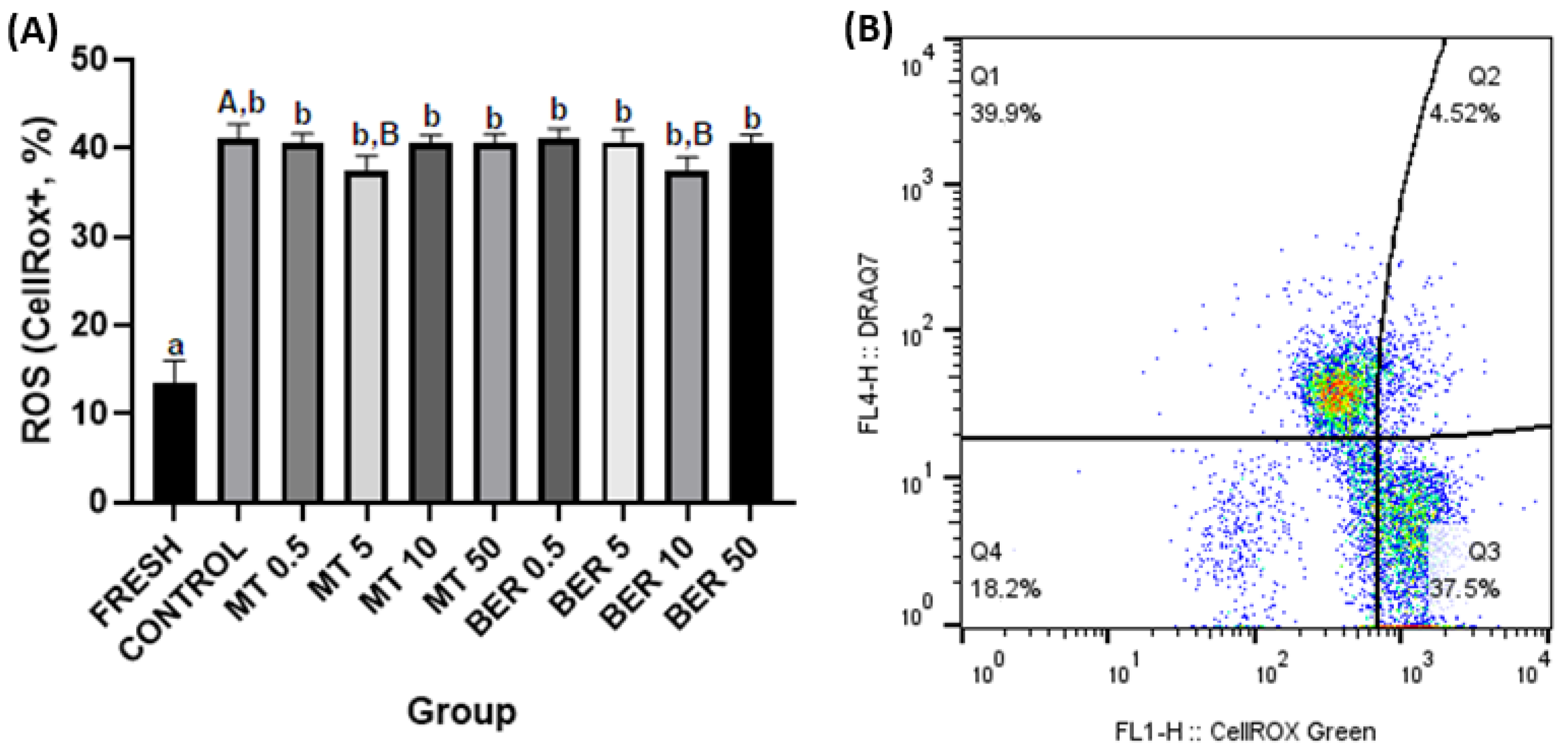
Intracellular ROS levels were assessed using the CellRox Green assay (Thermo Fisher Scientific, Waltham, MA, USA). A semen sample (1 × 10⁶ spermatozoa) was incubated with CellRox (2.5 μM) for 30 min at 37 °C, followed by DRAQ7 staining. The percentage of ROS-positive spermatozoa was calculated (Figure 3B).

Mitochondrial Activity Evaluation

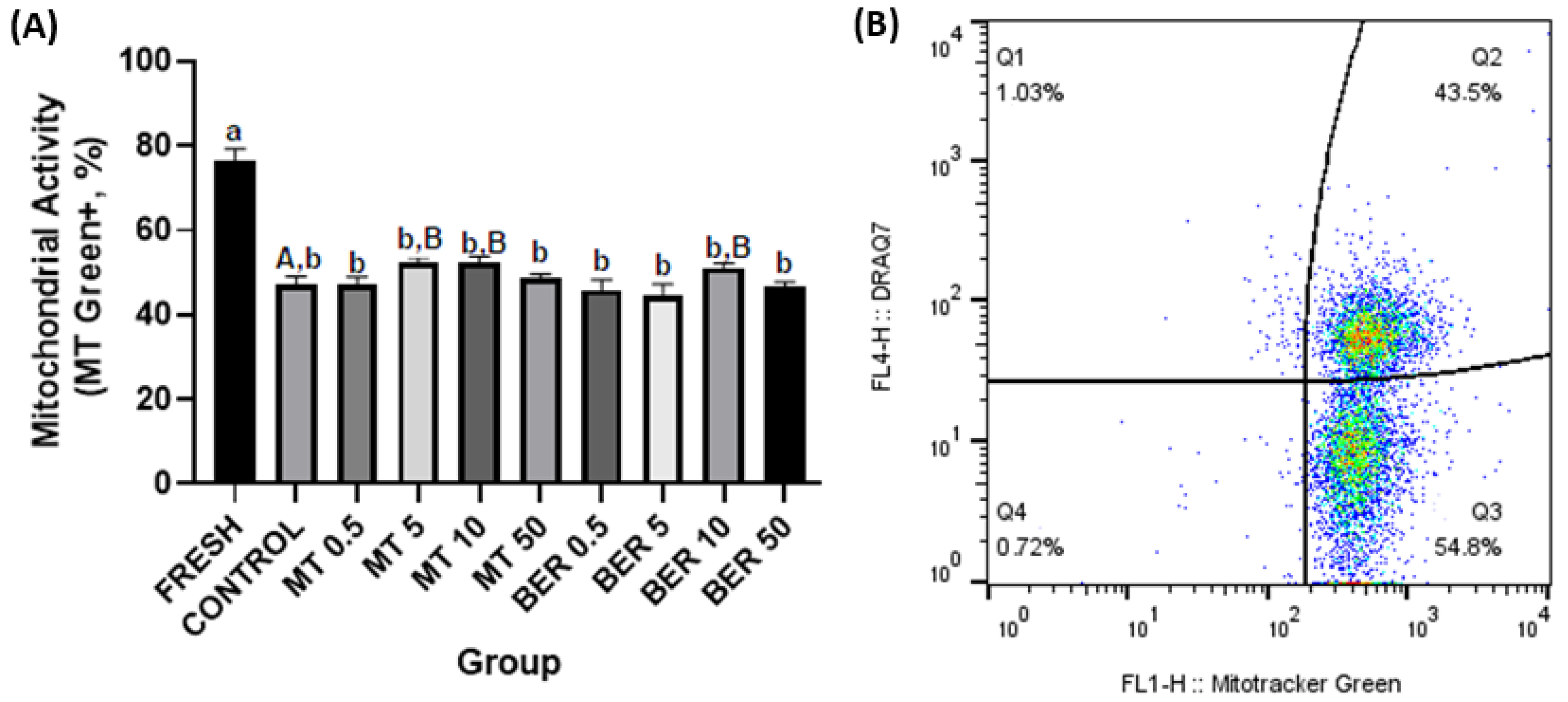
Mitochondrial activity was measured using MitoTracker® Green FM (300 nM, MT Green, Thermo Fisher Scientific, Waltham, MA, USA). A semen sample was incubated at 37 °C for 10 min, and then centrifuged and stained with DRAQ7. The proportion of spermatozoa positive for MitoTracker (MT Green+/DRAQ7) was assessed (Figure 4B).

Acrosome Integrity Evaluation

Acrosome integrity was assessed using peanut agglutinin (PNA, Thermo Fisher Scientific, Waltham, MA, USA) conjugated with Alexa Fluor 488 dye. One microliter of PNA solution was incubated with 1 × 10⁶ spermatozoa for 15 min in the dark. Following centrifugation and DRAQ7 staining, samples were analyzed to determine the percentage of acrosome-damaged spermatozoa (Figure 5B).

Apoptosis Assessment

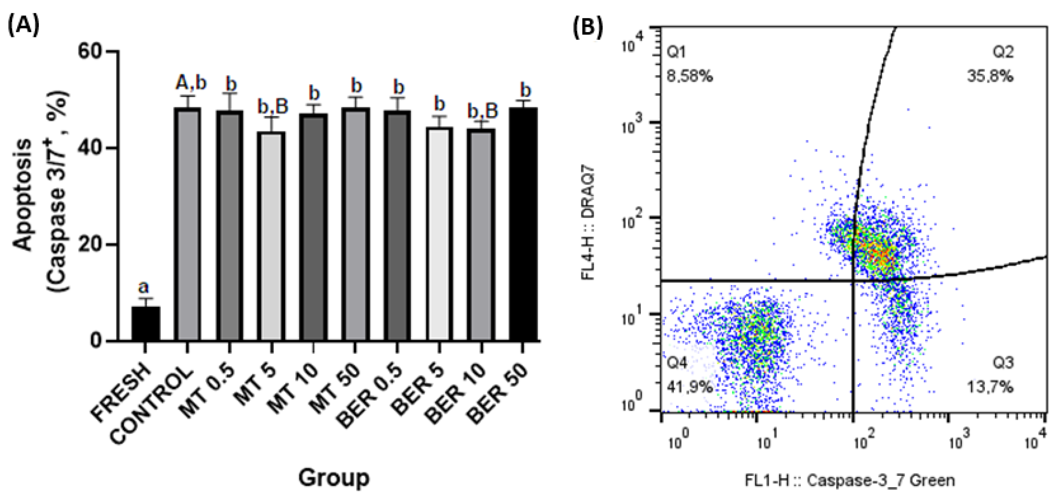
To determine apoptotic changes, spermatozoa were treated with Yo-Pro-1 (100 nM, Thermo Fisher Scientific, Waltham, MA, USA) and incubated for 15 min. After centrifugation, samples were stained with DRAQ7 and analyzed. Caspase activity was measured using the Caspase 3/7 reagent; (CellEventTM Caspase-3/7 Green Flow Cytometry Assay Kit, Thermo Fisher Scientific, Waltham, MA, USA). Caspase 3/7 specifically recognizes active caspase-3 and caspase-7 proteins [23]. Spermatozoa were incubated with the reagent and DRAQ7 before flow cytometric analysis. The proportion of Yo-Pro-1+ and Caspase 3/7+ spermatozoa was recorded (Figure 6B and Figure 7B, respectively).

2.4. Statistical Analyses

Experiments were repeated six times, and the Shapiro–Wilk test was used to evaluate normality. Data were analyzed using GraphPad Prism (version 9.3.1, GraphPad Software, San Diego, CA, USA) with two-way ANOVA and Sidak test for multiple comparisons. Results are expressed as mean ± SD, with statistical significance set at p < 0.05.

2.5. Use of Generative AI for Language Editing

The AI tool (OpenAI’s ChatGPT) was employed specifically for reviewing certain sections of the manuscript. This included grammar checks and overall language improvement to ensure the text met the publication standards. The AI-generated suggestions were carefully reviewed and incorporated as deemed appropriate by the authors.

3. Results

The quality parameters of the fresh pooled ejaculate showed high sperm motility and overall quality, which, as expected, significantly declined after thawing (Figures 1A–6A).

3.1. Effects of MT and BER on the Motility of Frozen-Thawed Sperm

CASA analyses revealed that concentrations of 5 and 10 μmol/L of MT and 10 μmol/L of BER significantly enhanced both total motility and progressive motility compared to the control group without antioxidants (p  <  0.05, Figure 1A,B).

3.2. Effects of MT and BER on the Viability of Frozen-Thawed Sperm

Flow cytometry analysis using SYBR-14 is presented in Figure 2A. The results showed that the percentage of viable sperm significantly increased with the supplementation of 5 and 10 μmol/L MT, reaching 54.28% and 54.89%, respectively, compared to 47.79% in the control group (p  <  0.05). Similarly, the addition of 10 μmol/L BER significantly boosted the viability to 53.02% compared to 47.79% in the control (p  <  0.05). Correspondingly, the percentage of dead sperm in rabbit ejaculates after thawing was notably reduced with the addition of 5 and 10 μmol/L MT and 10 μmol/L BER, with percentages of 45.79%, 45.24%, and 46.71%, respectively, compared to 51.55% in the control group (p  <  0.05, Figure 2B).

3.3. Effects of MT and BER on the ROS Generation of Frozen-Thawed Sperm

Flow cytometry revealed that supplementation with 5 μmol/L MT and 10 μmol/L BER led to significant reductions in sperm intracellular ROS levels, with percentages of 37.35% and 37.50%, respectively, compared to 41.13% in the control group (p  <  0.05, Figure 3A).

3.4. Effects of MT and BER on the Mitochondrial Activity of Frozen-Thawed Sperm

As presented in Figure 4A, a significant increase of frozen-thawed rabbit sperm with high mitochondrial activity was recorded if treated with 5 and 10 μmol/L of MT and 10 μmol/L of BER, 52.38%, 51.65%, 50.48%, respectively vs. 47.24% (p  <  0.05).

3.5. Effects of MT and BER on the Acrosome Integrity of Frozen-Thawed Sperm

MT supplementation did not result in significant improvements in sperm acrosome damage compared to the control across the tested doses. However, treatment with 10 µmol/L BER significantly reduced sperm acrosome damage to 38.28% in comparison to 42.62% of damage observed in the control group (p  <  0.05, Figure 5A).

3.6. Effects of MT and BER on the Apoptotic-like Changes of Frozen-Thawed Sperm

As shown in Figure 6A and Figure 7A, supplementation with 5 μmol/L MT and 10 μmol/L BER led to decreased expression of all apoptotic markers (Yo-Pro-1, and Caspase 3/7) compared to the control group (p  <  0.05).

4. Discussion

This study aimed to explore strategies for improving rabbit sperm resilience against oxidative stress and enhancing the quality of sperm after thawing. Oxidative damage during cryopreservation is a significant factor that reduces the fertilization capacity of frozen-thawed sperm [41,42]. Sperm are particularly susceptible to ROS generation, especially during temperature fluctuations and cryo-injuries, due to their high content of polyunsaturated fatty acids, which makes their membranes prone to oxidative damage [43]. Elevated levels of ROS can trigger apoptosis, impair cellular metabolism, and disrupt the acrosome reaction in sperm [44]. Therefore, it is crucial to enhance freezing extenders, for example by incorporating potent antioxidants, to counteract the negative effects of ROS during cryopreservation. Researchers have extensively investigated the detrimental effects of increased OS caused by semen cryopreservation. Studies have shown that the incorporation of such antioxidants into cryopreservation media can significantly improve the success rates of sperm preservation across different species [11,45,46,47]. We selected the antioxidants MT and BER, which could potentially serve as cryoprotectants for rabbit semen. Our goal was to evaluate their protective effects during sperm freezing and determine the optimal concentration for their addition.
Sperm motility and kinetics are crucial for effective sperm transport to the fertilization site and are commonly used as key indicators of semen quality [48,49]. Mitochondria play a vital role in maintaining normal sperm function and energy balance through oxidative phosphorylation and ATP synthesis [50]. Cryogenic damage to mitochondria has been shown to negatively impact sperm motility by disrupting ATP transport processes [41]. In our experiment, we confirmed that the cryopreservation process significantly reduces sperm motility parameters. Concentrations of 5 and 10 μmol/L of MT significantly enhanced total motility and progressive motility compared to the control group without antioxidant supplementation. In our study, supplementation of the extender with 5 and 10 μmol/L MT significantly improved mitochondrial activity, and viability compared to the control group. The 5 μmol/L dose also substantially reduced ROS levels and apoptosis rates compared to the control. The addition of 10 μmol/L of BER also showed a similar positive significant effect on total and progressive motility. Furthermore, treatment with 10 µmol/L BER significantly reduced sperm acrosome damage, ROS production, and apoptosis compared to the control group. BER enhances mitochondrial activity and energy production in rabbit sperm, which are vital for sustaining sperm motility and overall functionality. This effect is particularly pronounced at a concentration of 10 µmol/L for rabbit sperm, as higher or lower doses did not demonstrate significant differences from the control group.
Previous studies showed that MT enhances motility and velocity in frozen–thawed sperm from human [29,30], rooster [32], or bull [27]. This aligns with our findings as both MT and BER demonstrated comparable effectiveness in improving sperm motility in cryopreserved samples, which is consistent with the study by Chen et al. [51], which examined the effects of various concentrations of berberine (10−4 to 10−7 mol/L) on human sperm motility. Their study analyzed semen samples from both normozoospermic and asthenozoospermic individuals, demonstrating that all tested concentrations of berberine effectively sustained both total and progressive sperm motility. Notably, motility increased even at a concentration of 10−4 mol/L, which corresponds to the 100 μmol/L concentration used in the current study. In contrast, our results showed that a concentration of 50 μmol/L did not yield any significant improvements in sperm behavior. This difference might stem from the fact that, in both studies, the sperm were stored rather than cryopreserved.
The freeze–thaw process places significant stress on sperm, leading to the overproduction of ROS, which causes DNA fragmentation, membrane damage, and cell apoptosis. Prolonged oxidative stress during cryopreservation is a major contributor to reduced sperm viability and fertility potential. Studies have reported significant benefits of using MT in protecting sperm DNA during the cryopreservation process. MT has been shown to reduce ROS levels and minimize apoptosis, improving post-thaw sperm quality and enhancing overall sperm function by mitigating oxidative damage [27,29,30]. In research on human sperm freezing, supplementation with MT was found to enhance sperm quality after thawing, improve antioxidant enzyme activity, and maintain mitochondrial membrane potential [29]. Zhang et al. [30] demonstrated that 10 and 100 μM concentrations of MT could alleviate cryodamage during freezing by modulating oxidative metabolism in spermatozoa from patients with compromised spermatogenesis. The activity of sperm mitochondria is closely linked to various quality characteristics of post-thawed spermatozoa. This finding is consistent with the current study, which showed that using optimal doses of MT resulted in enhanced mitochondrial activity and improved other quality parameters in sperm. Adding MT to the freezing media is believed to prevent mitochondrial Bax translocation, a process associated with mitochondrial dysfunction and apoptosis. Bax, a pro-apoptotic protein, translocates to the mitochondrial membrane under stress conditions like cryopreservation, where it promotes the release of cytochrome c, leading to cell death. By inhibiting this translocation, Mito-TEMPO helps maintain mitochondrial integrity and function [52,53,54]. Additionally, MT mitigates the excessive generation of oxygen-free radicals associated with sperm freezing–thawing through its hydroxylamine-like structure [27,30].
BER, as an alkaloid [55], primarily accumulates in mitochondria, where it plays a pivotal role in its intracellular distribution in mammalian cells [56]. This mitochondrial accumulation is driven by the mitochondrial membrane potential, which promotes the cellular entry of BER and inhibits its efflux from the cell. This rapid uptake by mitochondria not only helps maintain a concentration gradient across the cell membrane but also preserves the negative charge of the inner mitochondrial membrane [39,57]. BER may help protect mammalian cells from apoptosis or necrosis by preserving cell communication channels [58]. Studies have demonstrated that BER significantly improves sperm viability while reducing oxidative stress markers, following both short- and long-term storage of spermatozoa [39,51]. Moreover, BER has shown protective effects in cases of gossypol-induced testicular damage by improving semen quality, oxidative profile, and reducing inflammatory markers, further emphasizing its potential as an antioxidant in mitigating testicular and sperm damage [59]. These findings support BER’s role in enhancing semen quality during cryopreservation and in response to various stressors. BER enhances mitochondrial activity and energy production in sperm, which are vital for sustaining sperm motility and overall functionality. The antioxidant properties of 10 µmol/L BER may help inhibit lipid peroxidation of rabbit sperm plasma membranes and mitochondria, thereby protecting sperm integrity and function. Sperm plasma membrane and acrosome integrity are critical indicators of rabbit sperm quality, assessing the sperm’s ability to manage intracellular and extracellular components. Loss of membrane integrity leads to altered membrane permeability, cholesterol efflux, and calcium influx, which trigger capacitation-like changes and acrosomal damage, ultimately rendering sperm nonviable [60,61].
These findings demonstrate the importance of mitochondria-targeted antioxidants, such as MT and BER, in cryopreservation protocols. Both antioxidants significantly improved rabbit sperm motility, mitochondrial activity, and viability, particularly under cryogenic stress conditions. Our study’s findings on the selective effectiveness of different BER and MT concentrations in enhancing rabbit sperm quality and reducing oxidative stress contribute to a more nuanced understanding of optimal antioxidant use in cryopreservation.

5. Conclusions

In conclusion, the addition of specific concentrations of antioxidants during rabbit semen cryopreservation proved beneficial, with 5 and 10 μmol/L for Mito-Tempo and 10 μmol/L for Berberine showing optimal effects. These antioxidants demonstrate strong potential as cryoprotectants for rabbit sperm, improving post-thaw viability and function. Future research should explore their mechanisms further and evaluate their broader application in cryopreservation protocols to enhance reproductive success across various species.

Author Contributions

L.K.: conceptualization, methodology, formal analysis, investigation, data curation, writing—original draft preparation, writing—review and editing, visualization, supervision. A.S.: methodology, formal analysis, writing—review and editing. A.B.: formal, analysis, investigation, writing—review and editing. J.V.: methodology, formal analysis, investigation, validation, writing—review and editing, visualization, funding acquisition. V.L.: validation, visualization. A.K.: conceptualization, methodology. P.S.: conceptualization, methodology. P.C.: writing—review and editing, supervision, project, administration, funding acquisition. All authors have read and agreed to the published version of the manuscript.

Funding

This research was funded Slovak Research and Development Agency (grant no. APVV 20-0006 and APVV 23-0089) and by the Scientific Grant Agency of the ministry of Education, Science, Research and Sport of the Slovak Republic and Slovak Academy of Science (grant no. VEGA 1/0002/23, VEGA 1/0011/23, KEGA-012UPJŠ-4/2023, KEGA-024SPU-4/2023) and INTERREG, HUSK/2302/1.2/018.

Institutional Review Board Statement

The experiments were conducted in compliance with the Code of Ethics established by the EU Directive 2010/63/EU regarding animal experimentation (https://ec.europa.eu/environment/chemicals/lab_animals/legislation_en.htm, accessed on 3 May 2022). Semen collection is routinely performed at the Research Institute for Animal Production in Nitra, and this process does not cause harm or discomfort to the animals; therefore, a specific Ethical Approval was not required for this type of study. This is also confirmed by the Ethics Committee of the National Agricultural and Food Centre for Protection of Animals Used for Scientific and Teaching Purposes. The experimental procedures were conducted under the guidelines of the approved experimental facility with registration number SK U 18021.

Informed Consent Statement

Not applicable.

Data Availability Statement

The datasets during and/or analyzed during the current study are available from the corresponding author on reasonable request.

Acknowledgments

We would like to acknowledge the use of (OpenAI’s ChatGPT) for assisting with language editing and improving the clarity of this manuscript. The authors reviewed and refined the suggestions provided by the AI tool to ensure the accuracy and quality of the content.

Conflicts of Interest

The authors declare no conflicts of interest.

References

  1. Nishijima, K.; Kitajima, S.; Matsuhisa, F.; Niimi, M.; Wang, C.C.; Fan, J. Strategies for Highly Efficient Rabbit Sperm Cryopreservation. Animals 2021, 11, 1220. [Google Scholar] [CrossRef] [PubMed]
  2. Kodzik, N.; Ciereszko, A.; Judycka, S.; Słowińska, M.; Szczepkowska, B.; Świderska, B.; Dietrich, M.A. Cryoprotectant-specific alterations in the proteome of Siberian sturgeon spermatozoa induced by cryopreservation. Sci. Rep. 2024, 14, 17707. [Google Scholar] [CrossRef] [PubMed]
  3. Sion, B.; Janny, L.; Boucher, D.; Grizard, G. Annexin V binding to plasma membrane predicts the quality of human cryopreserved spermatozoa. Int. J. Androl. 2004, 27, 108–114. [Google Scholar] [CrossRef]
  4. Chatterjee, S.; Gagnon, C. Production of reactive oxygen species by spermatozoa undergoing cooling, freezing, and thawing. Mol. Reprod. Dev. 2001, 59, 451–458. [Google Scholar] [CrossRef]
  5. Wang, A.W.; Zhang, H.; Ikemoto, I.; Anderson, D.J.; Loughlin, K.R. Reactive oxygen species generation by seminal cells during cryopreservation. Urology 1997, 49, 921–925. [Google Scholar] [CrossRef] [PubMed]
  6. Khan, I.M.; Cao, Z.; Liu, H.; Khan, A.; Rahman, S.U.; Khan, M.Z.; Sathanawongs, A.; Zhang, Y. Impact of cryopreservation on spermatozoa freeze-thawed traits and relevance OMICS to assess sperm cryo-tolerance in farm animals. Front. Vet. Sci. 2021, 8, 609180. [Google Scholar] [CrossRef]
  7. Bilodeau, J.F.; Chatterjee, S.; Sirard, M.A.; Gagnon, C. Levels of antioxidant defenses are decreased in bovine spermatozoa after a cycle of freezing and thawing. Mol. Reprod. Dev. 2000, 55, 282–288. [Google Scholar] [CrossRef]
  8. Lasso, J.L.; Noiles, E.E.; Alvarez, J.G.; Storey, B.T. Mechanism of superoxide dismutase loss from human sperm cells during cryopreservation. J. Androl. 1994, 15, 255–265. [Google Scholar] [CrossRef] [PubMed]
  9. Ball, B.A. Oxidative stress, osmotic stress, and apoptosis: Impacts on sperm function and preservation in the horse. Anim. Reprod. Sci. 2008, 107, 257–267. [Google Scholar] [CrossRef]
  10. Gadea, J.; Molla, M.; Selles, E.; Marco, M.A.; Garcia-Vazquez, F.A.; Gardon, J.C. Reduced glutathione content in human sperm is decreased after cryopreservation: Effect of the addition of reduced glutathione to the freezing and thawing extenders. Cryobiology 2011, 62, 40–46. [Google Scholar] [CrossRef]
  11. Amidi, F.; Pazhohan, A.; Nashtaei, M.S.; Khodarahmian, M.; Nekoonam, S. The role of antioxidants in sperm freezing: A review. Cell Tissue Bank. 2016, 17, 745–756. [Google Scholar] [CrossRef] [PubMed]
  12. Covarrubias, L.; Hernández-García, D.; Schnabel, D.; Salas-Vidal, E.; Castro-Obregón, S. Function of reactive oxygen species during animal development: Passive or active? Dev. Biol. 2008, 320, 1–11. [Google Scholar] [CrossRef] [PubMed]
  13. Baskaran, S.; Finelli, R.; Agarwal, A.; Henkel, R. Reactive oxygen species in male reproduction: A boon or a bane? Andrologia 2021, 53, e13577. [Google Scholar] [CrossRef] [PubMed]
  14. Qamar, A.Y.; Naveed, M.I.; Raza, S.; Fang, X.; Roy, P.K.; Bang, S.; Tanga, B.M.; Saadeldin, I.M.; Lee, S.; Cho, J. Role of antioxidants in fertility preservation of sperm—A narrative review. Anim. Biosci. 2023, 36, 385–403. [Google Scholar] [CrossRef] [PubMed]
  15. Mehdipour, M.; Daghigh-Kia, H.; Najafi, A.; Mehdipour, Z.; Mohammadi, H. Protective effect of rosiglitazone on microscopic and oxidative stress parameters of ram sperm after freeze-thawing. Sci. Rep. 2022, 12, 13981. [Google Scholar] [CrossRef]
  16. Santonastaso, M.; Mottola, F.; Iovine, C.; Colacurci, N.; Rocco, L. Protective effects of curcumin on the outcome of cryopreservation in human sperm. Reprod. Sci. 2021, 28, 2895–2905. [Google Scholar] [CrossRef]
  17. Akbari, B.; Baghaei-Yazdi, N.; Bahmaie, M.; Mahdavi Abhari, F. The role of plant-derived natural antioxidants in reduction of oxidative stress. Biofactors 2022, 48, 414–423. [Google Scholar] [CrossRef]
  18. Moslemi, M.K.; Tavanbakhsh, S. Selenium–vitamin E supplementation in infertile men: Effects on semen parameters and pregnancy rate. Int. J. Gen. Med. 2011, 4, 99–104. [Google Scholar] [CrossRef]
  19. Azawi, O.I.; Hussein, E.K. Effect of vitamins C or E supplementation to Tris diluent on the semen quality of Awassi rams preserved at 5 °C. Vet. Res. Forum 2013, 4, 157–160. [Google Scholar]
  20. Mistry, H.D.; Pipkin, F.B.; Redman, C.W.G.; Poston, L. Selenium in reproductive health. Am. J. Obstet. Gynecol. 2012, 206, 21–30. [Google Scholar] [CrossRef]
  21. Alsalman, A.R.S.; Almashhedy, L.A.; Alta’ee, A.H.; Hadwan, M.H. Effect of zinc supplementation on urate pathway enzymes in spermatozoa and seminal plasma of asthenozoospermic patients: A randomized controlled trial. Int. J. Fertil. Steril. 2020, 13, 315–323. [Google Scholar] [CrossRef]
  22. Kaltsas, A. Oxidative stress and male infertility: The protective role of antioxidants. Medicina 2023, 59, 1769. [Google Scholar] [CrossRef]
  23. Najafi, A.; Mohammadi, H.; Sharifi, S.D.; Rahimi, A. Apigenin supplementation substantially improves rooster sperm freezability and post-thaw function. Sci. Rep. 2024, 14, 4527. [Google Scholar] [CrossRef]
  24. Iovine, C.; Mottola, F.; Santonastaso, M.; Finelli, R.; Agarwal, A.; Rocco, L. In vitro ameliorative effects of ellagic acid on vitality, motility, and DNA quality in human spermatozoa. Mol. Reprod. Dev. 2021, 88, 167–174. [Google Scholar] [CrossRef]
  25. Grba, J.; Kuželová, L.; Makarevich, A.; Baláži, A.; Dragin, S.; Tekic, D.; Chrenek, P. The effect of ellagic acid on rabbit sperm in vitro parameters after cryopreservation. Czech J. Anim. Sci. 2024, 69, 110–117. [Google Scholar] [CrossRef]
  26. Zarei, F.; Daghigh Kia, H.; Masoudi, R.; Moghaddam, G.; Ebrahimi, M. Supplementation of ram’s semen extender with Mito-TEMPO I: Improvement in quality parameters and reproductive performance of cooled-stored semen. Cryobiology 2021, 98, 215–218. [Google Scholar] [CrossRef]
  27. Elkhawagah, A.R.; Ricci, A.; Bertero, A.; Poletto, M.L.; Nervo, T.; Donato, G.G.; Vincenti, L.; Martino, N.A. Supplementation with MitoTEMPO before cryopreservation improves sperm quality and fertility potential of Piedmontese beef bull semen. Front. Vet. Sci. 2024, 11, 1376057. [Google Scholar] [CrossRef]
  28. Trnka, J.; Blaikie, F.H.; Smith, R.A.J.; Murphy, M.P. A mitochondria-targeted nitroxide is reduced to its hydroxylamine by ubiquinol in mitochondria. Free Radic. Biol. Med. 2008, 44, 1406–1419. [Google Scholar] [CrossRef]
  29. Lu, X.; Zhang, Y.; Bai, H.; Liu, J.; Li, J.; Wu, B. Mitochondria-targeted antioxidant MitoTEMPO improves the post-thaw sperm quality. Cryobiology 2018, 80, 26–29. [Google Scholar] [CrossRef] [PubMed]
  30. Zhang, X.; Lu, X.; Li, J.; Xia, Q.; Gao, J.; Wu, B. Mito-Tempo alleviates cryodamage by regulating intracellular oxidative metabolism in spermatozoa from asthenozoospermic patients. Cryobiology 2019, 91, 18–22. [Google Scholar] [CrossRef]
  31. Kumar, A.; Ghosh, S.K.; Katiyar, R.; Rautela, R.; Bisla, A.; Ngou, A.A.; Pande, M.; Srivastava, N.; Bhure, S.K. Effect of Mito-Tempo incorporated semen extender on physico-morphological attributes and functional membrane integrity of frozen-thawed buffalo spermatozoa. Cryoletters 2021, 42, 111–119. [Google Scholar] [PubMed]
  32. Masoudi, R.; Asadzadeh, N.; Sharafi, M. Effects of freezing extender supplementation with mitochondria-targeted antioxidant Mito-TEMPO on frozen-thawed rooster semen quality and reproductive performance. Anim. Reprod. Sci. 2021, 225, 106671. [Google Scholar] [CrossRef] [PubMed]
  33. Barfourooshi, H.A.; Esmaeilkhanian, S.; Dadashpour Davachi, A.; Asadzadeh, N.; Masoudi, R. Effect of Mito-TEMPO on post-thawed semen quality in goats. Iran. J. Vet. Med. 2023, 17, 393–400. [Google Scholar] [CrossRef]
  34. Hassan, H.A.; Banchi, P.; Domain, G.; Vanderheyden, L.; Prochowska, S.; Nizański, W.; Van Soom, A. Mito-Tempo improves acrosome integrity of frozen-thawed epididymal spermatozoa in tomcats. Front. Vet. Sci. 2023, 10, 1170347. [Google Scholar] [CrossRef] [PubMed]
  35. Imanshahidi, M.; Hosseinzadeh, H. Pharmacological and therapeutic effects of Berberis vulgaris and its active constituent, berberine. Phytother. Res. 2008, 22, 999–1012. [Google Scholar] [CrossRef]
  36. Tillhon, M.; Guamán Ortiz, L.M.; Lombardi, P.; Scovassi, A.I. Berberine: New perspectives for old remedies. Biochem. Pharmacol. 2012, 84, 1260–1267. [Google Scholar] [CrossRef]
  37. Abudureheman, B.; Zhou, X.; Shu, X.; Chai, Z.; Xu, Y.; Li, S.; Tian, J.; Pan, H.; Ye, X. Evaluation of biochemical properties, antioxidant activities, and phenolic content of two wild-grown Berberis fruits: Berberis nummularia and Berberis atrocarpa. Foods 2022, 11, 2569. [Google Scholar] [CrossRef]
  38. Golubev, D.; Platonova, E.; Zemskaya, N.; Shevchenko, O.; Shaposhnikov, M.; Nekrasova, P.; Patov, S.; Ibragimova, U.; Valuisky, N.; Borisov, A.; et al. Berberis vulgaris L. extract supplementation exerts regulatory effects on the lifespan and healthspan of Drosophila through its antioxidant activity depending on the sex. Biogerontology 2024, 25, 507–528. [Google Scholar] [CrossRef]
  39. Tvrdá, E.; Greifová, H.; Ivanič, P.; Lukáč, N. In vitro effects of berberine on the vitality and oxidative profile of bovine spermatozoa. Int. J. Anim. Vet. Sci. 2019, 13, 244–249. [Google Scholar] [CrossRef]
  40. Vašíček, J.; Baláži, A.; Svoradová, A.; Vozaf, J.; Dujíčková, L.; Makarevich, A.V.; Bauer, M.; Chrenek, P. Comprehensive flow-cytometric quality assessment of ram sperm intended for gene banking using standard and novel fertility biomarkers. Int. J. Mol. Sci. 2022, 23, 5920. [Google Scholar] [CrossRef]
  41. Fang, L.; Bai, C.; Chen, Y.; Dai, J.; Xiang, Y.; Ji, X.; Huang, C.; Dong, Q. Inhibition of ROS production through mitochondria-targeted antioxidant and mitochondrial uncoupling increases post-thaw sperm viability in yellow catfish. Cryobiology 2014, 69, 386–393. [Google Scholar] [CrossRef]
  42. Chelewani, A.P.; Takahashi, E.; Nishimura, T.; Fujimoto, T. Optimizing the post-thaw quality of cryopreserved masu salmon (Oncorhynchus masou) sperm: Evaluating the effects of antioxidant-supplemented extender. Aquaculture 2024, 593, 741332. [Google Scholar] [CrossRef]
  43. Len, J.S.; Koh, W.S.D.; Tan, S.-X. The roles of reactive oxygen species and antioxidants in cryopreservation. Biosci. Rep. 2019, 39, BSR20191601. [Google Scholar] [CrossRef]
  44. Niżański, W.; Partyka, A.; Prochowska, S. Evaluation of spermatozoal function—Useful tools or just science. Reprod. Domest. Anim. 2016, 51 (Suppl. S1), 37–45. [Google Scholar] [CrossRef]
  45. Agarwal, A.; Durairajanayagam, D.; du Plessis, S.S. Utility of antioxidants during assisted reproductive techniques: An evidence-based review. Reprod. Biol. Endocrinol. 2014, 12, 112. [Google Scholar] [CrossRef]
  46. Berean, D.I.; Bogdan, L.M.; Cimpean, R. Advancements in Understanding and Enhancing Antioxidant-Mediated Sperm Cryopreservation in Small Ruminants: Challenges and Perspectives. Antioxidants 2024, 13, 624. [Google Scholar] [CrossRef]
  47. Yi, X.; Qiu, Y.; Tang, X.; Lei, Y.; Pan, Y.; Raza, S.H.A.; Althobaiti, N.A.; Albalawi, A.E.; Al Abdulmonem, W.; Makhlof, R.T.M.; et al. Effect of Five Different Antioxidants on the Effectiveness of Goat Semen Cryopreservation. Reprod. Sci. 2024, 31, 1958–1972. [Google Scholar] [CrossRef]
  48. Chakraborty, S.; Saha, S. Understanding sperm motility mechanisms and the implication of sperm surface molecules in promoting motility. Middle East Fertil. Soc. J. 2022, 27, 4. [Google Scholar] [CrossRef]
  49. Verstegen, J.; Iguer-Ouada, M.; Onclin, K. Computer assisted semen analyzers in andrology research and veterinary practice. Theriogenology 2002, 57, 149–179. [Google Scholar] [CrossRef] [PubMed]
  50. Ruiz-Pesini, E.; Díez-Sánchez, C.; López-Pérez, M.J.; Enríquez, J.A. The role of the mitochondrion in sperm function: Is there a place for oxidative phosphorylation or is this a purely glycolytic process? Curr. Top. Dev. Biol. 2007, 77, 3–19. [Google Scholar] [CrossRef] [PubMed]
  51. Chen, L.; Wang, T.; Liu, J. Effect of berberine on human sperm parameters in vitro. Transl. Androl. Urol. 2016, 5 (Suppl. S1), AB229. [Google Scholar] [CrossRef]
  52. Liang, H.L.; Sedlic, F.; Bosnjak, Z.; Nilakantan, V. SOD1 and MitoTEMPO partially prevent mitochondrial permeability transition pore opening, necrosis, and mitochondrial apoptosis after ATP depletion recovery. Free Radic. Biol. Med. 2010, 49, 1550–1560. [Google Scholar] [CrossRef] [PubMed]
  53. Hu, H.; Li, M. Mitochondria-targeted antioxidant MitoTEMPO protects mitochondrial function against amyloid beta toxicity in primary cultured mouse neurons. Biochem. Biophys. Res. Commun. 2016, 478, 174–180. [Google Scholar] [CrossRef] [PubMed]
  54. Smaili, S.; Hsu, Y.T.; Sanders, K.; Russell, C.; Youle, R.J. Bax translocation to mitochondria subsequent to a rapid loss of mitochondrial membrane potential. Cell Death Differ. 2001, 8, 909–920. [Google Scholar] [CrossRef] [PubMed]
  55. Grycová, L.; Dostál, J.; Marek, R. Quaternary protoberberine alkaloids. Phytochemistry 2007, 68, 150–175. [Google Scholar] [CrossRef]
  56. Li, Q.; Zhou, T.; Liu, C.; Wang, X.-Y.; Zhang, J.-Q.; Wu, F.; Lin, G.; Ma, Y.-M.; Ma, B.-L. Mitochondrial membrane potential played crucial roles in the accumulation of berberine in HepG2 cells. Biosci. Rep. 2019, 39, BSR20190477. [Google Scholar] [CrossRef]
  57. Pereira, G.C.; Branco, A.F.; Matos, J.A.; Pereira, S.L.; Parke, D.; Perkins, E.L.; Serafim, T.L.; Sardao, V.A.; Santos, M.S.; Moreno, A.J.; et al. Mitochondrially targeted effects of berberine [Natural Yellow 18, 5,6-dihydro-9,10-dimethoxybenzo(g)-1,3-benzodioxolo(5,6-a)quinolizinium] on K1735-M2 mouse melanoma cells: Comparison with direct effects on isolated mitochondrial fractions. J. Pharmacol. Exp. Ther. 2007, 323, 636–649. [Google Scholar] [CrossRef]
  58. Ye, J.; Le, J.; Sun, Y. Berberine improves mitochondrial function in colon epithelial cells to protect L-cells from necrosis in preservation of GLP-1 secretion. Diabetes 2018, 67 (Suppl. S1), 2451-PUB. [Google Scholar] [CrossRef]
  59. Saleh, S.R.; Attia, R.; Ghareeb, D.A. The ameliorating effect of berberine-rich fraction against gossypol-induced testicular inflammation and oxidative stress. Oxid. Med. Cell. Longev. 2018, 2018, 1056173. [Google Scholar] [CrossRef]
  60. Bailey, J.L.; Blodeau, J.F.; Cormier, N. Semen cryopreservation in domestic animals: A damaging and capacitating phenomenon minireview. J. Androl. 2000, 21, 1–7. [Google Scholar] [CrossRef]
  61. Palacín, I.; Santolaria, P.; Alquezar-Baeta, C.; Soler, C.; Yániz, J. Relationship of sperm plasma membrane and acrosomal integrities with sperm morphometry in Bos taurus. Asian J. Androl. 2020, 22, 578–582. [Google Scholar] [CrossRef] [PubMed]
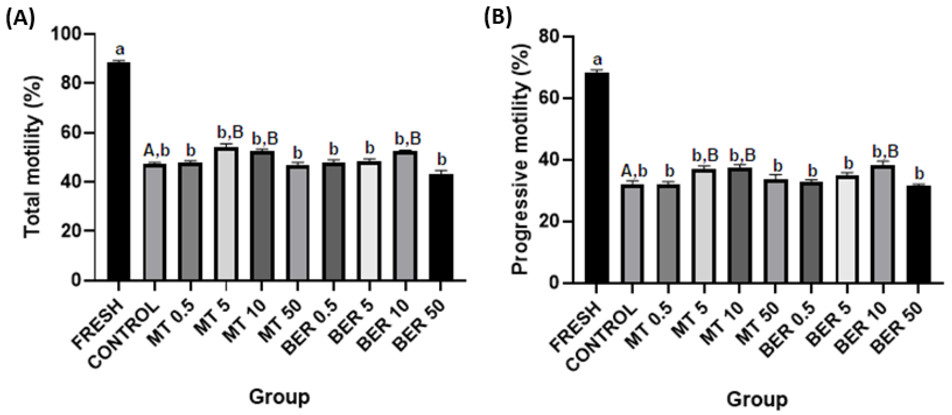
Figure 1. Effect of antioxidants MT and BER on rabbit sperm motility parameters after cryopreservation. (A) Total motility, (B) Progressive motility. Mean ± SD. Level of significance was set at 0.05. Columns labeled “a” and “b” indicate a statistically significant difference between the fresh sample and the thawed samples. Labels “A” and “B” represent a statistically significant difference between the control group without antioxidants and the experimental groups (p  <  0.05, a vs. b; A vs. B).
Antioxidants 13 01360 g001
Figure 2. (A) Effect of antioxidants MT and BER on rabbit sperm viability after cryopreservation. (B) Effect of antioxidants MT and BER on the incidence of dead rabbit sperm after cryopreservation. Mean ± SD. Level of significance was set at 0.05 Columns labeled “a” and “b” indicate a statistically significant difference between the fresh sample and the thawed samples. Labels “A” and “B” represent a statistically significant difference between the control group without antioxidants and the experimental groups (p  <  0.05, a vs. b; A vs. B). (C) Representative flow cytometry plots showing viable (Q3 quadrant) and dead spermatozoa (Q1 and Q2 quadrants).
Antioxidants 13 01360 g002
Figure 3. (A) Effect of antioxidants MT and BER on the rabbit sperm ROS generation after cryopreservation. Mean ± SD. Level of significance was set at 0.05. Columns labeled “a” and “b” indicate a statistically significant difference between the fresh sample and the thawed samples. Labels “A” and “B” represent a statistically significant difference between the control group without antioxidants and the experimental groups (p  <  0.05, a vs. b; A vs. B). (B) Representative flow cytometry plots showing viable ROS positive spermatozoa (Q2 and Q3 quadrants).
Antioxidants 13 01360 g003
Figure 4. (A) Effect of antioxidants MT and BER on rabbit sperm mitochondrial activity after cryopreservation. Mean ± SD. Level of significance was set at 0.05. Columns labeled “a” and “b” indicate a statistically significant difference between the fresh sample and the thawed samples. Labels “A” and “B” represent a statistically significant difference between the control group without antioxidants and the experimental groups (p  <  0.05, a vs. b; A vs. B). (B) Representative flow cytometry plots showing live spermatozoa with high mitochondrial activity (Q3 quadrant).
Antioxidants 13 01360 g004
Figure 5. (A) Effect of antioxidants MT and BER on rabbit sperm acrosome integrity after cryopreservation. Mean ± SD. Level of significance was set at 0.05 Columns labeled “a” and “b” indicate a statistically significant difference between the fresh sample and the thawed samples. Labels “A” and “B” represent a statistically significant difference between the control group without antioxidants and the experimental groups (p  <  0.05, a vs. b; A vs. B). (B) Representative flow cytometry plots showing PNA positive spermatozoa (Q2 and Q3 quadrants).
Antioxidants 13 01360 g005
Figure 6. (A) Effect of antioxidants MT and BER on rabbit sperm apoptotic-like changes after cryopreservation. Mean ± SD. Level of significance was set at 0.05. Columns labeled “a” and “b” indicate a statistically significant difference between the fresh sample and the thawed samples. Labels “A” and “B” represent a statistically significant difference between the control group without antioxidants and the experimental groups (p  <  0.05, a vs. b; A vs. B). (B) Representative flow cytometry plots showing Yo-Pro-1 positive spermatozoa (Q2 and Q3 quadrants).
Antioxidants 13 01360 g006
Figure 7. (A) Effect of antioxidants MT and BER on rabbit sperm apoptotic-like changes after cryopreservation. Mean ± SD. Level of significance was set at 0.05. Columns labeled “a” and “b” indicate a statistically significant difference between the fresh sample and the thawed samples. Labels “A” and “B” represent a statistically significant difference between the control group without antioxidants and the experimental groups (p  <  0.05, a vs. b; A vs. B). (B) Representative flow cytometry plots showing Caspase 3/7 positive spermatozoa (Q2 and Q3 quadrants).
Antioxidants 13 01360 g007
Disclaimer/Publisher’s Note: The statements, opinions and data contained in all publications are solely those of the individual author(s) and contributor(s) and not of MDPI and/or the editor(s). MDPI and/or the editor(s) disclaim responsibility for any injury to people or property resulting from any ideas, methods, instructions or products referred to in the content.