Review

Metal-Based Nanoparticles and Their Relevant Consequences on Cytotoxicity Cascade and Induced Oxidative Stress

1
Interdisciplinary Graduate Program in Advanced Convergence Technology and Science, Jeju National University, Jeju-si 63243, Republic of Korea
2
Department of Animal Biotechnology, Faculty of Biotechnology, College of Applied Life Sciences, Jeju National University, Jeju-si 63243, Republic of Korea
3
Department of Biotechnology, School of Bio, Chemical and Processing Engineering (SBCE), Kalasalingam Academy of Research and Educational, Krishnankoil 626126, India
4
Bio-Health Materials Core-Facility Center, Jeju National University, Jeju-si 63243, Republic of Korea
5
Practical Translational Research Center, Jeju National University, Jeju-si 63243, Republic of Korea
*
Authors to whom correspondence should be addressed.
Antioxidants 2023, 12(3), 703; https://doi.org/10.3390/antiox12030703
Submission received: 9 February 2023 / Revised: 6 March 2023 / Accepted: 10 March 2023 / Published: 12 March 2023
(This article belongs to the Special Issue Oxidative Stress in Environmental Toxicology)

Abstract

:
Emerging nanoscience allows us to take advantage of the improved evolutionary components and apply today’s advanced characterization and fabrication techniques to solve environmental and biological problems. Despite the promise that nanotechnology will improve our lives, the potential risks of technology remain largely uncertain. The lack of information on bio-impacts and the absence of consistent standards are the limitations of using metal-based nanoparticles (mNPs) for existing applications. To analyze the role played by the mNPs physicochemical characteristics and tactics to protect live beings, the field of nanotoxicology nowadays is focused on collecting and analyzing data from in vitro and in vivo investigations. The degree of reactive oxygen species (ROS) and oxidative stress caused by material nanoparticles (NPs) depends on many factors, such as size, shape, chemical composition, etc. These characteristics enable NPs to enter cells and interact with biological macromolecules and cell organelles, resulting in oxidative damage, an inflammatory response, the development of mitochondrial dysfunction, damage to genetic material, or cytotoxic effects. This report explored the mechanisms and cellular signaling cascades of mNPs-induced oxidative stress and the relevant health consequences.

1. Introduction

Throughout the twenty-first century, revolutionary nanotechnology has been rapidly expanding and intriguing scientists, medical professionals, and consumers [1,2]. Material nanoparticles (NPs) have distinct physicochemical qualities due to their nano-size (<100 nm), wide surface area, and rapid reactivity. Nevertheless, more than ~2800 commercial nanoparticulate-based applications are promptly available [3]. These NPs exhibit specific physicochemical characteristics and are manufactured for applications in biological and commercial functions, including cancer research [4], drug delivery, cosmetics, biosensors, environmental remediation, antimicrobial agents, and environmental remediation [5]. The population susceptible to metal-based nanoparticles (mNPs) is growing as their application develops. According to the allied market report (https://www.alliedmarketresearch.com/nanotechnology-market, published in July 2021), the worldwide nanotechnology market was valued at USD 1055.1 million in 2018 and is estimated to reach USD 2231.4 million by 2025, with a total value of USD 43.1 billion by 2030 with a CAGR of 18.05% from 2022 to 2030. However, with the fast growth of nanotechnology and the widespread usage of nano products, the hazard of human exposure is rapidly increasing [6]. mNPs are frequently consumed through food, drink, air, cosmetics, or pharmaceuticals and absorbed by the body before being redistributed to major organs, potentially causing tissue damage.
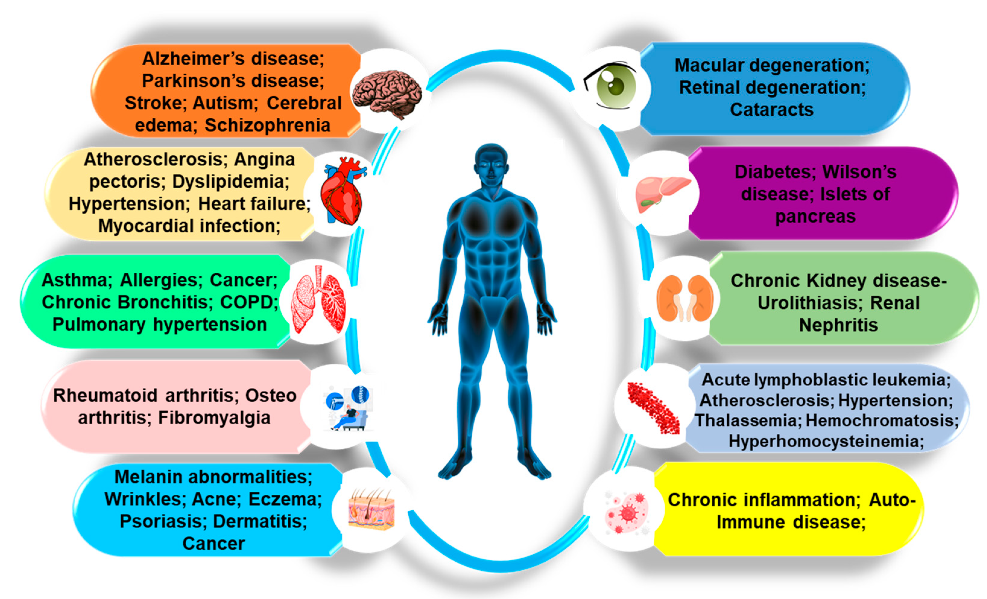
Despite the undeniable benefits of nanoscale materials’ potency, there are questions which still need to be solved regarding how NPs are implemented in everyday life and may or may not impact the environment. The direct and indirect effects of mNPs on the environment and air pollution may be researched from various perspectives. It may inevitably enter the environment by producing, using, and discarding nanoparticle products. These mNPs may accumulate at a certain “level to environmental risk assessment” that necessitates an understanding of the relationship due to their propensity to aggregate, agglomerate, and maybe adsorb or interact with biological matter. As a result, living creatures are constantly exposed to inorganic NPs, which enter the human body through various paths such as ingestion, inhalation, dermal penetration, and blood circulation [7] and are translocated in organs (liver, kidneys, spleen, heart, and brain) and tissues, depending on their physicochemical properties (Figure 1). To assess the threat of inorganic NPs, it will be crucial to comprehend the precise mechanism underlying their cell-specific cytotoxicity and the validity of toxicity analytical techniques.
The current focus of nanotoxicology is on gathering and interpreting data from in vivo and in vitro studies to ascertain how the physicochemical properties of NPs and mitigation strategies influence living beings. These variables affected the biokinetics and biological activity of NPs, influencing their translocation from epithelia into organs, intracellular localization, the stimulation of ROS generation, and interaction with receptors [8]. In this study, we discussed the mechanisms and cellular signaling cascades of mNPs-induced oxidative stress and their possible detrimental impacts on health. A comprehensive search was undertaken using relevant electronic databases such as PubMed, Scopus, Google Scholar, Web of Science, ProQuest, ERIC, WorldCat Discovery, JSTOR, and Directory of Open Access Journals (DOAJ). The search followed a strategy to select literature that addressed the review objectives.

2. Revolutionary Implications of mNPs in Pharmaceutical Research

The essential features of mNPs are their size, which lies in the transitional zone between individual atoms or molecules and comparable bulk materials [9,10]. The engineered physicochemical properties of mNPs allows them to create the opportunity for an increased uptake and interaction with biological tissues. Because of these features, mNPs have potential uses in biological diagnostics, cell labeling, targeted drug administration, medical imaging, cancer treatment, and biological sensors. They have also been employed as a gene carrier for selective tissue or cell distribution [11], transfection vectors [12], and contrast agents in medical imaging fluorescent labels [13]. mNPs comprised of titanium dioxide (TiO2), copper (Cu), iron (Fe), zinc (Zn), aluminum (Al), gold (Au), and silver (Ag) are also gaining popularity.
It is believed that the quantity of silver that individuals consume everyday by consumption is about 20–80 µg [7]. Numerous studies have been conducted on broadly used silver NPs (AgNPs) in healthcare as potential antimicrobial agents against more than ~650 different types of disease-causing organisms, including viruses, biocides, antibiotic treatment alternatives, and nanocomposite coatings [2,14,15,16,17]. Additionally, interactions between AgNPs and sulfur-containing proteins can impact the ability of bacteria to survive. AgNPs can impair cellular respiration and cell death by exchanging other essential metal ions from bacterial cells, such as Zn2+ and Ca2+ [18]. Furthermore, gold NPs (AuNPs) are employed to assault bacterial membranes and disrupt DNA replication, killing certain bacteria, including E. coli, Staphylococcus aureus, and Pseudomonas aeruginosa [19,20,21,22]. Peroxidase-like nanoparticles, e.g., AuNPs, can be employed as labels in various analytical techniques, such as virus detection. The immunochromatographic strip nanozyme-strip made of Fe3O4 magnetic NPs can identify the Ebola virus’ glycoprotein. The authors suggest that it may be feasible to detect additional pathogenic viruses, such as the Bunya virus [23]. Concerns regarding conflicting bio-effects are raised by the dual function of ZnO and TiO2 in protecting skin from sunlight and eradicating surface microorganisms, respectively [2].
The use of NPs in drug delivery is a potential technique for improving the administration of chemotherapies, radionuclides, and antibody medicines to target cells. Al NPs have been proposed as drug delivery systems, mainly by encapsulating medications with aluminum–magnesium hybrids to boost solubility, prevent clearance processes, and allow for the site-specific targeting of medications to cells. Anticancer medications delivered with zinc oxide and gold NPs are thought to have an antitumor effect by stimulating the immune system [24]. Research demonstrated that adding ZnO NPs to doxorubicin boosted the drug’s intracellular concentration in hepatocarcinoma cells, increasing its therapeutic impact compared to controls [25]. Recent advances in nanotechnology have made it feasible to combine NPs with biological components for a targeted treatment, enabling the administration of radiation doses with a greater specificity and limiting harm to healthy tissues [22].
The commercial applications of nanotechnology in medical research are mostly focused on the production of nano-agents for labeling, tissue engineering, and medication delivery systems [26]. Studies have primarily focused on cellular imaging with the imaging of in vivo macrophage activity. The fabrication of mNPs employed as markers in medical diagnostics, such as iron oxide (Fe2O3) or superparamagnetic-based nanocrystal technology and other noble metal plasmon-resonant particles (i.e., Au, Ag, Pt, and Pd), have dramatically increased in medical imaging approaches, such as magnetically targeted drug delivery [27], MRI contrast agents [28], magnetically assisted gene transfection [29], triggered drug release [30], and magnetic fluid hyperthermia (MFH) [31,32,33]. These NPs must have high magnetization values, be smaller than 100 nm, and have a narrow particle size distribution for all these biological and bioengineering applications [32]. PdNPs have been employed in medical diagnostics, such as glucose detection, because of their enzyme-like function. Ru, Au, and Pt nanoparticle-based biosensors were utilized to detect bilirubin, ultra-low levels of mercury, catalase (CAT) and superoxide dismutase (SOD), polyphenol oxidase, ferroxidase, and ascorbate oxidase mimics, accordingly [34].
The nanowires and nanospheres of iron, gadolinium, and NiO have shown excellent imaging signals [35]. It has been reported that 198Au NPs may be synthesized directly involving radionuclides [36]. Xie et al. described the process for radiolabeling gold nanoshells with the radionuclide labels copper-64 (64Cu) and indium-111 (111In) through a bifunctional PEG and chelating agent in live rats. They demonstrated head and neck squamous cell carcinoma xenografts using non-invasive PET and SPECT imaging [22,37]. These engineered mNPs-altered physicochemical and structural characteristics may cause various material interactions and toxicological consequences [38,39]. Transition metals, including Fe, Ni, Cu, and Cr, may be absorbed into the surface of mNPs due to their enormous specific surface area and catalyze Fenton reactions that directly damage DNA [40], which may lead to carcinoma. Hence, to prevent certain metals from leaching, non-permeable coatings are required.

3. Physio-Pathological Implications of mNPs

mNPs are extremely attractive in various applications due to their distinctive physicochemical characteristics, allowing them to influence cellular processes at the biological level [41,42]. The fact that mNPs have high surface-to-volume ratios makes them reactive or catalytic [1]. Due to their small size, they are more likely to be able to penetrate biological barriers such as cell membranes and cause cellular dysfunction in living organisms [42]. Indeed, the high toxicity of some transition metals can make it challenging to use mixed oxide NPs in biomedical uses. It triggers adverse effects on organisms, causing oxidative stress, stimulating the formation of ROS, mitochondrial perturbation, and the modulation of cellular functions, with fatal results in some cases [39,43,44] (Figure 2).
Size-dependent cellular interactions with known physiologically active silver nanoparticles reported a strong inflammatory response by emitting tumor necrosis factor-α (TNF-α), MIP-2, and interleukin-1β (IL-1β), and the oxidative stress might be the principal cause of cellular damages [45].The primary sources of mNPs-induced oxidative stress are (i) mNPs or transition metal pollutants employed as catalysts in the manufacture of nonmetal nanoparticles; (ii) intermediates of highly stable free radicals found on “reactive” surfaces of particles such as quartz and carbonaceous particles; and (iii) the functionalization of NPs to produce redox-active groups [6,46,47,48]. Since oxidative stress is a significant factor in NP-induced injury, it can result in a variety of physiopathologic effects, including genotoxicity, necrosis, inflammation, fibrosis, metaplasia, hypertrophy, lipid peroxidation, damage to macromolecules such as DNA, leading to mutations, and fostering the growth of tumors [49,50,51]. It is crucial to comprehend the underlying processes of nanoparticle-induced ROS and the accompanying oxidative stress they impose.

The Adverse Impacts of Various mNPs on the Organs

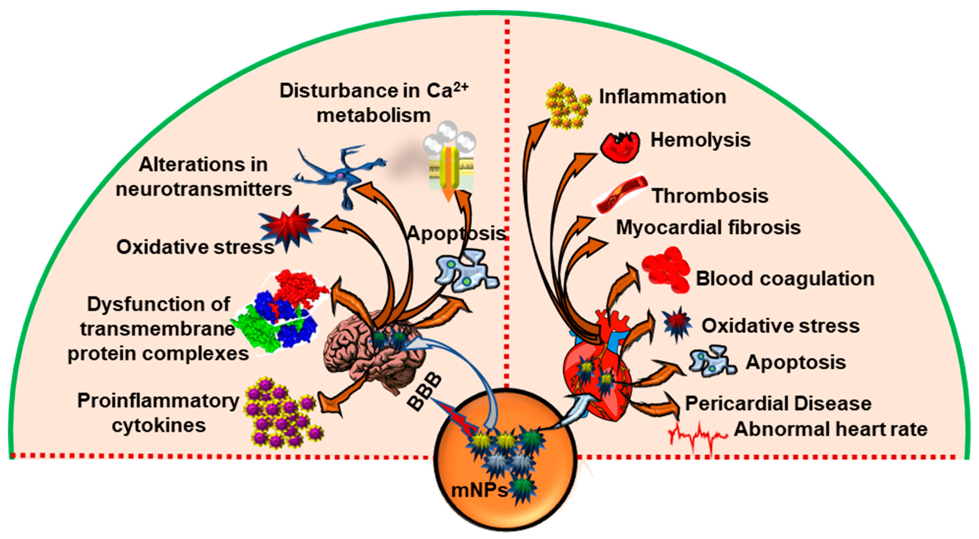
Since mNPs are used increasingly often in healthcare, the safety of employing them for the central nervous system (CNS) is receiving increased attention [52] (Figure 3). This CNS of vertebrates is separated from the rest of the body by the blood–brain barrier (BBB), responsible for the exchange of nutrients and metabolites between the blood and brain, xenobiotics penetration prevention, and restricting immune cell infiltration [53]. Several studies demonstrated the transport of various mNPs, e.g., ultrafine Ag NPs [54], Al NPs [55], CdSe/ZnS, quantum dots (CdSe/ZnS QDs) [56], copper oxide NPs (CuO NPs) [57], ultrafine manganese dioxide NPs (MnO2 NPs) [58], titanium dioxide NPs (TiO2 NPs) [59], and aluminum oxide NPs (Al2O3 NPs) [60,61], leading NPs (Pb NPs) [62] to the brain via the olfactory nerve, which is a direct route, circumventing the BBB [63]. After being exposed to Ag NPs, microvascular endothelial cells, astrocytes, and pericytes showed mitochondrial shrinkage, endoplasmic reticulum expansion, and vacuolation. There have also been alterations in 23 genes related to cell death, metabolic and biosynthetic processes, and response to stimuli [64]. Because of their outstanding biocompatibility and simple biodegradation in vivo, magnetite-based iron oxide NPs (below 30 nm) have gathered the most interest for various diagnostic and potential therapeutic applications in the CNS. In addition, after being metabolized, iron ions from these NPs are incorporated to the iron deposits or become absorbed by erythrocytes as part of the hemoglobin. It has been described that ultrasmall superparamagnetic iron oxide particles can cross the human BBB [65]. Hence, it is essential to precisely characterize the neurotoxic effects of mNPs in order to promote the creation of safer and more effective CNS-targeting nanomedicines.
Another concern is the influence of mNPs on offspring. Until now, studies have shown that NPs entering the maternal body during gestation may damage fetal development through direct or indirect processes. According to neurodevelopmental research, prenatal damages by mNPs lead in distinct phenotypes in both male and female offspring and influenced the expression of functional genes related to brain development in mice [66,67]. According to a study, subcutaneously injected TiO2 NPs into pregnant mice caused translocation to the offspring and had an adverse effect on the male offspring’s genital system by lowering the daily sperm production [66,68]. Additionally, it was shown that adult mice that inhaled TiO2 NPs experienced lengthy lung inflammation and, as a result, their progeny displayed aberrant neurobehavioral abnormalities [69].
Researchers have recently emphasized the potentially hazardous cardiovascular repercussions of exposure to mNPs, particularly engineered nanoparticles, due to their extensive applications in electronics, mechanical design, environmental remediation, and biomedicine (Figure 3). According to a study by Kan et al., there are three potential mechanisms for adverse effects on the cardiovascular system: (a) synthetic NPs may travel from the lungs to the bloodstream and cause pathological alterations in the tissue of the heart and arteries; (b) engineered NPs may cause a systemic oxidative stress/inflammatory reaction through the lungs that changes cardiovascular function; or (c) they could affect cardiovascular performance through the neurogenic pathway [70]. Au-NPs are recognized to have antioxidant potential in therapeutic strategies; nonetheless, in vitro studies have shown that Au-NPs were associated with autophagy when they entered rat heart muscle cells. The effects of Au-NPs on the heart are proportional to their size. The study found that Au-NPs with a diameter of 40 nm were harmful to the heart. Eventually, 5 nm Au-NPs exhibited no cardiac injury. As a result, larger Au-NPs (greater than 40 nm) are more likely to induce cardiac dysfunction [71]. The Impacts of Fe2O3 NPs (spherical; 50 nm particle size; 50–245 m2/g surface area) and AgNPs (spherical; 50 nm particle size; 5.0 m2/g surface area) on the heart and lungs of male rats showed toxicity when administered either alone or in combination [72]. Cardiotoxicity and lung toxicity were triggered through JNK, p53, and NF-κB pathways, which included oxidative DNA modification, the activation of inflammation, free radical production, and the inhibition of antioxidant defense. mNPs caused distinct metabolic changes that may have important implications as atherogenic causes. However, PON1 is a hydrolytic enzyme with a broad substrate range that can protect against lipid oxidation. PON1 was depleted in the plasma and cardiac tissues by mNPs, such as Fe2O3NPs and AgNPs [72]. The toxicity of TiO2 and AgNPs on Daphnia magna revealed that the heart rate dropped with increasing concentrations and that the ROS levels were elevated in comparison to the control group [73]. In order to effectively implement mNPs in a therapeutic context, it is necessary to efficiently measure their preventive efficacy in relevant cardiac pathological conditions.
Several mNPs have been demonstrated to cause hepatic steatosis due to residual NPs in the liver [74]. Despite the fact that 30–99% of specified NPs will accumulate and sequester in the liver, the accumulation of specific mNPs in the liver has been observed to induce oxidative stressors, which in turn disrupt the liver’s metabolism and homeostasis [75,76]. In vivo pulmonary toxicity experiments were performed using five distinct particle types, including (1) carbonyl iron (CI), (2) crystalline silica (CS) (Min-U-Sil 5, α-quartz), (3) precipitated amorphous silica (AS), (4) nano-sized zinc oxide (NZO), or (5) fine-sized zinc oxide (FZO). This study revealed that instilled carbonyl iron particles caused slight toxicity, whereas crystalline quartz silica particle exposure caused sustained inflammation and cytotoxicity, and amorphous silica particle exposure caused transient inflammatory responses that were reversible one week later. Intriguingly, the intratracheal intravenous administration of nano or fine-sized zinc oxide particles elicited strong but transitory inflammation/cytotoxic effects that were reversed by exposure one month after instillation [77,78]. Jia et al. demonstrated that the oral administration of 10 or 20 nm Ag NPs in normal mice promoted the progression of fatty liver disease from steatosis to steatohepatitis in obese mice, which can be attributed to the pro-inflammatory activation of KCs in the liver, increased hepatic inflammation, and the suppression of fatty acid oxidation [79]. It was reported that 14 ± 4 nm Au NPs with spherical forms were documented to induce hepatic steatosis in Wistar-Kyoto rats [80]. In contrast, 60 nm spherical Si NPs exacerbated hepatic steatosis in mice or a zebrafish model via the TLR5 signaling pathway, while SiO2 NPs caused oxidative damage and triggered the transforming growth factor-β1 (TGF-β1)/Smad3 signaling pathway, which accelerated the liver fibrosis process [81,82]. In addition to aggravating the liver, Fe2O3 NPs cause higher ALT levels, increased hepatocyte necrosis, hepatic inflammation, interstitial congestion, and fatty degeneration near the central vein [74], whereas Fe3O4 NPs cause higher ALT (alanine transaminase), AST (aspartate aminotransferase), and ALP (alkaline phosphatase) levels, central venous congestion, hepatocyte hypertrophy, regeneration, and necrosis, as well as increased Kupffer cell counts [83]. Furthermore, the co-administration of ZnO NPs with the xenobiotic chemical organophosphate dimethoate-enhanced hepatic deposition of zinc and dimethoate, led to increased liver oxidative stress and damage [84].
The kidneys are essential organs that filter all hazardous chemicals and metabolites through urine. Nonetheless, the key diseases that contribute to kidney disorders are oxidative stress, inflammation, apoptosis, and necrosis. However, the effects of NPs to address kidney dysfunction has not been extensively studied in in vivo and in vitro animal models. Most researches have pointed to the significance of ROS in SiNPs toxicity; as a result, SiNPs evoked an inflammatory response in macrophages and the kidney of mice [85,86]. The administration of mesoporous silica NPs (mSiNPs) increased the expression of NF-κB, MyD88, TLR4, caspase-3, and p65 in the liver and kidney of rats, along with the levels of ROS, lipid peroxidation, and nitric oxide, while suppressing antioxidants and Nrf2/HO-1 signaling [87]. The toxicity profile of nano-sized TiO2 delivered to mice has also been described in the lung, liver, and kidney [7,88,89]. Some important studies that have shown the toxic effects of AgNPs on different cell lines, including embryonic kidney cells (HEK293T) and porcine kidney cells (Pk 15) [90,91]. According to a previous study, AgNPs with diameters less than 100 nm are mostly taken up by endocytosis in epithelial cells and can cause oxidative stress, DNA damage, and inflammation in enterocytes [92]. Kidney damage inflicted by various AgNP doses was also studied on male Wistar rats after 28 days of oral administration [93]. A histological examination indicated damage to the lining tubular epithelial cells after treatment with 30 and 125 mg/kg of AgNPs, including vacuolization, hazy swelling, severe necrosis, and pyknotic nuclei, confirming the toxicity induced by these doses [93].

4. Effects of mNPs on Cytotoxicity and Cellular Damage

The most pressing worry is how hazardous mNPs are at regular doses [94]. Given the unique nature of nanoparticles, it is difficult to link the test results from diverse studies and evaluate whether the toxicity and cellular damage seen are physiologically significant. The cytotoxicity of mNPs is being studied using a variety of standardized techniques, including in vitro and in vivo studies, involving rats, humans, and aquatic species such as zebrafish, catfish, algae, and macrophages, as well as comprehensive genomic or biodistribution studies. The inherent toxicity of specific mNPs may alter biological behavior in terms of proteins, cells, subcellular structures, tissue, and organs [45,95,96,97,98,99,100,101]. Exposure to these mNPs is associated with oxidant production, macrophage activation, the extended release of inflammatory mediators and growth factors, and the fibroblast stimulation of the creation of the extracellular matrix at the cellular level.
An in vivo study on gastrointestinal persorption and the tissue distribution of differently sized colloidal gold NPs revealed that it could permeate the small intestine and spread to the blood, brain, lung, heart, kidney, spleen, liver, intestine, and stomach [102]. It was found that mNPs (such as AuNPs, AgNPs, and CuNPs) may not be detected by normal phagocytic defenses and cluster first outside the cell. Then, they entered the cell in an aggregated condition [94]. The cell was exposed to spherical gold NPs of varying sizes for 24 and 48 h, indicating that they are not fundamentally harmful to human cells [103]. The study by Goodman et al. stated that AuNPs are non-cytotoxic and reduce the levels of potentially hazardous ROS in the cells [104]. However, it is necessary to distinguish between cytotoxicity and cellular damage. The theories stated that NPs that exhibit little or no cytotoxicity might be capable of causing significant cellular damage indeed [94,105].
The report also revealed that the toxicity of Au NPs directly related to their shapes [102,103,106], coat on the mNPs, and surface charge [104]. The study has demonstrated that the cellular response is based on particle size, with 1.4 nm particles mainly causing fast cell death by necrosis within 12 h. In contrast, closely comparable particles 1.2 nm in diameter primarily induce planned cell death through apoptosis [107]. Gold nanoparticles’ cellular absorption and cytotoxicity has been widely researched in human leukemia cells and skin HaCaT keratinocytes. CTAB-coated Au nanorods were shown to be more hazardous to human HaCaT keratinocytes than spherical Au NPs (30 nm) [94,103]. According to another study, Au NPs did not trigger cytotoxicity in human astrocytes, but they instead boosted ROS generation, upregulated NF-kB activity, and decreased micronuclei development [108].
Ag NPs have been widely used as antimicrobials; however, the use of ~25 nm Ag NPs (at concentrations of >25 µg/mL) in the image analysis of neural tissue and cells, in particular, raises concerns about the possibility that they may be contributing to neurodegenerative diseases (such as Parkinson’s and Alzheimer’s), due to their capacity to produce ROS and oxidative stress [2,109]. The dose-dependent Ag NPs-induced toxicity experiment was carried out in zebrafish embryos [110]. An in vivo dose-dependent investigation of Ag NPs using adult Sprague Dawley rats revealed substantial alterations in plasma alkaline phosphatase (ALP) and blood cholesterol, indicating that these NPs might harm the liver [111]. In PC12 cells, Ag NPs enhanced ROS generation and upregulated the expression of genes associated with oxidative stress, including those encoding HO-1 and MMP-3 [112]. An incident involving a 71-year-old man who consumed colloidal silver every day for four months was reported to have developed myoclonic status epilepticus and fallen into a coma. It was observed that the patient’s plasma, erythrocytes, and CSF all had high quantities of silver [113].
Copper NPs (CuNPs) have also received significant attention as potential candidates for innovative antimicrobial applications such as biocides, antibiotic treatment alternatives, and nanocomposite coatings [114]. However, they have severe toxicological consequences through oxidative damage and/or an inflammatory reaction [115]. Aside from industrial-scale uses, transition metal oxide particles have revolutionized several sectors, including catalysis, sensors, optoelectronic materials, and drug delivery. The study suggested that transition metals, including Si, Zn, Cu, Fe, chromium (Cr), and vanadium (V), are linked to ROS production via Fenton and Haber–Weiss reaction pathways [46]. The toxicity of metal oxide nanoparticles, such as nano-TiO2 [116], nano-ZnO [117], nano-CuO [118], nano-Fe3O4 [119], Al2O3, and CrO3 of particle sizes ranging from 30 to 45 nm, is widely reported [120,121]. TiO2 toxicity studies have indicated that they cause inflammatory responses, cell death, an increase in ROS, and the stimulation of oxidative stress-related genes [122,123,124]. It has been demonstrated that these NPs interact directly with the liver and, as they migrate through the organ systems, might cause synaptic damage and neurotransmitter dysfunction called neurotoxicity [2,125,126]. According to the reports, if these NPs can efficiently distribute to specific tissues by penetrating the epidermis, entering via damaged skin, or being administered systemically, they may threaten the overall health of the body [127,128,129]. According to the study, nano ZnO can influence the diversity, metabolism, and functional pathways of the human gut microbiome and the gut resistome [117]. Considering the risk mentioned above, NPs may enter the bloodstream or come into direct contact with the respiratory system, which could lead to endothelial cell membrane toxicity; more even, transfer to the lymphatic system may result in secretory immunological responses.
Environmental factors such as air pollution can cause oxidative damage in the brain, which may result in neurodegenerative illnesses. NPs disrupt the tight junctions of the blood–brain barrier or enter the central nervous system (CNS), allowing them to access and induce neurotoxicity [130,131,132,133]; however, how NPs induce oxidative stress in the brain remains unclear. In vivo experiments on rats’ brains demonstrated that oxidative stress caused by TiO2 NPs may cause hippocampus apoptosis and spatial recognition memory impairment [134]. After being exposed to TiO2 NPs, the concentration of Ti in the mouse brain elevated, causing an increase in ROS generation, inhibition of antioxidant activities in hippocampal regions, and an increase in the proportion of apoptotic cells [135]. It appears to be a paradox that different types of mNPs induce a broad range of adverse effects in humans. Despite the fact that at the cellular/molecular level, mNPs have been found to alter analogous pathways and processes, the majority of which are based on oxidative stress.

4.1. Influence of mNPs on Excessive ROS Generation

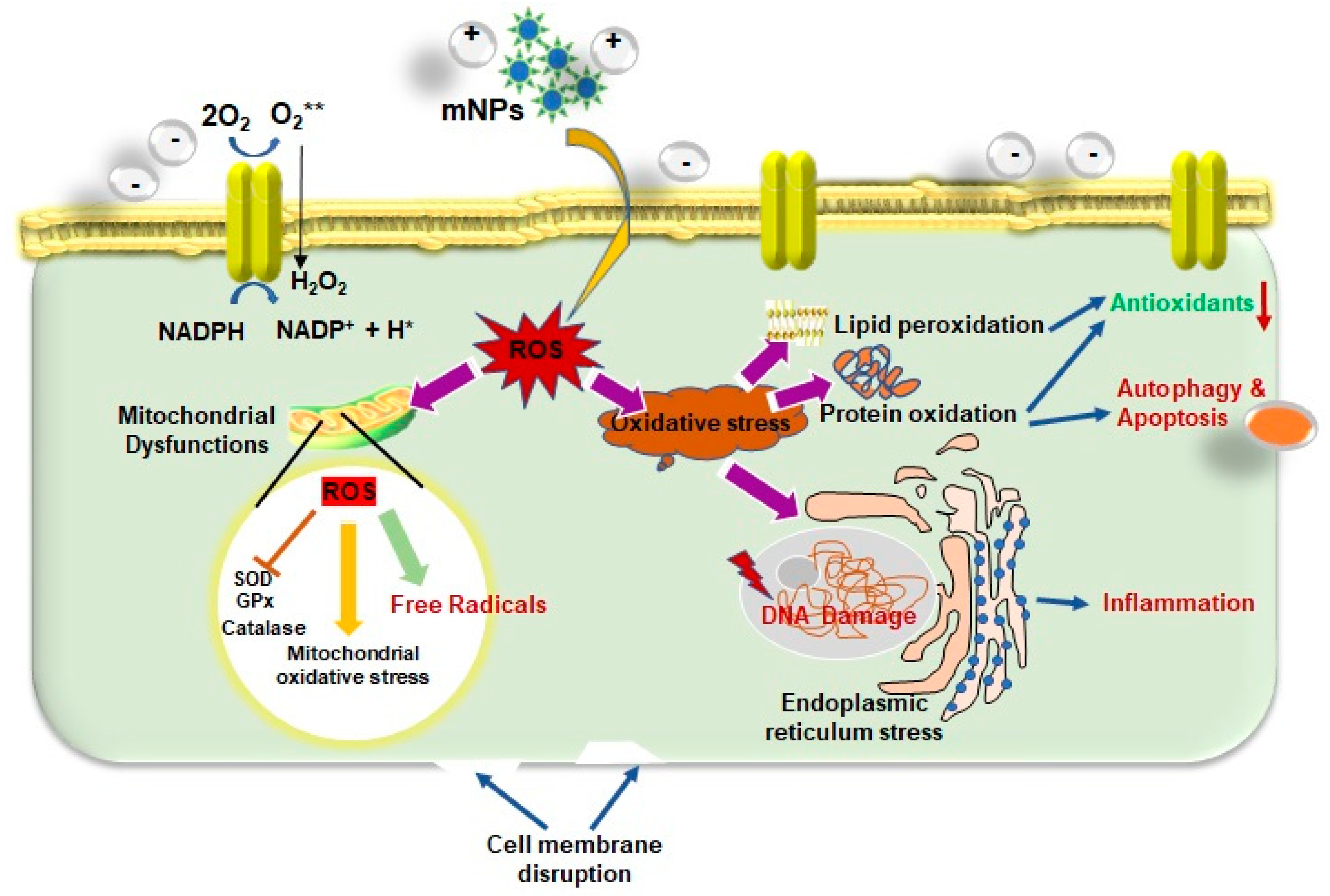
ROS serve as cell signaling agents for typical biological activities; nevertheless, the overproduction of ROS can induce oxidative stress, resulting in damage to several cellular organelles and functions, which can eventually cause disrupt normal physiology [136,137,138] as well as significant genotoxicity (Figure 4). The cellular oxidative stress is initiated by an imbalance between the generation of ROS (including the superoxide radical, hydroxyl radical, hydrogen peroxide, nitric oxide, and peroxynitrite) and lessened antioxidant defenses inside the cell, which can cause a wide range of diseases [139]. The toxicity of mNPs was assessed utilizing mitochondrial and cell membrane viability, as well as ROS, which revealed a dose-dependent reduction in cell viability. ROS can cause membrane damage, lipid denaturation, and DNA structural changes. This increases ROS levels in the mitochondria, lowering ATP that causes a flux in the tricarboxylic acid (TCA) cycle and the reduction in cardiolipin. There is evidence to suggest that mitochondria-derived ROS may be involved in the apoptosis that is brought on by TNF-α and IL-1β [140,141,142,143]. Excessive ROS generation triggers a cascade of proinflammatory cytokines and mediators through redox-sensitive MAPK and NF-B signaling pathways, which control the transcription of inflammatory genes including IL-1, IL-8, and TNF-α. Moreover, ROS directly regulate neuronal ion channels, kinases, and transcription factors, which is an essential aspect in brain development [144,145,146]. A lot of studies have found that ROS play an important role in neurodegenerative illnesses including Alzheimer’s and Parkinson’s disease.
The physical and chemical characteristics of mNPs, such as their size, chemical composition, surface area, and charge, can impact the mNPs-mediated ROS formation mechanism. The primary driving mechanisms underlying NP-induced ROS include prooxidant functional groups on the reactive surface, active redox cycling induced by transition of mNPs, and particle-cell interactions [46,147,148]. The nanoscale dimensions can enhance and change the electrical characteristics of the NP surface, producing reactive groups. The observation stated that cells exposed to 50 g/mL of 15 nm Ag NPs experienced a more than 10-fold rise in ROS levels implying that the toxicity is most likely caused by oxidative stress [2]. Typically, oxidative stress inhibits antioxidant enzymes such as catalase, superoxide dismutase, and glutathione peroxidase while depleting non-enzymatic antioxidants such as vitamin C, vitamin E, and glutathione [149,150].

4.2. Impact of mNPs-Induced Oxidative Stress

The mNPs could function as oxidants and frequently cause oxidative stress in biological systems. These mNPs can drive redox processes, which results in endogenous ROS production, leading to cause serious health hazards such as even genotoxicity. Several redox signaling pathways in cells are made up of signaling molecules like kinases and transcription factors that are negatively controlled by sensor proteins. According to conventional understanding, oxidative stress can trigger such cellular signaling by changing sensor protein thiol groups [151]. Due to their high surface-to-volume ratio, NP is reactive and susceptible to environmental stressors. After exposure to mNPs, alveolar macrophages (AM) and neutrophils triggered oxidative stress in the lungs. The immune system’s phagocytic cells, such as neutrophils, activate NADPH oxidase to produce ROS [152].
The impact of metal oxide NPs via airborne pollutants on the respiratory system was studied in vitro by exposing airway epithelial (HEp-2) cells to silicon oxide (SiO2), ferric oxide (Fe2O3), and copper oxide (CuO) nanoparticles. In comparison, CuO exposure resulted in a significant increase in the levels of 8-isoprostanes and the ratio of GSSG to total glutathione, which showed that the ROS it produced caused oxidative stress in HEp-2 cells [153]. In brief, the prooxidant attributes of copper NPs enable them to either inhibit antioxidants or produce more ROS, which increases oxidative stress. The Cu NPs inhibit cellular antioxidant enzymes like catalase and glutathione reductase while increasing glutathione peroxidase activity. This shows that copper NPs produce ROS and inhibit cell redox balance. The Cu ion’s toxicity causes severe liver damage, increased endoplasmic reticulum stress, and inducted neuronal apoptosis [154,155,156].
Iron NPs combine with hydrogen peroxide to form hydroxyl ions and redox-active iron, which releases hydroxyl radicals through the Fenton reaction. These free radicals may damage biological macromolecules and cell organelles and have been linked to various diseases. The iron NPs also promoted cytotoxicity by increasing LDH levels [157]. Due to excessive oxygen consumption, inadequate antioxidant defense, and an abundance of oxidation-sensitive lipids, the brain is particularly susceptible to oxidative damage. Neurons, unlike other damaged tissues, cannot be restored through regeneration. Moreover, neurotoxicity and increased protein aggregation were observed in mice with neuron-specific suppression of key autophagy proteins (i.e., Atg5, Atg7, and beclin-1) [158]. The activation of various pathways, such as the p38 member of the mitogen-activated protein kinases (MAPKs) family, which is directly impacted by iron accumulations and oxidative stress and could be the underlying cause of Alzheimer’s and Parkinson’s illnesses [159].
In particular, the Fenton-type reactions generate free radicals and interact with cellular macromolecules to cause oxidative stress. Fenton reactions typically involve a transition metal ion reacting with H2O2 to yield OH and an oxidized metal ion that is extremely reactive and toxic to biological molecules [143]. On the other hand, the Haber-Weiss reaction involves a reaction between an oxidized metal ion and H2O2 to generate OH. The brief investigation has shown that these free radicals produced directly or indirectly are activating the mitogen-activated protein kinase pathways. Indeed, the activation of pathways is mediated by an increase in the number of inflammatory mediators such NF-κB, signal transducer and activator of transcription (STATs), mitochondrial malfunction, and intracellular calcium [160]. Moreover, lipid hydroperoxides are produced as a result of the oxidation of polyunsaturated fatty acids at initial progress in generating ROS. Prooxidant metals like Cu and Fe subsequently react with these lipid hydroperoxides to produce DNA-damaging end products, including malondialdehyde (MDA) and 4-hydroxynonenal, which function as inflammatory mediators and carcinogenesis risk factors [161,162].
The studies demonstrated how mNPs affect oxidative DNA damage and gene expression, which can result in the development of tumors and/or have an impact on fertility. It has been shown that exposure to metal oxide NPs results in DNA fragmentation and the formation of oxidation-induced DNA adducts [160,163]. In human embryonic lung fibroblasts, it has been demonstrated that AuNPs (20 nm in size) at 1 nM concentration cause DNA damage via the formation of 8-hydroxyl-2′-deoxyguanosine (8-OHdG) DNA adduct, which is accompanied by a decreased expression of DNA repair and the cell cycle checkpoint genes MAD2, BRCA1, Hus1, ATLD/HNGS1, AT-V1/AT-V2, cyclin B1, and cyclin B2 [164]. The ionic copper (Cu2+) cytotoxicity has been correlated to DNA damage and apoptosis-mediated cell death. According to a study, Mytilus galloprovincialis short-term exposure to CuO NPs causes oxidative stress, further leading to genotoxicity and cancer development [165].

4.3. Effects of mNPs on Cellular Signaling and Immune Response

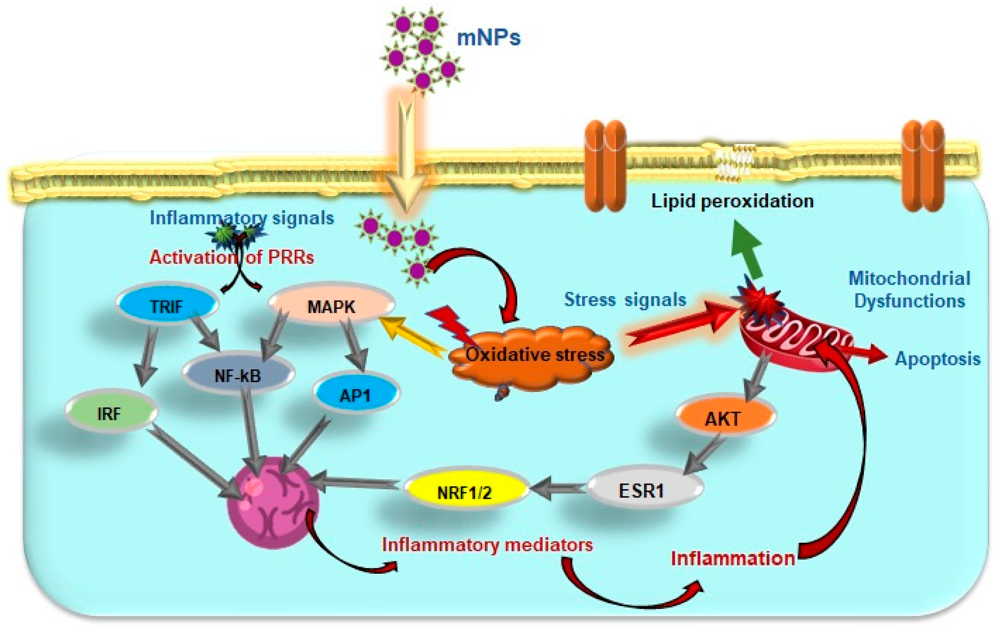
Excessive ROS production and oxidative stress can activate nuclear respiratory factor (NRF) 1 and 2 via AKT (Protein Kinase B), estrogen receptor (ESR) 1, and alter proteins function implicated in the stress response pathway such as SODs, CAT, GR, GPXs, PDI, and PRDXs [166]. The release of pro-inflammatory mediators through the cytokine cascades, and activation of signaling pathways including the NF-κB, MAPK, and interferon regulatory factor 3 (IRF3), and PI3-K pathways, indicating a reciprocal relationship between oxidative stress and inflammation (Figure 5). These active transcription factors stimulate the production of inflammatory mediators as TNFα, interferons (IFNs), IL1, nitric oxide (NO), and tumor growth factor (TGF) B3, which intensify inflammation. Through increased production of cytokines like ILs, kinase activation, and phosphatase inhibition, cells are known to suppress the overexpressed oxidative stress response, which affects the phosphorylation cascade.
Protein tyrosine phosphatases (PTP) have highly reactive cysteine residues that are susceptible to oxidative stress from H2O2, free radicals, or changes in the intracellular thiol/disulfide redox state. The mNPs, such as Mg2+, Zn2+, and V5+, may play a crucial role in the redox control of PTP by inhibiting MAPK and EGFR [167,168,169]. The effects of uncoated AuNPs and inflammatory response in rats have shown the modulation of IL-1β, IL-6, and TNF-α expression [170]. The subacute exposure to 20 nm ZnO-NPs affected the immune system in juvenile and adult BALB/c mice has shown an increase of IL-6, IFN-γ, TNF-α, and ROS in the adult mouse; however, the same molecule levels in juvenile mice did not change significantly [171].
Mitogen-activated protein kinase (MAPK) are serine-threonine protein kinases that comprise the stress-activated MAPK, c-Jun NH2-terminal kinases (JNK), and p38 MAPK. Studies on in vivo nanotoxicity using the model organism C. elegans revealed that excessive ROS production could activate MAPK pathways through the inhibition and/or degradation of MAPK phosphatases and produces free radicals that can lead to the oxidative modification of MAPK signaling proteins [172]. The studies also revealed that PMK-1, p38 MAPK, and hypoxia-inducible factors are highly expressed [173,174]. Additionally, it has been demonstrated that several metal oxide nanoparticles, including those made of iron, cadmium, silica, and zinc, cause the inflammatory cytokines NF-κB induces [175,176,177]. The inhibitor of κB (IκB) degrades during oxidative stress and activates NF-κB, which subsequently translocate into the nucleus to control the transcription of its target genes [178,179].

5. Conclusions and Further Prospective

The mNPs have a substantial beneficial effect due to their vast array of uses in the healthcare, cosmetics, and industrial sectors. Despite the prospective benefits of employing NPs in various applications, the potential health risks connected with human exposure to these mNPs have yet to be extensively reported or understood. This systematic review was presented to explore some of the critical aspects that impact the assessment of health and safety concerns associated with mNPs exposure. According to both in vivo and in vitro studies, an attempt was made to provide an overview of the cellular mechanisms of mNPs-induced oxidative stress and the adverse effects which can eventually cause disrupt normal physiology as well as significant genotoxicity. An approach called “green” synthesis of mNPs from plant extracts must be prioritized in hopes of reducing the cytotoxicity [180,181]. mNPs display significantly varied oxidative stress pathways depending on their size, surface chemistry, and structure. Thus, surface modification of nanoparticles, modified by binding different molecules to their structure to produce NPs may potentially lower nanotoxicity, however this remains under research.

Author Contributions

Conceptualization, resources, validation, Y.M.; formal analysis, investigation, G.G.D.S., Y.H. and M.K.; data curation, writing—original draft preparation, writing—review and editing, visualization, M.G.; supervision, project administration, funding acquisition, Y.-O.S. All authors have read and agreed to the published version of the manuscript.

Funding

This work was supported by the National Research Foundation of Korea (NRF), funded by the Korean government (MSIT; Grant No. 2020R1A2C2004128), and the Basic Science Research Program of the Research Institute for Basic Sciences (RIBS) of Jeju National University through the NRF funded by the Ministry of Education (Grant No. 2019R1A6A1A10072987). This research was also partly supported by the Brain Pool Program funded by the Ministry of Science and ICT through the NRF (Grant No. 2022H1D3A2A02053110).

Conflicts of Interest

The authors declare no conflict of interest.

References

  1. Fu, P.P.; Xia, Q.; Hwang, H.-M.; Ray, P.C.; Yu, H. Mechanisms of nanotoxicity: Generation of reactive oxygen species. J. Food Drug Anal. 2014, 22, 64–75. [Google Scholar] [CrossRef] [PubMed] [Green Version]
  2. Schrand, A.M.; Rahman, M.F.; Hussain, S.M.; Schlager, J.J.; Smith, D.A.; Syed, A.F. Metal-based nanoparticles and their toxicity assessment. Wiley Interdiscip. Rev. Nanomed. Nanobiotechnol. 2010, 2, 544–568. [Google Scholar] [CrossRef] [Green Version]
  3. Chusuei, C.C.; Wu, C.-H.; Mallavarapu, S.; Hou, F.Y.S.; Hsu, C.-M.; Winiarz, J.G.; Aronstam, R.S.; Huang, Y.-W. Cytotoxicity in the age of nano: The role of fourth period transition metal oxide nanoparticle physicochemical properties. Chem.-Biol. Interact. 2013, 206, 319–326. [Google Scholar] [CrossRef] [Green Version]
  4. Kreuter, J.; Gelperina, S. Use of Nanoparticles for Cerebral Cancer. Tumori J. 2008, 94, 271–277. [Google Scholar] [CrossRef]
  5. Aitken, R.J.; Chaudhry, M.; Boxall, A.; Hull, M. Manufacture and use of nanomaterials: Current status in the UK and global trends. Occup. Med. 2006, 56, 300–306. [Google Scholar] [CrossRef] [PubMed] [Green Version]
  6. Shvedova, A.A.; Pietroiusti, A.; Fadeel, B.; Kagan, V.E. Mechanisms of carbon nanotube-induced toxicity: Focus on oxidative stress. Toxicol. Appl. Pharm. 2012, 261, 121–133. [Google Scholar] [CrossRef] [PubMed] [Green Version]
  7. De Matteis, V. Exposure to Inorganic Nanoparticles: Routes of Entry, Immune Response, Biodistribution and In Vitro/In Vivo Toxicity Evaluation. Toxics 2017, 5, 29. [Google Scholar] [CrossRef] [Green Version]
  8. Casals, E.; Vázquez-Campos, S.; Bastús, N.G.; Puntes, V. Distribution and potential toxicity of engineered inorganic nanoparticles and carbon nanostructures in biological systems. TrAC Trends Anal. Chem. 2008, 27, 672–683. [Google Scholar] [CrossRef]
  9. Wang, S.-H.; Lee, C.-W.; Chiou, A.; Wei, P.-K. Size-dependent endocytosis of gold nanoparticles studied by three-dimensional mapping of plasmonic scattering images. J. Nanobiotechnol. 2010, 8, 33. [Google Scholar] [CrossRef] [PubMed] [Green Version]
  10. He, C.; Hu, Y.; Yin, L.; Tang, C.; Yin, C. Effects of particle size and surface charge on cellular uptake and biodistribution of polymeric nanoparticles. Biomaterials 2010, 31, 3657–3666. [Google Scholar] [CrossRef]
  11. Dizaj, A. A sight on the current nanoparticle-based gene delivery vectors. Nanoscale Res. Lett. 2014, 9, 1–9. [Google Scholar] [CrossRef] [PubMed] [Green Version]
  12. Tan, W.B.; Jiang, S.; Zhang, Y. Quantum-dot based nanoparticles for targeted silencing of HER2/neu gene via RNA interference. Biomaterials 2007, 28, 1565–1571. [Google Scholar] [CrossRef]
  13. Su, J.; Zhang, J.; Liu, L.; Huang, Y.; Mason, R.P. Exploring feasibility of multicolored CdTe quantum dots for in vitro and in vivo fluorescent imaging. J. Nanosci. Nanotechnol. 2008, 8, 1174–1177. [Google Scholar] [CrossRef] [PubMed]
  14. Hardman, S.a.; Cope, A.; Swann, A.; Bell, P.; Naylor, A.; Hayes, P. An in vitro model to compare the antimicrobial activity of silver-coated versus rifampicin-soaked vascular grafts. Ann. Vasc. Surg. 2004, 18, 308–313. [Google Scholar] [CrossRef] [PubMed]
  15. Lansdown, A.B. Silver in health care: Antimicrobial effects and safety in use. Biofunctional Text. Ski. 2006, 33, 17–34. [Google Scholar]
  16. Elechiguerra, J.L.; Burt, J.L.; Morones, J.R.; Camacho-Bragado, A.; Gao, X.; Lara, H.H.; Yacaman, M.J. Interaction of silver nanoparticles with HIV-1. J. Nanobiotechnol. 2005, 3, 6. [Google Scholar] [CrossRef] [Green Version]
  17. Raffi, M.; Hussain, F.; Bhatti, T.; Akhter, J.; Hameed, A.; Hasan, M. Antibacterial characterization of silver nanoparticles against E. coli ATCC-15224. J. Mater. Sci. Technol. 2008, 24, 192–196. [Google Scholar]
  18. Feng, Q.L.; Wu, J.; Chen, G.Q.; Cui, F.; Kim, T.; Kim, J. A mechanistic study of the antibacterial effect of silver ions on Escherichia coli and Staphylococcus aureus. J. Biomed. Mater. Res. 2000, 52, 662–668. [Google Scholar] [CrossRef] [PubMed]
  19. Guzman, M.; Dille, J.; Godet, S. Synthesis and antibacterial activity of silver nanoparticles against gram-positive and gram-negative bacteria. Nanomed. Nanotechnol. Biol. Med. 2012, 8, 37–45. [Google Scholar] [CrossRef] [PubMed]
  20. Chaloupka, K.; Malam, Y.; Seifalian, A.M. Nanosilver as a new generation of nanoproduct in biomedical applications. Trends Biotechnol. 2010, 28, 580–588. [Google Scholar] [CrossRef] [PubMed]
  21. Le Ouay, B.; Stellacci, F. Antibacterial activity of silver nanoparticles: A surface science insight. Nano Today 2015, 10, 339–354. [Google Scholar] [CrossRef] [Green Version]
  22. Mortezaee, K.; Najafi, M.; Samadian, H.; Barabadi, H.; Azarnezhad, A.; Ahmadi, A. Redox interactions and genotoxicity of metal-based nanoparticles: A comprehensive review. Chem.-Biol. Interact. 2019, 312, 108814. [Google Scholar] [CrossRef]
  23. Duan, D.; Fan, K.; Zhang, D.; Tan, S.; Liang, M.; Liu, Y.; Zhang, J.; Zhang, P.; Liu, W.; Qiu, X.; et al. Nanozyme-strip for rapid local diagnosis of Ebola. Biosens. Bioelectron. 2015, 74, 134–141. [Google Scholar] [CrossRef]
  24. Hanley, C.; Layne, J.; Punnoose, A.; Reddy, K.; Coombs, I.; Coombs, A.; Feris, K.; Wingett, D. Preferential killing of cancer cells and activated human T cells using ZnO nanoparticles. Nanotechnology 2008, 19, 295103. [Google Scholar] [CrossRef] [PubMed] [Green Version]
  25. Deng, Y.; Zhang, H. The synergistic effect and mechanism of doxorubicin-ZnO nanocomplexes as a multimodal agent integrating diverse anticancer therapeutics. Int. J. Nanomed. 2013, 8, 1835. [Google Scholar]
  26. Lee, S.; Yun, H.-S.; Kim, S.-H. The comparative effects of mesoporous silica nanoparticles and colloidal silica on inflammation and apoptosis. Biomaterials 2011, 32, 9434–9443. [Google Scholar] [CrossRef] [PubMed]
  27. Arruebo, M.; Fernández-Pacheco, R.; Ibarra, M.R.; Santamaría, J. Magnetic nanoparticles for drug delivery. Nano Today 2007, 2, 22–32. [Google Scholar] [CrossRef]
  28. Na, H.B.; Song, I.C.; Hyeon, T. Inorganic nanoparticles for MRI contrast agents. Adv. Mater. 2009, 21, 2133–2148. [Google Scholar] [CrossRef]
  29. McBain, S.C.; Yiu, H.H.; Dobson, J. Magnetic nanoparticles for gene and drug delivery. Int. J. Nanomed. 2008, 3, 169–180. [Google Scholar] [CrossRef] [Green Version]
  30. Hua, X.; Yang, Q.; Dong, Z.; Zhang, J.; Zhang, W.; Wang, Q.; Tan, S.; Smyth, H.D. Magnetically triggered drug release from nanoparticles and its applications in anti-tumor treatment. Drug Deliv. 2017, 24, 511–518. [Google Scholar] [CrossRef] [Green Version]
  31. Corot, C.; Robert, P.; Idée, J.-M.; Port, M. Recent advances in iron oxide nanocrystal technology for medical imaging. Adv. Drug Deliv. Rev. 2006, 58, 1471–1504. [Google Scholar] [CrossRef]
  32. Gupta, A.K.; Gupta, M. Synthesis and surface engineering of iron oxide nanoparticles for biomedical applications. Biomaterials 2005, 26, 3995–4021. [Google Scholar] [CrossRef] [PubMed]
  33. Canaparo, R.; Foglietta, F.; Limongi, T.; Serpe, L. Biomedical Applications of Reactive Oxygen Species Generation by Metal Nanoparticles. Materials 2021, 14, 53. [Google Scholar] [CrossRef]
  34. Ivanova, P.; Dzięgielewski, K.; Drozd, M.; Skorupska, S.; Grabowska-Jadach, I.; Pietrzak, M. Nanoparticles of chosen noble metals as reactive oxygen species scavengers. Nanotechnology 2021, 32, 055704. [Google Scholar] [CrossRef] [PubMed]
  35. Alaraby, M.; Hernández, A.; Marcos, R. Systematic in vivo study of NiO nanowires and nanospheres: Biodegradation, uptake and biological impacts. Nanotoxicology 2018, 12, 1027–1044. [Google Scholar] [CrossRef] [PubMed]
  36. Chanda, N.; Kan, P.; Watkinson, L.D.; Shukla, R.; Zambre, A.; Carmack, T.L.; Engelbrecht, H.; Lever, J.R.; Katti, K.; Fent, G.M. Radioactive gold nanoparticles in cancer therapy: Therapeutic efficacy studies of GA-198AuNP nanoconstruct in prostate tumor-bearing mice. In Nanomedicine in Cancer; Jenny Stanford Publishing: Dubai, United Arab Emirates, 2017; pp. 753–774. [Google Scholar]
  37. Xie, H.; Wang, Z.J.; Bao, A.; Goins, B.; Phillips, W.T. In vivo PET imaging and biodistribution of radiolabeled gold nanoshells in rats with tumor xenografts. Int. J. Pharm. 2010, 395, 324–330. [Google Scholar] [CrossRef]
  38. Oberdörster, G.; Oberdörster, E.; Oberdörster, J. Nanotoxicology: An emerging discipline evolving from studies of ultrafine particles. Env. Health Perspect 2005, 113, 823–839. [Google Scholar] [CrossRef] [PubMed]
  39. Nel, A.; Xia, T.; Madler, L.; Li, N. Toxic potential of materials at the nanolevel. Science 2006, 311, 622–627. [Google Scholar] [CrossRef] [PubMed] [Green Version]
  40. Petersen, E.J.; Nelson, B.C. Mechanisms and measurements of nanomaterial-induced oxidative damage to DNA. Anal. Bioanal. Chem. 2010, 398, 613–650. [Google Scholar] [CrossRef]
  41. Dhawan, A.; Sharma, V. Toxicity assessment of nanomaterials: Methods and challenges. Anal. Bioanal. Chem. 2010, 398, 589–605. [Google Scholar] [CrossRef] [PubMed]
  42. Makhdoumi, P.; Karimi, H.; Khazaei, M. Review on Metal-Based Nanoparticles: Role of Reactive Oxygen Species in Renal Toxicity. Chem. Res. Toxicol. 2020, 33, 2503–2514. [Google Scholar] [CrossRef]
  43. AshRrani, P.; Low Kah Mun, G.; Hande, M.; Valiyaveettil, S. Cytotoxicity and genotoxicity of silver nanoparticles in human cells. ACS Nano 2009, 3, 279–290. [Google Scholar] [CrossRef] [PubMed]
  44. Lee, Y.-H.; Cheng, F.-Y.; Chiu, H.-W.; Tsai, J.-C.; Fang, C.-Y.; Chen, C.-W.; Wang, Y.-J. Cytotoxicity, oxidative stress, apoptosis and the autophagic effects of silver nanoparticles in mouse embryonic fibroblasts. Biomaterials 2014, 35, 4706–4715. [Google Scholar] [CrossRef] [PubMed]
  45. Carlson, C.; Hussain, S.M.; Schrand, A.M.; Braydich-Stolle, L.K.; Hess, K.L.; Jones, R.L.; Schlager, J.J. Unique cellular interaction of silver nanoparticles: Size-dependent generation of reactive oxygen species. J. Phys. Chem. B 2008, 112, 13608–13619. [Google Scholar] [CrossRef] [PubMed]
  46. Knaapen, A.M.; Borm, P.J.; Albrecht, C.; Schins, R.P. Inhaled particles and lung cancer. Part A: Mechanisms. Int. J. Cancer 2004, 109, 799–809. [Google Scholar] [CrossRef]
  47. Kovacic, P.; Somanathan, R. Biomechanisms of nanoparticles (toxicants, antioxidants and therapeutics): Electron transfer and reactive oxygen species. J. Nanosci. Nanotechnol. 2010, 10, 7919–7930. [Google Scholar] [CrossRef]
  48. Li, J.J.; Muralikrishnan, S.; Ng, C.T.; Yung, L.Y.; Bay, B.H. Nanoparticle-induced pulmonary toxicity. Exp. Biol. Med. 2010, 235, 1025–1033. [Google Scholar] [CrossRef]
  49. Gojova, A.; Guo, B.; Kota, R.S.; Rutledge, J.C.; Kennedy, I.M.; Barakat, A.I. Induction of inflammation in vascular endothelial cells by metal oxide nanoparticles: Effect of particle composition. Environ. Health Perspect. 2007, 115, 403–409. [Google Scholar] [CrossRef] [PubMed]
  50. AshaRani, P.; Sethu, S.; Lim, H.K.; Balaji, G.; Valiyaveettil, S.; Hande, M.P. Differential regulation of intracellular factors mediating cell cycle, DNA repair and inflammation following exposure to silver nanoparticles in human cells. Genome Integr. 2012, 3, 2. [Google Scholar] [CrossRef] [PubMed] [Green Version]
  51. Yu, Z.; Li, Q.; Wang, J.; Yu, Y.; Wang, Y.; Zhou, Q.; Li, P. Reactive Oxygen Species-Related Nanoparticle Toxicity in the Biomedical Field. Nanoscale Res. Lett. 2020, 15, 115. [Google Scholar] [CrossRef]
  52. Liu, Y.; Li, J.; Xu, K.; Gu, J.; Huang, L.; Zhang, L.; Liu, N.; Kong, J.; Xing, M.; Zhang, L. Characterization of superparamagnetic iron oxide nanoparticle-induced apoptosis in PC12 cells and mouse hippocampus and striatum. Toxicol. Lett. 2018, 292, 151–161. [Google Scholar] [CrossRef] [PubMed]
  53. Chen, L.; Zhang, B.; Toborek, M. Autophagy is involved in nanoalumina-induced cerebrovascular toxicity. Nanomed. Nanotechnol. Biol. Med. 2013, 9, 212–221. [Google Scholar] [CrossRef] [Green Version]
  54. Takenaka, S.; Karg, E.; Roth, C.; Schulz, H.; Ziesenis, A.; Heinzmann, U.; Schramel, P.; Heyder, J. Pulmonary and systemic distribution of inhaled ultrafine silver particles in rats. Environ. Health Perspect. 2001, 109, 547–551. [Google Scholar] [PubMed] [Green Version]
  55. Kakkar, V.; Kaur, I.P. Evaluating potential of curcumin loaded solid lipid nanoparticles in aluminium induced behavioural, biochemical and histopathological alterations in mice brain. Food Chem. Toxicol. 2011, 49, 2906–2913. [Google Scholar] [CrossRef] [PubMed]
  56. Hopkins, L.E.; Patchin, E.S.; Chiu, P.-L.; Brandenberger, C.; Smiley-Jewell, S.; Pinkerton, K.E. Nose-to-brain transport of aerosolised quantum dots following acute exposure. Nanotoxicology 2014, 8, 885–893. [Google Scholar] [CrossRef] [Green Version]
  57. Liu, Y.; Gao, Y.; Liu, Y.; Li, B.; Chen, C.; Wu, G. Oxidative stress and acute changes in murine brain tissues after nasal instillation of copper particles with different sizes. J. Nanosci. Nanotechnol. 2014, 14, 4534–4540. [Google Scholar] [CrossRef]
  58. Elder, A.; Gelein, R.; Silva, V.; Feikert, T.; Opanashuk, L.; Carter, J.; Potter, R.; Maynard, A.; Ito, Y.; Finkelstein, J. Translocation of inhaled ultrafine manganese oxide particles to the central nervous system. Environ. Health Perspect. 2006, 114, 1172–1178. [Google Scholar] [CrossRef] [PubMed] [Green Version]
  59. Li, Y.; Li, J.; Yin, J.; Li, W.; Kang, C.; Huang, Q.; Li, Q. Systematic influence induced by 3 nm titanium dioxide following intratracheal instillation of mice. J. Nanosci. Nanotechnol. 2010, 10, 8544–8549. [Google Scholar] [CrossRef]
  60. Li, X.-b.; Zheng, H.; Zhang, Z.-r.; Li, M.; Huang, Z.-y.; Schluesener, H.J.; Li, Y.-y.; Xu, S.-q. Glia activation induced by peripheral administration of aluminum oxide nanoparticles in rat brains. Nanomed. Nanotechnol. Biol. Med. 2009, 5, 473–479. [Google Scholar] [CrossRef] [PubMed]
  61. Takács, S.; Szabó, A.; Oszlánczi, G.; Pusztai, P.; Sápi, A.; Kónya, Z.; Papp, A. Repeated simultaneous cortical electrophysiological and behavioral recording in rats exposed to manganese-containing nanoparticles. Acta Biol. Hung. 2012, 63, 426–440. [Google Scholar] [CrossRef]
  62. Oszlánczi, G.; Papp, A.; Szabó, A.; Nagymajtényi, L.; Sápi, A.; Kónya, Z.; Paulik, E.; Vezér, T. Nervous system effects in rats on subacute exposure by lead-containing nanoparticles via the airways. Inhal. Toxicol. 2011, 23, 173–181. [Google Scholar] [CrossRef]
  63. De Simone, U.; Roccio, M.; Gribaldo, L.; Spinillo, A.; Caloni, F.; Coccini, T. Human 3D cultures as models for evaluating magnetic nanoparticle CNS cytotoxicity after short-and repeated long-term exposure. Int. J. Mol. Sci. 2018, 19, 1993. [Google Scholar] [CrossRef] [PubMed] [Green Version]
  64. Xu, F.; Piett, C.; Farkas, S.; Qazzaz, M.; Syed, N.I. Silver nanoparticles (AgNPs) cause degeneration of cytoskeleton and disrupt synaptic machinery of cultured cortical neurons. Mol. Brain 2013, 6, 29. [Google Scholar] [CrossRef] [PubMed] [Green Version]
  65. Thomsen, L.B.; Linemann, T.; Pondman, K.M.; Lichota, J.; Kim, K.S.; Pieters, R.J.; Visser, G.M.; Moos, T. Uptake and transport of superparamagnetic iron oxide nanoparticles through human brain capillary endothelial cells. ACS Chem. Neurosci. 2013, 4, 1352–1360. [Google Scholar] [CrossRef] [PubMed] [Green Version]
  66. Feng, X.; Chen, A.; Zhang, Y.; Wang, J.; Shao, L.; Wei, L. Central nervous system toxicity of metallic nanoparticles. Int. J. Nanomed. 2015, 10, 4321–4340. [Google Scholar] [CrossRef] [Green Version]
  67. Shimizu, M.; Tainaka, H.; Oba, T.; Mizuo, K.; Umezawa, M.; Takeda, K. Maternal exposure to nanoparticulate titanium dioxide during the prenatal period alters gene expression related to brain development in the mouse. Part. Fibre Toxicol. 2009, 6, 20. [Google Scholar] [CrossRef] [PubMed] [Green Version]
  68. Takeda, K.; Suzuki, K.-i.; Ishihara, A.; Kubo-Irie, M.; Fujimoto, R.; Tabata, M.; Oshio, S.; Nihei, Y.; Ihara, T.; Sugamata, M. Nanoparticles transferred from pregnant mice to their offspring can damage the genital and cranial nerve systems. J. Health Sci. 2009, 55, 95–102. [Google Scholar] [CrossRef] [Green Version]
  69. Hougaard, K.S.; Jackson, P.; Jensen, K.A.; Sloth, J.J.; Löschner, K.; Larsen, E.H.; Birkedal, R.K.; Vibenholt, A.; Boisen, A.-M.Z.; Wallin, H.; et al. Effects of prenatal exposure to surface-coated nanosized titanium dioxide (UV-Titan). A study in mice. Part. Fibre Toxicol. 2010, 7, 16. [Google Scholar] [CrossRef] [Green Version]
  70. Kan, H.; Pan, D.; Castranova, V. Engineered nanoparticle exposure and cardiovascular effects: The role of a neuronal-regulated pathway. Inhal. Toxicol. 2018, 30, 335–342. [Google Scholar] [CrossRef] [PubMed]
  71. Zhang, J.; Xue, Y.; Ni, Y.; Ning, F.; Shang, L.; Ma, A. Size dependent effects of Gold Nanoparticles in ISO-induced Hyperthyroid Rats. Sci. Rep. 2018, 8, 10960. [Google Scholar] [CrossRef] [Green Version]
  72. Yousef, M.I.; Abuzreda, A.A.; Kamel, M.A. Cardiotoxicity and lung toxicity in male rats induced by long-term exposure to iron oxide and silver nanoparticles. Exp. Ther. Med. 2019, 18, 4329–4339. [Google Scholar] [CrossRef] [PubMed] [Green Version]
  73. Park, J.; Park, C.; Lee, Y.; Ryu, C.; Park, J.; Kim, Y. Acute Adverse Effects of Metallic Nanomaterials on Cardiac and Behavioral Changes in Daphnia magna. Environments 2022, 9, 26. [Google Scholar] [CrossRef]
  74. Li, J.; Chen, C.; Xia, T. Understanding Nanomaterial-Liver Interactions to Facilitate the Development of Safer Nanoapplications. Adv. Mater. 2022, 34, e2106456. [Google Scholar] [CrossRef] [PubMed]
  75. Zhang, Y.N.; Poon, W.; Tavares, A.J.; McGilvray, I.D.; Chan, W.C.W. Nanoparticle-liver interactions: Cellular uptake and hepatobiliary elimination. J. Control. Release 2016, 240, 332–348. [Google Scholar] [CrossRef]
  76. Thomson, A.W.; Knolle, P.A. Antigen-presenting cell function in the tolerogenic liver environment. Nat. Rev. Immunol. 2010, 10, 753–766. [Google Scholar] [CrossRef] [PubMed]
  77. Sayes, C.M.; Reed, K.L.; Warheit, D.B. Assessing toxicity of fine and nanoparticles: Comparing in vitro measurements to in vivo pulmonary toxicity profiles. Toxicol. Sci. 2007, 97, 163–180. [Google Scholar] [CrossRef] [Green Version]
  78. Warheit, D.B.; Sayes, C.M.; Reed, K.L.; Swain, K.A. Health effects related to nanoparticle exposures: Environmental, health and safety considerations for assessing hazards and risks. Pharmacol. Ther. 2008, 120, 35–42. [Google Scholar] [CrossRef]
  79. Jia, J.; Li, F.; Zhou, H.; Bai, Y.; Liu, S.; Jiang, Y.; Jiang, G.; Yan, B. Oral Exposure to Silver Nanoparticles or Silver Ions May Aggravate Fatty Liver Disease in Overweight Mice. Env. Sci. Technol. 2017, 51, 9334–9343. [Google Scholar] [CrossRef] [PubMed]
  80. Abdelhalim, M.A.; Jarrar, B.M. Gold nanoparticles administration induced prominent inflammatory, central vein intima disruption, fatty change and Kupffer cells hyperplasia. Lipids Health Dis. 2011, 10, 133. [Google Scholar] [CrossRef] [Green Version]
  81. Duan, J.; Liang, S.; Feng, L.; Yu, Y.; Sun, Z. Silica nanoparticles trigger hepatic lipid-metabolism disorder in vivo and in vitro. Int. J. Nanomed. 2018, 13, 7303–7318. [Google Scholar] [CrossRef] [Green Version]
  82. Yu, Y.; Duan, J.; Li, Y.; Li, Y.; Jing, L.; Yang, M.; Wang, J.; Sun, Z. Silica nanoparticles induce liver fibrosis via TGF-β(1)/Smad3 pathway in ICR mice. Int. J. Nanomed. 2017, 12, 6045–6057. [Google Scholar] [CrossRef] [PubMed] [Green Version]
  83. Parivar, K.; Fard, F.M.; Bayat, M.; Alavian, S.M.; Motavaf, M. Evaluation of iron oxide nanoparticles toxicity on liver cells of BALB/c rats. Iran. Red Crescent Med. J. 2016, 18, e28939. [Google Scholar] [CrossRef] [PubMed] [Green Version]
  84. Yan, X.; Xu, X.; Guo, M.; Wang, S.; Gao, S.; Zhu, S.; Rong, R. Synergistic toxicity of zno nanoparticles and dimethoate in mice: Enhancing their biodistribution by synergistic binding of serum albumin and dimethoate to zno nanoparticles. Environ. Toxicol. 2017, 32, 1202–1212. [Google Scholar] [CrossRef] [PubMed]
  85. Waters, K.M.; Masiello, L.M.; Zangar, R.C.; Tarasevich, B.J.; Karin, N.J.; Quesenberry, R.D.; Bandyopadhyay, S.; Teeguarden, J.G.; Pounds, J.G.; Thrall, B.D. Macrophage responses to silica nanoparticles are highly conserved across particle sizes. Toxicol. Sci. 2009, 107, 553–569. [Google Scholar] [CrossRef] [PubMed] [Green Version]
  86. Chen, X.; Zhouhua, W.; Jie, Z.; Xinlu, F.; Jinqiang, L.; Yuwen, Q.; Zhiying, H. Renal interstitial fibrosis induced by high-dose mesoporous silica nanoparticles via the NF-κB signaling pathway. Int. J. Nanomed. 2015, 10, 1–22. [Google Scholar] [CrossRef] [PubMed] [Green Version]
  87. Mahmoud, A.M.; Desouky, E.M.; Hozayen, W.G.; Bin-Jumah, M.; El-Nahass, E.-S.; Soliman, H.A.; Farghali, A.A. Mesoporous Silica Nanoparticles Trigger Liver and Kidney Injury and Fibrosis Via Altering TLR4/NF-κB, JAK2/STAT3 and Nrf2/HO-1 Signaling in Rats. Biomolecules 2019, 9, 528. [Google Scholar] [CrossRef] [PubMed] [Green Version]
  88. Jia, X.; Wang, S.; Zhou, L.; Sun, L. The potential liver, brain, and embryo toxicity of titanium dioxide nanoparticles on mice. Nanoscale Res. Lett. 2017, 12, 478. [Google Scholar] [CrossRef]
  89. Chen, J.; Dong, X.; Zhao, J.; Tang, G. In vivo acute toxicity of titanium dioxide nanoparticles to mice after intraperitioneal injection. J. Appl. Toxicol. 2009, 29, 330–337. [Google Scholar] [CrossRef]
  90. Milić, M.; Leitinger, G.; Pavičić, I.; Zebić Avdičević, M.; Dobrović, S.; Goessler, W.; Vinković Vrček, I. Cellular uptake and toxicity effects of silver nanoparticles in mammalian kidney cells. J. Appl. Toxicol. 2015, 35, 581–592. [Google Scholar] [CrossRef] [PubMed] [Green Version]
  91. Ferdous, Z.; Nemmar, A. Health Impact of Silver Nanoparticles: A Review of the Biodistribution and Toxicity Following Various Routes of Exposure. Int. J. Mol. Sci. 2020, 21, 2375. [Google Scholar] [CrossRef] [Green Version]
  92. Axson, J.L.; Stark, D.I.; Bondy, A.L.; Capracotta, S.S.; Maynard, A.D.; Philbert, M.A.; Bergin, I.L.; Ault, A.P. Rapid kinetics of size and pH-dependent dissolution and aggregation of silver nanoparticles in simulated gastric fluid. J. Phys. Chem. C 2015, 119, 20632–20641. [Google Scholar] [CrossRef] [Green Version]
  93. Nosrati, H.; Hamzepoor, M.; Sohrabi, M.; Saidijam, M.; Assari, M.J.; Shabab, N.; Gholami Mahmoudian, Z.; Alizadeh, Z. The potential renal toxicity of silver nanoparticles after repeated oral exposure and its underlying mechanisms. BMC Nephrol. 2021, 22, 228. [Google Scholar] [CrossRef]
  94. Ray, P.C.; Yu, H.; Fu, P.P. Toxicity and environmental risks of nanomaterials: Challenges and future needs. J. Environ. Sci. Health Part C 2009, 27, 1–35. [Google Scholar] [CrossRef] [Green Version]
  95. Connor, E.E.; Mwamuka, J.; Gole, A.; Murphy, C.J.; Wyatt, M.D. Gold nanoparticles are taken up by human cells but do not cause acute cytotoxicity. Small 2005, 1, 325–327. [Google Scholar] [CrossRef]
  96. Griffitt, R.J.; Luo, J.; Gao, J.; Bonzongo, J.C.; Barber, D.S. Effects of particle composition and species on toxicity of metallic nanomaterials in aquatic organisms. Environ. Toxicol. Chem. Int. J. 2008, 27, 1972–1978. [Google Scholar] [CrossRef] [PubMed]
  97. He, W.; Zhou, Y.-T.; Wamer, W.G.; Boudreau, M.D.; Yin, J.-J. Mechanisms of the pH dependent generation of hydroxyl radicals and oxygen induced by Ag nanoparticles. Biomaterials 2012, 33, 7547–7555. [Google Scholar] [CrossRef] [PubMed]
  98. Özel, R.E.; Alkasir, R.S.; Ray, K.; Wallace, K.N.; Andreescu, S. Comparative evaluation of intestinal nitric oxide in embryonic zebrafish exposed to metal oxide nanoparticles. Small 2013, 9, 4250–4261. [Google Scholar] [CrossRef]
  99. Wang, Y.; Aker, W.G.; Hwang, H.-m.; Yedjou, C.G.; Yu, H.; Tchounwou, P.B. A study of the mechanism of in vitro cytotoxicity of metal oxide nanoparticles using catfish primary hepatocytes and human HepG2 cells. Sci. Total Environ. 2011, 409, 4753–4762. [Google Scholar] [CrossRef] [Green Version]
  100. Wang, Z.; Li, J.; Zhao, J.; Xing, B. Toxicity and internalization of CuO nanoparticles to prokaryotic alga Microcystis aeruginosa as affected by dissolved organic matter. Environ. Sci. Technol. 2011, 45, 6032–6040. [Google Scholar] [CrossRef] [PubMed]
  101. Sohaebuddin, S.K.; Thevenot, P.T.; Baker, D.; Eaton, J.W.; Tang, L. Nanomaterial cytotoxicity is composition, size, and cell type dependent. Part. Fibre Toxicol. 2010, 7, 22. [Google Scholar] [CrossRef] [PubMed] [Green Version]
  102. Hillyer, J.F.; Albrecht, R.M. Gastrointestinal persorption and tissue distribution of differently sized colloidal gold nanoparticles. J. Pharm. Sci. 2001, 90, 1927–1936. [Google Scholar] [CrossRef] [PubMed]
  103. Wang, S.; Lu, W.; Tovmachenko, O.; Rai, U.S.; Yu, H.; Ray, P.C. Challenge in Understanding Size and Shape Dependent Toxicity of Gold Nanomaterials in Human Skin Keratinocytes. Chem. Phys. Lett. 2008, 463, 145–149. [Google Scholar] [CrossRef] [Green Version]
  104. Goodman, C.M.; McCusker, C.D.; Yilmaz, T.; Rotello, V.M. Toxicity of Gold Nanoparticles Functionalized with Cationic and Anionic Side Chains. Bioconj. Chem. 2004, 15, 897–900. [Google Scholar] [CrossRef] [PubMed]
  105. Pernodet, N.; Fang, X.; Sun, Y.; Bakhtina, A.; Ramakrishnan, A.; Sokolov, J.; Ulman, A.; Rafailovich, M. Adverse effects of citrate/gold nanoparticles on human dermal fibroblasts. Small 2006, 2, 766–773. [Google Scholar] [CrossRef]
  106. Yen, H.J.; Hsu, S.H.; Tsai, C.L. Cytotoxicity and immunological response of gold and silver nanoparticles of different sizes. Small 2009, 5, 1553–1561. [Google Scholar] [CrossRef] [PubMed]
  107. Pan, Y.; Neuss, S.; Leifert, A.; Fischler, M.; Wen, F.; Simon, U.; Schmid, G.; Brandau, W.; Jahnen-Dechent, W. Size-dependent cytotoxicity of gold nanoparticles. Small 2007, 3, 1941–1949. [Google Scholar] [CrossRef] [PubMed]
  108. Fukami, G.; Hashimoto, K.; Koike, K.; Okamura, N.; Shimizu, E.; Iyo, M. Effect of antioxidant N-acetyl-L-cysteine on behavioral changes and neurotoxicity in rats after administration of methamphetamine. Brain Res. 2004, 1016, 90–95. [Google Scholar] [CrossRef] [PubMed]
  109. Cai, D.; Blair, D.; Dufort, F.J.; Gumina, M.R.; Huang, Z.; Hong, G.; Wagner, D.; Canahan, D.; Kempa, K.; Ren, Z.F.; et al. Interaction between carbon nanotubes and mammalian cells: Characterization by flow cytometry and application. Nanotechnology 2008, 19, 345102. [Google Scholar] [CrossRef] [Green Version]
  110. Asharani, P.V.; Lian Wu, Y.; Gong, Z.; Valiyaveettil, S. Toxicity of silver nanoparticles in zebrafish models. Nanotechnology 2008, 19, 255102. [Google Scholar] [CrossRef] [PubMed]
  111. Kim, Y.S.; Kim, J.S.; Cho, H.S.; Rha, D.S.; Kim, J.M.; Park, J.D.; Choi, B.S.; Lim, R.; Chang, H.K.; Chung, Y.H.; et al. Twenty-eight-day oral toxicity, genotoxicity, and gender-related tissue distribution of silver nanoparticles in Sprague-Dawley rats. Inhal. Toxicol. 2008, 20, 575–583. [Google Scholar] [CrossRef]
  112. Kim, T.H.; Kim, M.; Park, H.S.; Shin, U.S.; Gong, M.S.; Kim, H.W. Size-dependent cellular toxicity of silver nanoparticles. J. Biomed. Mater. Res. A 2012, 100, 1033–1043. [Google Scholar] [CrossRef] [PubMed]
  113. Mirsattari, S.; Hammond, R.; Sharpe, M.; Leung, F.; Young, G. Myoclonic status epilepticus following repeated oral ingestion of colloidal silver. Neurology 2004, 62, 1408–1410. [Google Scholar] [CrossRef]
  114. Cioffi, N.; Torsi, L.; Ditaranto, N.; Tantillo, G.; Ghibelli, L.; Sabbatini, L.; Bleve-Zacheo, T.; D’Alessio, M.; Zambonin, P.G.; Traversa, E. Copper nanoparticle/polymer composites with antifungal and bacteriostatic properties. Chem. Mater. 2005, 17, 5255–5262. [Google Scholar] [CrossRef]
  115. Chen, Z.; Meng, H.; Xing, G.; Chen, C.; Zhao, Y.; Jia, G.; Wang, T.; Yuan, H.; Ye, C.; Zhao, F.; et al. Acute toxicological effects of copper nanoparticles in vivo. Toxicol. Lett. 2006, 163, 109–120. [Google Scholar] [CrossRef] [PubMed]
  116. Suzuki, H.; Toyooka, T.; Ibuki, Y. Simple and easy method to evaluate uptake potential of nanoparticles in mammalian cells using a flow cytometric light scatter analysis. Environ. Sci. Technol. 2007, 41, 3018–3024. [Google Scholar] [CrossRef]
  117. Zhang, T.; Zhu, X.; Guo, J.; Gu, A.Z.; Li, D.; Chen, J. Toxicity Assessment of Nano-ZnO Exposure on the Human Intestinal Microbiome, Metabolic Functions, and Resistome Using an In Vitro Colon Simulator. Environ. Sci. Technol. 2021, 55, 6884–6896. [Google Scholar] [CrossRef] [PubMed]
  118. Du, Z.; Chai, X.; Li, X.; Ren, G.; Yang, X.; Yang, Z. Nano-CuO causes cell damage through activation of dose-dependent autophagy and mitochondrial lncCyt b-AS/ND5-AS/ND6-AS in SH-SY5Y cells. Toxicol. Mech. Methods 2022, 32, 37–48. [Google Scholar] [CrossRef] [PubMed]
  119. Lin, Z.; Ma, L.; X, Z.-g.; Zhang, H.; Lin, B. A comparative study of lung toxicity in rats induced by three types of nanomaterials. Nanoscale Res. Lett. 2013, 8, 521. [Google Scholar] [CrossRef] [Green Version]
  120. Khanna, P.; Ong, C.; Bay, B.H.; Baeg, G.H. Nanotoxicity: An Interplay of Oxidative Stress, Inflammation and Cell Death. Nanomaterials 2015, 5, 1163–1180. [Google Scholar] [CrossRef] [PubMed] [Green Version]
  121. Jeng, H.A.; Swanson, J. Toxicity of metal oxide nanoparticles in mammalian cells. J. Environ. Sci. Health Part A 2006, 41, 2699–2711. [Google Scholar] [CrossRef]
  122. Grassian, V.H.; O’Shaughnessy, P.T.; Adamcakova-Dodd, A.; Pettibone, J.M.; Thorne, P.S. Inhalation exposure study of titanium dioxide nanoparticles with a primary particle size of 2 to 5 nm. Environ. Health Perspect. 2007, 115, 397–402. [Google Scholar] [CrossRef] [PubMed] [Green Version]
  123. Sayes, C.M.; Wahi, R.; Kurian, P.A.; Liu, Y.; West, J.L.; Ausman, K.D.; Warheit, D.B.; Colvin, V.L. Correlating nanoscale titania structure with toxicity: A cytotoxicity and inflammatory response study with human dermal fibroblasts and human lung epithelial cells. Toxicol. Sci. 2006, 92, 174–185. [Google Scholar] [CrossRef] [PubMed] [Green Version]
  124. Gurr, J.R.; Wang, A.S.; Chen, C.H.; Jan, K.Y. Ultrafine titanium dioxide particles in the absence of photoactivation can induce oxidative damage to human bronchial epithelial cells. Toxicology 2005, 213, 66–73. [Google Scholar] [CrossRef]
  125. Kim, J.S.; Yoon, T.J.; Yu, K.N.; Kim, B.G.; Park, S.J.; Kim, H.W.; Lee, K.H.; Park, S.B.; Lee, J.K.; Cho, M.H. Toxicity and tissue distribution of magnetic nanoparticles in mice. Toxicol. Sci. 2006, 89, 338–347. [Google Scholar] [CrossRef]
  126. Lee, J.W.; Choi, H.; Hwang, U.K.; Kang, J.C.; Kang, Y.J.; Kim, K.I.; Kim, J.H. Toxic effects of lead exposure on bioaccumulation, oxidative stress, neurotoxicity, and immune responses in fish: A review. Env. Toxicol. Pharm. 2019, 68, 101–108. [Google Scholar] [CrossRef]
  127. Lademann, J.; Weigmann, H.; Rickmeyer, C.; Barthelmes, H.; Schaefer, H.; Mueller, G.; Sterry, W. Penetration of titanium dioxide microparticles in a sunscreen formulation into the horny layer and the follicular orifice. Ski. Pharm. Appl. Ski. Physiol. 1999, 12, 247–256. [Google Scholar] [CrossRef]
  128. Zhou, Y.; Yokel, R.A. The chemical species of aluminum influences its paracellular flux across and uptake into Caco-2 cells, a model of gastrointestinal absorption. Toxicol. Sci. 2005, 87, 15–26. [Google Scholar] [CrossRef] [Green Version]
  129. Hoet, P.H.; Brüske-Hohlfeld, I.; Salata, O.V. Nanoparticles–known and unknown health risks. J. Nanobiotechnol. 2004, 2, 12. [Google Scholar] [CrossRef] [PubMed] [Green Version]
  130. Sharma, H.S.; Sharma, A. Nanoparticles aggravate heat stress induced cognitive deficits, blood-brain barrier disruption, edema formation and brain pathology. Prog. Brain Res. 2007, 162, 245–273. [Google Scholar] [CrossRef]
  131. Chen, D.; Xi, T.; Bai, J. Biological effects induced by nanosilver particles: In vivo study. Biomed. Mater. 2007, 2, S126–S128. [Google Scholar] [CrossRef] [PubMed]
  132. Chen, J.; Patil, S.; Seal, S.; McGinnis, J.F. Rare earth nanoparticles prevent retinal degeneration induced by intracellular peroxides. Nat. Nanotechnol. 2006, 1, 142–150. [Google Scholar] [CrossRef]
  133. Kashiwada, S. Distribution of nanoparticles in the see-through medaka (Oryzias latipes). Environ. Health Perspect. 2006, 114, 1697–1702. [Google Scholar] [CrossRef] [PubMed] [Green Version]
  134. Meena, R.; Kumar, S.; Paulraj, R. Titanium oxide (TiO2) nanoparticles in induction of apoptosis and inflammatory response in brain. J. Nanopart. Res. 2015, 17, 49. [Google Scholar] [CrossRef]
  135. Hu, R.; Zheng, L.; Zhang, T.; Gao, G.; Cui, Y.; Cheng, Z.; Cheng, J.; Hong, M.; Tang, M.; Hong, F. Molecular mechanism of hippocampal apoptosis of mice following exposure to titanium dioxide nanoparticles. J. Hazard Mater. 2011, 191, 32–40. [Google Scholar] [CrossRef] [PubMed]
  136. Auten, R.L.; Davis, J.M. Oxygen Toxicity and Reactive Oxygen Species: The Devil Is in the Details. Pediatr. Res. 2009, 66, 121–127. [Google Scholar] [CrossRef] [PubMed] [Green Version]
  137. Halliwell, B. The chemistry of oxygen radicals and other derived species. Free Radic. Biol. Med. 1989, 22–85. [Google Scholar]
  138. Valko, M.; Rhodes, C.; Moncol, J.; Izakovic, M.; Mazur, M. Free radicals, metals and antioxidants in oxidative stress-induced cancer. Chem.-Biol. Interact. 2006, 160, 1–40. [Google Scholar] [CrossRef]
  139. Tee, J.K.; Ong, C.N.; Bay, B.H.; Ho, H.K.; Leong, D.T. Oxidative stress by inorganic nanoparticles. Wiley Interdiscip. Rev. Nanomed. Nanobiotechnol. 2016, 8, 414–438. [Google Scholar] [CrossRef]
  140. Wissing, D.; Mouritzen, H.; Jäättelä, M. TNF-induced mitochondrial changes and activation of apoptotic proteases are inhibited by A20. Free Radic. Biol. Med. 1998, 25, 57–65. [Google Scholar] [CrossRef]
  141. Singh, I.; Pahan, K.; Khan, M.; Singh, A.K. Cytokine-mediated induction of ceramide production is redox-sensitive. Implications to proinflammatory cytokine-mediated apoptosis in demyelinating diseases. J. Biol. Chem. 1998, 273, 20354–20362. [Google Scholar] [CrossRef] [PubMed] [Green Version]
  142. Sidoti-de Fraisse, C.; Rincheval, V.; Risler, Y.; Mignotte, B.; Vayssière, J.L. TNF-alpha activates at least two apoptotic signaling cascades. Oncogene 1998, 17, 1639–1651. [Google Scholar] [CrossRef] [Green Version]
  143. Thannickal, V.J.; Fanburg, B.L. Reactive oxygen species in cell signaling. Am. J. Physiol. Lung Cell Mol. Physiol. 2000, 279, L1005–L1028. [Google Scholar] [CrossRef] [Green Version]
  144. Sorce, S.; Krause, K.H. NOX enzymes in the central nervous system: From signaling to disease. Antioxid. Redox Signal. 2009, 11, 2481–2504. [Google Scholar] [CrossRef] [PubMed]
  145. Thomas, M.P.; Chartrand, K.; Reynolds, A.; Vitvitsky, V.; Banerjee, R.; Gendelman, H.E. Ion channel blockade attenuates aggregated alpha synuclein induction of microglial reactive oxygen species: Relevance for the pathogenesis of Parkinson’s disease. J. Neurochem. 2007, 100, 503–519. [Google Scholar] [CrossRef]
  146. Schäppi, M.G.; Jaquet, V.; Belli, D.C.; Krause, K.-H. Hyperinflammation in chronic granulomatous disease and anti-inflammatory role of the phagocyte NADPH oxidase. Semin. Immunopathol. 2008, 30, 255–271. [Google Scholar] [CrossRef] [PubMed] [Green Version]
  147. Vallyathan, V.; Shi, X. The role of oxygen free radicals in occupational and environmental lung diseases. Environ. Health Perspect. 1997, 105, 165–177. [Google Scholar]
  148. Risom, L.; Møller, P.; Loft, S. Oxidative stress-induced DNA damage by particulate air pollution. Mutat. Res./Fundam. Mol. Mech. Mutagen. 2005, 592, 119–137. [Google Scholar] [CrossRef] [PubMed]
  149. Jones, D.P. Redefining oxidative stress. Antioxid. Redox Signal. 2006, 8, 1865–1879. [Google Scholar] [CrossRef]
  150. Song, B.; Zhang, Y.; Liu, J.; Feng, X.; Zhou, T.; Shao, L. Is Neurotoxicity of Metallic Nanoparticles the Cascades of Oxidative Stress? Nanoscale Res. Lett. 2016, 11, 291. [Google Scholar] [CrossRef] [Green Version]
  151. Zheng, F.; Gonçalves, F.M.; Abiko, Y.; Li, H.; Kumagai, Y.; Aschner, M. Redox toxicology of environmental chemicals causing oxidative stress. Redox Biol. 2020, 34, 101475. [Google Scholar] [CrossRef]
  152. Khalili Fard, J.; Jafari, S.; Eghbal, M.A. A Review of Molecular Mechanisms Involved in Toxicity of Nanoparticles. Adv. Pharm. Bull. 2015, 5, 447–454. [Google Scholar] [CrossRef]
  153. Fahmy, B.; Cormier, S.A. Copper oxide nanoparticles induce oxidative stress and cytotoxicity in airway epithelial cells. Toxicol. Vitr. 2009, 23, 1365–1371. [Google Scholar] [CrossRef] [Green Version]
  154. Chan, H.W.; Liu, T.; Verdile, G.; Bishop, G.; Haasl, R.J.; Smith, M.A.; Perry, G.; Martins, R.N.; Atwood, C.S. Copper Induces Apoptosis of Neuroblastoma Cells Via Post-translational Regulation of the Expression of Bcl-2-family Proteins and the tx Mouse is a Better Model of Hepatic than Brain Cu Toxicity. Int. J. Clin. Exp. Med. 2008, 1, 76–88. [Google Scholar]
  155. Zou, L.; Cheng, G.; Xu, C.; Liu, H.; Wang, Y.; Li, N.; Fan, X.; Zhu, C.; Xia, W. Copper Nanoparticles Induce Oxidative Stress via the Heme Oxygenase 1 Signaling Pathway in vitro Studies. Int. J. Nanomed. 2021, 16, 1565–1573. [Google Scholar] [CrossRef] [PubMed]
  156. Liu, H.; Lai, W.; Liu, X.; Yang, H.; Fang, Y.; Tian, L.; Li, K.; Nie, H.; Zhang, W.; Shi, Y.; et al. Exposure to copper oxide nanoparticles triggers oxidative stress and endoplasmic reticulum (ER)-stress induced toxicology and apoptosis in male rat liver and BRL-3A cell. J. Hazard. Mater. 2021, 401, 123349. [Google Scholar] [CrossRef] [PubMed]
  157. Ahamed, M.; Alhadlaq, H.A.; Alam, J.; Khan, M.A.; Ali, D.; Alarafi, S. Iron oxide nanoparticle-induced oxidative stress and genotoxicity in human skin epithelial and lung epithelial cell lines. Curr. Pharm. Des. 2013, 19, 6681–6690. [Google Scholar] [CrossRef]
  158. Podila, R.; Brown, J.M. Toxicity of engineered nanomaterials: A physicochemical perspective. J. Biochem. Mol. Toxicol. 2013, 27, 50–55. [Google Scholar] [CrossRef] [PubMed] [Green Version]
  159. Lin, M.T.; Beal, M.F. Mitochondrial dysfunction and oxidative stress in neurodegenerative diseases. Nature 2006, 443, 787–795. [Google Scholar] [CrossRef]
  160. Sarkar, A.; Ghosh, M.; Sil, P.C. Nanotoxicity: Oxidative stress mediated toxicity of metal and metal oxide nanoparticles. J. Nanosci. Nanotechnol. 2014, 14, 730–743. [Google Scholar] [CrossRef] [PubMed]
  161. Howden, P.J.; Faux, S.P. Fibre-induced lipid peroxidation leads to DNA adduct formation in Salmonella typhimurium TA104 and rat lung fibroblasts. Carcinogenesis 1996, 17, 413–419. [Google Scholar] [CrossRef] [PubMed] [Green Version]
  162. Manke, A.; Wang, L.; Rojanasakul, Y. Mechanisms of Nanoparticle-Induced Oxidative Stress and Toxicity. BioMed Res. Int. 2013, 2013, 942916. [Google Scholar] [CrossRef] [Green Version]
  163. Onuma, K.; Sato, Y.; Ogawara, S.; Shirasawa, N.; Kobayashi, M.; Yoshitake, J.; Yoshimura, T.; Iigo, M.; Fujii, J.; Okada, F. Nano-scaled particles of titanium dioxide convert benign mouse fibrosarcoma cells into aggressive tumor cells. Am. J. Pathol. 2009, 175, 2171–2183. [Google Scholar] [CrossRef] [PubMed] [Green Version]
  164. Li, J.J.; Zou, L.; Hartono, D.; Ong, C.N.; Bay, B.H.; Lanry Yung, L.Y. Gold nanoparticles induce oxidative damage in lung fibroblasts in vitro. Adv. Mater. 2008, 20, 138–142. [Google Scholar] [CrossRef]
  165. Ruiz, P.; Katsumiti, A.; Nieto, J.A.; Bori, J.; Jimeno-Romero, A.; Reip, P.; Arostegui, I.; Orbea, A.; Cajaraville, M.P. Short-term effects on antioxidant enzymes and long-term genotoxic and carcinogenic potential of CuO nanoparticles compared to bulk CuO and ionic copper in mussels Mytilus galloprovincialis. Mar. Environ. Res. 2015, 111, 107–120. [Google Scholar] [CrossRef] [PubMed]
  166. Cameron, S.J.; Sheng, J.; Hosseinian, F.; Willmore, W.G. Nanoparticle Effects on Stress Response Pathways and Nanoparticle-Protein Interactions. Int. J. Mol. Sci. 2022, 23, 7962. [Google Scholar] [CrossRef] [PubMed]
  167. Kim, Y.-M.; Reed, W.; Wu, W.; Bromberg, P.A.; Graves, L.M.; Samet, J.M. Zn2+-induced IL-8 expression involves AP-1, JNK, and ERK activities in human airway epithelial cells. Am. J. Physiol.-Lung Cell. Mol. Physiol. 2006, 290, L1028–L1035. [Google Scholar] [CrossRef] [PubMed] [Green Version]
  168. Tal, T.; Graves, L.; Silbajoris, R.; Bromberg, P.; Wu, W.; Samet, J. Inhibition of protein tyrosine phosphatase activity mediates epidermal growth factor receptor signaling in human airway epithelial cells exposed to Zn2+. Toxicol. Appl. Pharmacol. 2006, 214, 16–23. [Google Scholar] [CrossRef]
  169. Bellomo, E.; Abro, A.; Hogstrand, C.; Maret, W.; Domene, C. Role of Zinc and Magnesium Ions in the Modulation of Phosphoryl Transfer in Protein Tyrosine Phosphatase 1B. J. Am. Chem. Soc. 2018, 140, 4446–4454. [Google Scholar] [CrossRef] [Green Version]
  170. Khan, H.A.; Abdelhalim, M.A.K.; Alhomida, A.S.; Al-Ayed, M.S. Effects of naked gold nanoparticles on proinflammatory cytokines mRNA expression in rat liver and kidney. BioMed Res. Int. 2013, 2013, 590730. [Google Scholar] [CrossRef] [Green Version]
  171. Senapati, V.A.; Gupta, G.S.; Pandey, A.K.; Shanker, R.; Dhawan, A.; Kumar, A. Zinc oxide nanoparticle induced age dependent immunotoxicity in BALB/c mice. Toxicol. Res. 2017, 6, 342–352. [Google Scholar] [CrossRef] [Green Version]
  172. Lim, D.; Roh, J.y.; Eom, H.j.; Choi, J.Y.; Hyun, J.; Choi, J. Oxidative stress-related PMK-1 P38 MAPK activation as a mechanism for toxicity of silver nanoparticles to reproduction in the nematode Caenorhabditis elegans. Environ. Toxicol. Chem. 2012, 31, 585–592. [Google Scholar] [CrossRef] [PubMed]
  173. Guyton, K.Z.; Liu, Y.; Gorospe, M.; Xu, Q.; Holbrook, N.J. Activation of Mitogen-activated Protein Kinase by H2O2: ROLE IN CELL SURVIVAL FOLLOWING OXIDANT INJURY (∗). J. Biol. Chem. 1996, 271, 4138–4142. [Google Scholar] [CrossRef] [Green Version]
  174. Tournier, C.; Thomas, G.; Pierre, J.; Jacquemin, C.; Pierre, M.; Saunier, B. Mediation by arachidonic acid metabolites of the H2O2-induced stimulation of mitogen-activated protein kinases (extracellular-signal-regulated kinase and c-Jun NH2-terminal kinase). Eur. J. Biochem. 1997, 244, 587–595. [Google Scholar] [CrossRef]
  175. Pujalté, I.; Passagne, I.; Brouillaud, B.; Tréguer, M.; Durand, E.; Ohayon-Courtès, C.; L’Azou, B. Cytotoxicity and oxidative stress induced by different metallic nanoparticles on human kidney cells. Part. Fibre Toxicol. 2011, 8, 10. [Google Scholar] [CrossRef] [Green Version]
  176. Murray, A.R.; Kisin, E.R.; Tkach, A.V.; Yanamala, N.; Mercer, R.; Young, S.-H.; Fadeel, B.; Kagan, V.E.; Shvedova, A.A. Factoring-in agglomeration of carbon nanotubes and nanofibers for better prediction of their toxicity versus asbestos. Part. Fibre Toxicol. 2012, 9, 10. [Google Scholar] [CrossRef] [PubMed] [Green Version]
  177. Hubbard, A.K.; Timblin, C.R.; Shukla, A.; Rincón, M.; Mossman, B.T. Activation of NF-κB-dependent gene expression by silica in lungs of luciferase reporter mice. Am. J. Physiol.-Lung Cell. Mol. Physiol. 2002, 282, L968–L975. [Google Scholar] [CrossRef] [PubMed] [Green Version]
  178. Allen, R.; Tresini, M. Oxidative stress and gene regulation. Free Radic. Biol. Med. 2000, 28, 463–499. [Google Scholar] [CrossRef] [PubMed]
  179. Ye, J.; Zhang, X.; Young, H.A.; Mao, Y.; Shi, X. Chromium (VI)-induced nuclear factor-κB activation in intact cells via free radical reactions. Carcinogenesis 1995, 16, 2401–2405. [Google Scholar] [CrossRef]
  180. Gorshkova, Y.; Barbinta-Patrascu, M.-E.; Bokuchava, G.; Badea, N.; Ungureanu, C.; Lazea-Stoyanova, A.; Răileanu, M.; Bacalum, M.; Turchenko, V.; Zhigunov, A.; et al. Biological Performances of Plasmonic Biohybrids Based on Phyto-Silver/Silver Chloride Nanoparticles. Nanomaterials 2021, 11, 1811. [Google Scholar] [CrossRef]
  181. Barbinta-Patrascu, M.-E.; Gorshkova, Y.; Ungureanu, C.; Badea, N.; Bokuchava, G.; Lazea-Stoyanova, A.; Bacalum, M.; Zhigunov, A.; Petrovic, S. Characterization and Antitumoral Activity of Biohybrids Based on Turmeric and Silver/Silver Chloride Nanoparticles. Materials 2021, 14, 4726. [Google Scholar] [CrossRef]
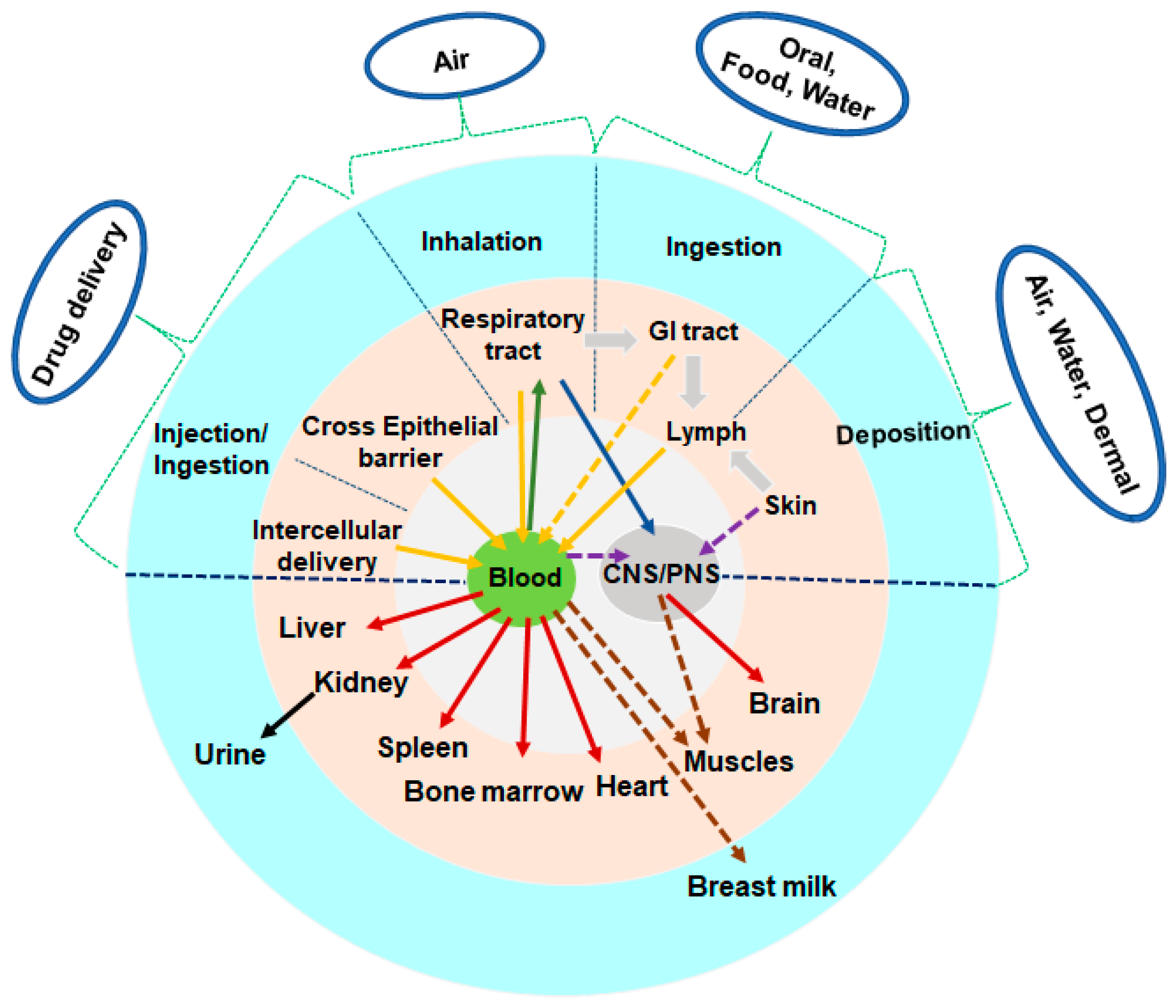
Figure 1. Nanoparticle and its bio interaction. The physicochemical characteristics of the surface and core of nano-sized particles will mostly determine the potential harmful consequences. Though several absorption and translocation pathways have been demonstrated, others remain hypothetical and must be studied.
Antioxidants 12 00703 g001
Figure 2. The overall negative impacts of mNPs on the human body. mNPs are oxidants that typically induce oxidative damage. mNPs can drive redox processes, resulting in endogenous ROS generation and significant health risks such as mutagenicity. (Image sources: skin anatomy, blood cells flow images by macrovector on Freepik; liver image by Freepik; arthritis image and infection image by Storyset on Freepik; human body image from brgfx on Freepik; the other images were downloaded from Pixabay). COPD—chronic obstructive pulmonary disease.
Antioxidants 12 00703 g002
Figure 3. The systematic illustration of mNPs induced effects on heart and brain. Overview of the neurotoxic effects of metal based nanoplastics. mNPs can enter the systemic circulation, and eventually the brain, via penetrating the blood–brain barrier (BBB), and may cause oxidative stress and potentially cause cellular damage and neuroinflammation. The possible impact of mNPs on cardiovascular performance. Synthetic NPs may enter the circulation from the lungs and produce pathological changes in the heart and artery tissues. Moreover, this causes a systemic oxidative stress/inflammatory response via the lungs, altering cardiovascular function.
Antioxidants 12 00703 g003
Figure 4. A schematic representation of the numerous triggers involved in forming reactive oxygen species (ROS) and the ROS induced pathways that lead to cell damage. Oxidative stress has a significant impact on nanotoxicity. The active surface of the nanoparticle, the size of the nanoparticle, photoactivation, toxins, metal ion dissolution, and nanoparticle interactions with biomolecules are all features that contribute to the generation of nanoparticle induced ROS.
Antioxidants 12 00703 g004
Figure 5. Cellular pathways connecting excess ROS production, oxidative stress and inflammation. The activated PRRs trigger downstream signalling pathways and activate transcription factors. (Image source: mitochondria and nucleus images from brgfx on Freepik).
Antioxidants 12 00703 g005
Disclaimer/Publisher’s Note: The statements, opinions and data contained in all publications are solely those of the individual author(s) and contributor(s) and not of MDPI and/or the editor(s). MDPI and/or the editor(s) disclaim responsibility for any injury to people or property resulting from any ideas, methods, instructions or products referred to in the content.