Article

Impact of Lactic Acid Bacteria Fermentation on Phenolic Compounds and Antioxidant Activity of Avocado Leaf Extracts

by
Soumi De Montijo-Prieto
1,†,
María del Carmen Razola-Díaz
2,3,†,
Federica Barbieri
2,4,
Giulia Tabanelli
4,5,
Fausto Gardini
4,5,
Maria Jiménez-Valera
1,
Alfonso Ruiz-Bravo
1,
Vito Verardo
2,3,*,‡ and
Ana Mª Gómez-Caravaca
3,6,‡
1
Campus of Cartuja, Department of Microbiology, University of Granada, 18071 Granada, Spain
2
Campus of Cartuja, Department of Nutrition and Food Science, University of Granada, 18071 Granada, Spain
3
Biomedical Research Center, Institute of Nutrition and Food Technology ‘José Mataix’, University of Granada, Avda del Conocimiento sn, 18100 Granada, Spain
4
Department of Agricultural and Food Sciences, University of Bologna, 40127 Bologna, Italy
5
Interdepartmental Centre for Industrial Agri-Food Research, University of Bologna, 47521 Cesena, Italy
6
Department of Analytical Chemistry, Faculty of Sciences, University of Granada, Avd. Fuentenueva s/n, 18071 Granada, Spain
*
Author to whom correspondence should be addressed.
These authors contributed equally to the work sharing first co-authorship.
These authors are joint senior authors on this work.
Antioxidants 2023, 12(2), 298; https://doi.org/10.3390/antiox12020298
Submission received: 31 December 2022 / Revised: 20 January 2023 / Accepted: 25 January 2023 / Published: 28 January 2023

Abstract

:
The growing global consumption of avocados, associated with contents including bioactive compounds with numerous health-promoting properties, is producing a large amount of agro wastes around the world. Different management approaches are available for the recovery of bioactive compounds from wastes as potential ingredients for use in the production of functional foods and nutraceuticals. Lactic acid fermentation can be used to exploit nutritional potential and add value to agro wastes. In this study, fermentations with lactic acid bacteria were carried out in avocado leaves, and the total phenolic content and the antioxidant activity were determined by DPPH and FRAP assays from hydroalcoholic extracts obtained from fermented avocado leaves. Fifteen new phenolic compounds were identified for the first time in avocado leaves by HPLC-ESI-TOF-MS. L. plantarum CECT 748T and P. pentosaceus CECT 4695T showed the highest antioxidant activity. The sum of phenolic compounds was increased by 71, 62, 55 and 21% in fermentations with P. pentosaceus CECT 4695T, L. brevis CECT 5354, P. acidilactici CECT 5765T and L. plantarum CECT 9567, respectively, while it was reduced in the fermentation with L. plantarum 748T by 21% as demonstrated by HPLC-ESI-TOF-MS. Biotransformations induced by bacterial metabolism modified the phenolic compound profile of avocado leaves in a strain-specific-dependent manner. P. pentosaceus CECT 4695T significantly increased kaempferol, P. pentosaceus 4695T, L. brevis 5354 and L. plantarum 9567 increased rutin, and dihydro-p-coumaric acid was increased by the five selected lactic acid bacteria. Total flavonoids were highly increased after fermentations with the five selected lactic acid bacteria but flavonoid glucosides were decreased by L. plantarum 748T, which was related to its higher antioxidant activity. Our results suggest that lactic acid bacteria led the hydrolysis of compounds by enzymatic activity such as glycosidases or decarboxylase and the release of phenolics bound to the plant cell wall, thus improving their bioavailability.

1. Introduction

Avocado (Persea americana Mill., Lauraceae) is native to southern Mexico, but it is cultivated around the world including in Central and South America, Indonesia, the United States, Australia, South Africa and Spain [1,2], with the Hass variety the most cultivated in the world [3]. In 2021, the Food and Agriculture Organization of the United Nations (FAO) calculated that global avocado production was 8.69 million metric tonnes, overcoming the previous year’s production of 8.06 million metric tonnes (https://www.fao.org; accessed on 29 December 2022). Due to the increase in the avocado fruit’s production and consumption, the amount of agro waste is increasingly becoming a major problem for the environment [1,4]. During harvesting, avocado leaves are discarded without any industry application [5]; however, the extracts obtained from avocado leaves contain appreciable amounts of bioactive compounds such as alkaloids, triterpenoids, saponins, carbohydrates, fatty acids and polyphenols [6,7,8,9,10]. Plant phenolic compounds are found in high concentrations in plant leaves and the green stems, with them being affected by genetic factors, environmental conditions, geographic location or physiological variations [11]. Redox properties of phenolic compounds are related with their ability to neutralize free radicals, contributing to the amelioration of some degenerative diseases associated to oxidative stress, such as neurodegenerative and cardiovascular diseases, cancer or skin aging through photoprotection from UV rays [12]. They have been also associated with antibacterial and antifungal activities. Hydroalcoholic leaf extracts of seven Mexican cultivars of P. americana var. drymifolia showed strong antioxidant activity by DPPH and ABTS assays [13], and purified phenolic fractions of avocado leaf extract showed a concentration-dependent antibacterial effect [5]. Plant phenolics are found covalently bonded to plant cell walls and their functional activities depend on their chemical structure and bioavailability. In the human gut, dietary phenolics are transformed by fermentative microbiota, which plays an important role in their absorption and in the modulation of their functional properties with benefits to health [14,15].
Lactic acid bacteria (LAB) fermentation is used in the food industry for the enhancement of the nutritional quality of foods by increasing protein digestibility, mineral availability and the release of peptides and amino acids [16,17]. However, fermentation with selected lactic acid bacteria also increases the antimicrobial and antioxidant activities of foods [18,19]. LAB are part of the microbiota of raw vegetables and can be isolated from spontaneous vegetable fermentations [20]. Therefore, they are characterized by a good adaptation to different environments by the utilization of different substances [21,22] including plant niches [23]. The capability of LAB to metabolize plant material is species- and strain-specific [24], and it is affected by several factors such as the presence of fermentable substrates or the presence of inhibitory factors such as phenol compounds [23]. In fact, the different phenolic compounds and their concentrations can affect the metabolism and viability of LAB [25]; therefore, tolerance to high levels of phenols is required to utilise and to biotransform plant material [23]. The ability of the microorganisms to produce enzymes such as glucosidase, amylase, cellulase, tannase, chitinase or lipase during fermentations can break down/hydrolyse the bound compounds that are released during the extraction [14]. Moreover, the metabolization of phenolics by LAB is recognised as a stress tolerance mechanism for the detoxification and bioconversion of polyphenols [26,27].
For this reason, this study aimed to evaluate the effect of submerged fermentation with LAB on the phenolic profile of avocado leaves as a strategy to obtain phenolic-compound-enriched extracts with improved antioxidant properties.

2. Materials and Methods

2.1. Chemicals and Samples

HPLC (High-Performance Liquid Chromatography)-grade water and other reagents and solvents were purchased from Merck KGaA (Darmstadt, Germany). Water was purified using a Milli-Q system (Millipore, Bedford, MA, USA). All the analytical standards were purchased from Sigma-Aldrich (St. Louis, MO, USA).
Avocado leaf samples of variety Hass were collected from Salobreña (Spain, 36°44′48″ N 3°35′13″ W) in April 2022. The fresh leaves were air dried at room temperature in a dark environment and milled and sieved to 100 µm particle size; after that, they were frozen at −18 °C until the analyses.

2.2. Lactic Acid Bacteria Strains and Culture Media

Lactic acid bacteria (LAB) strains used in fermentations were obtained from the Spanish Collection of Type Cultures (CECT): Pediococcus acidilactici 5765T, Pediococcus acidilactici 98, Pediococcus pentosaceus 4695T, Pediococcus pentosaceus 923, Leuconostoc mesenteroides subsp. mesenteroides 219T, Leuconostoc mesenteroides subsp. mesenteroides 215, Levilactobacillus brevis 4121T, Levilactobacillus brevis 5354, Lactiplantibacillus plantarum subsp. plantarum 748T and Lactiplantibacillus plantarum 9567 (formerly strain C4). Strains were reactivated by incubation in MRS broth and agar, at 26 °C for 24–48 h, and stocks were maintained in glycerol at −20 °C.

2.3. Preparation of the Inocula

Strain stocks were cultivated in MRS broth and incubated at 26 °C for 24 h. After growth, the cells were harvested by centrifugation (3000× g for 20 min) to remove the medium and resuspended in 10 mL of sterile saline solution (0.85% NaCl). The bacterial concentration was estimated by turbidimetry and the suspensions were used as inocula for each fermentation. An aliquot was taken from each suspension for counting viable bacteria by enumeration of colonies on MRS agar plates.

2.4. Fermentation of Avocado Leaves

Fermentations were carried out as follows: 1 g of the dried ground avocado leaves were submerged in 8 mL of sterile water previously heated to 90 °C. After mixing and cooling, the mixture was supplemented with 1 mL sterile medium containing glucose and yeast extract to obtain a concentration of 0.4% w/v of each. Inocula were added to obtain a concentration between 106 and 107 cell/mL. CFU/mL counting on MRS agar and pH values were determined at 0, 24, 48, 72 and 96 h of incubation at 26 °C. A control without the addition of LAB strains was included. Two replicates were prepared for each bacteria and control. At the end of incubations, samples and control were stored at −20 °C and then freeze-dried for further determinations.

2.5. Polar Compound Extraction

Briefly, 0.2 g of lyophilized avocado leaf fermented powder was dissolved in a 6 mL solution of ethanol/water 80/20, v/v. The mixture was placed in an ultrasonic bath for 15 min, and then it was centrifuged for 10 min at 9000 rpm. The extracting procedure was repeated twice more and all the supernatants were collected, evaporated and reconstituted in 1 mL of methanol/water (50:50, v/v). The final extracts were filtered with regenerated cellulose filters 0.2 µm (Millipore, Bedford, MA, USA) and stored at −18 °C until the analyses.

2.6. Determination of Folin–Ciocalteu Reacting Substances

Folin–Ciocalteu spectrophotometric method was used to determine the total Folin–Ciocalteu reacting substances (FCRS) for the first screening in all the fermented avocado leaves [28]. Briefly, 500 µL of the Folin–Ciocalteu reagent was added to 100 µL of the extract. It was added to 6 mL of bi-distilled water and the flask was agitated for a minute. After that, it was added to 2 mL of 15% (w/v) Na2CO3 and filled up to 10 mL with bi-distilled water. The flasks were kept in darkness for 2 h and the measures were carried out at 750 nm and 25 °C with a UV–visible spectrophotometer (Spectrophotometer 300 Array, UV–Vis, single beam, Shimadzu, Duisburg, Germany). Calibration curve was carried out with gallic acid from 1 to 1000 ppm and the equation obtained was y = 0.0012x − 0.0164 (R² = 0.9984). Analyses were performed in triplicate and the results are expressed as mg gallic acid equivalents (GAE)/g dry weight (d.w.).

2.7. Determination of Antioxidant Activity: DPPH and FRAP Assays

DPPH and FRAP assays were carried out to determine the antioxidant capacity of the avocado leaf fermented by different strains’ extracts by the procedures described in previous research [29,30]. Briefly, for the DPPH antioxidant assay, it was mixed with 100 µL of extract with 2.9 mL of the DPPH reagent and the decrease in absorbance was measured after 30 min at 517 nm. Otherwise, for FRAP it was mixed with 30 µL of extract with 90 µL of distilled water and 900 µL of FRAP reagent, kept at 37 °C for 30 min and the absorbance was measured at 595 nm. The measurements were performed using a UV–visible spectrophotometer (Spectrophotometer 300 Array, UV–Vis, single beam, Shimadzu, Duisburg, Germany). In both assays, Trolox was used as the standard for the calibration curves from 1 to 1000 ppm and the equations obtained were y = 0.0027x + 0.0495 (R² = 0.9989) and y = 0.0031x + 0.0016 (R² = 0.9934) for the DPPH and FRAP assays, respectively. The analyses were performed in triplicate and the results are expressed in mg of Trolox equivalents (TE)/g of dry weight (d.w.).

2.8. Determination of Polar Compounds by HPLC-ESI-TOF-MS

Phenolic compounds present in the fermented and non-fermented avocado leaf extracts were analysed using an Acquity Ultra Performance Liquid Chromatography (UPLC) system (Waters Corporation, Milford, MA, USA) coupled to an electrospray ionization (ESI) source operating in the negative mode and a mass detector time of flight (TOF) micro mass spectrometer (Waters). The compounds of interest were separated on an ACQUITY UPLC BEH Shield RP18 column (1.7 μm, 2.1 × 100 mm; Waters Corporation, Milford, MA, USA) at 40 °C using the conditions and gradient previously stated [31]. H2O acidified with 1% of acetic acid and acetonitrile were used as phase A and B, respectively. Analyses were performed in triplicate. The identification of the phenolic compounds was made according to the literature. For ensuring the mass accuracy, the tolerances chosen had a score higher than 90% and error lower than 5 ppm. To quantify the phenolic compounds identified in the avocado leaf extracts, calibration curves were used for vanillic acid (y = 8.1947x + 122.91; R² = 0.9976), chlorogenic acid (y = 85.138x + 135.16; R² = 0.9978), ferulic acid (y = 16.507x + 92.06; R² = 0.9980), quercetin (y = 112.8x + 287.12; R² = 0.9957), catechin (y = 41.108x + 335.6; R² = 0.9959) and rutin (y = 26.176x + 403.46; R² = 0.9924). The results are expressed as µg/g d.w.

2.9. Data Processing

The data for the identification of polar compounds in the avocado leaves by HPLC-ESI-TOF-MS were elaborated using MassLynx 4.1 software (Waters Corporation, Milford, MA, USA). Statistical differences (Tukey test) by one-way ANOVA analysis, and Pearson correlations were performed using Statistica 7.0 package (StatSoft, Tulsa, OK, USA). The rest of the statistical analyses were performed using MetaboAnalyst 5.0.

3. Results and Discussion

3.1. Screening of Lactic Acid Bacteria in Fermentation of Avocado Leaves

As described above, a minimal medium (dextrose plus yeast extract) was added to stimulate the initiation of growth of the inoculated LAB. To check if the treatment with hot water was able to eliminate the microorganisms present in the avocado leaves, viable microorganism counts were performed on MacConkey agar (selective medium for enterobacteria), Tryptic soy agar (TSA, enriched medium for bacteria), and Sabouraud agar (medium for fungi). After incubation of the media, the counting was under the limit of detection of the tests.
As shown in Table S1, avocado leaves did not support the growth of most of the inoculated LAB strains. The number of viable bacteria dropped during the first hours of incubation and continued to decrease gradually throughout the fermentation, with the exception of the two L. plantarum strains, which increased their viable counts. While L. plantarum 748T reached its growth peak at 24 h of incubation (8.44 ± 0.01 log CFU/mL), the exponential phase of growth of L. plantarum 9567 was prolonged until 48 h reaching similar counts (8.41 ± 0.05 log CFU/mL). The growth of the two strains of L. plantarum caused the acidification of the medium (Figure S1). After 24 h of incubation, the pH dropped from 5.94 to 5.31 with L. plantarum 9567 and to 3.93 with L. plantarum 748T. After 48 h, both pH values were around 4. The pH values for the rest of the strains remained similar to the initial values. Although P. acidilactici strains did not increase their concentrations, they were kept viable during fermentations similar to the initial concentrations.
Poor bacterial growth in avocado leaves can be due to their composition. Some compounds present in fermentation, such as phenolics, can affect the viability and metabolism of LAB [23,32]. Avocado leaves contain glycosides, alkaloids, tannins, saponins, flavonoids, terpenoids and steroids [7,8,9,33] and represent a potential source of antibacterial molecules [5]. Nevertheless, high tolerance to phenolic compounds is found in LAB, especially in members of Lactobacilli [34], which can be isolated from fermented products with a high content of phenolic compounds [35]. L. plantarum has been widely studied for its adaptation to plant habits and capability to metabolise phenolics [24], and it is used as starter in food fermentation.
All the fermented avocado leaves at different hours of incubation were analysed in terms of Folin–Ciocalteu reacting substances (FCRS) and antioxidant activity, and the results are presented in Table 1.
As can be seen from the results, the FCRS content ranged from 17.34 to 30.72 mg GAE/g d.w. Otherwise, the antioxidant activity was in the range of 25.56–53.88 and 50.34–96.61 for DPPH and FRAP, respectively. In our study, we applied a heat treatment on the avocado leaves in order to eliminate contaminants that could affect the fermentation process. Thus, avocado leaves were submerged into hot water and then allowed to cool spontaneously. This treatment was carried out both on avocado leaves fermented with lactic bacteria and on the unfermented control. Yamassaki et al. previously reported no decrease in the phenolic content or antioxidant activity when heating avocado leaf hydroalcoholic solutions at 40–100 °C for more than 8 h, and the total phenolic content or antioxidant activity of the extracts did not decrease [36].
An increase in the FCRS content was detected after the fermentation for some microorganisms; however, antioxidant activity by DPPH and FRAP assays was lower after fermentations with LAB strains. For most strains, the highest antioxidant activity was found at 24 and 48 h of fermentation. According to the results, the highest antioxidant activity was obtained after 24 h fermentation with P. acidilactici 5765T in the DPPH assay with 50.01 ± 0.23 mg TE/g d.w. and after 48 h fermentation with L. plantarum 748T with 96.61 ± 1.60 mg TE/g d.w. in FRAP assay. It was previously reported that fermentation of avocado puree with L. plantarum resulted in high levels of total free amino acids and a marked increase in antioxidant activity [37]. However, studies of lactic acid fermentations of avocado leaves are scarce.
Among all the microorganisms tested, the two varieties of L. mesenteroides showed the most minor results at all the different hours evaluated, so they were discarded for the next steps. Regarding the two L. brevis strains, a significant reduction in the FCRS and the antioxidant activity for the 4121T strain was seen; conversely, the 5254 strain produced an increase in FCRS content. Comparing P. acidilactici 5765T and P. acidilactici 98, in the first case, a higher recovery of FCRS was found compared to the control; in the second case, a low amount of FCRS was noticed. When fermenting the avocado leaves with P. pentosaceus strains, the highest results were found at 24 h of fermentation with P. pentosaceus 4695T. Finally, L. plantarum 748T had the best FCRS at 48 h and L. plantarum 9567 at 24 h. Both strains of L. plantarum showed very good performances with significant differences to the control.
The FCRS content was related to the antioxidant activity. A significant positive correlation (p < 0.05) was found between total phenolic content and DPPH (r = 0.7857) and FRAP (r = 0.8069) assays. Likewise, the DPPH assay showed a significant positive correlation with the FRAP assay (r = 0.6107).
Based on the FCRS content and antioxidant capacity, the strains P. acidilactici 5765T (24 h), P. pentosaceus 4695T (24 h), L. brevis 5354 (24 h), L. plantarum 748T (48 h) and L. plantarum 9567 (24 h) were selected for studying the phenolic composition.

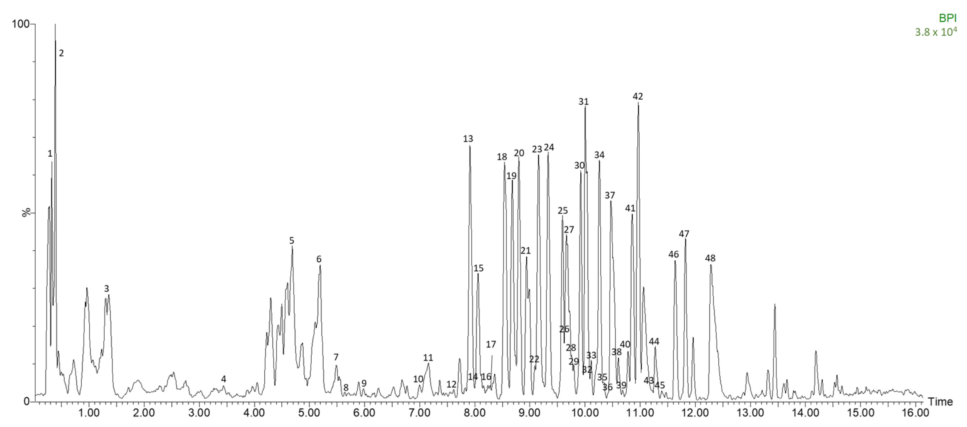
3.2. Identification of Polar Compounds in Fermented Avocado Leaves by HPLC-ESI-TOF-MS

The selected fermented avocado leaves and a control were characterized by HPLC-ESI-TOF-MS and a total of 48 polar compounds were identified. Among them were seven phenolic acids, thirty-seven flavonoids and four other compounds. They are presented in Table 2 with their experimental and calculated m/z, time (min), error (ppm), score (%), molecular formula and tentative name for each compound. The peaks presented in Table 2 correspond to the numbers shown in Figure 1, which is a representative chromatogram of a fermented avocado leaf. To the best of our knowledge, 15 polar compounds are identified here for the first time in avocado leaves.
Phenolic acids. Corresponding to peaks 3, 4, 5 and 7 were detected protocatechuic acid-4-glucoside, p-coumaric acid, chlorogenic acid and sinapic acid-C-hexoside in concordance with López-Cobo et al. [38] who previously identified them in avocado by-products. Moreover, at times 6.02 and 7.14, and with the m/z 221 and 165, two coumaric acid derivatives were found named as p-coumaroyl glycolic acid and dihydro-p-coumaric acid, respectively, according to the phenol explorer database [39]. The first one was previously quantified in lentils seeds [40] and the second one in olives [41], and both were found in avocado leaves here for the first time. In addition, a ferulic acid derivative tentatively named as dihydroferulic acid 4-O-glucuronide was detected with the m/z 371, the m/z in source fragment 195 and the predicted molecular formula C16H20O10 in agreement with Hu et al. [42] who found it in sweet cherries. Previously, Fan et al. [43] reported a similar compound named ferulic acid 4-O-glucoside in rejected avocados, but it is the first time this compound has been found in avocado leaves.
Flavonoids. Flavan-3-ols are a well-known group of flavonoids usually found in avocado samples, and in this case catechin derivatives were detected corresponding to peaks 8, 11 and 41, named as procyanidin dimer, procyanidin trimer and catechin diglucopyranoside, respectively [13]. Moreover, special attention was paid to the compounds detected at peaks 14 and 17. They were tentatively named as cinchonain-1a-(4beta->8)-catechin isomer a and b according to their m/z in source fragments 289 [C15H13O6] and 245 [M–3H]3− (PubChem CID: 442686), and are described in avocado leaves here for the first time. Moreover, the flavonolignan cinchonain was identified at 8.15 min with the m/z 451 [13]. At time 7.57, the compound quercetin (m/z 301) was identified, and a total of 12 quercetin derivatives were detected. With the molecular formula C27H30O17, two isomers named as quercetin-diglucoside isomers a and b were found. Three isomers of quercetin 3-O-arabinosyl-glucoside (a, b and c) were detected with the m/z 595. Corresponding to peaks 23 and 24, two other isomers of a quercetin derivative were identified and named as quercetin-3-glucoside isomers a and b, respectively. In addition, quercetin glucuronide and quercetin-O-deoxyhesoxide were found at times 9.65 and 10.45. All of them were identified in agreement with Castro-López et al. [13] who previously described them in avocado leaves. Also they were found three other quercetin derivatives not reported previously in avocado samples. At 11.02 min the compound named as quercetin 3-O-acetyl-rhamnoside was identified according to Mi et al. [44] who found it in berries. With the m/z 565 and the molecular formula C25H26O15, the compound quercetin 3-xilosyl-(1->2)-alpha-L-arabinopyranoside, and with the m/z 505 and the formula C23H22O13, the compound quercetin 3-O-glucose-6″-acetate, were tentatively identified according to the PubChem database (PubChem CID: 44259231 and 24211981, respectively) with their m/z in source fragment 301. There were three isomers of the flavonoid rutin named as isomer a, b and c detected, corresponding to peaks 20, 21 and 31, respectively, with the m/z 609. Four isomers of a luteolin derivative were found with the molecular formula C26H28O15 that were called luteolin 7-O-(2″-O-pentosyl)-hexoside isomers a, b, c and d. At 11.81 min with the m/z 563, the compound apigenin-C-hexoside-C-pentoside was identified [13]. Otherwise, kaempferol was detected at 12.3 min with the molecular formula C15H10O6. Its derivatives, kaempferol-O-hexoside isomers a and b, kaempferol 3-O-acetyl-glucoside and kaempferol-O-coumaroyl, were identified corresponding to peaks 30, 34, 44 and 46, respectively [13]. With the m/z 461, the compound kaempferol 3-glucuronide that was previously quantified in strawberry [45] and endive [46] was tentatively identified. Additionally, two isomers (a and b) of kaempferol 3,4′-dixyloside were detected at 10.49 and 10.57 min, respectively, in agreement with Nakane et al. [47] who identified them in leaves of Allium macrostemon. In addition, the compounds detected at peaks 43 and 45 were tentatively named as luteolin 7-[6-O-(2-methylbutyryl)-beta-glucoside] isomers a and b, respectively, according to its fragment 285 that corresponds to C15H10O6 (luteolin), and the other to its source fragments 191 and 339 in agreement with Xiong et al. [48] who identified it in sorghum.
Other. Three organic acids and two quinones were detected. Corresponding to peaks 1 and 2, two isomers (a and b) of quinic acid, respectively, were identified [38,49]. Moreover, with the m/z 299, the compounds found at 9.62 and 10.06 min were tentatively identified as two isomers of a trihydroxyanthraquinones named as emodic acid according to its m/z in source fragments 255 [C14H8O5], 243 [C13H8O5] and 227 [C13H8O4], and the PubChem database (PubChem CID: 361510).

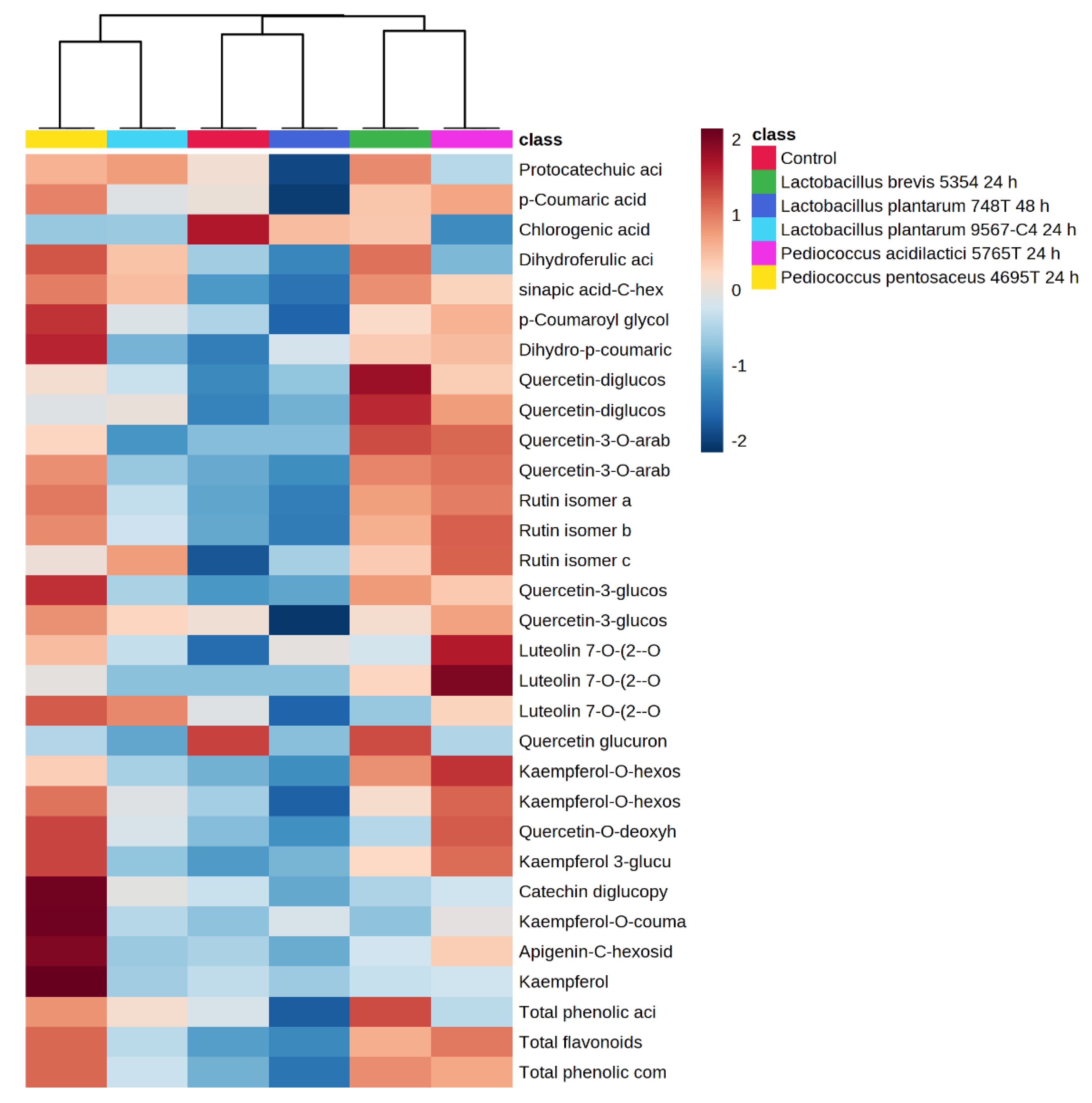
3.3. Quantification of Phenolic Compounds by HPLC-ESI-TOF-MS and Its Biotransformations during Fermentation in Avocado Leaves

The phenolic acids and flavonoids identified in the unfermented and selected fermented avocado leaves were quantified and the results are summarized in Table 3.
Fermentations of avocado leaves with the five selected LAB resulted in modifications in their phenolic profile (Figure S2). Total phenolic compounds’ content was increased by 71, 62, 55 and 21% in fermentations with P. pentosaceus 4695T, L. brevis 5354, P. acidilactici 5765T and L. plantarum 9567, respectively, while it was reduced in the fermentation with L. plantarum 748T by 21% in comparison to the unfermented control. Total phenolic acids were increased by 27, 40 and 43% in P. pentosaceus 4695T, L. brevis 5354 and L. plantarum 9567 fermentations, respectively, but decreased with L. plantarum 748T and P. acidilactici 5765T. With regard to total flavonoids, their content was highly increased by 91, 96 and 75% in P. acidilactici 5765T, P. pentosaceus 4695T and L. brevis 5354 fermentations, respectively.
LAB are able to degrade and biotransform food phenolic compounds by tannase, amylase, esterase, β-glucosidase, phenolic acid decarboxylase (PAD), reductase, or benzyl alcohol dehydrogenase enzymes [14,20]. Hydroxycinnamic acids such as caffeic, p-coumaric or ferulic acids can be reduced into dihydrocaffeic, phloretic or dihydroferulic acids, respectively, or decarboxylated into vinyl derivatives by a phenolic acid decarboxylase enzyme (PAD), and subsequently reduced into ethyl derivatives [50]. While p-coumaric acid was increased in fermentations with P. acidilactici 5765T, P. pentosaceus 4698T and L. brevis 5354, it was reduced after fermentation by L. plantarum 9567 and consumed by L. plantarum 748T (<LOQ). Trans-p-coumaric and cis-ferulic acids were decreased by L. plantarum in cowpeas (Vigna sinensis L.) depending on the isomeric form of the acids; however, spontaneous fermentation increased them [51]. Likewise, L. brevis, L. plantarum and P. pentosaceus were able to metabolize p-coumaric and ferulic acids through decarboxylation [52]. In addition, p-coumaric acid was degraded until p-vinyl-phenol and it reduced derivative dihydro-p-coumaric acid (phloretic acid) by L. plantarum in cherry juice exhibiting a strain-specific metabolism [24]. We found a significant increase in the concentration of dihydro-p-coumaric acid with the five selected strains, especially with P. acidilactici 5765T, P. pentosaceus 4695T and L. brevis 5354, but the decrease in p-coumaric acid in L. plantarum 748T fermentation did not correspond to a higher accumulation of this reduced metabolite compared to the last three bacteria.
Hydroxycinnamic acids are found glycosylated in plants (esterified), covalently attached to the cell wall and as a soluble form in cytoplasm. The breakdown of the ester linkages between polymers release the free phenolic acids [14,53]. Cinnamoyl ester hydrolases, also known as cinnamoyl esterase, catalyse the hydrolysis reaction of hydroxycinnamoyl esters releasing free acids that will be new substrates for phenolic acid decarboxylases PAD [53]. In our study, p-coumaroyl glycolic acid, an ester of p-coumaric acid, was decreased (<LOQ) in the fermentation with L. plantarum 748T. As we mentioned above, this microorganism also consumed p-coumaric acid, suggesting an initial hydrolysis of the ester by a cinnamoyl esterase followed by a decarboxylase activity. Chlorogenic acid, also known as 3-caffeoylquinic acid, is the ester of caffeic and quinic acids, and was significantly decreased in fermentations with all bacteria, especially with P. acidilactici 5765T. Conversely to our results, L. plantarum consumed caffeic, p-coumaric, ferulic, protocatechuic and p-hydroxybenzoic acids except for chlorogenic acid [26]. The ability to hydrolyse cinnamoyl esters of some L. plantarum strains was related to the presence of two esterases with differences in their substrate range: Lp_0796 that hydrolyses esters of caffeic, p-coumaric, ferulic and sinapic acids, while Est_1092 was able to hydrolyse both hydroxycinnamoyl and hydroxybenzoyl esters [54]. This cinnamoyl esterase activity is important in the de-esterification process of dietary fiber in human and ruminal digestion, improving the antioxidant, antiinflammatory and antimicrobial activity of complex dietary compounds [14].
Rutin isomers were increased by P. pentosaceus 4695T, L. brevis 5354 and L. plantarum 9567, while they were reduced by P. acidilactici 5765T (<LOQ). Nevertheless, in fermentation with L. plantarum 748T, rutin isomers a and b were reduced, but isomer c was increased significantly. Kaempferol, kaempferol-3-glucoside, quercetin and quercetin-3-glucoside were released from rutin after fermentation with Aspergillus awamori in Litchi pericarp [55]. Our results showed a significant increase in quercetin-3-glucoside isomers in all fermentations with the exception of L. plantarum 748T. Likewise, a significant increase in kaempferol was found only in the fermentation with P. pentosaceus 4695T, suggesting that this microorganism led to a biotransformation towards this compound. The hydrolysis of rutin to kaempferol-3-rutinoside or quercetin-3-glucoside is catalysed by α-rhamnosidases and further hydrolysed by β-glucosidases to free kaempferol or quercetin [32]. However, free quercetin was found under the limit of quantification (<LOQ) in both fermented and unfermented avocado leaves.
The enzymatic activity of the bacteria can break down vegetable cell walls and release bound phenolics, improving their bioavailability and facilitating their extraction [14,22]. Flavonoids were the main phenolic compound found in avocado leaves and were highly increased after fermentations with LAB. Flavonoids are found predominantly as glycosylated conjugates in plants, mostly as quercetin and kaempferol [56]. With the exception of L. plantarum 748T, the concentrations of flavonoid glucosides such as luteolin-7-O-(2″-O-pentosyl)-hexoside isomer a, quercetin-diglucoside and quercetin-3-O-arabinosyl-glucoside were increased compared to the unfermented control. Conversely, luteolin-7-O-(2″-O-pentosyl)-hexoside isomer d, quercetin-3-O-arabinosyl-glucoside isomer b and quercetin-3-glucoside isomer b, were reduced significantly by L. plantarum 748T. In soybeans’ and mung beans’ fermentation with L. plantarum 748T, glycosylated isoflavones were deglycosylated into their respective aglycones increasing their bioavailability [57]. Likewise, cultures of L. plantarum 748T transformed food aryl glycosides: phloridzin, esculin, daidzin and salicin into aglycones with the exception of quercetin glucoside, which remained glycosylated after incubation. The deglycosylation was associated with an increase in the antioxidant activity [58]. Our results showed a decrease in the concentration of quercetin glucoside isomer b but not in isomer a, suggesting a glycosyl hydrolase activity dependent on the isomeric form. In the gut, conjugated glucosides are hydrolysed by the intestinal microbiota to be absorbed into their corresponding aglycone, which show higher activity than their precursor glycosides [23]. β-glycosidase activity is widespread among LAB and have a significant positive impact on fermented products, improving their flavour and fragrance [59]. L. plantarum is commonly found in the human gastrointestinal tract and is used as a starter in the fermentation of dairy products, vegetables and meats [20]. Glycosidase activity of L. plantarum has been associated with an improvement in the bioaccessibility and bioavailability of food phenolic compounds as well as with an increase in their antioxidant activity [58]. Although L. plantarum 748T decreased the total phenolic content of avocado leaves, it showed a significant decrease in aryl-glucosides such as: quercetin-3-glucoside isomer b, protocatechuic acid 4-glucoside, quercetin-3-O-arabinosyl-glucoside isomer b, luteolin 7-O-(2″-O-pentosyl)-hexoside isomer d and kaempferol-O-hexoside isomer b, suggesting a higher glycosidase activity than the rest of the strains. Otherwise, L. plantarum 9567 increased total phenolic compounds by 21% but did not show such marked glucosidase activity as L. plantarum 748T. The higher glycoside deglycosylation shown by L. plantarum 748T, and to a lesser extent by L. plantarum 9567, may be related to the high antioxidant activities found in the DPPH and FRAP assays in comparison with the rest of the bacteria.
A hierarchical clustering heatmap was performed to provide an intuitive visualization of all of the phenolic compounds quantified by HPLC-ESI-TOF-MS in the fermented avocado leaves by the selected strains and a non-fermented control. The features were previously normalized, the distance measure was the Pearson statistical meaning and the clustering method was the average. Therefore, the clustering result for the features (rows) and samples (columns) is shown in Figure 2. Each colour cell on the map corresponds to a concentration value normalized from 2 (intense red) to −2 (intense blue). Moreover, each sample has an associated colour (legend).
As can be seen from the figure, the avocado leaf fermented by L. plantarum 748T at 48 h was clustered with the control sample, which shows the minor differences among them in the polyphenol profile, and it had the lowest total phenolic content but with the highest content of chlorogenic acid. Close to them, L. brevis 5354 and P. acidilactici 5765T were also clustered according to their phenolic profile, being the group that showed higher contents of luteolin and quercetin derivatives. Finally, the group that was clustered furthest from the control was the one composed by P. pentosaceus 4695T and L. plantarum 9567. It seems to be a heterogeneous group in terms of amounts but with a similar profile and proportions between the individual phenolic compounds. Among them, P. pentosaceus 4695T was the strain that led the avocado leaf to release the highest content of coumaric acid derivatives and kaempferol derivatives with the highest total phenolic content. This clustering analysis confirms the strain-specific metabolism of LAB on the phenolic compounds present in avocado leaves, which is dependent on the capability of strains to tolerate and hydrolyse them.

4. Conclusions

This study allowed us to identify the chemical biotransformations induced by LAB strains in avocado leaves using submerged fermentations. A total of 48 polar compounds were identified by HPLC-ESI-TOF-MS and, to our knowledge, 15 of them were identified for the first time. We found a strain-specific metabolism of the phenolic compounds of avocado leaves, which was dependent on the tolerance of LAB strains to the phenolics’ concentration and their capacity to hydrolyse them. Fermentations with P. acidilactici CECT 5765T, P. pentosaceus CECT 4695T, L. plantarum CECT 9567 and L. brevis CECT 5354 led to an increase in the total phenolic content, with the exception of L. plantarum CECT 748T, which decreased it. The phenolic content in fermented leaf extracts was from 21 (L. plantarum CECT 9567) to 71% (P. pentosaceus CECT 4695T), higher than in the control. Briefly, submerged fermentation with lactic acid bacteria can be used in the exploitation and valorisation of avocado agro wastes for the production of enriched phenolic extracts.

Supplementary Materials

The following supporting information can be downloaded at: https://www.mdpi.com/article/10.3390/antiox12020298/s1, Figure S1: pH values of lactic acid bacteria cultures during fermentation of avocado leaves; Figure S1: Phenolic profile in fermented and unfermented avocado leaves by HPLC-ESI-TOF-MS; Table S1: Log CFU/mL of lactic acid bacteria in avocado leaves.

Author Contributions

Conceptualization, G.T., F.G., V.V. and A.M.G.-C.; methodology, M.J.-V., A.R.-B., V.V. and A.M.G.-C.; formal analysis, S.D.M.-P., M.d.C.R.-D. and F.B.; investigation, S.D.M.-P., M.d.C.R.-D. and F.B.; resources, V.V., A.M.G.-C., M.J.-V. and A.R.-B.; data curation, G.T., M.J.-V., A.R.-B. and V.V.; writing—original draft preparation, S.D.M.-P., M.d.C.R.-D. and F.B.; writing—review and editing, G.T., F.G., M.J.-V., A.R.-B., V.V. and A.M.G.-C.; supervision, G.T., F.G., M.J.-V., A.R.-B., V.V. and A.M.G.-C.; project administration, V.V. and A.M.G.-C.; funding acquisition, V.V. and A.M.G.-C. All authors have read and agreed to the published version of the manuscript.

Funding

This study is part of the national project RTI2018-099835-A-I00 financed by MCIN/AEI/10.13039/501100011033/ FEDER “Una manera de hacer Europa”.

Institutional Review Board Statement

Not applicable.

Informed Consent Statement

Not applicable.

Data Availability Statement

Data are contained within the article and Supplementary Materials.

Acknowledgments

Authors thank Carla van Coller for English reviewing and editing.

Conflicts of Interest

The authors declare no conflict of interest.

References

  1. Mora-Sand, A.; Ram, A.; Castillo-Henr, L.; Lopretti-Correa, M.; Vega-Baudrit, J.R. Persea americana Agro-Industrial Waste Biorefinery for Sustainable High-Value-Added Products. Polymers 2021, 13, 1727. [Google Scholar] [CrossRef] [PubMed]
  2. Sommaruga, R.; Eldridge, H.M. Avocado Production: Water Footprint and Socio-economic Implications. EuroChoices 2021, 20, 48–53. [Google Scholar] [CrossRef]
  3. Ramírez-Mejía, D.; Levers, C.; Mas, J.F. Spatial patterns and determinants of avocado frontier dynamics in Mexico. Reg. Environ. Chang. 2022, 22, 28. [Google Scholar] [CrossRef] [PubMed]
  4. Capanoglu, E.; Nemli, E.; Tomas-Barberan, F. Novel Approaches in the Valorization of Agricultural Wastes and Their Applications. J. Agric. Food Chem. 2022, 70, 6787–6804. [Google Scholar] [CrossRef] [PubMed]
  5. Solís-Salas, L.M.; Sierra-Rivera, C.A.; Cobos-Puc, L.E.; Ascacio-Valdés, J.A.; Silva-Belmares, S.Y. Antibacterial potential by rupture membrane and antioxidant capacity of purified phenolic fractions of Persea americana leaf extract. Antibiotics 2021, 10, 508. [Google Scholar] [CrossRef]
  6. Nathaniel, O.B.; Selina, A.S.; John, K.M.; Mercy, B.; Sylvester, A.A.; Michael, B.M. Phytoconstituents, antimicrobial and antioxidant properties of the leaves of Persea americana Mill cultivated in Ghana. J. Med. Plants Res. 2015, 9, 933–939. [Google Scholar] [CrossRef] [Green Version]
  7. Park, S.J.; Nam, Y.H.; Rodriguez, I.; Park, J.H.; Kwak, H.J.; Oh, Y.; Oh, M.; Park, M.S.; Lee, K.W.; Lee, J.S.; et al. Chemical constituents of leaves of Persea americana (avocado) and their protective effects against neomycin-induced hair cell damage. Rev. Bras. Farmacogn. 2019, 29, 739–743. [Google Scholar] [CrossRef]
  8. Yamassaki, F.T.; Campestrini, L.H.; Zawadzki-Baggio, S.F.; Maurer, J.B.B. Chemical characterization and complement modulating activities of an arabinogalactan-protein-rich fraction from an aqueous extract of avocado leaves. Int. J. Biol. Macromol. 2018, 120, 513–521. [Google Scholar] [CrossRef]
  9. Rincón-Hernández, C.A.; De La Pérez, J.L.S.; Espinosa-García, F.J. Caracterización química foliar de los árboles de aguacate criollo (Persea americana var. drymifolia) en los bancos de germoplasma de Michoacán, México. Rev. Mex. Biodivers. 2011, 82, 395–412. [Google Scholar] [CrossRef]
  10. Jimenez, P.; Garcia, P.; Quitral, V.; Vasquez, K.; Parra-Ruiz, C.; Reyes-Farias, M.; Garcia-Diaz, D.F.; Robert, P.; Encina, C.; Soto-Covasich, J. Pulp, Leaf, Peel and Seed of Avocado Fruit: A Review of Bioactive Compounds and Healthy Benefits. Food Rev. Int. 2021, 37, 619–655. [Google Scholar] [CrossRef]
  11. Martins, S.; Mussatto, S.I.; Martínez-Avila, G.; Montañez-Saenz, J.; Aguilar, C.N.; Teixeira, J.A. Bioactive phenolic compounds: Production and extraction by solid-state fermentation. A review. Biotechnol. Adv. 2011, 29, 365–373. [Google Scholar] [CrossRef] [PubMed] [Green Version]
  12. Rojas-García, A.; Fuentes, E.; Cádiz-Gurrea, M.d.l.L.; Rodriguez, L.; Villegas-Aguilar, M.D.C.; Palomo, I.; Arráez-Román, D.; Segura-Carretero, A. Biological Evaluation of Avocado Residues as a Potential Source of Bioactive Compounds. Antioxidants 2022, 11, 1049. [Google Scholar] [CrossRef] [PubMed]
  13. Castro-López, C.; Bautista-Hernández, I.; González-Hernández, M.D.; Martínez-Ávila, G.C.G.; Rojas, R.; Gutiérrez-Díez, A.; Medina-Herrera, N.; Aguirre-Arzola, V.E. Polyphenolic Profile and Antioxidant Activity of Leaf Purified Hydroalcoholic Extracts from Seven Mexican Persea americana Cultivars. Molecules 2019, 24, 173. [Google Scholar] [CrossRef] [Green Version]
  14. Muñoz, R.; de las Rivas, B.; López de Felipe, F.; Reverón, I.; Santamaría, L.; Esteban-Torres, M.; Curiel, J.A.; Rodríguez, H.; Landete, J.M. Biotransformation of Phenolics by Lactobacillus plantarum in Fermented Foods; Elsevier Inc.: Amsterdam, The Netherlands, 2017; ISBN 9780128023099. [Google Scholar]
  15. Selma, M.V.; Espín, J.C.; Tomás-Barberán, F.A. Interaction between phenolics and gut microbiota: Role in human health. J. Agric. Food Chem. 2009, 57, 6485–6501. [Google Scholar] [CrossRef] [PubMed]
  16. Verni, M.; De Mastro, G.; De Cillis, F.; Gobbetti, M.; Rizzello, C.G. Lactic acid bacteria fermentation to exploit the nutritional potential of Mediterranean faba bean local biotypes. Food Res. Int. 2019, 125, 108571. [Google Scholar] [CrossRef] [PubMed]
  17. Bergillos-Meca, T.; Cabrera-Vique, C.; Artacho, R.; Moreno-Montoro, M.; Navarro-Alarcón, M.; Olalla, M.; Giménez, R.; Seiquer, I.; Ruiz-López, M.D. Does Lactobacillus plantarum or ultrafiltration process improve Ca, Mg, Zn and P bioavailability from fermented goats’ milk? Food Chem. 2015, 187, 314–321. [Google Scholar] [CrossRef] [PubMed]
  18. Curiel, J.A.; Pinto, D.; Marzani, B.; Filannino, P.; Farris, G.A.; Gobbetti, M.; Rizzello, C.G. Lactic acid fermentation as a tool to enhance the antioxidant properties of Myrtus communis berries. Microb. Cell Fact. 2015, 14, 67. [Google Scholar] [CrossRef] [Green Version]
  19. Pontonio, E.; Montemurro, M.; Pinto, D.; Marzani, B.; Trani, A.; Ferrara, G.; Mazzeo, A.; Gobbetti, M.; Rizzello, C.G. Lactic acid fermentation of pomegranate juice as a tool to improve antioxidant activity. Front. Microbiol. 2019, 10, 1550. [Google Scholar] [CrossRef] [Green Version]
  20. Rodríguez, H.; Curiel, J.A.; Landete, J.M.; de las Rivas, B.; de Felipe, F.L.; Gómez-Cordovés, C.; Mancheño, J.M.; Muñoz, R. Food phenolics and lactic acid bacteria. Int. J. Food Microbiol. 2009, 132, 79–90. [Google Scholar] [CrossRef] [Green Version]
  21. Hatti-Kaul, R.; Chen, L.; Dishisha, T.; El Enshasy, H. Lactic acid bacteria: From starter cultures to producers of chemicals. FEMS Microbiol. Lett. 2018, 365, fny213. [Google Scholar] [CrossRef]
  22. Wang, Y.; Wu, J.; Lv, M.; Shao, Z.; Hungwe, M.; Wang, J.; Bai, X.; Xie, J.; Wang, Y.; Geng, W. Metabolism Characteristics of Lactic Acid Bacteria and the Expanding Applications in Food Industry. Front. Bioeng. Biotechnol. 2021, 9, 612285. [Google Scholar] [CrossRef] [PubMed]
  23. Filannino, P.; Di Cagno, R.; Gobbetti, M. Metabolic and functional paths of lactic acid bacteria in plant foods: Get out of the labyrinth. Curr. Opin. Biotechnol. 2018, 49, 64–72. [Google Scholar] [CrossRef] [PubMed]
  24. Filannino, P.; Bai, Y.; Di Cagno, R.; Gobbetti, M.; Gänzle, M.G. Metabolism of phenolic compounds by Lactobacillus spp. during fermentation of cherry juice and broccoli puree. Food Microbiol. 2015, 46, 272–279. [Google Scholar] [CrossRef] [PubMed]
  25. Filannino, P.; Gobbetti, M.; De Angelis, M.; Di Cagno, R. Hydroxycinnamic acids used as external acceptors of electrons: An energetic advantage for strictly heterofermentative lactic acid bacteria. Appl. Environ. Microbiol. 2014, 80, 7574–7582. [Google Scholar] [CrossRef] [Green Version]
  26. Sánchez-Maldonado, A.F.; Schieber, A.; Gänzle, M.G. Structure-function relationships of the antibacterial activity of phenolic acids and their metabolism by lactic acid bacteria. J. Appl. Microbiol. 2011, 111, 1176–1184. [Google Scholar] [CrossRef]
  27. Reverón, I.; de las Rivas, B.; Muñoz, R.; López de Felipe, F. Genome-wide transcriptomic responses of a human isolate of Lactobacillus plantarum exposed to p-coumaric acid stress. Mol. Nutr. Food Res. 2012, 56, 1848–1859. [Google Scholar] [CrossRef]
  28. Lamuela-Raventós, R.M. Folin–Ciocalteu method for the measurement of total phenolic content and antioxidant capacity. In Measurement of Antioxidant Activity & Capacity; John Wiley & Sons, Ltd.: Hoboken, NJ, USA, 2017; pp. 107–115. ISBN 9781119135388. [Google Scholar]
  29. Razola-Díaz, M.d.C.; Guerra-Hernández, E.J.; Rodríguez-Pérez, C.; Gómez-Caravaca, A.M.; García-Villanova, B.; Verardo, V. Optimization of Ultrasound-Assisted Extraction via Sonotrode of Phenolic Compounds from Orange By-Products. Foods 2021, 10, 1120. [Google Scholar] [CrossRef]
  30. Razola-Díaz, M.d.C.; Gómez-Caravaca, A.M.; Guerra-Hernández, E.J.; Garcia-Villanova, B.; Verardo, V. New Advances in the Phenolic Composition of Tiger Nut (Cyperus esculentus L.) by-Products. Foods 2022, 11, 343. [Google Scholar] [CrossRef]
  31. Verni, M.; Pontonio, E.; Krona, A.; Jacob, S.; Pinto, D.; Rinaldi, F.; Verardo, V.; Díaz-de-Cerio, E.; Coda, R.; Rizzello, C.G. Bioprocessing of Brewers’ Spent Grain Enhances Its Antioxidant Activity: Characterization of Phenolic Compounds and Bioactive Peptides. Front. Microbiol. 2020, 11, 1831. [Google Scholar] [CrossRef]
  32. Leonard, W.; Zhang, P.; Ying, D.; Adhikari, B.; Fang, Z. Fermentation transforms the phenolic profiles and bioactivities of plant-based foods. Biotechnol. Adv. 2021, 49, 107763. [Google Scholar] [CrossRef]
  33. García-Rodríguez, Y.M.; Torres-Gurrola, G.; Meléndez-González, C.; Espinosa-García, F.J. Phenotypic Variations in the Foliar Chemical Profile of Persea americana Mill. cv. Hass. Chem. Biodivers. 2016, 13, 1767–1775. [Google Scholar] [CrossRef] [PubMed]
  34. Hervert-Hernández, D.; Goñi, I. Dietary polyphenols and human gut microbiota: A review. Food Rev. Int. 2011, 27, 154–169. [Google Scholar] [CrossRef]
  35. Ghabbour, N.; Lamzira, Z.; Thonart, P.; Cidalia, P.; Markaoui, M.; Asehraou, A. Selection of oleuropein-degrading lactic acid bacteria strains isolated from fermenting moroccan green olives. Grasas Aceites 2011, 62, 84–89. [Google Scholar] [CrossRef] [Green Version]
  36. Yamassaki, F.T.; Campestrini, L.H.; Faria, S.; Bello, J.; Maurer, B. Avocado leaves: Influence of drying process, thermal incubation, and storage conditions on preservation of polyphenolic compounds and antioxidant activity. Int. J. Food Prop. 2017, 20, 2280–2293. [Google Scholar] [CrossRef] [Green Version]
  37. Filannino, P.; Tlais, A.Z.A.; Morozova, K.; Cavoski, I.; Scampicchio, M.; Gobbetti, M.; Di Cagno, R. Lactic acid fermentation enriches the profile of biogenic fatty acid derivatives of avocado fruit (Persea americana Mill.). Food Chem. 2020, 317, 126384. [Google Scholar] [CrossRef]
  38. López-Cobo, A.; Gómez-Caravaca, A.M.; Pasini, F.; Caboni, M.F.; Segura-Carretero, A.; Fernández-Gutiérrez, A. HPLC-DAD-ESI-QTOF-MS and HPLC-FLD-MS as valuable tools for the determination of phenolic and other polar compounds in the edible part and by-products of avocado. LWT—Food Sci. Technol. 2016, 73, 505–513. [Google Scholar] [CrossRef]
  39. Rothwell, J.A.; Perez-Jimenez, J.; Neveu, V.; Medina-Remón, A.; M’hiri, N.; García-Lobato, P.; Manach, C.; Knox, C.; Eisner, R.; Wishart, D.S.; et al. Phenol-Explorer 3.0: A major update of the Phenol-Explorer database to incorporate data on the effects of food processing on polyphenol content. Database 2013, 2013, bat070. [Google Scholar] [CrossRef]
  40. Dueñas, M.; Hernández, T.; Estrella, I. Changes in the content of bioactive polyphenolic compounds of lentils by the action of exogenous enzymes. Effect on their antioxidant activity. Food Chem. 2007, 101, 90–97. [Google Scholar] [CrossRef]
  41. Boskou, G.; Salta, F.N.; Chrysostomou, S.; Mylona, A.; Chiou, A.; Andrikopoulos, N.K. Antioxidant capacity and phenolic profile of table olives from the Greek market. Food Chem. 2006, 94, 558–564. [Google Scholar] [CrossRef]
  42. Hu, T.; Subbiah, V.; Wu, H.; Amrit, B.K.; Rauf, A.; Alhumaydhi, F.A.; Suleria, H.A.R. Determination and Characterization of Phenolic Compounds from Australia-Grown Sweet Cherries (Prunus avium L.) and Their Potential Antioxidant Properties. ACS Omega 2021, 6, 34687–34699. [Google Scholar] [CrossRef]
  43. Fan, S.; Qi, Y.; Shi, L.; Giovani, M.; Zaki, N.A.A.; Guo, S.; Suleria, H.A.R. Screening of Phenolic Compounds in Rejected Avocado and Determination of Their Antioxidant Potential. Processes 2022, 10, 1747. [Google Scholar] [CrossRef]
  44. Mi, J.C.; Howard, L.R.; Prior, R.L.; Clark, J.R. Flavonoid glycosides and antioxidant capacity of various blackberry, blueberry and red grape genotypes determined by high-performance liquid chromatography/mass spectrometry. J. Sci. Food Agric. 2004, 84, 1771–1782. [Google Scholar] [CrossRef]
  45. Wang, S.Y.; Zheng, W.; Galletta, G.J. Cultural system affects fruit quality and antioxidant capacity in strawberries. J. Agric. Food Chem. 2002, 50, 6534–6542. [Google Scholar] [CrossRef] [PubMed]
  46. DuPont, M.S.; Mondin, Z.; Williamson, G.; Price, K.R. Effect of variety, processing, and storage on the flavonoid glycoside content and composition of lettuce and endive. J. Agric. Food Chem. 2000, 48, 3957–3964. [Google Scholar] [CrossRef]
  47. Nakane, R.; Iwashina, T. Flavonol glycosides from the leaves of allium macrostemon. Nat. Prod. Commun. 2015, 10, 1381–1382. [Google Scholar] [CrossRef] [Green Version]
  48. Xiong, Y.; Zhang, P.; Warner, R.D.; Shen, S.; Johnson, S.; Fang, Z. HPLC-DAD-ESI-QTOF-MS/MS qualitative analysis data and HPLC-DAD quantification data of phenolic compounds of grains from five Australian sorghum genotypes. Data Brief 2020, 33, 106584. [Google Scholar] [CrossRef]
  49. Figueroa, J.G.; Borrás-Linares, I.; Del Pino-García, R.; Curiel, J.A.; Lozano-Sánchez, J.; Segura-Carretero, A. Functional ingredient from avocado peel: Microwave-assisted extraction, characterization and potential applications for the food industry. Food Chem. 2021, 352, 129300. [Google Scholar] [CrossRef]
  50. Filannino, P.; Cardinali, G.; Rizzello, C.G.; Buchin, S.; De Angelis, M.; Gobbetti, M.; Di Cagno, R. Metabolic responses of Lactobacillus plantarum strains during fermentation and storage of vegetable and fruit juices. Appl. Environ. Microbiol. 2014, 80, 2206–2215. [Google Scholar] [CrossRef] [Green Version]
  51. Dueñas, M.; Fernández, D.; Hernández, T.; Estrella, I.; Muñoz, R. Bioactive phenolic compounds of cowpeas (Vigna sinensis L). Modifications by fermentation with natural microflora and with Lactobacillus plantarum ATCC 14917. J. Sci. Food Agric. 2005, 85, 297–304. [Google Scholar] [CrossRef]
  52. Cavin, J.F.; Andioc, V.; Etiévant, P.X.; Divies, C. Ability of wine lactic acid bacteria to metabolize phenol carboxylic acids. Am. J. Enol. Vitic. 1993, 44, 76–80. [Google Scholar] [CrossRef]
  53. Landete, J.M.; Rodríguez, H.; Curiel, J.A.; De Las Rivas, B.; Mancheño, J.M.; Muñoz, R. Gene cloning, expression, and characterization of phenolic acid decarboxylase from Lactobacillus brevis RM84. J. Ind. Microbiol. Biotechnol. 2010, 37, 617–624. [Google Scholar] [CrossRef] [PubMed] [Green Version]
  54. Esteban-Torres, M.; Landete, J.M.; Reverón, I.; Santamaría, L.; de las Rivas, B.; Muñoz, R. A Lactobacillus plantarum esterase active on a broad range of phenolic esters. Appl. Environ. Microbiol. 2015, 81, 3235–3242. [Google Scholar] [CrossRef] [Green Version]
  55. Lin, S.; Zhu, Q.; Wen, L.; Yang, B.; Jiang, G.; Gao, H.; Chen, F.; Jiang, Y. Production of quercetin, kaempferol and their glycosidic derivatives from the aqueous-organic extracted residue of litchi pericarp with Aspergillus awamori. Food Chem. 2014, 145, 220–227. [Google Scholar] [CrossRef] [PubMed]
  56. Lee, N.K.; Jeong, J.H.; Oh, J.; Kim, Y.; Ha, Y.S.; Jeong, Y.S. Conversion of Flavonols Kaempferol and Quercetin in Mulberry (Morus alba L.) Leaf Using Plant-Fermenting Lactobacillus plantarum. J. Food Biochem. 2015, 39, 765–770. [Google Scholar] [CrossRef]
  57. Landete, J.M.; Hernández, T.; Robredo, S.; Dueñas, M.; De Las Rivas, B.; Estrella, I.; Muñoz, R. Effect of soaking and fermentation on content of phenolic compounds of soybean (Glycine max cv. Merit) and mung beans (Vigna radiata [L] Wilczek). Int. J. Food Sci. Nutr. 2015, 66, 203–209. [Google Scholar] [CrossRef] [PubMed] [Green Version]
  58. Landete, J.M.; Curiel, J.A.; Rodríguez, H.; de las Rivas, B.; Muñoz, R. Aryl glycosidases from Lactobacillus plantarum increase antioxidant activity of phenolic compounds. J. Funct. Foods 2014, 7, 322–329. [Google Scholar] [CrossRef] [Green Version]
  59. Michlmayr, H.; Kneifel, W. β-Glucosidase activities of lactic acid bacteria: Mechanisms, impact on fermented food and human health. FEMS Microbiol. Lett. 2014, 352, 1–10. [Google Scholar] [CrossRef]
Figure 1. Base peak chromatogram of fermented avocado leaf by HPLC-ESI-TOF-MS.
Antioxidants 12 00298 g001
Figure 2. Clustering heatmap of the avocado leaves fermented by the selected strains and control.
Antioxidants 12 00298 g002
Table 1. Folin–Ciocalteu reacting substances’ content and antioxidant activity of all the strains tested from 24–96 h in avocado leaves and a control. Results are expressed as average ± standard deviation.
MicroorganismTime (min)FCRS
(mg GAE/g d.w.)
Antioxidant Activity
(mg TE/g d.w.)
DPPHFRAP
L. mesenteroides 2152417.34 ± 0.30 h27.84 ± 0.82 a,b57.86 ± 0.65 c,d
4818.94 ± 0.13 g30.30 ± 0.06 b–e58.40 ± 0.97 d–f
7220.23 ± 0.42 f33.34 ± 1.25 g–i72.96 ± 1.23 j
9618.84 ± 0.33 g32.18 ± 1.27 e–g70.95 ± 1.21 j
L. brevis 4121T2420.06 ± 0.16 f35.68 ± 0.52 i,j62.31 ± 1.04 e–h
4819.85 ± 0.18 g33.17 ± 0.01 f– h58.29 ± 0.99 d,e
7217.83 ± 0.19 h30.32 ± 0.70 c–e58.53 ± 0.99 d–f
9619.31 ± 0.18 g29.23 ± 0.33 b–d60.57 ± 1.02 d–g
L. brevis 53542429.39 ± 0.60 a47.20 ± 1.54 r,s91.58 ± 1.53 p,q
4827.62 ± 0.50 b43.78 ± 1.27 o–q86.34 ± 1.49 m–o
7225.67 ± 0.42 c,d40.29 ± 1.36 l,m81.48 ± 1.39 k,l
9619.87 ± 0.24 g31.64 ± 0.89 d–g63.43 ± 1.08 g,h
L. plantarum 748T2421.98 ± 0.12 e25.56 ± 0.46 a71.58 ± 1.24 j
4830.72 ± 0.52 a44.53 ± 1.00 p,q96.61 ± 1.60 r
7230.07 ± 0.52 a43.35 ± 0.92 o–q92.01 ± 1.55 p,q
9626.62 ± 0.40 c38.59 ± 1.04 k,l85.14 ± 1.45 l–n
L. plantarum 95672429.09 ± 0.02 a49.68 ± 0.78 t–v90.38 ± 1.57 o,p,q
4827.65 ± 0.10 b,c42.74 ± 0.39 m,n93.23 ± 1.54 q,r
7228.50 ± 0.30 b43.59 ± 0.66 o–q89.72 ± 1.51 o–q
9627.08 ± 0.32 c40.82 ± 0.75 l–n89.10 ± 1.50 n–p
P. acidilactici 5765T2429.56 ± 0.37 a51.32 ± 0.37 v78.50 ± 1.35 k
4825.87 ± 0.08 c,d50.01 ± 0.23 u,v70.31 ± 1.19 j
7224.49 ± 0.09 d42.85 ± 0.27 n–p50.34 ± 0.84 a
9622.80 ± 0.05 e45.45 ± 0.37 q,r71.12 ± 1.20 j
P. acidilactici 982417.77 ± 0.01 h28.90 ± 0.07 b,c53.59 ± 0.90 a,b
4820.10 ± 0.04 f33.09 ± 0.07 f–h56.66 ± 0.96 b–d
7218.35 ± 0.53 g32.11 ± 1.28 e–g72.28 ± 1.21 j
9618.58 ± 0.21 g31.33 ± 0.36 c–g64.51 ± 1.08 g,h
P. pentosaceus 4695T2427.70 ± 0.20 b,c50.04 ± 0.56 u,v93.33 ± 1.57 q,r
4821.49 ± 0.24 e48.03 ± 0.55 s–u82.79 ± 1.41 l,m
7221.22 ± 0.16 e36.45 ± 0.41 j,k65.64 ± 1.11 h,i
9617.46 ± 0.13 h36.63 ± 0.94 j,k53.82 ± 0.93 a–c
P. pentosaceus 9232420.21 ± 0.02 f42.56 ± 0.74 m–p62.30 ± 1.03 e–h
4821.60 ± 0.08 e41.38 ± 0.28 m–o68.89 ± 1.14 i,j
7223.41 ± 0.21 d,e48.79 ± 0.28 s–u78.61 ± 1.35 k
9623.53 ± 0.59 d49.70 ± 0.27 t–v83.41 ± 1.41 l,m
L. mesenteroides 219T2420.49 ± 0.43 f47.47 ± 0.32 r–t72.51 ± 1.20 j
4818.94 ± 0.25 g43.39 ± 1.09 o–q64.97 ± 1.08 h,i
7220.08 ± 0.01 f35.38 ± 0.29 h–j62.42 ± 1.04 f–h
9618.88 ± 0.11 g30.79 ± 0.50 c–f57.49 ± 1.00 b–d
Control-26.90 ± 0.04 b,c53.88 ± 0.58 w92.10 ± 1.26 p,q
Different letters in the same column mean statistically different (p < 0.05) values.
Table 2. Identified compounds in fermented and non-fermented avocado leaf by HPLC-ESI-TOF-MS.
PeakTime (min)m/z
Experimental
m/z
Calculated
Error (ppm)Score (%)Molecular
Formula
Compound
10.324191.0554191.0556−1.0100C7H12O6Quinic acid isomer a
20.394191.0549191.0556−3.7100C7H12O6Quinic acid isomer b
31.362315.0703315.0716−4.199.97C13H16O9Protocatechuic acid-4-glucoside
43.406163.0398163.03951.8100C9H8O3p-coumaric acid
54.688353.0865353.0873−2.399.97C16H18O9Chlorogenic acid
65.172371.0977371.0978−0.395.46C16H20O10Dihydroferulic acid 4-O-glucuronide
75.478385.1153385.11354.790.68C17H22O10sinapic acid-C-hexoside
85.644577.1351577.13460.999.3C30H26O12Procyanidin dimer
96.016221.044221.045−4.5100C11H10O5p-Coumaroyl glycolic acid
107.138165.0547165.0552−3.0100C9H10O3Dihydro-p-coumaric acid
117.199865.1981865.1980.191.8C45H38O18Procyanidin trimer
127.568301.0342301.0348−2.096.02C15H10O7Quercetin
137.903625.1406625.14050.298.96C27H30O17Quercetin-diglucoside isomer a
147.973739.1682739.16632.690.78C39H32O15Cinchonain-1a-(4beta->8)-catechin isomer a
158.048625.1428625.14053.799.53C27H30O17Quercetin-diglucoside isomer b
168.147451.1015451.1029−3.199.51C24H20O9Cinchonain
178.263739.1646739.1663−2.395.25C39H32O15Cinchonain-1a-(4beta->8)-catechin isomer b
188.531595.1297595.1299−0.394.55C26H28O16Quercetin-3-O-arabinosyl-glucoside isomer a
198.668595.1292595.1299−1.1795.67C26H28O16Quercetin-3-O-arabinosyl-glucoside isomer b
208.788609.146609.14560.799.01C27H30O16Rutin isomer a
218.966609.1456609.14560.096.23C27H30O16Rutin isomer b
229.07595.1286595.1299−2.299.57C26H28O16Quercetin-3-O-arabinosyl-glucoside isomer c
239.144463.0862463.0877−3.294.2C21H20O12Quercetin-3-glucoside isomer a
249.314463.0866463.0877−2.499.16C21H20O12Quercetin-3-glucoside isomer b
259.57579.1331579.135−3.396.32C26H28O15Luteolin 7-O-(2″-O-pentosyl)-hexoside isomer a
269.624299.0183299.0192−3.098.09C15H8O7Emodic acid isomer a
279.645477.0653477.0669−3.499.22C21H18O13Quercetin glucuronide
289.715579.135579.1350.091.62C26H28O15Luteolin 7-O-(2″-O-pentosyl)-hexoside isomer b
299.765565.1204565.11931.992.13C25H26O15Quercetin 3-xilosyl-(1->2)-alpha-L-arabinopyranoside
309.901447.0918447.0927−2.094.92C21H20O11Kaempferol-O-hexoside isomer a
319.992609.1456609.14560.099.88C27H30O16Rutin isomer c
3210.06299.0192299.01920.099.87C15H8O7Emodic acid isomer b
3310.087579.1343579.135−1.292.58C26H28O15Luteolin 7-O-(2″-O-pentosyl)-hexoside isomer c
3410.244447.0915447.0927−2.793.19C21H20O11Kaempferol-O-hexoside isomer b
3510.311505.096505.0982−4.496.8C23H22O13Quercetin 3-O-glucose-6″-acetate
3610.451447.0913447.0927−3.193.55C21H20O11Quercetin-O-deoxyhesoxide
3710.493461.0706461.0779−3.099.87C21H18O12Kaempferol 3-glucuronide
3810.567549.124549.1244−0.792.41C25H26O14Kaempferol 3,4′-dixyloside isomer b
3910.77549.1262549.12443.393.06C25H26O14Kaempferol 3,4′-dixyloside isomer a
4010.845579.136579.1351.794.44C26H28O15Luteolin 7-O-(2″-O-pentosyl)-hexoside isomer d
4110.944593.1521593.15062.599.34C27H30O15Catechin diglucopyranoside
4211.022489.1032489.1033−0.293.58C23H22O12Quercetin 3-O-acetyl-rhamnoside
4311.266531.1507531.15030.899.48C26H28O12Luteolin 7-[6-O-(2-methylbutyryl)-beta-glucoside] isomer a
4411.291489.1024489.1033−1.898.79C23H22O12Kaempferol 3-O-acetyl-glucoside
4511.374531.15531.1503−0.694.94C26H28O12Luteolin 7-[6-O-(2-methylbutyryl)-beta-glucoside] isomer b
4611.618431.0989431.09782.693.85C21H20O10Kaempferol-O-coumaroyl
4711.808563.1408563.14011.294.47C26H28O14Apigenin-C-hexoside-C-pentoside
4812.313285.0394285.0399−1.890.15C15H10O6Kaempferol
Table 3. Phenolic compounds quantified by HPLC-ESI-TOF-MS in the fermented avocado leaves and a control. Results are expressed as average ± standard deviation.
µg/g d.w.
CompoundP. acidilactici
CECT 5765T
P. pentosaceus
CECT 4695T
L. brevis
CECT 5354
L. plantarum
CECT 748T
L. plantarum
CECT 9567
Control
Protocatechuic acid-4-glucoside235.18 ± 10.3 a364.60 ± 7.40 c401.68 ± 7.01 d<LOQ384.61 ± 7.15 d301.93 ± 2.66 b
Chlorogenic acid37.84 ± 1.25 a93.16 ± 0.66 b194.14 ± 3.75 c201.66 ± 3.66 c95.63 ± 2.39 b310.69 ± 5.89 d
Dihydroferulic acid 4-O-glucuronide486.77 ± 6.16 b766.37 ± 16.91 d741.21 ± 6.43 d419.82 ± 14.40 a657.75 ± 6.57 c516.74 ± 13.19 b
Sinapic acid-C-hexoside115.99 ± 2.61 c132.64 ± 2.93 d129.81 ± 4.43 d71.78 ± 1.16 a120.71 ± 2.40 c81.34 ± 1.54 b
p-Coumaric acid185.67 ± 5.25 d200.73 ± 0.05 e169.85 ± 2.25 c<LOQ140.29 ± 2.42 a147.97 ± 1.55 b
p-Coumaroyl glycolic acid42.16 ± 1.94 d56.81 ± 3.41 e35.88 ± 0.36 c<LOQ30.27 ± 0.74 b24.05 ± 0.55 a
Dihydro-p-coumaric acid191.22 ± 4.13 d230.99 ± 2.98 e186.30 ± 2.94 d165.49 ± 0.42 c140.21 ± 5.34 b121.23 ± 1.94 a
Cinchonain<LOQ<LOQ<LOQ<LOQ<LOQ<LOQ
Cinchonain-1a-(4beta->8)-catechin isomer a<LOQ<LOQ<LOQ<LOQ<LOQ<LOQ
Cinchonain-1a-(4beta->8)-catechin isomer b<LOQ<LOQ<LOQ<LOQ<LOQ<LOQ
Procyanidin dimer<LOQ<LOQ<LOQ<LOQ<LOQ<LOQ
Procyanidin trimer<LOQ<LOQ<LOQ<LOQ<LOQ<LOQ
Catechin diglucopyranoside174.76 ± 2.87 a,b231.20 ± 14.79 c168.59 ± 8.56 a,b156.56 ± 2.79 a179.05 ± 0.94 b173.43 ± 2.46 a,b
Quercetin<LOQ<LOQ<LOQ<LOQ<LOQ<LOQ
Quercetin-diglucoside isomer a396.74 ± 5.37 e370.98 ± 11.08 d558.21 ± 4.90 f277.86 ± 9.81 b324.16 ± 5.66 c214.69 ± 4.12 a
Quercetin-diglucoside isomer b217.72 ± 10.52 d170.00 ± 3.00 c262.31 ± 3.76 e124.72 ± 7.42 b176.52 ± 1.49 c98.53 ± 6.75 a
Quercetin-3-O-arabinosyl-glucoside isomer a498.00 ± 79.73 d394.58 ± 8.20 c519.29 ± 8.56 d270.03 ± 14.03 b223.42 ± 10.62 a269.02 ± 0.79 b
Quercetin-3-O-arabinosyl-glucoside isomer b302.52 ± 10.79 d284.07 ± 8.30 d291.30 ± 7.83 d103.57 ± 8.68 a152.27 ± 10.32 c126.63 ± 2.70 b
Quercetin-3-O-arabinosyl-glucoside isomer c<LOQ<LOQ<LOQ<LOQ<LOQ<LOQ
Quercetin-3-glucoside isomer a248.31 ± 10.50 c317.37 ± 16.93 e272.94 ± 2.03 d161.98 ± 4.52 ab191.52 ± 4.31153.37 ± 1.88 a
Quercetin-3-glucoside isomer b281.07 ± 16.50 c291.79 ± 15.11 c225.80 ± 2.41 b15.13 ± 2.87 a237.81 ± 0.13 b221.66 ± 12.70 b
Quercetin glucuronide244.54 ± 4.62 a245.23 ± 9.88 a310.17 ± 15.05 b234.06 ± 6.78 a225.18 ± 3.68 a312.69 ± 11.19 b
Quercetin 3-apiosyl-(1->2)-alpha-L-arabinopyranoside<LOQ<LOQ<LOQ<LOQ<LOQ<LOQ
Quercetin-O-deoxyhesoxide223.65 ± 8.65 e233.55 ± 2.55 e123.24 ± 1.33 c75.82 ± 6.74 a140.25 ± 3.27 d102.90 ± 2.56 b
Quercetin 3-O-glucose-6″-acetate<LOQ<LOQ<LOQ<LOQ<LOQ<LOQ
Quercetin 3-O-acetyl-rhamnoside<LOQ<LOQ<LOQ<LOQ<LOQ<LOQ
Rutin isomer a348.26 ± 1.27 e350.88 ± 3.37 e324.72 ± 5.94 d117.66 ± 8.92 a219.94 ± 9.95 c155.12 ± 10.53 b
Rutin isomer b281.89 ± 0.47 f257.55 ± 5.62 e232.75 ± 6.29 d54.63 ± 5.25 a157.58 ± 7.31 c94.67 ± 0.40 b
Rutin isomer c557.92 ± 1.75 e473.91 ± 11.52 c498.48 ± 11.19 c426.23 ± 10.28 b527.58 ± 3.41 d332.33 ± 13.44 a
Luteolin 7-O-(2″-O-pentosyl)-hexoside isomer a184.54 ± 11.24 d124.92 ± 9.70 c88.77 ± 1.71 b97.85 ± 0.37 b82.09 ± 2.83 b17.24 ± 3.20 a
Luteolin 7-O-(2″-O-pentosyl)-hexoside isomer b<LOQ<LOQ<LOQ<LOQ<LOQ<LOQ
Luteolin 7-O-(2″-O-pentosyl)-hexoside isomer c<LOQ<LOQ<LOQ<LOQ<LOQ<LOQ
Luteolin 7-O-(2″-O-pentosyl)-hexoside isomer d146.00 ± 45.85 c181.60 ± 14.25 d108.99 ± 3.12 b69.94 ± 7.67 a169.59 ± 1.49 d130.81 ± 3.40 c
Luteolin 7-[6-O-(2-methylbutyryl)-beta-glucoside] isomer a<LOQ<LOQ<LOQ<LOQ<LOQ<LOQ
Luteolin 7-[6-O-(2-methylbutyryl)-beta-glucoside] isomer b<LOQ<LOQ<LOQ<LOQ<LOQ<LOQ
Kaempferol32.04 ± 3.57 b232.24 ± 25.93 c26.53 ± 4.71 a,b<LOQ2.82 ± 0.78 a,b23.49 ± 0.99 a,b
Kaempferol-O-hexoside isomer a309.41 ± 8.65 f219.59 ± 9.05 d258.68 ± 2.99 e94.00 ± 6.02 a148.73 ± 0.92 c120.91 ± 7.55 b
Kaempferol-O-hexoside isomer b258.40 ± 7.65 c250.72 ± 11.78 c177.49 ± 2.89 b<LOQ156.92 ± 9.50 b116.48 ± 6.57 a
Kaempferol 3-glucuronide154.26 ± 4.36 e171.12 ± 1.78 f101.26 ± 0.92 d31.31 ± 5.45 b40.93 ± 2.45 c15.95 ± 2.62 a
Kaempferol 3,4′-dixyloside isomer a<LOQ<LOQ<LOQ<LOQ<LOQ<LOQ
Kaempferol 3,4′-dixyloside isomer b<LOQ<LOQ<LOQ<LOQ<LOQ<LOQ
Kaempferol 3-O-acetyl-glucoside<LOQ<LOQ<LOQ<LOQ<LOQ<LOQ
Kaempferol-O-coumaroyl38.94 ± 4.85 c135.91 ± 3.08 d8.34 ± 1.69 a34.16 ± 5.92 c21.29 ± 7.208.27 ± 1.39 ab
Apigenin-C-hexoside-C-pentoside63.37 ± 8.24 c137.46 ± 3.42 d36.65 ± 4.60 b<LOQ17.34 ± 4.26 a22.80 ± 1.98 a
Sum of phenolic acids1445.14 ± 32.50 b1976.73 ± 30.92 e2185.23 ± 27.17 f886.58 ± 19.65 a1686.24 ± 27.02 d1557.08 ± 27.32 c
Sum of flavonoids4937.17 ± 113.27 c,d5026.32 ± 190.43 d4592.16 ± 105.17 c2355.58 ± 113.52 a3417.55 ± 90.51 b2606.36 ± 97.24 a
Sum of phenolic compounds6382.32 ± 145.78 d7003.05 ± 221.35 e6777.39 ± 132.34 d,e3242.17 ± 133.17 a5103.78 ± 117.53 c4163.45 ± 124.56 b
Different letters in the same row (a–f) mean statistical differences (p < 0.05); LOQ: limit of quantification.
Disclaimer/Publisher’s Note: The statements, opinions and data contained in all publications are solely those of the individual author(s) and contributor(s) and not of MDPI and/or the editor(s). MDPI and/or the editor(s) disclaim responsibility for any injury to people or property resulting from any ideas, methods, instructions or products referred to in the content.

