Article

Anserine and Carnosine Induce HSP70-Dependent H2S Formation in Endothelial Cells and Murine Kidney

by
Charlotte Wetzel
1,
Tilman Pfeffer
1,
Ruben Bulkescher
2,
Johanna Zemva
2,
Sergio Modafferi
3,
Alessandra Polimeni
3,
Angela Trovato Salinaro
3,
Vittorio Calabrese
3,†,
Claus Peter Schmitt
1,† and
Verena Peters
1,*,†
1
Centre for Pediatric and Adolescent Medicine, University Hospital Heidelberg, 69120 Heidelberg, Germany
2
Department of Medicine I and Clinical Chemistry, Heidelberg University Hospital, 69120 Heidelberg, Germany
3
Department of Biomedical and Biotechnological Sciences, University of Catania, 95125 Catania, Italy
*
Author to whom correspondence should be addressed.
These authors share senior authorship.
Antioxidants 2023, 12(1), 66; https://doi.org/10.3390/antiox12010066
Submission received: 2 November 2022 / Revised: 19 December 2022 / Accepted: 24 December 2022 / Published: 28 December 2022

Abstract

:
Anserine and carnosine have nephroprotective actions; hydrogen sulfide (H2S) protects from ischemic tissue damage, and the underlying mechanisms are debated. In view of their common interaction with HSP70, we studied possible interactions of both dipeptides with H2S. H2S formation was measured in human proximal tubular epithelial cells (HK-2); three endothelial cell lines (HUVEC, HUAEC, MCEC); and in renal murine tissue of wild-type (WT), carnosinase-1 knockout (Cndp1-KO) and Hsp70-KO mice. Diabetes was induced by streptozocin. Incubation with carnosine increased H2S synthesis capacity in tubular cells, as well as with anserine in all three endothelial cell lines. H2S dose-dependently reduced anserine/carnosine degradation rate by serum and recombinant carnosinase-1 (CN1). Endothelial Hsp70-KO reduced H2S formation and abolished the stimulation by anserine and could be restored by Hsp70 transfection. In female Hsp70-KO mice, kidney H2S formation was halved. In Cndp1-KO mice, kidney anserine concentrations were several-fold and sex-specifically increased. Kidney H2S formation capacity was increased 2–3-fold in female mice and correlated with anserine and carnosine concentrations. In diabetic Cndp1-KO mice, renal anserine and carnosine concentrations as well as H2S formation capacity were markedly reduced compared to non-diabetic Cndp1-KO littermates. Anserine and carnosine induce H2S formation in a cell-type and Hsp70-specific manner within a positive feedback loop with CN1.

1. Introduction

The histidine-containing dipeptide carnosine is synthesized in muscle and kidney by carnosine synthase and is methylated to anserine via carnosine methyltransferase [1]. Both dipeptides are degraded by carnosinase 1 (CN1), a product of the Cndp1 gene [2]. Diabetic patients with Cndp1 gene variants, which are associated with a lower serum CN1 activity, have a lower risk of nephropathy [3]. Serum CN1 concentrations have been correlated with renal fibrosis, oxidative stress and tubular injury [4]. In rodent models of diabetes type 1 and 2, carnosine treatment reduced oxidative stress, carbonyl stress and advanced glycation end-product (AGE) formation, and improved glucose homeostasis, all of which are associated with less structural and functional renal damage [5,6]. In contrast to rodents, anserine and carnosine are rapidly metabolized by serum CN1 in humans; still, the first clinical trials yielded some positive results [7,8], possibly by carnosine delivery to the kidney via erythrocytes [9]. Carnosine has been approved as a nutritional supplement and is well tolerated [10]. The mode of action of anserine and carnosine, however, is not fully understood. Anti-inflammatory, antioxidant, anti-glycation and reactive carbonyl-quenching properties have been reported [11,12]. Depending on the experimental setting, quenching of highly toxic methylglyoxal is low [12,13].
Carnosine induces heat shock protein (HSP) 70 in podocytes, also in the presence of metabolic stress [14], and anserine induces HSP70 in human proximal tubular epithelial cells (HK-2) cells together with reactive metabolite stress [15]. HSP70 mediates a variety of cellular functions, prominently including protein folding and regulatory processes of cell repair [16,17]. HSP70 expression is altered in diabetic patients, and in clinical trials, circulating levels of HSP70 were associated with diabetic complications [18]. HSP70 increases in response to hydrogen sulfide (H2S) [19], which has a broad spectrum of biological activities, including antioxidant, cytoprotective, vasodilatory, anti-inflammatory and antifibrotic effects [17,20]. H2S is primarily synthesized from homocysteine and L-cysteine by cystathionine-β-synthase (CBS) and cystathionine-γ-lyase (CSE) [21]. CBS is the major H2S-producing enzyme in brain, while CSE is abundant in the mammalian cardiovascular and respiratory system and possibly the main H2S-forming enzyme in the liver, kidney and pancreas [22]. Lower H2S levels were observed in many renal pathologies, including diabetic nephropathy [20,23], and plasma H2S levels correlate with glomerular filtration rate in patients with chronic kidney disease [24]. In animal models of renal disorders, H2S donors restored H2S levels and improved renal function [25,26]. H2S donors such as thiosulfate are well-tolerated, and their therapeutic potential is currently studied in clinical trials [27,28,29,30].
Considering the protective effects of anserine and carnosine and of H2S and their common interaction with HSP70, we studied possible interactions of the two dipeptides with H2S synthesis. Anserine and carnosine increase H2S in a cell-specific, HSP70-dependent mode, findings which shed novel light on current therapeutic approaches involving histidine-containing dipeptides and H2S donors.

2. Materials and Methods

2.1. Cell Culture

Immortalized human proximal tubular epithelial cells (HK-2, American Type Culture Collection CRL-2190, Manassas, VA, USA) were grown in RPMI 1640 Glutamax Medium (ThermoFisher scientific, Waltham, MA, USA) with 10% fetal calf serum (ThermoFisher scientific, Waltham, MA, USA) (v/v), 1% penicillin and streptomycin (ThermoFisher scientific, Waltham, MA, USA) (v/v) at 37 °C and 5% CO2. Immortalized murine cardiac endothelial cells (MCEC, CLU510 Cedarlane Laboratories, Burlington, ON, Canada) were grown in Dulbecco’s Modified Eagle Medium (DMEM) (ThermoFisher scientific, Waltham, MA, USA) with 5% fetal calf serum (ThermoFisher scientific, Waltham, MA, USA) (v/v), 1% penicillin and streptomycin (v/v), 1% Ampicillin B (v/v) and 1% HEPES (ThermoFisher Scientific, Waltham, MA, USA) (v/v) at 37 °C and 5% CO2. Primary human umbilical vein endothelial cells (HUVEC, PromoCell, Heidelberg, GER) and primary human umbilical artery endothelial cells (HUAEC, PromoCell, Heidelberg, GER) were grown in Endothelial Cell Growth Medium with SupplementMix (PromoCell, Heidelberg, GER) and 1% penicillin and streptomycin (v/v) at 37 °C and 5% CO2. Cells were split by using 0.25% EDTA trypsin (Thermo Fisher Scientific, Waltham, MA, USA) for dissociation. Medium LDH measurements excluded anserine- and carnosine-induced cytotoxicity, and the applied concentrations were below the previously published EC 50 [13].

2.2. Hsp70-Knockout MCEC

Hsp70-knockout was generated by transfection (Neon Transfection System, Invitrogen, Waltham, MA, USA) of 106 with a vector from Sigma-Aldrich, targeting the stress-inducible Hsp70 variant Hspa1a (Gene ID: 193740; targeting sequence of the gRNA: TGTGCTCAGACCTGTTCCG). The vector contained the respective gRNA target sequences, the Cas9 endonuclease gene and a fluorescent reporter gene (GFP for Hspa1a), that was used for single cell isolation by FACS. Clones were cultured, and genome, mRNA and protein analysis were performed to confirm successful knockout of Hspa1a.

2.3. Maximal H2S Production Capacity

H2S was identified by detection of Ag2S according to Ahn et al. [31]. Plates were coated with AgNO3/Nafion/PVP solution and dried at room temperature (1 h) before incubation start. Cells were seeded on 96-well plates (2 × 10⁴ per well for HK-2, 5 × 10⁴ per well for HUAEC, HUVEC and MCEC) followed by a 22 h growth period and treated with L-homocysteine, anserine and carnosine for 48 h; L-homocysteine was renewed after 24 h. Incubation with a CBS inhibitor (O-(Carboxymethyl)-hydroxylamin-hemihydrochlorid; AOAA) and a CSE inhibitor (DL-proparglycine; PAG) for 48 h was used to demonstrate the involvement of those enzymes in H2S production capacity.
Murine kidney tissue (20 mg in protease-inhibited DPBS buffer) was homogenized and centrifuged at 4 °C and 10,000× g for 10 min. Supernatant was treated for 6 h with L-cysteine.
Absorbance was measured at 310 nm, and Ag2S production was normalized to protein concentration (DC™ Protein Assay; Bio-Rad Laboratories, Hercules, CA, USA).

2.4. Anserine and Carnosine Concentrations

Anserine and carnosine concentrations in kidney tissue of WT, Cndp1-KO and Hsp70-KO mice were measured fluorometrically using HPLC as described previously [32]. Next, 50 mg of frozen renal tissue was homogenized, diluted and subsequently derivatized with carbazole-9-carbonyl chloride (CFC) and measured by fluorescence detection (RF-20A, Shimadzu, Kyoto, Japan; Jupiter column C18, 300 Å, 5 µm particle size, 250 × 4.6 mm, Phenomenex, Aschaffenburg, Germany). Mobile phase consisted of a binary gradient with 82% solution A (50 mmol/L acetate buffer in distilled water; pH 4.37) and 18% solution B (acetonitrile, methanol and tetrahydrofuran 70:25:5 (v/v/v)).

2.5. Western Blotting

Cell samples were lysed in RIPA buffer (radio-immunoprecipitation assay buffer: 150 mM NaCl, 0.1% Triton X-100, 0.5% sodium deoxycholate, 0.1% SDS and 50 mM Tris-HCl; pH 8.0) and protease inhibitor (cOmplete tablets, Mini EASYpack, Roche Diagnostics, Mannheim, Germany) and separated by SDS-PAGE in 8% polyacrylamide gels. Samples were transferred to a nitrocellulose membrane by semi-dry blot. The membrane was then blocked with 5% milk (1 h at room temperature) and incubated with anti-Hsp70-antibody (HSP70 Polyclonal antibody, ProteinTech, Rosemont, IL, USA, 1:10,000 in 5% milk; 1.5 h at room temperature). After washing with Tris-buffered saline with Tween 20 (TBS-T), the membrane was incubated with a secondary HRP-conjugated antibody (1:1000 in 5% milk) for 1 h at room temperature. Protein expression of the target protein was normalized to β-Actin expression of the representative sample.
Western blots were developed with Clarity Western ECL Substrate (Biorad, Hercules, CA, USA), imaged via a fluorescence imaging system (PEQLAB fusion, PEQLAB, Erlangen, Germany) and quantified via ImageJ program (NIH).

2.6. Carnosinase Activity

CN1 activity was assayed according to the method described by Teufel et al. (2003) [2]. The reaction was initiated by addition of carnosine to human serum carnosinase or recombinant enzyme (rCN1; R&D Systems, Minneapolis, MN, USA) at pH of 7. The reaction was stopped after defined periods by adding 1% trichloracetic acid (final concentration in the test 0.3%). Liberated histidine was derivatized by adding o-phtaldialdehyde (OPA), and fluorescence was read using a plate reader (MicroTek International Inc., Hsinchu, Taiwan).

2.7. Mice

Global Cndp1-KO mice were used for the performed experiments (strain C57B6/6J; 129S5-Cndp1tm1Lex; #032215; MMRRC UC, Davis, CA, USA; https://mmrrc.ucdavis.edu/protocols/032215Geno_Protocol.pdf (accessed on 1 July 2018), and global Hsp70-KO mice (strain C57B6/6N; 129S7-Hspa1/Hspa1btm1Dix; #000371-UCD; MMRRC UC, Davis, CA, USA) and their respective wild types (WT; strain C57B6/6J and C57B6/6N). Animals were housed at the Interfaculty Biomedical Facility (IBF) at Heidelberg University with a daily cycle of 12 h light and 12 h darkness at 24 °C as well as ad libitum supply of food and water. The global Cndp1-KO was confirmed by gene expression via PCR (PhireTM Tissue Direct PCR Master Mix, ThermoFisher Scientific). The global Hsp70-KO was confirmed by gene expression via genome-PCR, qPCR and Western blot analysis. Diabetes mellitus Type 1 (DMT1) was induced by 5–6 intraperitoneal injections of Streptozocin (STZ, 50 mg/kg BW) at the age of 9 to 10 weeks. After manifestation of DMT1, Cndp1-KO mice were kept for 12 weeks. Blood sugar levels were measured three times per week. Insulin (Lantus, Sanofi-Aventis, Frankfurt, Germany) was injected s.c. according to blood sugar levels. Blood glucose levels were maintained stable, and urinary ketones were not elevated at any time point (Strip tests, Macherey-Nagel, Düren, Germany). At sacrifice, organs were harvested and cryo-preserved. The studies were approved by the respective authorities (Regierungspräsidium Karlsruhe, Germany, 35-9165.81/G-209/16 and 35-9185.81/G-103/18).

2.8. Statistical Analysis

Data were obtained from at least three independent experiments and are given as mean ± standard deviation (SD). Statistical analysis was performed with GraphPad prism 9 using analysis of variance (ANOVA) with Tukey’s test. p-values of <0.05 were considered significant.

3. Results

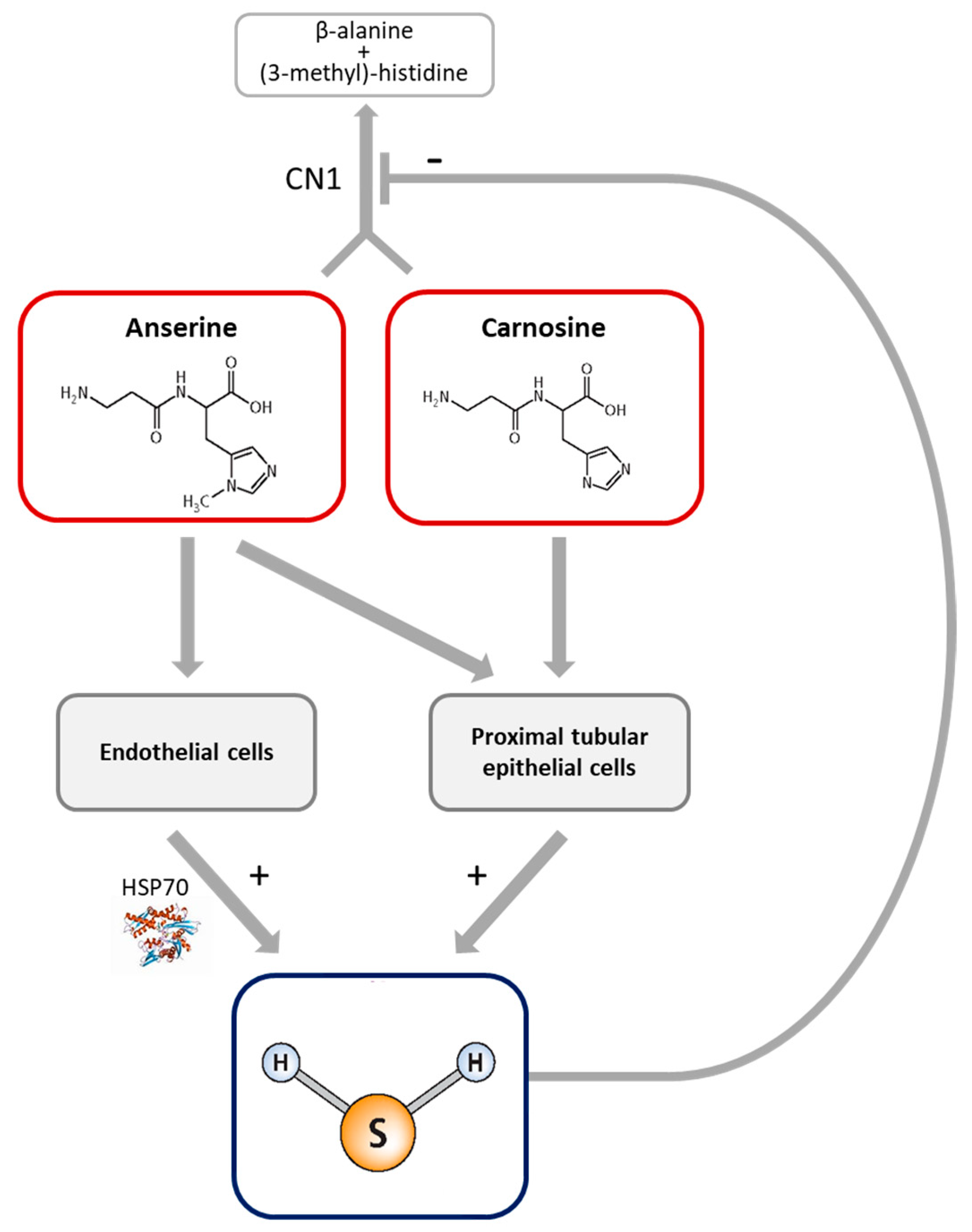
3.1. Dipeptide-Induced H2S Formation in Tubular Epithelial and Endothelial Cells

Homocysteine dose-dependently increased H2S formation in HK-2 cells (Supplementary Figure S1, Supplementary Table S1). At a homocysteine concentration of 5 mM, the H2S formation rate in HK-2, HUVEC, HUAEC and MCEC varied between 15 and 73 pmol H2S/mg protein (Figure 1, Supplementary Table S2). The addition of anserine dose-dependently increased H2S formation in HK-2 cells (Figure 1A, Supplementary Table S3); H2S formation from cysteine was below the detection limit in HK-2. Addition of 1 mM anserine increased H2S formation in all cell types, and the addition of 1 mM carnosine increased H2S formation in HK-2 cells only. Combined anserine and carnosine exposure had no additive effect beyond the anserine-mediated effects (Figure 1).
To study the interaction of H2S on anserine and carnosine availability, CN1 activity was measured at increasing concentrations of the H2S donor sodium disulfide (Na2S). Na2S dose-dependently reduced recombinant CN1 and human serum CN1 carnosine and anserine degradation activity (Figure 2, Supplementary Table S4).

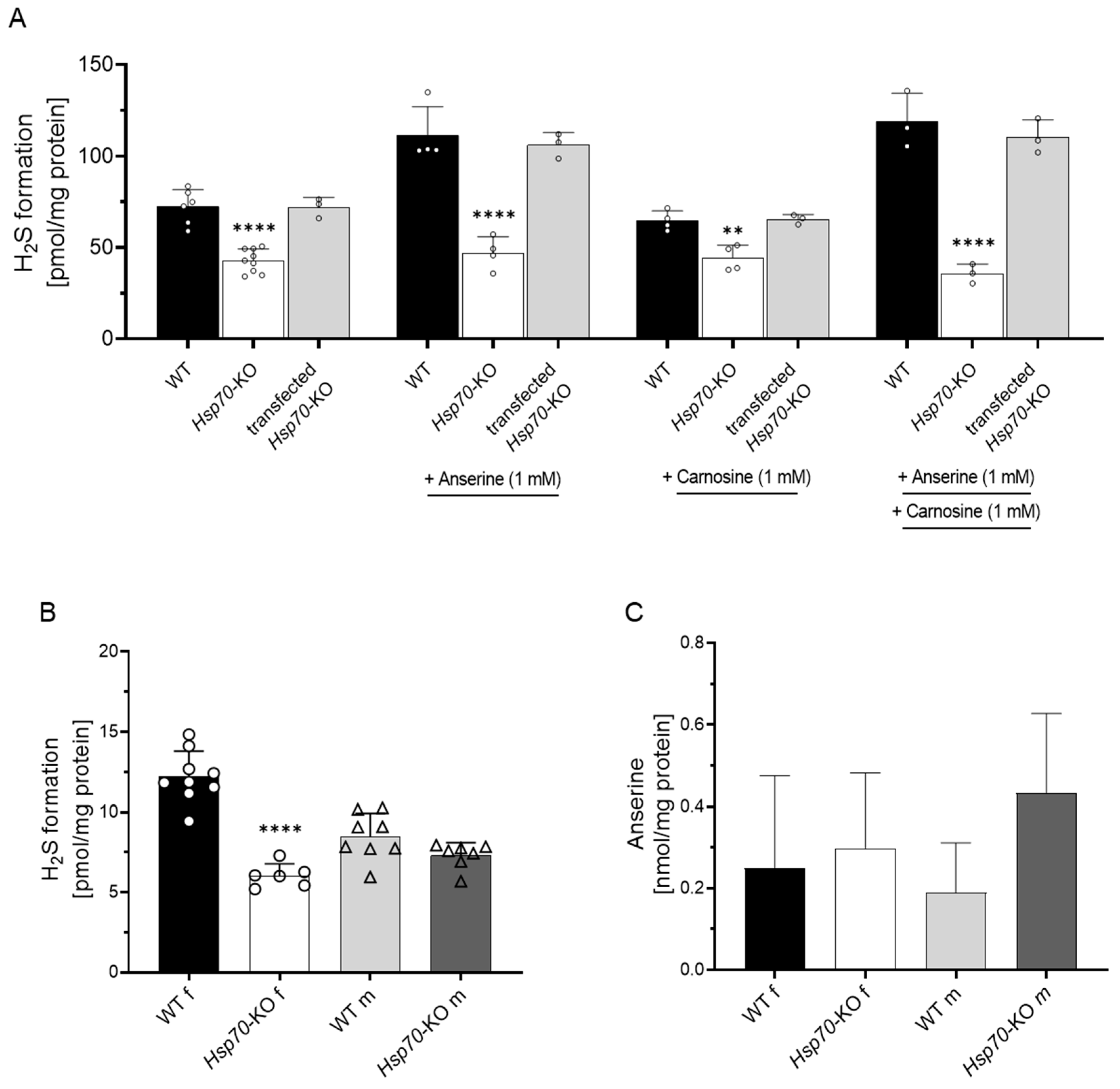
3.2. Anserine-Induced H2S Formation Depends on HSP70

Since anserine, carnosine and H2S have previously been described to modulate Hsp70 expression, we studied H2S formation capacity in MCEC with Hsp70-KO. H2S formation was 60% lower in Hsp70-KO MCEC compared to WT MCEC. Anserine and carnosine both did not increase H2S formation in the Hsp70-KO MCEC (Figure 3A). Rescue of the Hsp70-KO by Hsp70 transfection restored H2S formation to the level of WT MCEC (Figure 3A). Successful transfection of Hsp70 could be demonstrated for two further vectors by detection of total protein concentration of HSPA1A via Western blot (Supplementary Figures S2 and S3). In all three rescued cell clones, anserine increased H2S formation to a similar degree as in WT MCEC (Supplementary Figure S2A, Supplementary Table S5).

3.3. H2S Formation in Murine Kidney Tissue

We then studied ex vivo H2S formation capacity in kidney tissue of WT mice. Addition of homocysteine and cysteine at equimolar concentrations to kidney tissue homogenate both increased H2S formation, and homocysteine to a smaller extent (Supplementary Figure S4A, Supplementary Table S6). H2S formation was not influenced by five hours of fasting (Supplementary Figure S4B, Supplementary Table S6) and was markedly reduced by inhibition of both CBS and CSE activity, indicating that both enzymes are involved in kidney H2S formation (Supplementary Figure S5, Supplementary Table S7). To demonstrate the role of renal HSP70 on H2S formation, kidney tissues of mice with a global Hsp70-KO were studied in 23- to 28-week-old animals. Ex vivo kidney H2S formation capacity was 50% lower in female Hsp70-KO kidneys compared to WT mice (Figure 3B). In male Hsp70-KO mice, H2S formation was within the range of WT littermates. Kidney anserine concentrations were similar in all four groups (Figure 3C, Supplementary Table S8).

3.4. Kidney Anserine Abundance and H2S Formation

Incubation of kidney tissue with anserine and carnosine for six hours did not increase H2S formation (Supplementary Figure S6; Supplementary Table S9). We then studied the effect of a persistent increase of endogenous kidney anserine and carnosine concentrations on H2S formation in global Cndp1-KO mice. Male Cndp1-KO mice had 3-fold higher renal anserine concentrations, and female Cndp1-KO mice had 5- to 12-fold higher concentrations than respective WT controls, depending on age (Table 1).
Renal carnosine concentrations were below the detection limit in WT and markedly increased in Cndp1-KO mice. H2S formation was 2- to 3-fold higher in female Cndp1-KO mice compared to their respective WT controls, but not different between male Cndp1-KO and WT animals. H2S formation and anserine/carnosine concentrations were correlated in 23- to 25- and 47- to 51-week-old mice (Figure 4).
We then studied the impact of diabetes on kidney anserine and carnosine concentrations and H2S formation. Kidney anserine and carnosine concentrations were similar in type 1 diabetic and non-diabetic WT mice, and carnosine concentrations were below detection level. In diabetic Cndp1-KO mice, kidney anserine and carnosine concentrations, however, were lower compared to non-diabetic Cndp1-KO littermates and in diabetic mice not related with H2S formation (Table 1, Figure 5). In female diabetic Cndp1-KO mice, kidney H2S formation was reduced compared to female non-diabetic Cndp1-KO mice and similar to diabetic WT mice. In male diabetic Cndp1-KO mice, kidney H2S formation was even reduced compared to the male diabetic WT littermates.

4. Discussion

Anserine and carnosine are experimentally well-established compounds mitigating diabetic nephropathy, and the first clinical trials yielded beneficial effects [7,33,34,35]. Similarly, H2S improves outcome in acute and chronic kidney impairment, experimental sepsis, hemorrhagic shock and following ischemia/reperfusion [20]. The first clinical trials investigating the effects of the H2S donor Na2S2O3 in myocardial infarct in humans are ongoing [27].
Both anserine/carnosine and H2S act via multiple mechanisms, and activation of HSP70 is a prominent common pathway [14,15,19,36]. We now demonstrate the cell type-specific increase in H2S synthesis capacity by anserine and carnosine, mediated by HSP70 activation (Figure 6). Ex vivo studies of kidney tissue reconfirm a correlation of H2S synthesis with tissue anserine and carnosine concentrations and demonstrate the mechanistic role of HSP70 in kidney H2S synthesis.
There is growing evidence that histidine-containing dipeptides exert major protective functions in various disease states by interfering with specific pathomechanisms [17]. Carnosine acts as a carbonyl scavenger [12,37], an ion-chelating agent [10], as an Angiotensin-Converting Enzyme (ACE) inhibitor [38,39] and multifunctional antioxidant [40,41]. In HK-2 cells, anserine has a higher antioxidative capacity than carnosine [15]. These activities are mediated via hormetic processes involving Nrf2, Sirt-1, Trx, Hsp70, the glutathione system [12,17,41] and modulation of the nitric oxide formation and metabolism [42,43]. Anserine and carnosine increase HSP70 expression depending on the cell type. Carnosine increases HSP70 in podocytes [14], independent of glucose concentrations, but not in HK-2 cells. Anserine increases HSP70 in HK-2 cells in the presence of oxidative and glucose stress [14,15], and incubation with carnosine and anserine are well tolerated by cells [13]. Since H2S increases myocardial and cerebral HSP70 [19,34], we investigated a putative interaction of anserine and carnosine with H2S synthesis in vitro and demonstrated that carnosine dose-dependently increases H2S synthesis capacity in HK-2 cells, and anserine increases this capacity in capillary, aortic and umbilical vein endothelial cells, i.e., in endothelial cells derived from capillaries and large arterial and venous vasculature. The induction of H2S synthesis in endothelial cells by anserine was entirely HSP70-dependent. Hsp70-KO in capillary endothelial cells abolished the stimulatory effect of anserine on H2S formation, and transfection of Hsp70 into Hsp70-KO cells restored it. In line with this, in Hsp70-KO mice, kidney H2S synthesis was reduced by 50%. In contrast, the carnosine-induced upregulation of H2S synthesis in HK-2 cells should be HSP70-independent, since carnosine does not activate Hsp70 in HK-2 cells [15].
We then studied the impact of H2S within the highly regulated metabolism of anserine and carnosine via CN1. Carnosine is provided by nutrition and tissue carnosine synthase (Carns1), but the impact of the latter is uncertain. In Carns1-KO mice, brain and muscle are carnosine-deficient, and stores can be replenished by oral intake [44], but the Carns1-KO had no impact on kidney and brain markers of carbonyl and oxidative stress in healthy and diabetic mice [45]. In contrast, anserine and carnosine are rapidly degraded by CN1, and kidney tissue CN1 activity in healthy and in type 2 diabetic mice is correlated with kidney anserine and carnosine concentrations and the tissue carbonyl and oxidative stress level [46]. These findings suggest a gatekeeping role and protective function of CN1 for kidney histidine-containing dipeptide concentration. In line with this, diabetic patients, who are homozygous carriers of the Cndp1 gene variant CTG5, have significantly lower serum CN1 concentrations and activity, and its concentrations independently predict eGFR [47]. Previous enzyme kinetic studies and molecular dynamic simulations revealed inhibition of carnosine degrading CN1 activity by competitive inhibition with anserine [48], and by thiol-containing compounds due to allosteric interactions [49]; the latter interaction might be the underlying mechanism of the CN1 inhibition by H2S. In the same direction, we now demonstrate dose-dependent CN1 inhibition by H2S donor Na2S, suggesting interaction of anserine and of H2S in a positive feedback loop, i.e., mutual reinforcement of both protective mechanisms.
To demonstrate the impact of the interaction of anserine and carnosine with H2S synthesis capacity in vivo, we studied Cndp1-KO mice. These mice exhibit a kidney-selective, age- and gender0dependent 2- to 9-fold increase in kidney tissue anserine and carnosine concentrations, and kidney function is unaltered [32]. The increased kidney anserine and carnosine concentrations could be reconfirmed, underlying molecular mechanisms of sex-specific differences, such as the influence of estrogens, have not yet been studied in detail. In these mice, kidney anserine and carnosine concentrations correlated with the kidney tissue H2S synthesis capacity, underpinning the significance of the mechanistic interactions demonstrated in vitro in the in vivo setting. Experimental studies demonstrated the upregulation of vascular endothelial cell H2S synthesis by estrogen [50]. H2S has been previously recognized as a toxic gas, but has emerged as an important gaseous signaling molecule, and administration of the H2S donor thiosulfate (2 × 15 g) is well tolerated by humans [51]. The action of H2S involves a variety of molecular mechanisms, such as activation of PI3K/Akt/eNOS pathway, suppressing ferroptosis or the antioxidant effect mediated by Nrf2 signaling [52,53,54]. In line with the in vitro findings of carnosine inducing H2S synthesis only in proximal tubular epithelial cells, but anserine in all cell lines studied, correlations of kidney H2S synthesis capacity were higher with tissue anserine than carnosine concentrations. Of note, short-term incubation of kidney tissue with anserine did not increase H2S synthesis capacity, suggesting slower anserine-induced actions than observed in vitro, but unspecific alterations in the ex vivo tissue homogenate devoid of blood supply cannot be excluded.
The impact of diabetes mellitus was demonstrated in type 1 diabetic Cndp1-KO mice. While kidney anserine and carnosine concentrations were in a similar range in diabetic and non-diabetic WT mice, concentrations of both histidine-containing dipeptides were 3-fold lower in the kidney of diabetic versus non-diabetic Cndp1-KO mice. In the diabetic mice, H2S formation rate was not correlated with the low kidney anserine and carnosine. Thus, the type 1 diabetes mellitus Cndp1-KO model does not allow for firm conclusions on their interaction. In type 2 diabetes db/db mice of similar age, kidney anserine and carnosine concentrations were also reduced [46]. Plasma and kidney H2S levels have repeatedly been reported to be low in diabetes mellitus [20], except for one study with double transgenic Balb/c mice sacrificed at the age of 13.5 weeks that had increased kidney H2S [55], a finding which could be reproduced here in male, but not in female STZ mice with a similar duration of diabetes. In female STZ Cndp1-KO mice, H2S formation was lower compared to female non-diabetic KO littermates, presumably due to the 3-fold lower kidney anserine and carnosine concentrations.
Our findings do not exclude beneficial effects of pharmacological doses of anserine and carnosine in diabetes via induction of H2S. Numerous studies in rodents demonstrated kidney protection in diabetic mice by carnosine administration [5,6,7,56,57]. Only two studies reported on the administration of anserine [15,58], even though nephroprotective effects of carnosine may at least in part be exerted by methylation to anserine [59]. We recently demonstrated several-fold increased kidney anserine concentrations after oral carnosine supplementation in healthy and db/db mice for four weeks [60]. None of these studies evaluated the interaction of therapeutic anserine or carnosine supplementation with H2S metabolism in diabetic mice, but in view of our finding, it deserves validation in animal models and in clinical trials. Likewise, our findings are of interest with regard to the potential therapeutic impact of combined treatment such as in patients with ischemic heart disease treated with the H2S donor thiosulfate. Anserine may not only exert direct beneficial effects, but also increase H2S availability.
A limitation of our study is the measurement of H2S synthesis capacity, based on an endpoint quantification with Ag2S, providing a high specificity and reproducibility with reasonable sensitivity. This, however, does not provide information on actual cell and tissue H2S concentrations. Current methods to quantify H2S concentrations comprise colorimetric assays, gas chromatography, fluorescence probes and electrochemical technics, and the advantages and limitations have recently been summarized [61]. Future studies combining different methods may further increase insights and validity of our findings. In the same direction, we demonstrated the synthesis activity of CBS and CSE in murine kidney tissue, and the functional role of HSP70, but not the specific mode of action of HSP70 on enzyme expression and activity.

5. Conclusions

In conclusion, we demonstrate a novel mechanism of action of the histidine-containing dipeptides anserine and carnosine, i.e., the induction of H2S synthesis in proximal tubular epithelial cells and capillary, venous and aortic endothelial cells, which in endothelial cells is exerted via HSP70. The in vivo relevance of these interactions is demonstrated in Hsp70- and Cndp1-KO mice, but awaits clarification in diabetes mellitus and ischemic heart disease. The positive feedback interaction of H2S, inhibiting anserine and carnosine degradation by CN1 together with the H2S synthesis-inducing effect of both dipeptides, should enhance the efficacy of therapeutic interventions with anserine, carnosine and with H2S donors.

Supplementary Materials

The following supporting information can be downloaded at: https://www.mdpi.com/article/10.3390/antiox12010066/s1, Supplementary Table S1: H2S formation from homocysteine increases H2S formation dose-dependently in HK-2 cells (n = 3); Supplementary Table S2: H2S formation from homocysteine (5 mM) in HK-2 cells and different endothelial cell types (n = 3–6); Supplementary Table S3: Anserine increases H2S formation from homocysteine (5 mM) dose-dependently in HK-2 cells (n = 3); Supplementary Table S4: Addition of Na2S to serum inhibits CN1 activity dose-dependently (n = 3–8); Supplementary Table S5: Rescue of the Hsp70-KO by Hsp70-transfection (clone 1–3) restored H2S formation to the level of WT MCEC (n = 3–9); Supplementary Table S6: Renal H2S formation in Cndp1-KO and WT mice from homocysteine (47- to 51-week-old mice, n = 3) and form cysteine under fasting and non-fasting conditions (23- to 25-week-old mice, n = 3–5); Supplementary Table S7: H2S formation was reduced by CSE and CBS inhibitors in kidney tissue lysate of WT and Cndp1-KO mice (47- to 51-week-old, n = 3–5); Supplementary Table S8: Renal H2S formation and anserine concentrations in Hsp70-KO mice (23- to 28-week-old, n = 6–9); Supplementary Table S9: Incubation of kidney tissue lysate with anserine and carnosine (6h) did not increase renal H2S formation in WT mice (23- to 26-week-old, n = 3–5); Supplementary Figure S1: Homocysteine dependent H2S formation in human proximal tubular epithelial cells (HK-2); Supplementary Figure S2: H2S formation depends on HSP70; Supplementary Figure S3: Original western blot membranes of protein expression of HSPA1A and β-ACTIN in cell clones; Supplementary Figure S4: Ex vivo renal H2S formation in Cndp1-KO mice; Supplementary Figure S5: Cystathionine-γ-lyase (CSE) and cystathionine-β-synthase (CBS) activity dependent kidney H2S formation; Supplementary Figure S6: Exogenous anserine and carnosine did not affect H2S formation

Author Contributions

Conceptualization, C.W., T.P., C.P.S. and V.P.; methodology, C.W., T.P. and R.B.; S.M. and A.P.; software, C.W. and T.P.; validation, C.W. and T.P.; formal analysis, C.W. and T.P.; investigation, C.W. and T.P.; resources, C.W., T.P., R.B. and J.Z.; data curation, C.W. and T.P.; writing—original draft preparation, C.W. and T.P.; writing—review and editing, C.W., T.P., A.T.S., V.C., C.P.S. and V.P.; visualization, C.W., T.P., C.P.S. and V.P.; supervision, V.P. and C.P.S.; project administration, V.P. and C.P.S.; funding acquisition, V.P. and C.P.S. All authors have read and agreed to the published version of the manuscript.

Funding

This study was funded by grants from the Deutsche Forschungsgemeinschaft (DFG, German Research Foundation)—Project number 236360313—CRC 1118.

Institutional Review Board Statement

The animal studies were approved by the respective authorities (Regierungspräsidium Karlsruhe, Germany, 35-9165.81/G-209/16 and 35-9185.81/G-103/18) and performed according to guidelines (ARRIVE).

Informed Consent Statement

Not applicable.

Data Availability Statement

The data are contained within the article and Supplementary Materials.

Conflicts of Interest

The authors declare no conflict of interest.

References

  1. Kwiatkowski, S.; Kiersztan, A.; Drozak, J. Biosynthesis of carnosine and related dipeptides in vertebrates. Curr. Protein Pept. Sci. 2018, 19, 771–789. [Google Scholar] [CrossRef] [PubMed]
  2. Teufel, M.; Saudek, V.; Ledig, J.P.; Bernhardt, A.; Boularand, S.; Carreau, A.; Cairns, N.J.; Carter, C.; Cowley, D.J.; Duverger, D.; et al. Sequence identification and characterization of human carnosinase and a closely related non-specific dipeptidase. J. Biol. Chem. 2003, 278, 6251–6531. [Google Scholar] [CrossRef] [PubMed] [Green Version]
  3. Janssen, B.; Hohenadel, D.; Brinkkoetter, P.; Peters, V.; Rind, N.; Fischer, C.; Rychlik, I.; Cerna, M.; Romzova, M.; de Heer, E.; et al. Carnosine as a protective factor in diabetic nephropathy: Association with a leucine repeat of the carnosinase gene CNDP1. Diabetes 2005, 54, 2320–2327. [Google Scholar] [CrossRef] [PubMed] [Green Version]
  4. Zhou, Z.; Liu, X.Q.; Zhang, S.Q.; Qi, X.M.; Zhang, Q.; Yard, B.; Wu, Y.G. Correlation between serum carnosinase concentration and renal damage in diabetic nephropathy patients. Amino Acids 2021, 53, 687–700. [Google Scholar] [CrossRef]
  5. Peters, V.; Yard, B.; Schmitt, C.P. Carnosine and diabetic nephropathy. Curr. Med. Chem. 2019, 27, 1801–1812. [Google Scholar] [CrossRef]
  6. Kilis-Pstrusinska, K. Carnosine and Kidney Diseases: What We Currently Know? Curr. Med. Chem. 2020, 27, 1764–1781. [Google Scholar] [CrossRef]
  7. Matthews, J.J.; Dolan, E.; Swinton, P.A.; Santos, L.; Artioli, G.G.; Turner, M.D.; Elliott-Sale, K.J.; Sale, C. Effect of Carnosine or β-Alanine Supplementation on Markers of Glycemic Control and Insulin Resistance in Humans and Animals: A Systematic Review and Meta-analysis. Adv. Nutr. 2021, 12, 2216–2231. [Google Scholar] [CrossRef]
  8. Caruso, G. Unveiling the Hidden Therapeutic Potential of Carnosine, a Molecule with a Multimodal Mechanism of Action: A Position Paper. Molecules 2022, 27, 3303. [Google Scholar] [CrossRef]
  9. Oppermann, H.; Elsel, S.; Birkemeyer, C.; Meixensberger, J.; Gaunitz, F. Erythrocytes Prevent Degradation of Carnosine by Human Serum Carnosinase. Int. J. Mol. Sci. 2021, 22, 12802. [Google Scholar] [CrossRef]
  10. Holeček, M. Histidine in Health and Disease: Metabolism, Physiological Importance, and Use as a Supplement. Nutrients 2020, 12, 848. [Google Scholar] [CrossRef]
  11. Boldyrev, A.A.; Aldini, G.; Derave, W. Physiology and pathophysiology of carnosine. Physiol. Rev. 2013, 93, 1803–1845. [Google Scholar] [CrossRef] [PubMed]
  12. Aldini, G.; de Courten, B.; Regazzoni, L.; Gilardoni, E.; Ferrario, G.; Baron, G.; Altomare, A.; D’Amato, A.; Vistoli, G.; Carini, M. Understanding the antioxidant and carbonyl sequestering activity of carnosine: Direct and indirect mechanisms. Free Radic. Res. 2021, 55, 321–330. [Google Scholar] [CrossRef] [PubMed]
  13. Weigand, T.; Singler, B.; Fleming, T.; Nawroth, P.; Klika, K.D.; Thiel, C.; Baelde, H.; Garbade, S.F.; Wagner, A.H.; Hecker, M.; et al. Carnosine Catalyzes the Formation of the Oligo/Polymeric Products of Methylglyoxal. Cell Physiol. Biochem. 2018, 46, 713–726. [Google Scholar] [CrossRef] [PubMed]
  14. Scuto, M.; Trovato Salinaro, A.; Modafferi, S.; Polimeni, A.; Pfeffer, T.; Weigand, T.; Calabrese, V.; Schmitt, C.P.; Peters, V. Carnosine Activates Cellular Stress Response in Podocytes and Reduces Glycative and Lipoperoxidative Stress. Biomedicines 2020, 8, 177. [Google Scholar] [CrossRef]
  15. Peters, V.; Calabrese, V.; Forsberg, E.; Volk, N.; Fleming, T.; Baelde, H.; Weigand, T.; Thiel, C.; Trovato, A.; Scuto, M.; et al. Protective Actions of Anserine Under Diabetic Conditions. Int. J. Mol. Sci. 2018, 19, 2751. [Google Scholar] [CrossRef] [Green Version]
  16. Chebotareva, N.; Bobkova, I.; Shilov, E. Heat shock proteins and kidney disease: Perspectives of HSP therapy. Cell Stress Chaperones 2017, 22, 319–343. [Google Scholar] [CrossRef] [Green Version]
  17. Calabrese, V.; Scuto, M.; Salinaro, A.T.; Dionisio, G.; Modafferi, S.; Ontario, M.L.; Greco, V.; Sciuto, S.; Schmitt, C.P.; Calabrese, E.J.; et al. Hydrogen Sulfide and Carnosine: Modulation of Oxidative Stress and Inflammation in Kidney and Brain Axis. Antioxidants 2020, 9, 1303. [Google Scholar] [CrossRef]
  18. Bellini, S.; Barutta, F.; Mastrocola, R.; Imperatore, L.; Bruno, G.; Gruden, G. Heat Shock Proteins in Vascular Diabetic Complications: Review and Future Perspective. Int. J. Mol. Sci. 2017, 18, 2709. [Google Scholar] [CrossRef] [Green Version]
  19. Calvert, J.W.; Jha, S.; Gundewar, S.; Elrod, J.W.; Ramachandran, A.; Pattillo, C.B.; Kevil, C.G.; Lefer, D.J. Hydrogen sulfide mediates cardioprotection through Nrf2 signaling. Circ. Res. 2009, 105, 365–374. [Google Scholar] [CrossRef] [Green Version]
  20. Peleli, M.; Zampas, P.; Papapetropoulos, A. Hydrogen Sulfide and the Kidney: Physiological Roles, Contribution to Pathophysiology, and Therapeutic Potential. Antioxid. Redox Signal. 2022, 36, 220–243. [Google Scholar] [CrossRef]
  21. Kabil, O.; Banerjee, R. Enzymology of H2S biogenesis, decay and signaling. Antioxid. Redox Signal. 2014, 20, 770–782. [Google Scholar] [CrossRef] [PubMed] [Green Version]
  22. Zhao, K.; Li, H.; Li, S.; Yang, G. Regulation of cystathionine gamma-lyase/H₂S system and its pathological implication. Front. Biosci. 2014, 19, 1355–1369. [Google Scholar] [CrossRef] [PubMed]
  23. Beck, K.F.; Pfeilschifter, J. The Pathophysiology of H(2)S in Renal Glomerular Diseases. Biomolecules 2022, 12, 207. [Google Scholar] [CrossRef] [PubMed]
  24. Kuang, Q.; Xue, N.; Chen, J.; Shen, Z.; Cui, X.; Fang, Y.; Ding, X. Low Plasma Hydrogen Sulfide Is Associated with Impaired Renal Function and Cardiac Dysfunction. Am. J. Nephrol. 2018, 47, 361–371. [Google Scholar] [CrossRef]
  25. Ngowi, E.E.; Sarfraz, M.; Afzal, A.; Khan, N.H.; Khattak, S.; Zhang, X.; Li, T.; Duan, S.F.; Ji, X.Y.; Wu, D.D. Roles of Hydrogen Sulfide Donors in Common Kidney Diseases. Front. Pharmacol. 2020, 11, 564281. [Google Scholar] [CrossRef]
  26. Koning, A.M.; Frenay, A.R.; Leuvenink, H.G.; van Goor, H. Hydrogen sulfide in renal physiology, disease and transplantation--the smell of renal protection. Nitric Oxide Biol. Chem. 2015, 46, 37–49. [Google Scholar] [CrossRef]
  27. de Koning, M.L.; van Dorp, P.; Assa, S.; Hartman, M.H.; Voskuil, M.; Anthonio, R.L.; Veen, D.; Pundziute-Do Prado, G.; Leiner, T.; van Goor, H.; et al. Rationale and Design of the Groningen Intervention Study for the Preservation of Cardiac Function with Sodium Thiosulfate after St-segment Elevation Myocardial Infarction (GIPS-IV) trial. Am. Heart J. 2022, 243, 167–176. [Google Scholar] [CrossRef]
  28. Lu, P.H.; Chuo, H.E.; Kuo, K.L.; Liao, J.F.; Lu, P.H. Clinical Efficacy and Safety of Sodium Thiosulfate in the Treatment of Uremic Pruritus: A Meta-Analysis of Randomized Controlled Trials. Toxins 2021, 13, 769. [Google Scholar] [CrossRef]
  29. Merz, T.; McCook, O.; Brucker, C.; Waller, C.; Calzia, E.; Radermacher, P.; Datzmann, T. H(2)S in Critical Illness-A New Horizon for Sodium Thiosulfate? Biomolecules 2022, 12, 543. [Google Scholar] [CrossRef]
  30. Zhou, X.; Feng, Y.; Zhan, Z.; Chen, J. Hydrogen sulfide alleviates diabetic nephropathy in a streptozotocin-induced diabetic rat model. J. Biol. Chem. 2014, 289, 28827–28834. [Google Scholar] [CrossRef]
  31. Ahn, Y.J.; Lee, Y.J.; Lee, J.; Lee, D.; Park, H.K.; Lee, G.J. Colorimetric detection of endogenous hydrogen sulfide production in living cells. Spectrochim. Acta Part A Mol. Biomol. Spectrosc. 2017, 177, 118–124. [Google Scholar] [CrossRef] [PubMed]
  32. Weigand, T.; Colbatzky, F.; Pfeffer, T.; Garbade, S.F.; Klingbeil, K.; Colbatzky, F.; Becker, M.; Zemva, J.; Bulkescher, R.; Schürfeld, R.; et al. A Global Cndp1-Knock-Out Selectively Increases Renal Carnosine and Anserine Concentrations in an Age- and Gender-Specific Manner in Mice. Int. J. Mol. Sci. 2020, 21, 4887. [Google Scholar] [CrossRef] [PubMed]
  33. Elbarbary, N.S.; Ismail, E.A.R.; El-Naggar, A.R.; Hamouda, M.H.; El-Hamamsy, M. The effect of 12 weeks carnosine supplementation on renal functional integrity and oxidative stress in pediatric patients with diabetic nephropathy: A randomized placebo-controlled trial. Pediatr. Diabetes 2017, 19, 470–477. [Google Scholar] [CrossRef] [PubMed]
  34. Siriwattanasit, N.; Satirapoj, B.; Supasyndh, O. Effect of Oral carnosine supplementation on urinary TGF-β in diabetic nephropathy: A randomized controlled trial. BMC Nephrol. 2021, 22, 236. [Google Scholar] [CrossRef]
  35. Menon, K.; Marquina, C.; Liew, D.; Mousa, A.; de Courten, B. Histidine-containing dipeptides reduce central obesity and improve glycaemic outcomes: A systematic review and meta-analysis of randomized controlled trials. Obes. Rev. Off. J. Int. Assoc. Study Obes. 2020, 21, e12975. [Google Scholar] [CrossRef]
  36. Ji, K.; Xue, L.; Cheng, J.; Bai, Y. Preconditioning of H2S inhalation protects against cerebral ischemia/reperfusion injury by induction of HSP70 through PI3K/Akt/Nrf2 pathway. Brain Res. Bull. 2016, 121, 68–74. [Google Scholar] [CrossRef]
  37. Lavilla, C.J.; Billacura, M.P.; Hanna, K.; Boocock, D.J.; Coveney, C.; Miles, A.K.; Foulds, G.A.; Murphy, A.; Tan, A.; Jackisch, L.; et al. Carnosine protects stimulus-secretion coupling through prevention of protein carbonyl adduction events in cells under metabolic stress. Free Radic. Biol. Med. 2021, 175, 65–79. [Google Scholar] [CrossRef]
  38. Saadah, L.M.; Deiab, G.I.A.; Al-Balas, Q.; Basheti, I.A. Carnosine to Combat Novel Coronavirus (nCoV): Molecular Docking and Modeling to Cocrystallized Host Angiotensin-Converting Enzyme 2 (ACE2) and Viral Spike Protein. Molecules 2020, 25, 5605. [Google Scholar] [CrossRef]
  39. Nakagawa, K.; Ueno, A.; Nishikawa, Y. Interactions between carnosine and captopril on free radical scavenging activity and angiotensin-converting enzyme activity in vitro. Yakugaku Zasshi 2006, 126, 37–42. [Google Scholar] [CrossRef] [Green Version]
  40. Calabrese, V.; Cornelius, C.; Mancuso, C.; Pennisi, G.; Calafato, S.; Bellia, F.; Bates, T.E.; Giuffrida Stella, A.M.; Schapira, T.; Dinkova Kostova, A.T.; et al. Cellular stress response: A novel target for chemoprevention and nutritional neuroprotection in aging, neurodegenerative disorders and longevity. Neurochem. Res. 2008, 33, 2444–2471. [Google Scholar] [CrossRef]
  41. Caruso, G.; Privitera, A.; Antunes, B.M.; Lazzarino, G.; Lunte, S.M.; Aldini, G.; Caraci, F. The Therapeutic Potential of Carnosine as an Antidote against Drug-Induced Cardiotoxicity and Neurotoxicity: Focus on Nrf2 Pathway. Molecules 2022, 27, 4452. [Google Scholar] [CrossRef] [PubMed]
  42. Fresta, C.G.; Fidilio, A.; Lazzarino, G.; Musso, N.; Grasso, M.; Merlo, S.; Amorini, A.M.; Bucolo, C.; Tavazzi, B.; Lazzarino, G.; et al. Modulation of Pro-Oxidant and Pro-Inflammatory Activities of M1 Macrophages by the Natural Dipeptide Carnosine. Int. J. Mol. Sci. 2020, 21, 776. [Google Scholar] [CrossRef] [PubMed] [Green Version]
  43. Zhao, J.M.; Chen, X.; Cheng, K.; Shi, Q.; Peng, K. Anserine and glucosamine supplementation attenuates the levels of inflammatory markers in rats with rheumatoid arthritis. AMB Express 2020, 10, 57. [Google Scholar] [CrossRef] [PubMed] [Green Version]
  44. Sakano, T.; Egusa, A.S.; Kawauchi, Y.; Wu, J.; Nishimura, T.; Nakao, N.; Kuramoto, A.; Kawashima, T.; Shiotani, S.; Okada, Y.; et al. Pharmacokinetics and tissue distribution of orally administrated imidazole dipeptides in carnosine synthase gene knockout mice. Biosci. Biotechnol. Biochem. 2022, 86, 1276–1285. [Google Scholar] [CrossRef]
  45. Wang-Eckhardt, L.; Becker, I.; Wang, Y.; Yuan, J.; Eckhardt, M. Absence of endogenous carnosine synthesis does not increase protein carbonylation and advanced lipoxidation end products in brain, kidney or muscle. Amino Acids 2022, 54, 1013–1023. [Google Scholar] [CrossRef]
  46. Peters, V.; Lanthaler, B.; Amberger, A.; Fleming, T.; Forsberg, E.; Hecker, M.; Wagner, A.H.; Yue, W.W.; Hoffmann, G.F.; Nawroth, P.; et al. Carnosine metabolism in diabetes is altered by reactive metabolites. Amino Acids 2015, 47, 2367–2376. [Google Scholar] [CrossRef]
  47. Zhang, S.; Albrecht, T.; Rodriguez-Niño, A.; Qiu, J.; Schnuelle, P.; Peters, V.; Schmitt, C.P.; van den Born, J.; Bakker, S.J.L.; Lammert, A.; et al. Carnosinase concentration, activity, and CNDP1 genotype in patients with type 2 diabetes with and without nephropathy. Amino Acids 2019, 51, 611–617. [Google Scholar] [CrossRef]
  48. Peters, V.; Jansen, E.E.; Jakobs, C.; Riedl, E.; Janssen, B.; Yard, B.A.; Wedel, J.; Hoffmann, G.F.; Zschocke, J.; Gotthardt, D.; et al. Anserine inhibits carnosine degradation but in human serum carnosinase (CN1) is not correlated with histidine dipeptide concentration. Clin. Chim Acta 2011, 412, 263–267. [Google Scholar] [CrossRef]
  49. Peters, V.; Schmitt, C.P.; Weigand, T.; Klingbeil, K.; Thiel, C.; van den Berg, A.; Calabrese, V.; Nawroth, P.; Fleming, T.; Forsberg, E.; et al. Allosteric inhibition of carnosinase (CN1) by inducing a conformational shift. J. Enzym. Inhib Med. Chem 2017, 32, 1102–1110. [Google Scholar] [CrossRef] [Green Version]
  50. Teoh, J.P.; Li, X.; Simoncini, T.; Zhu, D.; Fu, X. Estrogen-Mediated Gaseous Signaling Molecules in Cardiovascular Disease. Trends Endocrinol. Metab. TEM 2020, 31, 773–784. [Google Scholar] [CrossRef]
  51. de Koning, M.L.Y.; Assa, S.; Maagdenberg, C.G.; van Veldhuisen, D.J.; Pasch, A.; van Goor, H.; Lipsic, E.; van der Harst, P. Safety and Tolerability of Sodium Thiosulfate in Patients with an Acute Coronary Syndrome Undergoing Coronary Angiography: A Dose-Escalation Safety Pilot Study (SAFE-ACS). J. Interv. Cardiol. 2020, 2020, 6014915. [Google Scholar] [CrossRef] [PubMed]
  52. Lin, F.; Yang, Y.; Wei, S.; Huang, X.; Peng, Z.; Ke, X.; Zeng, Z.; Song, Y. Hydrogen Sulfide Protects Against High Glucose-Induced Human Umbilical Vein Endothelial Cell Injury Through Activating PI3K/Akt/eNOS Pathway. Drug Des. Dev. Ther. 2020, 14, 621–633. [Google Scholar] [CrossRef] [PubMed] [Green Version]
  53. Wang, Y.; Liao, S.; Pan, Z.; Jiang, S.; Fan, J.; Yu, S.; Xue, L.; Yang, J.; Ma, S.; Liu, T.; et al. Hydrogen sulfide alleviates particulate matter-induced emphysema and airway inflammation by suppressing ferroptosis. Free Radic. Biol. Med. 2022, 186, 1–16. [Google Scholar] [CrossRef]
  54. Zhao, H.; Qiu, Y.; Wu, Y.; Sun, H.; Gao, S. Protective Effects of GYY4137 on Renal Ischaemia/Reperfusion Injury through Nrf2-Mediated Antioxidant Defence. Kidney Blood Press. Res. 2021, 46, 257–265. [Google Scholar] [CrossRef]
  55. Uyy, E.; Suica, V.I.; Boteanu, R.M.; Safciuc, F.; Cerveanu-Hogas, A.; Ivan, L.; Stavaru, C.; Simionescu, M.; Antohe, F. Diabetic nephropathy associates with deregulation of enzymes involved in kidney sulphur metabolism. J. Cell. Mol. Med. 2020, 24, 12131–12140. [Google Scholar] [CrossRef]
  56. Rodriguez-Niño, A.; Pastene, D.O.; Hettler, S.A.; Qiu, J.; Albrecht, T.; Vajpayee, S.; Perciaccante, R.; Gretz, N.; Bakker, S.J.L.; Krämer, B.K.; et al. Influence of carnosine and carnosinase-1 on diabetes-induced afferent arteriole vasodilation: Implications for glomerular hemodynamics. Am. J. Physiol. Ren. Physiol. 2022, 323, F69–F80. [Google Scholar] [CrossRef] [PubMed]
  57. Liu, X.Q.; Jiang, L.; Lei, L.; Nie, Z.Y.; Zhu, W.; Wang, S.; Zeng, H.X.; Zhang, S.Q.; Zhang, Q.; Yard, B.; et al. Carnosine alleviates diabetic nephropathy by targeting GNMT, a key enzyme mediating renal inflammation and fibrosis. Clin. Sci. 2020, 134, 3175–3193. [Google Scholar] [CrossRef] [PubMed]
  58. Everaert, I.; Van der Stede, T.; Stautemas, J.; Hanssens, M.; van Aanhold, C.; Baelde, H.; Vanhaecke, L.; Derave, W. Oral anserine supplementation does not attenuate type-2 diabetes or diabetic nephropathy in BTBR ob/ob mice. Amino Acids 2021, 53, 1269–1277. [Google Scholar] [CrossRef]
  59. Drozak, J.; Chrobok, L.; Poleszak, O.; Jagielski, A.K.; Derlacz, R. Molecular identification of carnosine N-methyltransferase as chicken histamine N-methyltransferase-like protein (hnmt-like). PLoS ONE 2013, 8, e64805. [Google Scholar] [CrossRef] [Green Version]
  60. Peters, V.; Schmitt, C.P.; Zschocke, J.; Gross, M.L.; Brismar, K.; Forsberg, E. Carnosine treatment largely prevents alterations of renal carnosine metabolism in diabetic mice. Amino Acids 2012, 42, 2411–2416. [Google Scholar] [CrossRef]
  61. Luo, Y.; Zuo, Y.; Shi, G.; Xiang, H.; Gu, H. Progress on the reaction-based methods for detection of endogenous hydrogen sulfide. Anal. Bioanal. Chem. 2022, 414, 2809–2839. [Google Scholar] [CrossRef] [PubMed]
Figure 1. H2S formation in proximal tubular epithelial (HK-2) and endothelial cells (HUVEC, HUAEC, MCEC). (A) Anserine and carnosine dose-dependently increased H2S formation in HK-2 cells. (B) Treatment of HUVEC, HUAEC and MCEC with anserine but not carnosine (each 1 mM) increased H2S formation compared to untreated cells. **: p < 0.01; ***: p < 0.001; ****: p < 0.0001 (one-way ANOVA with Tukey’s test).
Antioxidants 12 00066 g001
Figure 2. Effect of H2S on human serum CN1 activity and on recombinant CN1 activity (rCN1). (A) Addition of H2S donor sodium disulfide (Na2S) reduced human serum CN1 activity dose-dependently for carnosine (left panel) and anserine (right panel) as the substrate. (B) Degradation rate for carnosine was also reduced by Na2S for the recombinant carnosinase 1 (rCN1). **: p < 0.01; ****: p < 0.0001 (one-way ANOVA with Tukey’s test).
Antioxidants 12 00066 g002
Figure 3. Decreased H2S formation in Hsp70 knockout MCEC and mice. (A) H2S formation was lower in Hsp70-KO MCEC compared to WT MCEC; Hsp70-transfection restored H2S formation. (B) Renal H2S formation was lower in female Hsp70-KO compared to female WT mice. (C) Renal anserine concentrations were similar in all four groups. **: p < 0.01; ****: p < 0.0001 (one-way ANOVA with Tukey’s test).
Antioxidants 12 00066 g003
Figure 4. Renal H2S formation in WT and Cndp1-KO mice. (AC) H2S formation in kidney tissue of 23-to 25-week-old and (DF) 47–51-week-old mice. H2S formation was increased in female Cndp1-KO vs. female control mice. Kidney H2S formation correlated with renal anserine concentrations (B,E) and with renal carnosine concentrations (C,F) in Cndp1-KO (green symbols) and WT (blue symbols) mice. Circles depict female and triangle male mice. ****: p < 0.0001 (one-way ANOVA with Tukey’s test).
Antioxidants 12 00066 g004
Figure 5. Renal H2S formation in diabetic WT and Cndp1-KO mice. (A) In female diabetic Cndp1-KO mice, kidney H2S formation was reduced compared to non-diabetic female Cndp1-KO mice and similar to diabetic WT mice. In male diabetic Cndp1-KO mice, kidney H2S formation was even reduced compared to male diabetic WT mice. (B) Kidney anserine and (C) carnosine concentrations were markedly reduced in diabetic Cndp1-KO mice (beige symbols) compared to diabetic controls (pink symbols) and not related with H2S formation. Circles depict female and triangle male mice. *: p < 0.05; **: p < 0.01; ****: p < 0.0001 (one-way ANOVA with Tukey’s test).
Antioxidants 12 00066 g005
Figure 6. Anserine and carnosine induce H2S formation in a cell type- and Hsp70-specific manner within a positive feedback loop with CN1.
Antioxidants 12 00066 g006
Table 1. Renal anserine and carnosine concentrations in kidney tissue of WT and Cndp1-KO mice (n = 3–17).
9Age of Mice
(Weeks)
GenderAnserine
(nmol/mg)
Carnosine
(nmol/mg)
H2S Formation
(pmol/mg)
WT23–25f0.8 ± 0.4<0.1 nmol10.74 ± 1.21
WT23–25m0.7 ± 0.5<0.1 nmol9.44 ± 1.78
Cndp1-KO23–25f3.0 ± 1.33.8 ± 2.3621.33 ± 4.46 a
Cndp1-KO23–25m1.8 ± 0.8 a0.9 ± 0.479.62 ± 1.79
WT47–51f0.3 ± 0.04<0.1 nmol10.48 ± 2.20
WT47–51m0.3 ± 0.2<0.1 nmol9.88 ± 1.11
Cndp1-KO47–51f3.1 ± 1.3 a1.81 ± 0.7229.22 ± 6.24 a
Cndp1-KO47–51m1.1 ± 0.3 a0.32 ± 0.196.38 ± 1.14 a
STZ WT23–25f0.7 ± 0.4<0.1 nmol13.20 ± 4.62
STZ WT23–25m0.8 ± 0.4<0.1 nmol13.43 ± 1.67 b
STZ Cndp1-KO23–25f1.0 ± 0.4 b0.80 ± 0.29 b13.84 ± 2.53 b
STZ Cndp1-KO23–25m1.2 ± 0.5 a,b0.68 ± 0.318.38 ± 3.21 a
a = significantly different compared to respective WT group, b = significantly different compared to respective non-diabetic group. Statistical analysis was done by unpaired Student’s t-test.
Disclaimer/Publisher’s Note: The statements, opinions and data contained in all publications are solely those of the individual author(s) and contributor(s) and not of MDPI and/or the editor(s). MDPI and/or the editor(s) disclaim responsibility for any injury to people or property resulting from any ideas, methods, instructions or products referred to in the content.

