Article

An In Vitro and In Silico Study of Antioxidant Properties of Curcuminoid N-alkylpyridinium Salts: Initial Assessment of Their Antitumoral Properties

by
Oscar Forero-Doria
1,
Luis Guzmán
2,
Felipe Jiménez-Aspee
3,
Javier Echeverría
4,
Sergio Wehinger
5,
Claudio Valenzuela
6,
Ramiro Araya-Maturana
7,8,* and
Maximiliano Martínez-Cifuentes
9,*
1
Departamento de Ciencias Básicas, Facultad de Ciencias, Universidad Santo Tomás, Avenida Carlos Schorr 255, Talca 3460000, Chile
2
Departamento de Bioquímica Clínica e Inmunohematología, Facultad de Ciencias de la Salud, Universidad de Talca, P.O. Box 747, Talca 3460000, Chile
3
Institute of Nutritional Sciences, Department of Food Biofunctionality, University of Hohenheim, Garbenstrasse 28, 70599 Stuttgart, Germany
4
Departamento de Ciencias del Ambiente, Facultad de Química y Biología, Universidad de Santiago de Chile, Santiago 9170022, Chile
5
Thrombosis Research Center, Department of Clinical Biochemistry and Immunohematology, Faculty of Health Sciences, Medical Technology School, Universidad de Talca, Talca 3460000, Chile
6
Center for Medical Research, School of Medicine, University of Talca, Talca 3460000, Chile
7
Instituto de Química de Recursos Naturales, Universidad de Talca, P.O. Box 747, Talca 3460000, Chile
8
MIBI: Interdisciplinary Group on Mitochondrial Targeting and Bioenergetics, Universidad de Talca, P.O. Box 747, Talca 3460000, Chile
9
Departamento de Química Orgánica, Facultad de Ciencias Químicas, Universidad de Concepción, Edmundo Larenas 129, Concepción 4070371, Chile
*
Authors to whom correspondence should be addressed.
Antioxidants 2022, 11(6), 1104; https://doi.org/10.3390/antiox11061104
Submission received: 4 May 2022 / Revised: 27 May 2022 / Accepted: 29 May 2022 / Published: 1 June 2022

Abstract

:
In this work, we report the synthesis of curcuminoids with ionic liquid characteristics, obtained by incorporating alkyl-substituted pyridinium moiety rather than one phenyl group through a two-step process. The antioxidant capacity of the obtained compounds was evaluated in vitro by 1,1-diphenyl-picrylhydrazyl (DPPH) free radical scavenging and ferric reducing antioxidant power (FRAP) assays, showing that some derivatives are more potent than curcumin. Pyridine curcuminoids (group 4) and curcuminoid N-alkylpyridinium salts with two methoxyl groups in the phenyl ring (group 7), presented the best antioxidant capacity. The experimental results were rationalized by density functional theory (DFT) calculations of the bond dissociation enthalpy (BDE) for O–H in each compound. The computational calculations allowed for insight into the structural–antioxidant properties relationship in these series of compounds. BDEs, obtained in the gas phase and water, showed a notable impact of water solvation on the stabilization of some radicals. The lower values of BDEs in the water solution correspond to the structurally related compounds curcuminoid-pyridine 4c and curcuminoid pyridinium salt 7a, which is consistent with the experimental results. Additionally, an assessment of cell viability and cell migration assays was performed for human colon cancer (HT29), human breast cancer (MCF7) cells, in addition to NIH3T3 murine fibroblast, as a model of non-cancer cell type. These compounds mainly cause inhibition of the cell migration observed in MCF7 cancer cells without affecting the non-tumoral NIH3T3 cell line: Neither in viability nor in migration.

Graphical Abstract

1. Introduction

Curcumin (diferuloylmethane or 1,7-bis(4-hydroxy-3-methoxyphenyl)-1,6-heptadiene-3,5-dione) (Figure 1) has attracted considerable interest due to its diverse biological activities, which include antioxidant, antitumoral, antimicrobial, anti-inflammatory, antihepatotoxic, antihyperlipidemic, antiviral, and anti-Alzheimer’s disease. However, its poor solubility and poor bioavailability during oral administration result in limited clinical applicability [1]. Different strategies have been used to improve it, including encapsulating or incorporating curcumin in a nanoparticle or microparticle drug delivery system, synthesizing more stable curcumin analogs that resist metabolism while retaining the pharmacological properties of curcumin, adding another natural product that has bio-enhancing properties to curcumin or a combination of two of these strategies [2]. Compared with native curcumin, micellar curcumin and curcumin-γ-cyclodextrin complexes exhibit greater bioavailability in humans, indicating that the post-digestive solubility is the most important factor to improve its pharmacokinetics [3].
Hybrid curcumin molecules are currently used in medicinal chemistry to search for drugs aimed at treating multifactorial diseases. Compounds with radical scavenger properties of curcumin have shown remarkable activities related to the activation of the antioxidant response element (ARE). This controls the expression of genes, whose protein products have key roles in cellular defense against the toxicity of electrophiles and ROS, providing neuroprotective effects [4]. In addition, several compounds have shown significant inhibitory activities against α-glucosidase (α-Gls) [5]. Moreover, curcumin derivatives have attracted considerable attention in the search for compounds with antioxidant and antibacterial activities combined in one molecule, which have been proposed for chronic infections associated with epithelial damage by pulmonary stress [6]. The reported anticancer activities of modified curcumin, with varying structural features, such as aromatic side chains, di-keto functionality, active methylene groups, carbon linker chain, and mono-carbonyl analogs, show the high potential of these types of compounds [7]. Among them, the antitumor activity of curcumin derivatives against triple-negative breast cancer, a highly metastatic breast cancer cell line, was inversely correlated with their antioxidant activity, as measured by the DPPH assay [8].
Recently, anticancer therapies target the uncontrolled proliferation of cancer cells. However, these strategies have proved highly inefficient in treating metastatic solid tumors, since the mechanisms of migration are not inhibited by conventional anticancer drugs. Therefore, the search for compounds that inhibit migration and invasion of solid cancers is highly relevant [9,10,11]. In this context, the antimigratory activity of some curcumin derivatives has been reported [12,13].
A promising strategy for obtaining curcuminoids with ionic liquid characteristics can overcome the main drawbacks associated with curcumin. Ionic liquids are composed of bulky organic cations, in combination with a wide variety of anions, ranging from simple inorganic anions to more complex organic species [14], and have melting points below 100 °C [15]. Moreover, some ionic liquids contain nitrogen and those with anticancer properties have been reported. For example, imidazolium salts containing long alkyl chains exhibit high growth inhibition and variable cytotoxicity against a wide panel of cancer cell lines [16]. Di-pyridinium salts containing octyl chains have cytotoxic effects against glioma cells [17]. In addition, the synthesis of various heterocyclic curcumin analogs of pharmacological interest has been reported [18,19].
In this work, we have obtained new analogs of curcumin with ionic liquid characteristics, through the replacement of one phenyl group with an alkyl-substituted pyridinium group. The in vitro antioxidant properties and computational calculations allowed for insight into the structural–antioxidant properties relationship in these series of compounds. To assess the anticancer potential of these compounds, cell viability and cell migration assays were performed for human colon cancer (HT29), human breast cancer (MCF7) cells, in addition to NIH3T3 murine fibroblast, as a model of non-tumoral cell type.

2. Materials and Methods

2.1. Chemical Synthesis
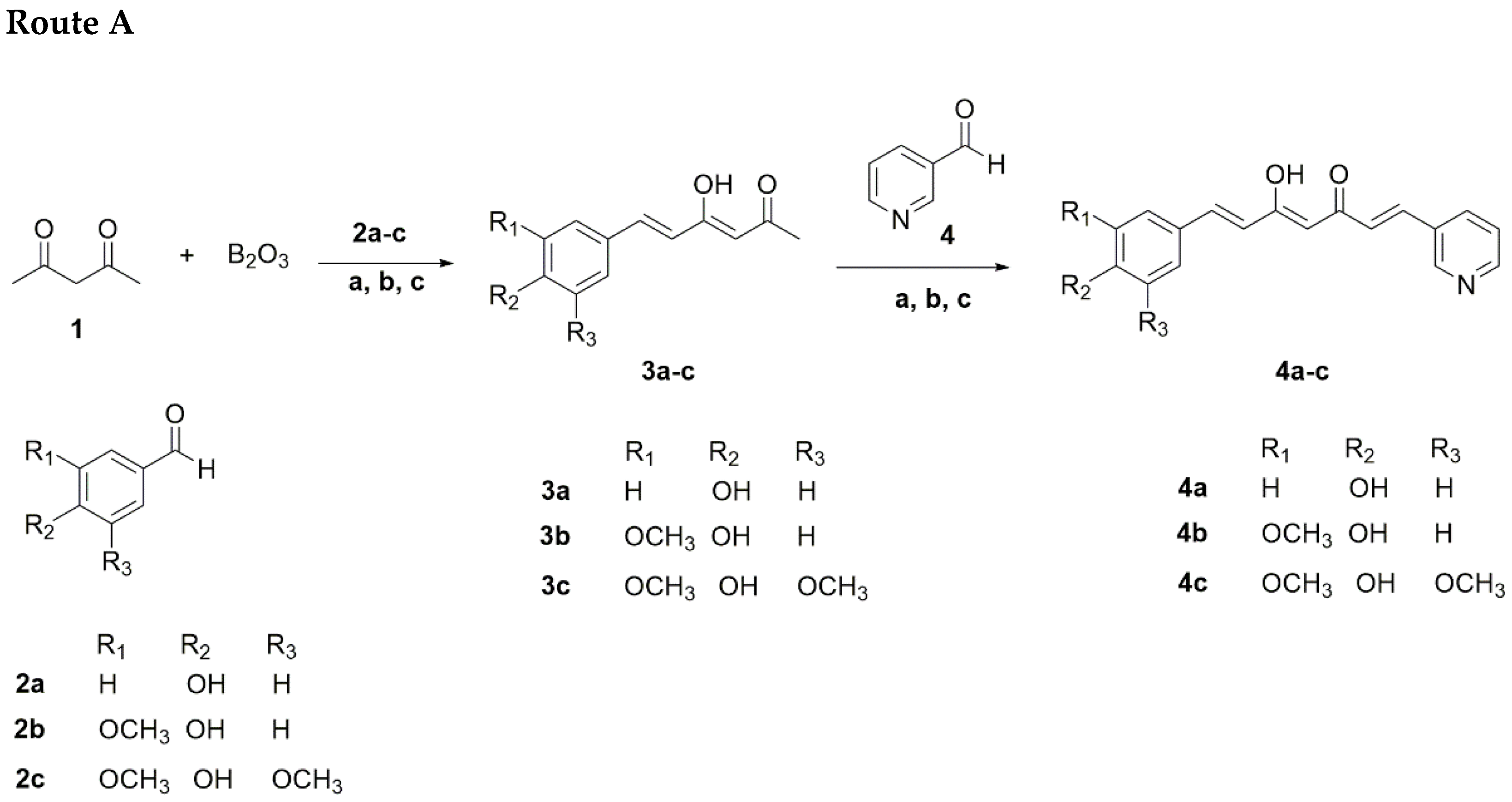
All of the reagents were purchased from Merck or Sigma-Aldrich (St. Louis, MO, USA) and used without further purification. The solvents used for reactions, such as dichloromethane, ethyl acetate, hexane, and dimethylformamide were of HPLC grade ≥ 99.7%. The synthetic plan for the construction of the curcumin-analogs pyridinium salts is described in Scheme 1 and Scheme 2.

2.1.1. Route A

Initially, aldehyde (2a-c) (9 mmol) and tri-n-butyl borate (20 mmol) in ethyl acetate (5 mL) were added to a stirring suspension of acetylacetone (10 mmol) and boric anhydride (10 mmol) in ethyl acetate (50 mL), which was vigorously stirred for 30 min at 80 °C. The reaction mixture was stirred at 80 °C for an additional 30 min, and then a solution of n-butylamine (4 mmol in 1.0 mL of EtOAc) was added dropwise over 15 min. The mixture was maintained at 80 °C for 8 h, cooled to room temperature, acidified with 0.5 N HCl (30 mL), and stirred at 80 °C for 30 min. The organic layer was separated and the aqueous layer was extracted with ethyl acetate (3 × 30 mL). The combined organic fractions were sequentially washed with saturated aqueous NaHCO3 and brine, dried over anhydrous Na2SO4, filtered, and concentrated under reduced pressure. The crude residue was purified by flash column chromatography (L × D.I × D.E: 70 cm × 24 mm × 28 mm) using a mixture of hexane/ethyl acetate 7:3 as eluent, followed by crystallization from a suitable solvent to obtain the monoaryl curcuminoids (3a-c). Following the same procedure, these compounds were used to obtain the pyridine curcuminoids 4a-c by reaction with nicotinaldehyde (4) in a 1:1 molar ratio. The pyridine curcuminoids 4a-c were obtained in yield ranges of 1.0–6.8% (Scheme 1, Table 1).
Scheme 1. Synthesis of different pyridine curcuminoids 4a-c. Reagents and conditions: (a) B(n-BuO)3; (b) n-BuNH2, 80 °C, refluxed for 1 h; (c) HCl, 80 °C.
Antioxidants 11 01104 sch001aAntioxidants 11 01104 sch001b
Table 1. Yields of different pyridine curcuminoid 4a-c.
R1R2R3Yield Route AYield Route B
4aHOHH<1.019.5
4bOCH3OHH6.820.2
4cOCH3OHOCH35.020.8

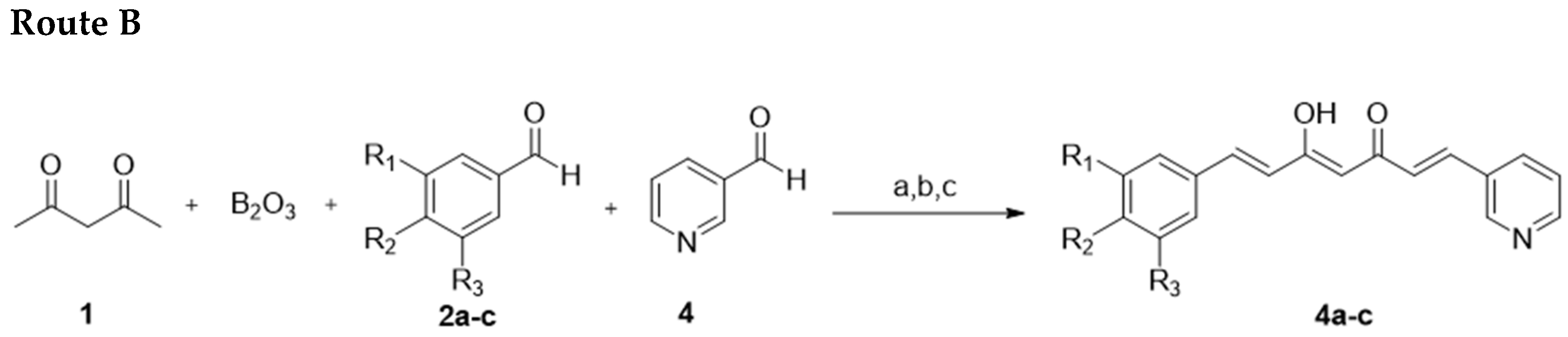
2.1.2. Route B “Mixed Pabon Reaction”

Briefly, aldehyde (2a-c) (10 mmol), nicotinaldehide (4) (10 mmol), and tri-n-butyl borate (20 mmol) dissolved in ethyl acetate (5 mL) were added to a stirring suspension of acetylacetone (10 mmol) and boric anhydride (10 mmol) in ethyl acetate (50 mL), which was vigorously stirred for 30 min at 80 °C. The reaction mixture was stirred at 80 °C for additional 30 min, then a solution of n-butylamine (4 mmol in 1.0 mL of EtOAc) was added dropwise over 15 min. The mixture was maintained at 80 °C for 8 h, cooled to room temperature, acidified with 0.5 N HCl (30 mL), and stirred at 80 °C for an additional 30 min. The organic phase was separated, and the aqueous layer was extracted with EtOAc (3 × 30 mL). The combined organic fractions were sequentially washed with saturated aqueous NaHCO3 and brine, dried over anhydrous Na2SO4, filtered, and concentrated under reduced pressure. The crude residue was purified by countercurrent chromatography (CCC). The solvent system selected for the purification was composed of hexane–ethyl acetate–methanol–water (5:5:5:5 v/v/v/v), acidified with 0.1% trifluoroacetic acid. In addition, the mobile phase operated at a flow rate of 4 mL/min in a head-to-tail mode, at a revolution speed of 700 rpm. The pyridine-curcuminoids 4a-c were obtained in yield ranges of 19.5–20.8% (Scheme 1, Table 1).

2.1.3. Curcuminoid-Derived Pyridinium Salts
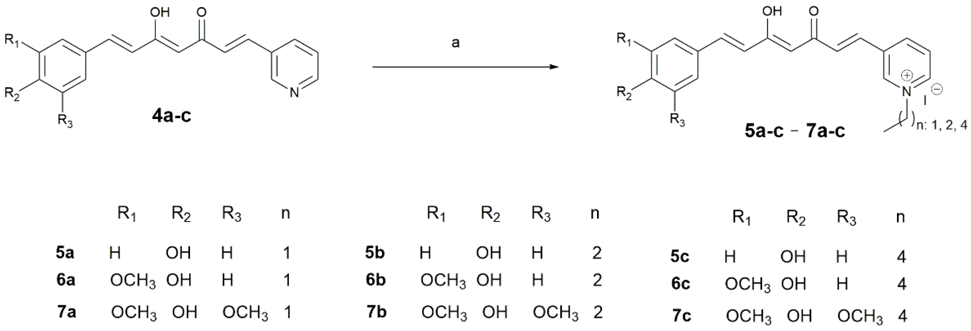
The quaternization reaction was performed by the reaction of pyridine curcuminoids (4a-c) (1 mmol) with alkyl bromides (ethyl, propyl, and pentyl iodides) (1 mmol) in dimethylformamide (DMF), using microwave irradiation (250 MW) at 80 °C for 10 min (Scheme 2). The end of the reaction was marked by adding water (1 mL) to the reaction mixture, resulting in the formation of an oily precipitate, which was subsequently washed with diethyl ether (4 × 2 mL) and separated by decantation. In addition, some of these salts were purified by CC using cellulose as stationary phase and 6:4 ethyl acetate–methanol as mobile phase. Finally, the purified compounds were maintained under a high vacuum for 4 h to obtain alkyl pyridinium salts (5a-c7a-c). These salts were obtained in the form of creamy solids with yield ranges of 6.2–34.3% (Table 2).
Scheme 2. Synthesis of curcuminoid-derived pyridinium salts 5a-c7a-c. Reagents and conditions: (a) 4a-c (1 equivalent), alkyl (ethyl, propyl, and pentyl) iodide (1 equivalent), DMF, MW, 200 W, 5 min.
Antioxidants 11 01104 sch002
Table 2. Yields of curcuminoid-derived pyridinium salts 5a-c7a-c.
Pyridinium SaltYield
5a, n: 111.1
b, n: 219.3
c, n: 416.4
6a, n: 134.0
b, n: 225.1
c, n: 46.2
7a, n: 134.3
b, n: 230.7
c, n: 427.3
(1E,4Z,6E)-5-hydroxy-7-(4-hydroxyphenyl)-1-(pyridine-3-yl)-hepta-1,4,6-trien-3-one (4a). 1H NMR (400 MHz, DMSO-d6) δ: 8.84 (s, 1H), 8.55 (d, J = 4.4 Hz, 1H), 8.12 (d, J = 7.5 Hz, 2H), 7.54 (d, J = 8.2 Hz, 4H), 7.44 (dd, J = 7.7, 4.8 Hz, 1H), 6.81 (d, J = 8.3 Hz, 3H), 6.73 (dd, J = 16.7, 8.1 Hz, 2H), 6.11–5.81 (m, 1H). These spectroscopic data correspond to the previously reported data [20].
(1E,4Z,6E)-5-hydroxy-7-(4-hydroxy-3-methoxyphenyl)-1-(pyridine-3-yl)-hepta-1,4,6-trien-3-one (4b). 1H NMR (400 MHz, DMSO-d6) δ: 9.21 (s, 1H), 8.81 (dd, J = 18.1, 6.8 Hz, 2H), 8.00 (dd, J = 7.9, 5.7 Hz, 1H), 7.69 (d, J = 16.1 Hz, 1H), 7.57 (d, J = 15.8 Hz, 1H), 7.27 (d, J = 16.1 Hz, 1H), 7.20 (s, 1H), 7.15 (d, J = 8.3 Hz, 1H), 6.96 (d, J = 8.4 Hz, 1H), 6.72 (d, J = 15.8 Hz, 1H), 6.19 (s, 1H), 3.81 (s, 3H).These spectroscopic data correspond to the previously reported data [21].
(1E,4Z,6E)-5-hydroxy-7-(4-hydroxy-3,5-dimethoxyphenyl)-1-(pyridine-3-yl)-hepta-1,4,6-trien-3-one (4c). 1H NMR (400 MHz, DMSO-d6) δ: 9.10 (s, 1H), 8.87 (s, 1H), 8.57 (d, J = 4.6 Hz, 1H), 8.15 (d, J = 8.0 Hz, 1H), 7.62 (dd, J = 15.9, 6.6 Hz, 2H), 7.46 (dd, J = 7.9, 4.8 Hz, 1H), 7.06 (s, 2H), 7.03 (d, J = 5.0 Hz, 1H), 6.86 (d, J = 15.8 Hz, 1H), 6.14 (s, 1H), 3.83 (s, 6H). 13C NMR (101 MHz, DMSO) δ: 185.58, 180.81, 150.73, 149.86, 148.34, 142.44, 138.86, 136.24, 134.72, 130.86, 126.36, 125.23, 124.28, 121.69, 106.60, 101.94, 56.31, 39.52. HRMS (ESI) m/z calculated for C20H19NO5 [M]+; 353.1263, found 353.1263.
1-ethyl-3-((1E,4Z,6E)-5-hydroxy-7-(4-hydroxyphenyl)-3-oxohepta-1,4,6-trien-1-yl)-pyridin-1-ium iodide (5a). 1H NMR (400 MHz, methanol-d4) δ: 9.41 (s, 1H), 8.98 (d, J = 8.0 Hz, 1H), 8.87 (d, J = 8.0 Hz, 1H), 8.15 (m, 1H), 8.17 (m, 1H) 7.71 (m, 1H), 7.56 (dd, J = 17.4 Hz, 9.7 Hz, 2H), 6.86 (m, 2H), 7.32 (d, J = 16.0 Hz, 1H), 7.0 (d, J = 16.0, 1H), 6.74 (d, J = 16.0 Hz, 1H), 4.71 (m, 2H), 1.24–0.76 (m, 3H). HRMS (ESI) m/z calculated for C20H20NO3+ [M-I]+; 322.1438, found 322.1452.
3-((1E,4Z,6E)-5-hydroxy-7-(4-hydroxyphenyl)-3-oxohepta-1,4,6-trien-1-yl)-1-propylpyridin-1-ium iodide (5b). 1H NMR (400 MHz, methanol-d4) δ: 9.41 (s, 1H), 8.99 (d, J = 8.0 Hz, 1H), 8.88 (d, J = 8.0 Hz, 1H), 8.25 (m, 1H), 8.17 (m, 1H) 7.72 (m, 1H), 7.56 (dd, J = 17.4 Hz, 9.7 Hz, 2H), 6.86 (m, 2H), 7.32 (d, J = 16.0 Hz, 1H), 7.01 (d, J = 16.0, 1H), 6.74 (d, J = 16.0 Hz, 1H), 4.71 (m, 2H), 1.69 (p, J = 7.6 Hz, 1H), 1.48 (dq, J = 14.9, 7.3 Hz, 1H), 1.24–0.76 (m, 3H). HRMS (ESI) m/z calculated for C21H22NO3+ [M-I]+; 336.1594, found 336.1590.
3-((1E,4Z,6E)-5-hydroxy-7-(4-hydroxyphenyl)-3-oxohepta-1,4,6-trien-1-yl)-1-pentylpyridin-1-ium iodide (5c). 1H NMR (400 MHz, methanol-d4) δ: 9.41 (s, 1H), 8.99 (d, J = 4.0 Hz, 1H), 8.87 (d, J = 4.0 Hz, 1H), 8.14 (m, 1H), 7.70 (d, J = 16.0 Hz, 2H), 7.57 (d, J = 8.0 Hz, 2H), 7.32 (d, J = 16.0 Hz, 1H), 6.87 (d, J = 8.0, 3H), 6.71 (d, J = 16.0 Hz, 1H), 4.70 (t, J = 7.5 Hz, 2H), 2.28–1.92 (m, 2H), 1.44 (s, 4H), 1.04–0.80 (m, 3H). HRMS (ESI) m/z calculated for C23H26NO3+ [M-I]+; 364.1907, found 364.1917.
1-ethyl-3-((1E,4Z,6E)-5-hydroxy-7-(4-hydroxy-3-methoxyphenyl)-3-oxohepta-1,4,6-trien-1-yl)-pyridin-1-ium iodide (6a). 1H NMR (400 MHz, DMSO-d6) δ: 9.40 (s, 1H), 9.18 (s, 1H), 8.97 (d, J = 5.9 Hz, 1H), 8.80 (d, J = 8.2 Hz, 2H), 8.11 (dd, J = 8.0, 6.2 Hz, 1H), 7.59 (d, J = 16.0 Hz, 1H), 7.52 (d, J = 15.8 Hz, 1H), 7.24 (d, J = 16.0 Hz, 1H), 7.09 (s, 1H), 6.91 (d, J = 8.8 Hz, 1H), 6.70 (d, J = 15.8 Hz, 1H), 6.15 (s, 1H), 4.56 (q, J = 7.2 Hz, 2H), 1.51 (t, J = 7.3 Hz, 3H). HRMS (ESI) m/z calculated for C21H22NO4+ [M-I]+; 352.1543, found 352.1547.
3-((1E,4Z,6E)-5-hydroxy-7-(4-hydroxy-3-methoxyphenyl)-3-oxohepta-1,4,6-trien-1-yl)-1-propylpyridin-1-ium iodide (6b). 1H NMR (400 MHz, DMSO-d6) δ: 9.43 (s, 1H), 8.94 (dd, J = 6.8 Hz, 3H), 8.26–8.12 (m, 1H), 7.64 (dd, J = 15.8, 3.6 Hz, 2H), 7.34 (s, 1H), 7.28 (d, J = 16.0 Hz, 1H), 7.18 (d, J = 8.1 Hz, 1H), 6.83 (dd, J = 19.5, 12.0 Hz, 2H), 6.16 (s, 1H), 4.54 (t, J = 7.0 Hz, 2H), 3.81 (s, 3H), 1.97 (dq, J = 14.1, 7.1 Hz, 2H), 0.89 (t, J = 7.2 Hz, 3H). HRMS (ESI) m/z calculated for C22H24NO4+ [M-I]+; 366.1700, found 366.1705.
3-((1E,4Z,6E)-5-hydroxy-7-(4-hydroxy-3-methoxyphenyl)-3-oxohepta-1,4,6-trien-1-yl)-1-pentylpyridin-1-ium iodide (6c). 1H NMR (400 MHz, DMSO-d6) δ: 9.76 (s, 1H), 9.60 (s, 1H), 9.10 (d, J = 5.6 Hz, 1H), 8.92 (d, J = 7.9 Hz, 1H), 8.20 (t, J = 6.9 Hz, 1H), 7.66 (dd, J = 15.8, 6.1 Hz, 2H), 7.37 (t, J = 7.9 Hz, 2H), 7.19 (d, J = 8.0 Hz, 1H), 6.94–6.80 (m, 2H), 6.20 (s, 1H), 4.63 (t, J = 7.0 Hz, 2H), 3.84 (s, 3H), 2.09–1.84 (m, 2H), 1.44–1.11 (m, 4H), 0.88 (t, J = 6.6 Hz, 3H). HRMS (ESI) m/z calculated for C24H28NO4+ [M-I]+; 394.2013, found 394.2016.
1-ethyl-3-((1E,4Z,6E)-5-hydroxy-7-(4-hydroxy-3,5-dimethoxyphenyl)-3-oxohepta-1,4,6-trien-1-yl)-pyridin-1-ium iodide (7a). 1H NMR (400 MHz, DMSO-d6) δ: 9.57 (s, 1H), 9.09 (d, J = 5.6 Hz, 1H), 8.90 (d, J = 7.7 Hz, 1H), 8.28–8.11 (m, 1H), 7.78–7.55 (m, 2H), 7.36 (d, J = 18.0 Hz, 1H), 7.09 (s, 2H), 6.95 (d, J = 15.9 Hz, 1H), 6.21 (s, 1H), 4.66 (q, J = 6.9 Hz, 2H), 3.83 (s, 6H), 1.59 (t, J = 7.2 Hz, 3H). 13C NMR (101 MHz, DMSO) δ: 187.26, 177.80, 174.18, 148.18, 144.03, 143.33, 142.67, 139.00, 135.26, 131.57, 130.44, 128.03, 124.93, 121.61, 119.63, 106.78, 102.88, 59.53, 56.77, 56.26, 16.15. HRMS (ESI) m/z calculated for C22H24NO5+ [M-I] +; 382.1649, found 382.1658.
3-((1E,4Z,6E)-5-hydroxy-7-(4-hydroxy-3,5-dimethoxyphenyl)-3-oxohepta-1,4,6-trien-1-yl)-1-propylpyridin-1-ium iodide (7b). 1H NMR (400 MHz, DMSO-d6) δ: 9.50 (s, 1H), 9.14 (s, 1H), 9.05 (d, J = 6.0 Hz, 1H), 8.92 (d, J = 8.2 Hz, 1H), 8.30–8.12 (m, 1H), 7.67 (dd, J = 15.9, 3.3 Hz, 2H), 7.32 (d, J = 16.0 Hz, 1H), 7.09 (s, 2H), 6.94 (d, J = 15.8 Hz, 1H), 6.20 (s, 1H), 4.58 (t, J = 7.3 Hz, 2H), 3.83 (s, 6H), 2.00 (h, J = 7.3 Hz, 2H), 0.92 (t, J = 7.3 Hz, 3H). 13C NMR (101 MHz, DMSO) δ: 187.26, 177.80, 174.18, 148.18, 144.03, 143.33, 142.67, 139.00, 135.26, 131.57, 130.44, 128.03, 124.93, 121.61, 119.63, 106.78, 102.88, 59.53, 56.77, 56.26, 16.15. HRMS (ESI) m/z calculated for C23H26NO5+ [M-I]+; 396.1805, found 396.1815.
3-((1E,4Z,6E)-5-hydroxy-7-(4-hydroxy-3,5-dimethoxyphenyl)-3-oxohepta-1,4,6-trien-1-yl)-1-pentylpyridin-1-ium iodide (7c). 1H NMR (400 MHz, DMSO-d6) δ: 9.50 (s, 1H), 9.14 (s, 1H), 9.05 (d, J = 6.0 Hz, 1H), 8.92 (d, J = 8.2 Hz, 1H), 8.30–8.12 (m, 1H), 7.67 (dd, J = 15.9, 3.3 Hz, 2H), 7.32 (d, J = 16.0 Hz, 1H), 7.09 (s, 2H), 6.94 (d, J = 15.8 Hz, 1H), 6.20 (s, 1H), 4.58 (t, J = 7.3 Hz, 2H), 3.83 (s, 6H), 2.00 (h, J = 7.3 Hz, 2H), 0.92 (t, J = 7.3 Hz, 3H). 13C NMR (101 MHz, DMSO) δ: 187.22, 177.71, 148.13, 144.17, 143.31, 142.66, 138.99, 135.23, 131.51, 130.40, 127.98, 124.86, 121.55, 106.76, 102.78, 61.11, 56.20, 30.14, 27.53, 21.57, 13.73. HRMS (ESI) m/z calculated for C25H30NO5+ [M-I]+; 424.2118, found 424.2121.

2.2. GC–MS Analysis

GC–MS analysis was performed with Trace 1300 gas chromatograph coupled to ISQ mass spectrometer (Thermo Fisher Scientific 713100331, ISQ 130729 2215, Grand Avenue Parkway Autin, TX, USA) series equipment equipped with an Rtx-5MS w/integra-guard capillary column (Crossbond 5% diphenyl–95% dimethyl polysiloxane) 30 m in length, 0.25 mm internal diameter, and 0.25 mm thick. Briefly, 3 µL of the sample at a temperature of 250 °C were injected into the injection port under splitless mode with the following conditions: 60 °C with a ramp of 10 °C/min to 300, waiting time of 5 min, drag flow of 1.5 mL/min. All of the assays were performed in scan mode.

2.3. Computational Calculations

In the present work, density functional theory (DFT) calculations were carried out at the M06-2X/6-311+G(d,p) level, using the Gaussian 09 software [22]. No imaginary vibrational frequencies were found at the optimized geometries, indicating that they are the true minimal of the potential energy surface. Solvent effects were taken into account by the polarizable continuum model (PCM), using parameters for water [23]. The formation of free radical species from phenols, and then their radical scavenging properties, occur in the biological systems mainly by the hydrogen atom transfer (HAT) mechanism, which is governed by the homolytic bond dissociation enthalpy (BDE) [24]. Therefore, we calculated the BDE of O–H bonds in our molecules, as follows:
H–O → H + O•      BDE = H(H) + H(O)–H(O–H)
where H(H), H(O), and H(O–H) are the corresponding enthalpies. The lower the BDE values, the easier the dissociation of the O–H bond, and the easier the radical is formed.

2.4. In Vitro Antioxidant Activity

2.4.1. DPPH Radical Scavenging Method

The 1,1-diphenyl-2-picrylhydrazyl (DPPH) assay was performed according to the microwell-adopted procedure described by Brand-Williams et al., 1995 [25]. Briefly, a stock solution of DPPH (20 mg/L) in methanol was prepared, then 75 μL of the compounds or control (80% methanol) were mixed with 150 μL of DPPH reagent (several dilutions of the extracts 1:2 were carried out using methanol). After 15 min, the absorbance was measured at 515 nm using a microplate reader spectrophotometer. The results were expressed as the half-maximal inhibitory concentration (IC50, µM) by a mean of three determinations, in which the percentage of antioxidant activity was first calculated for each dilution as follows:
% Scavenging DPPH free radical = 100 × (1 − AE/AD)
where AE is the absorbance of the solution after adding the IL and AD is the absorbance of DPPH solution. Then, the half-maximal (IC50) inhibitory concentration value was calculated in mg/ml by the linear regression analysis, and the results were expressed in µM using the MW of each compound.

2.4.2. Ferric Reducing Antioxidant Power (FRAP) Assay

The FRAP assay was performed according to the procedure described by Benzie and Strain 1996 [26]. The reagent was prepared by mixing 25 mL of 300 mM of acetate buffer (pH 3.6) and 2.5 mL of 2,4,6-tris(2-pyridyl)-s-triazine in 40 mM of HCl and 2.5 mL of 20 mM FeCl3·6H2O. Then, 0.05 mL of the compounds or standard (FeSO4·7H2O, Fe2+ concentration vs. absorbance) were added to 1.5 mL of FRAP solution and 0.15 mL of water. Thereafter, the mixture was incubated at room temperature for 15 min. The final results of the product (ferrous tripyridyl triazine complex) were read at a wavelength of 593 nm, using a spectrophotometer. The percentage of Fe3+ scavenging (reduction to Fe2+) was calculated by comparison with the standard curve.

2.5. Cell Lines and Culture

Three cell lines were used to assess the antitumor activities of the compounds studied in this work: Human colon cancer-derived HT29, human breast cancer-derived cells MCF7, and murine fibroblast NIH3T3. The cell lines were cultured in Dulbecco’s Modified Eagle’s Medium (GIBCO) supplemented with bovine calf serum to a final concentration of 10%, glucose (4500 mg/L), L-glutamine (2 mM), penicillin (50 U/L), and streptomycin (50 µg/mL). The cultures were incubated at 37 °C and 5% CO2 in a humidified atmosphere.

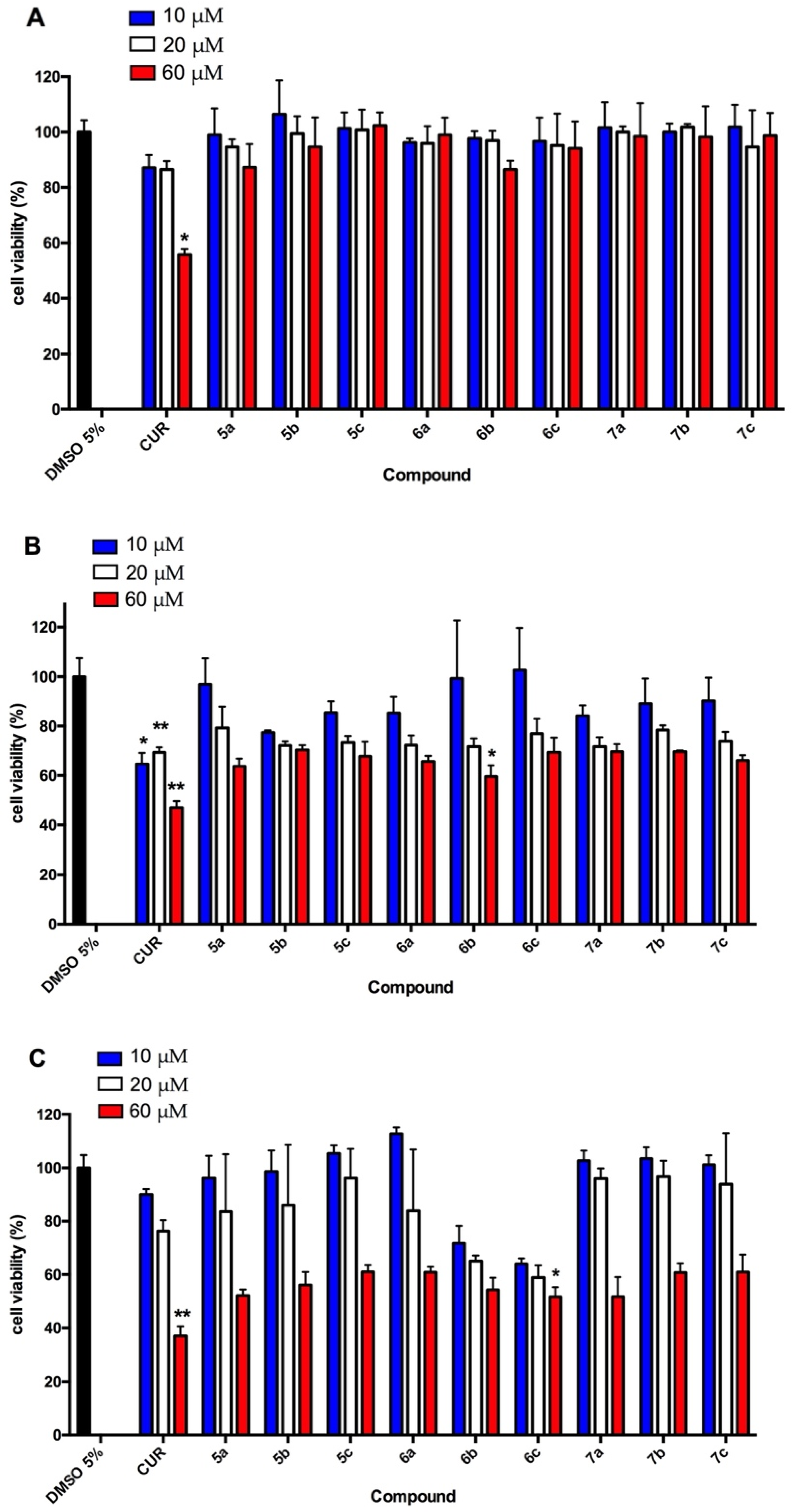
2.5.1. Viability Assay

Viability was assessed using a tetrazolium salt, the 3-(4,5-dimethylthiazol-2-yl)-2,5-diphenyl tetrazolium bromide (MTT) (Cayman Chemical Company, Ann Arbor, MI, USA) [27]. For this assay, 1.5 × 10⁴ cells per well were seeded in a 96-well plate and incubated for 24 h with different concentrations of the curcumin-derived compounds (10, 20, and 60 μM dissolved in water with 0.5% DMSO). In addition, DMSO was included as a vehicle control at 0.5%. Following this treatment, the culture medium of the cells was changed and the MTT reagent was added and incubated for 3 h. Then, cells were lysed and the crystals formed were dissolved in isopropanol (Cayman Chemical Company), for reading in a multiplate reader (Thermo Scientific, Waltham, MA, USA) at 570 nm. The results are expressed as a percentage of vehicle control.

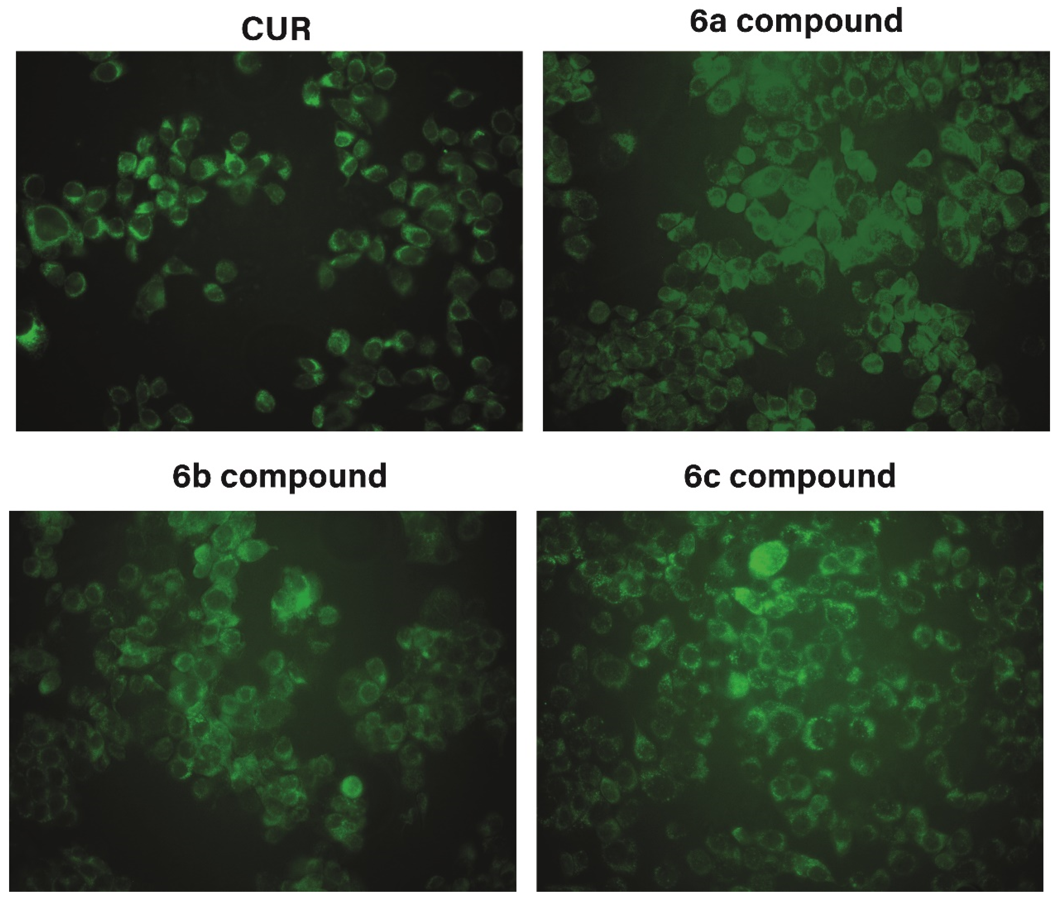
2.5.2. Fluorescence Images

MCF7 cells were seeded at 50–60% confluence on coverslips in a 12-well plate. Then, the cells were treated with 20 µM of compounds, incubated for 1 h, and washed twice with phosphate-buffered saline to remove the compounds. Images were digitally acquired with an Olympus BX53 fluorescence microscope (Olympus) coupled to a CCD camera. Digital images were taken using Q-Capture Pro 7 software (Qimagine, Inc. Surrey, BC, Canada).

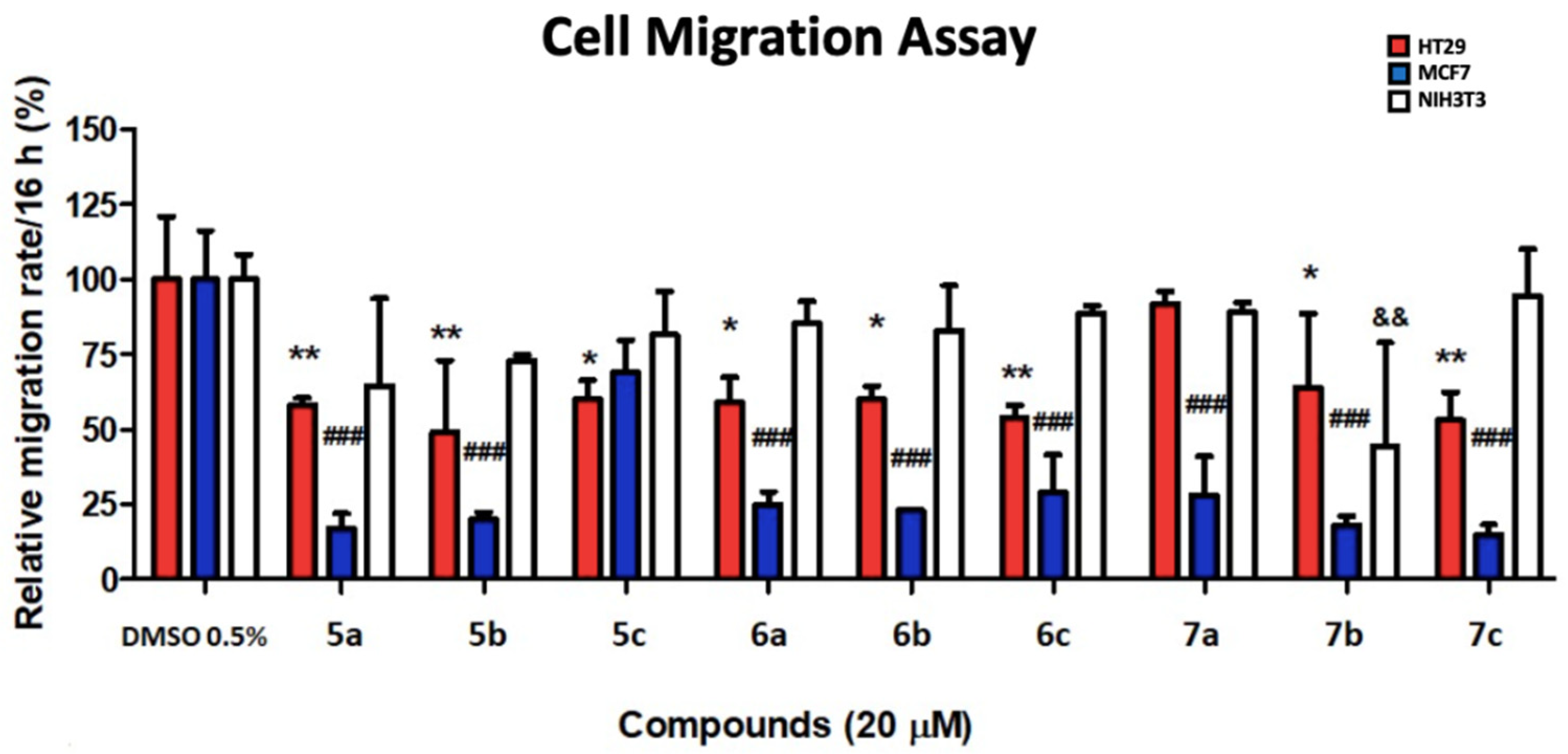
2.5.3. Cell Migration Assay

Cells from different cell lines (HT29, MCF7 or NIH3T3) were seeded at 70–80% confluence in polystyrene plates of 60 mm. A vertical scratch was made on the plate using a micropipette tip of 200 μL. Then, the cells were washed twice with phosphate-buffered saline to remove the non-adhered cells. Thereafter, 20 μM of compounds were added in addition to a control plate with only 0.5% DMSO as a vehicle and the plates were incubated for 16 h. Photographs of the scratch were taken before adding the extracts and after incubation with a Canon® Model Eos Rebel XSI camera. Migration was determined by measuring the delta of the area of wound closure (0–16 h area) obtained by the ImageJ computer program. The results were expressed as the relative migration rate within 16 h, which is the percentage of migration to the vehicle control in that period (control: 100% of migration/16 h).

2.5.4. Statistical Analysis

For statistical analysis, the results represent measurements of at least three independent experiments. The data from the groups were compared using the analysis of variance with Tukey’s post hoc test. A p-value < 0.05 was considered as statistically significant. The GraphPad Prism 5 software program (San Diego, CA, USA) was used and data were expressed as the mean ± standard deviation (SD).

3. Results and Discussion

3.1. Chemical Synthesis

As shown in Scheme 1 and Scheme 2, the synthesis of curcumin-analog alkyl pyridinium salts was achieved with a total of three reaction steps, according to the Pabon method [28]. At first, phenyl diketones 3a-e were obtained by the reaction of 2,4-pentadione 1, boron trioxide (B2O3) at reflux in a 1:1 molar ratio, 0.9 equivalent of aldehyde 2a-e, 2 equivalent of tributyl borate (B(n-BuO)3), and butylamine (0.4 M in AcOEt). Column chromatography (CC) using hexane–ethyl acetate mixtures in 8:2, 7:3, 6:4, and 1:1 as eluents allowed us to obtain the phenyl diketones 3a-e, respectively as yellow solids and 13.4–49.0% reaction yields. To obtain the curcuminoids 4a-c, a second Pabon condensation, using the same procedure, was carried out, maintaining an equivalent molar ratio of 1:1 between the previously synthesized phenyl diketones 3a-e and the nicotinaldehyde 4 (Route A). The low yields revealed by TLC and gas chromatography (GC–MS), indicating low consumption of the starting reagents, led us to explore the effect of some changes in the reaction and purification methodologies. Curcuminoids 4a-c, which were performed by CC using petroleum ether–ethyl acetate mixtures in 7:3, 6:4, 1:1, 4:6, and 3:7, obtained 4a-c as orange solids. A “one-pot” process, involving the double-condensation of two aldehydes of different nature in a single synthetic step (Route B), named by us as “mixed Pabon reaction”, allowed for the reduction of reaction times and purification processes (Scheme 1). In addition, these changes improved two fundamental parameters of green chemistry, namely, the atom economy (EA) and atom efficiency (EfA). For example, the linear synthesis of pyridine curcuminoid 4c led to EA and EfA values of 54.03 and 2.0, while the mixed Pabon reaction led to values of 90.66 and 19, respectively. The yields obtained by this procedure, for the 4a-c series were in the range of 19.5–20.8%, and the purification was efficiently carried out by countercurrent chromatography (CCC).
CCC is a separation technique with several advantages, including the lack of solid support, which could result in irreversible adsorption of target compounds [29]. For example, the solvent system selected for the purification of 4b was composed of 5:5:5:5 HEMW at v/v/v/v (hexane–ethyl acetate–methanol-water), which provided a KD value of 1.72 for compound 4b with Rf 0.9 and 0.74 for by-product 4b. A similar solvent system was used for the separation of curcuminoids from curcumin and turmeric powder [30].
Finally, the reaction of curcuminoids 4a-c with alkyl iodides (ethyl, propyl, and pentyl) to obtain the alkyl pyridinium salts, in dimethylformamide (DMF) and a 1:1 molar ratio, was carried out under 10 min of microwave irradiation (MW), as previously described [31,32] (Scheme 2). Some of these salts were purified by CC using cellulose as the stationary phase and 6:4 mixtures of ethyl acetate/methanol as eluent, obtaining yield ranges of 6.2–34.3%.

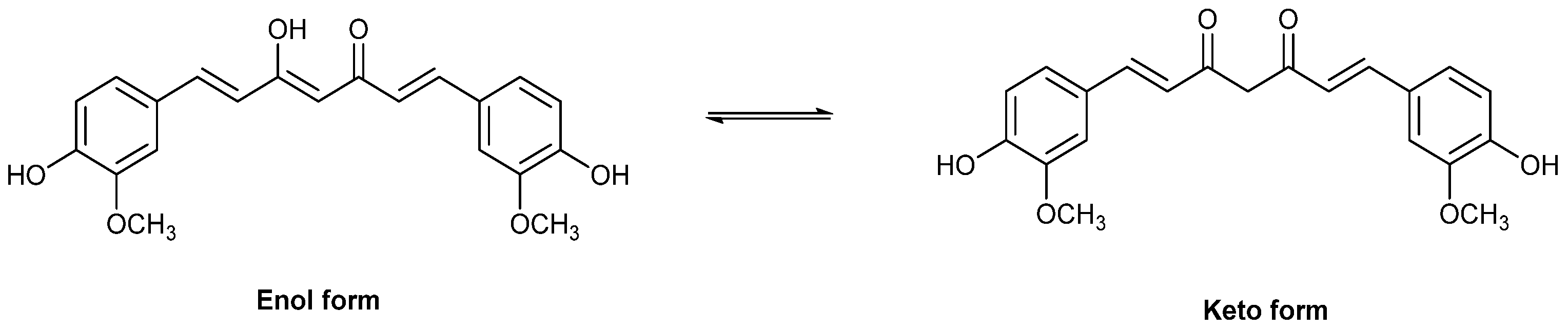
3.2. In Vitro and In Silico Assessment of Antioxidant Capacity

The in vitro antioxidant capacity of compounds was evaluated through the DPPH and FRAP assays. In addition, computational analyses were performed for insight into the structure of compounds and to rationalize the experimental results. Table 3 shows the results for all of the synthetized compounds and curcumin, which were used as a standard to compare the antioxidant capacity.
Among the derivatives studied, 4a-c curcumin analogs exhibited the highest DPPH quenching capacity, which is similar to or better than curcumin. Pyridinium salts 5a-c and 6a-c showed DPPH quenching capacities lower than curcumin. However, in the third group of pyridinium salts (7a-c) possessing two methoxy groups, the derivatives 7b and 7c showed higher DPPH quenching capacities than curcumin in the DPPH and FRAP assays.
Notably, the increase in the length of the N-alkyl chain has different effects on the antioxidant capacity for each group of salts. On the one hand, group 5 diminishes the antioxidant capacity, with an increase in the length of the chain. On the other hand, group 7 shows a drastic increase in the antioxidant capacity with an increase in the number of carbon atoms in the chain. Unlike groups 5 and 7, group 6 does not show any tendency.
Moreover, the FRAP assay results showed that groups 5 and 6 have the lowest reducing power, with no significant influence with the change in the length of the N-alkyl chain. The group curcuminoids-pyridine 4a-c presented higher values than groups 5 and 6, but they were below the reducing power of curcumin. Group 7 showed a drastic drop with an increase in the number of carbon atoms in the chain. The highest values of reducing power, following the same tendency as DPPH in regards to the length of the alkyl chain, showed that 7c is the curcuminoid pyridinium salt with the highest reducing power. Both assays showed that alkyl pyridiniums of group 7 (two methoxyl groups in phenyl ring) exhibit the best antioxidant capacity, which increases along with the length of the N-alkyl chain.
To rationalize our results, bond dissociation enthalpy (BDE) calculations in gas- and aqueous phases were conducted. Initially, we studied non-ionic compounds 4a-c as well as one ionic compound of each family 5a, 6a, and 7a. For our calculation study, we selected all of the compounds of group 4 to examine the influence of methoxy groups in the phenyl ring. In particular, we selected the compounds that possessed the shortest N-alkyl chain of each group (5a, 6a, and 7a), considering that the length of the alkyl chain should not significantly affect the electronic density of OH bonds in the compounds.
Table 4 shows the BDE values for the hydroxyl groups of the compounds. For the calculation, we have considered both possible tautomers for the keto-enol equilibrium, as shown in Figure 2. Therefore, four BDEs were calculated for each one of the six compounds studied, both in gas phase and water as solvent.
For the compounds of group 4, the results show that all of the O–H BDEs in the tautomer II were lower than their equivalents in tautomer I. Compounds 5a, 6a, and 7a showed remarkably high values of BDE for tautomer II compared with I. Notably, water solvation has a different impact on the BDE of each O–H. For instance, for the tautomer I of the group 4 compounds, the solvation effect of water reduced the O–H BDEs values in the gas phase. However, the opposite effect occurs for their tautomers II, except for 4b. The most significant effect observed is the remarkable reduction of the O–H BDE values when water solvation was considered for 5a, 6a, and 7a. This indicates a relevant stabilization of the radicals formed in water compared with the gas phase, as seen for tautomer II of these ionic compounds. The lower values of BDEs in the water solution correspond to the structurally related curcuminoid-pyridine 4c and the curcuminoid pyridinium salt 7a, which is consistent with the obtained experimental results.

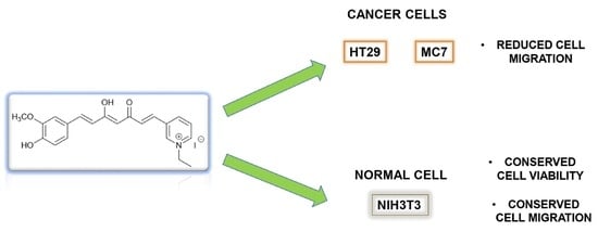
3.3. Preliminary Antitumor Evaluation

To evaluate the biological activity of the compounds on cells, an MTT assay was performed by incubating HT29, MCF7, and NIH3T3 cells in the presence of the compounds and vehicle control for 24 h. Although curcumin-derived compounds have been reported to exert an antiproliferative inhibitory effect in the range of 10–20 μM (IC50), we decided to test a wide range of concentrations to observe effects not only in proliferation and cell migration, but also in cell viability. The following reports illustrate the range of concentrations used in the antitumor assessment of related compounds [33,34,35]. This is especially important in our trials, since we test previously unreported compounds and use cell lines of diverse origins, such as colon cancer (HT29), breast cancer (MCF7), and mouse fibroblast (NIH3T3).
As shown in Figure 3, a significant concentration-dependent decrease in the cell viability was observed in the tumoral cell lines when they were incubated with curcumin (CUR) as a reference compound. The effect was more noticeable over HT29. Notably, the viability of the non-tumoral cell line NIH3T3 was not significantly affected by CUR (p > 0.05).
All of the compounds produced a significant decrease in the viability of HT29 and MCF7 cells only at the highest concentration (60 µM). In general, a more noticeable effect over MCF7 is observed, since six of the nine compounds (7a, 7b, 7c, 5b, 5c, and 6a) significantly decreased the cell viability at 10 or 20 µM. For HT29, the effects were significant only at 60 µM. For this cell line, compounds 6b and 6c showed the highest cytotoxic effects. Notably, all of the compounds showed no significant in vitro effect over the non-tumoral cell line NIH3T3, similar to CUR. However, none of them decrease viability in HT29 cells as effectively as CUR at the highest concentration (32.8 ± 6.4% viability), with compound 6c as the more active (51.6 ± 3.8%). Related to MCF7, compound 6b generates a very similar effect (60.0 ± 4.6%) to CUR (60.2 ± 1.5%) at 60 µM, but only CUR was found to be significantly active from 20 µM (68.1 ± 5.2%).
Moreover, to visualize the capacity of the derivates for incorporation inside the cell, we took advantage of fluorescence emission for these compounds. Series 6 (6a-c) were selected, specifically for their different results in the MCF7 cell line at 20 μM. Interestingly, all of the treatments showed a perinuclear distribution, and the three analyzed compounds of series 6 showed a major fluorescence compared with the control CUR. However, compound 6a showed a higher signal compared with the other compounds (Figure 4).
Recently, the intracellular localization of curcumin and polysorbate-80 micelles of curcumin were studied in Caco-2 cells. The authors reported that after 30 min, curcumin from each formulation was associated with mitochondria and lysosomes. After 180 min, native curcumin was associated with mitochondria and peroxisomes, while micellar curcumin was associated only with peroxisomes. Interestingly, the subcellular localization was not associated with the differences in bioavailability in humans [36].
Cell migration is a very fundamental aspect of cancer since it is one of the initial steps of tumor metastasis [37]. We evaluated the effects of curcumin-analogs on the in vitro 2-D cell migration ability through a wound-healing assay at 20 µM of final concentration. The results showed a marked negative effect of CUR on cell migration in the three cell lines (Figure 5). Most of the 10 compounds significantly inhibited the in vitro tumor cell migration, except for compounds 7a in HT29 and 5c in MCF7. The strongest inhibition was observed for 7c (53.0 ± 7.4% in HT29 and 15.0 ± 3.3% in MCF7 cell migration), but it was less than CUR (39.0 ± 12.5% in HT29 and 23.0 ± 5.2% in MCF7 cell migration). In general, a higher effect on in vitro cell migration was observed in MCF7 cells. Noteworthy, compound 6b showed a good combination of effects, since it significantly reduced cell migration in both HT29 and MCF7 cells, but also showed a significant decrease in the cell viability at 20 µM (Figure 5), without affecting the non-tumoral NIH3T3 cell line: Neither in viability nor in migration, unlike the CUR compound.

4. Conclusions

In conclusion, using the two-step process described herein, it has been possible to obtain curcumin with ionic liquid characteristics. Initially, a “one-pot” reaction involving the double-condensation of two aldehydes of different nature in a single synthetic step, named by us as “mixed Pabon reaction”, allowed us to obtain pyridine curcumoids 4a-c. Then, curcuminoid N-alkylpyridinium salts were obtained by microwave assisted synthesis in moderate global yields. The in vitro antioxidant properties demonstrated that group 4, pyridine curcumoids, and group 7, curcuminoid N-alkylpyridinium salts with two methoxy groups in phenyl ring, presented the best antioxidant capacity. For group 7, an increase in the antioxidant capacity was observed as the length of the N-alkyl chain increased. We rationalized the experimental results by density functional theory (DFT) calculations of the bond dissociation enthalpy (BDE) for O–H in each compound. BDEs, obtained in the gas phase and water, showed a notable impact of water solvation on the stability of radicals. The lower values of BDEs in the water solution correspond to the structurally related curcuminoid-pyridine 4c and the curcuminoid pyridinium salts 7a, which is consistent with the experimental results. Additionally, a preliminary assessment of cell viability and cell migration assays showed that these compounds mainly cause inhibition of the cell migration observed in MCF7 cancer cells, without affecting the non-tumoral NIH3T3 cell line: Neither in viability nor in migration. Finally, these results suggest that some of these compounds could be selective antimetastatic agents, and we hope to carry out future studies to unveil the cellular mechanisms involved.

Author Contributions

Conceptualization, O.F.-D., M.M.-C. and R.A.-M.; methodology, O.F.-D., M.M.-C., R.A.-M. and L.G.; validation, M.M.-C., R.A.-M. and O.F.-D.; investigation, O.F.-D., M.M.-C., L.G., S.W. and F.J.-A.; data curation, M.M.-C., O.F.-D., S.W. and C.V.; writing—original draft preparation, M.M.-C., O.F.-D., L.G. and R.A.-M.; writing—review and editing, M.M.-C., O.F.-D., J.E., L.G. and R.A.-M.; visualization, M.M.-C., R.A.-M. and O.F.-D.; supervision, M.M.-C., O.F.-D. and R.A.-M.; project administration, O.F.-D., M.M.-C., and R.A.-M.; funding acquisition, M.M.-C., O.F.-D. and R.A.-M. All authors have read and agreed to the published version of the manuscript.

Funding

This research was funded by FONDECYT 3170757 (O.F.-D.), FONDECYT 11170142 (M.M.-C.) and was supported by ACT210097-ANID. Powered@NLHPC: this research was partially supported by the supercomputing infrastructure of the NLHPC (ECM-02).

Institutional Review Board Statement

Not applicable.

Informed Consent Statement

Not applicable.

Data Availability Statement

All of the data is contained within the article.

Acknowledgments

The authors would like to thank Andrew Quest (CEMC, Universidad de Chile, Chile) for providing the HT29, MCF7, and NIH3T3 cells.

Conflicts of Interest

The authors declare no conflict of interest.

References

  1. Noureddin, S.A.; El-Shishtawy, R.M.; Al-Footy, K.O. Curcumin Analogues and Their Hybrid Molecules as Multifunctional Drugs. Eur. J. Med. Chem. 2019, 182, 111631. [Google Scholar] [CrossRef] [PubMed]
  2. Mahran, R.I.; Hagras, M.M.; Sun, D.; Brenner, D.E. Bringing Curcumin to the Clinic in Cancer Prevention: A Review of Strategies to Enhance Bioavailability and Efficacy. AAPS J. 2017, 19, 54–81. [Google Scholar] [CrossRef]
  3. Flory, S.; Sus, N.; Haas, K.; Jehle, S.; Kienhöfer, E.; Waehler, R.; Adler, G.; Venturelli, S.; Frank, J. Increasing Post-Digestive Solubility of Curcumin Is the Most Successful Strategy to Improve Its Oral Bioavailability: A Randomized Cross-Over Trial in Healthy Adults and In Vitro Bioaccessibility Experiments. Mol. Nutr. Food Res. 2021, 65, 2100613. [Google Scholar] [CrossRef] [PubMed]
  4. Hirata, Y.; Ito, Y.; Takashima, M.; Yagyu, K.; Oh-Hashi, K.; Suzuki, H.; Ono, K.; Furuta, K.; Sawada, M. Novel Oxindole–Curcumin Hybrid Compound for Antioxidative Stress and Neuroprotection. ACS Chem. Neurosci. 2019, 11, 76–85. [Google Scholar] [CrossRef] [PubMed]
  5. Tavaf, Z.; Dangolani, S.K.; Yousefi, R.; Panahi, F.; Shahsavani, M.B.; Khalafi-Nezhad, A. Synthesis of New Curcumin Derivatives as Influential Antidiabetic α-Glucosidase and α-Amylase Inhibitors with Anti-Oxidant Activity. Carbohydr. Res. 2020, 494, 108069. [Google Scholar] [CrossRef] [PubMed]
  6. Martelli, G.; Giacomini, D. Antibacterial and Antioxidant Activities for Natural and Synthetic Dual-Active Compounds. Eur. J. Med. Chem. 2018, 158, 91–105. [Google Scholar] [CrossRef] [PubMed]
  7. Rodrigues, F.C.; Kumar, N.V.A.; Thakur, G. Developments in the Anticancer Activity of Structurally Modified Curcumin: An up-to-Date Review. Eur. J. Med. Chem. 2019, 177, 76–104. [Google Scholar] [CrossRef]
  8. Bonaccorsi, P.M.; Labbozzetta, M.; Barattucci, A.; Salerno, T.M.G.; Poma, P.; Notarbartolo, M. Synthesis of Curcumin Derivatives and Analysis of Their Antitumor Effects in Triple Negative Breast Cancer (TNBC) Cell Lines. Pharmaceuticals 2019, 12, 161. [Google Scholar] [CrossRef] [Green Version]
  9. Gandalovičová, A.; Rosel, D.; Fernandes, M.; Veselý, P.; Heneberg, P.; Čermák, V.; Petruželka, L.; Kumar, S.; Sanz-Moreno, V.; Brábek, J. Migrastatics—Anti-Metastatic and Anti-Invasion Drugs: Promises and Challenges. Trends Cancer 2017, 3, 391–406. [Google Scholar] [CrossRef] [Green Version]
  10. Riggi, N.; Aguet, M.; Stamenkovic, I. Cancer Metastasis: A Reappraisal of Its Underlying Mechanisms and Their Relevance to Treatment. Annu. Rev. Pathol. Mech. Dis. 2018, 13, 117–140. [Google Scholar] [CrossRef]
  11. Urra, F.A.; Muñoz, F.; Córdova-Delgado, M.; Ramírez, M.P.; Peña-Ahumada, B.; Rios, M.; Cruz, P.; Ahumada-Castro, U.; Bustos, G.; Silva-Pavez, E. FR58P1a; a New Uncoupler of OXPHOS That Inhibits Migration in Triple-Negative Breast Cancer Cells via Sirt1/AMPK/Β1-Integrin Pathway. Sci. Rep. 2018, 8, 13190. [Google Scholar] [CrossRef] [PubMed]
  12. Koroth, J.; Nirgude, S.; Tiwari, S.; Gopalakrishnan, V.; Mahadeva, R.; Kumar, S.; Karki, S.S.; Choudhary, B. Investigation of Anti-Cancer and Migrastatic Properties of Novel Curcumin Derivatives on Breast and Ovarian Cancer Cell Lines. BMC Complement. Altern. Med. 2019, 19, 273. [Google Scholar] [CrossRef] [PubMed]
  13. Zhou, G.-Z.; Li, A.-F.; Sun, Y.-H.; Sun, G.-C. A Novel Synthetic Curcumin Derivative MHMM-41 Induces ROS-Mediated Apoptosis and Migration Blocking of Human Lung Cancer Cells A549. Biomed. Pharmacother. 2018, 103, 391–398. [Google Scholar] [CrossRef]
  14. Plechkova, N.V.; Seddon, K.R. Applications of Ionic Liquids in the Chemical Industry. Chem. Soc. Rev. 2008, 37, 123–150. [Google Scholar] [CrossRef] [PubMed]
  15. Ranke, J.; Mölter, K.; Stock, F.; Bottin-Weber, U.; Poczobutt, J.; Hoffmann, J.; Ondruschka, B.; Filser, J.; Jastorff, B. Biological Effects of Imidazolium Ionic Liquids with Varying Chain Lengths in Acute Vibrio Fischeri and WST-1 Cell Viability Assays. Ecotoxicol. Environ. Saf. 2004, 58, 396–404. [Google Scholar] [CrossRef]
  16. Malhotra, S.V.; Kumar, V. A Profile of the in Vitro Anti-Tumor Activity of Imidazolium-Based Ionic Liquids. Bioorg. Med. Chem. Lett. 2010, 20, 581–585. [Google Scholar] [CrossRef]
  17. Fahs, S.; Rowther, F.B.; Dennison, S.R.; Patil-Sen, Y.; Warr, T.; Snape, T.J. Development of a Novel, Multifunctional, Membrane-Interactive Pyridinium Salt with Potent Anticancer Activity. Bioorg. Med. Chem. Lett. 2014, 24, 3430–3433. [Google Scholar] [CrossRef]
  18. Martinez-Cifuentes, M.; Weiss-Lopez, B.; Santos, S.L.; Araya-Maturana, R. Heterocyclic Curcumin Derivatives of Pharmacological Interest: Recent Progress. Curr. Top. Med. Chem. 2015, 15, 1663–1672. [Google Scholar] [CrossRef]
  19. Bairwa, K.; Grover, J.; Kania, M.; Jachak, S.M. Recent Developments in Chemistry and Biology of Curcumin Analogues. RSC Adv. 2014, 4, 13946–13978. [Google Scholar] [CrossRef]
  20. Takahashi, T.; Hijikuro, I.; Sugimoto, H.; Kihara, T.; Shimmyo, Y.; Niidome, T. Preparation of Novel Curcumin Derivatives as βsecretase Inhibitors. PCT International Patent WO 2008066151 A1, 5 June 2008. [Google Scholar]
  21. Kim, B.R.; Park, J.-Y.; Jeong, H.J.; Kwon, H.-J.; Park, S.-J.; Lee, I.-C.; Ryu, Y.B.; Lee, W.S. Design, Synthesis, and Evaluation of Curcumin Analogues as Potential Inhibitors of Bacterial Sialidase. J. Enzyme Inhib. Med. Chem. 2018, 33, 1256–1265. [Google Scholar] [CrossRef] [Green Version]
  22. Frisch, M.J.; Trucks, G.W.; Schlegel, H.B.; Scuseria, G.E.; Robb, M.A.; Cheeseman, J.R.; Scalmani, G.; Barone, V.; Mennucci, B.; Petersson, G.A. Gaussian 09; Gaussian, Inc.: Wallingford, CT, USA, 2009; Volume 32, pp. 5648–5652. [Google Scholar]
  23. Lipparini, F.; Mennucci, B. Perspective: Polarizable Continuum Models for Quantum-Mechanical Descriptions. J. Chem. Phys. 2016, 144, 160901. [Google Scholar] [CrossRef] [PubMed] [Green Version]
  24. Galano, A.; Raúl Alvarez-Idaboy, J. Computational Strategies for Predicting Free Radical Scavengers’ Protection against Oxidative Stress: Where Are We and What Might Follow? Int. J. Quantum Chem. 2019, 119, e25665. [Google Scholar] [CrossRef] [Green Version]
  25. Brand-Williams, W.; Cuvelier, M.E.; Berset, C. Use of a Free Radical Method to Evaluate Antioxidant Activity. LWT—Food Sci. Technol. 1995, 28, 25–30. [Google Scholar] [CrossRef]
  26. Benzie, I.F.F.; Strain, J.J. The Ferric Reducing Ability of Plasma (FRAP) as a Measure of “Antioxidant Power”: The FRAP Assay. Anal. Biochem. 1996, 239, 70–76. [Google Scholar] [CrossRef] [PubMed] [Green Version]
  27. Yamaguchi, H.; Wyckoff, J.; Condeelis, J. Cell Migration in Tumors. Curr. Opin. Cell Biol. 2005, 17, 559–564. [Google Scholar] [CrossRef]
  28. Pabon, H.J.J. A Synthesis of Curcumin and Related Compounds. Recl. Des Trav. Chim. Pays-Bas 1964, 83, 379–386. [Google Scholar] [CrossRef]
  29. Ito, Y. Golden Rules and Pitfalls in Selecting Optimum Conditions for High-Speed Counter-Current Chromatography. J. Chromatogr. A 2005, 1065, 145–168. [Google Scholar] [CrossRef]
  30. Patel, K.; Krishna, G.; Sokoloski, E.; Ito, Y. Preparative Separation of Curcuminoids from Crude Curcumin and Turmeric Powder by PH-Zone-Refining Countercurrent Chromatography; Taylor & Francis: Milton Park, UK, 2000. [Google Scholar]
  31. Forero-Doria, O.; Araya-Maturana, R.; Barrientos-Retamal, A.; Morales-Quintana, L.; Guzmán, L. N-Alkylimidazolium Salts Functionalized with p-Coumaric and Cinnamic Acid: A Study of Their Antimicrobial and Antibiofilm Effects. Molecules 2019, 24, 3484. [Google Scholar] [CrossRef] [Green Version]
  32. Doria, O.F.; Castro, R.; Gutierrez, M.; Valenzuela, D.G.; Santos, L.; Ramirez, D.; Guzman, L. Novel Alkylimidazolium Ionic Liquids as an Antibacterial Alternative to Pathogens of the Skin and Soft Tissue Infections. Molecules 2018, 23, 2354. [Google Scholar] [CrossRef] [Green Version]
  33. Dorai, T.; Gehani, N.; Katz, A. Therapeutic potential of curcumin in human prostate cancer— I. curcumin induces apoptosis in both androgen-dependent and androgen-independent prostate cancer cells. Prostate Cancer Prostatic Dis. 2000, 3, 84–93. [Google Scholar] [CrossRef] [Green Version]
  34. Termini, D.; Den Hartogh, D.J.; Jaglanian, A.; Tsiani, E. Curcumin against Prostate Cancer: Current Evidence. Biomolecules 2020, 10, 1536. [Google Scholar] [CrossRef] [PubMed]
  35. Belluti, S.; Orteca, G.; Semeghini, V.; Rigillo, G.; Parenti, F.; Ferrari, E.; Imbriano, C. Potent Anti-Cancer Properties of Phthalimide-Based Curcumin Derivatives on Prostate Tumor Cells. Int. J. Mol. Sci. 2019, 20, 28. [Google Scholar] [CrossRef] [PubMed] [Green Version]
  36. Flory, S.; Benz, A.; Frank, J. Uptake and Time-dependent Subcellular Localization of Native and Micellar Curcumin in Intestinal Cells. BioFactors 2022, 1. [Google Scholar] [CrossRef] [PubMed]
  37. Mosmann, T. Rapid Colorimetric Assay for Cellular Growth and Survival: Application to Proliferation and Cytotoxicity Assays. J. Immunol. Methods 1983, 65, 55–63. [Google Scholar] [CrossRef]
Figure 1. Structure of curcumin (enol and keto forms).
Antioxidants 11 01104 g001
Figure 2. Identification of bond dissociation enthalpies (BDEs) in the possible tautomers (I and II) of the compounds studied.
Antioxidants 11 01104 g002
Figure 3. Effect of different concentrations of curcumin-analogs on the viability of two tumor cell lines: HT29 (C), MCF7 (B), and the non-tumoral NIH3T3 (A) cell line. Cells were incubated in the presence or not of the distinct compounds at three final concentrations (10, 20, and 60 µM) for 24 h. Then, an MTT assay was used to evaluate the cell viability. Results are expressed as mean ± SD of three independent experiments. The statistical significance shown is with respect to the untreated controls (DMSO 0.5%). * p < 0.05; ** p < 0.01.
Antioxidants 11 01104 g003
Figure 4. Incorporation of curcumin-derived compounds for the tumor cell line MCF7. The cells were incubated with the different curcumin-derived compounds 6a-c with 20 µM for 1 h with curcumin as the control.
Antioxidants 11 01104 g004
Figure 5. Effect of curcumin-derived compounds on the migration capacity of two tumor cell lines: HT29 and MCF7, and the non-tumoral NIH3T3 cell line. Cells were seeded in 24-well plates and a wound-healing assay was conducted in the presence of the distinct compounds (20 µM of final concentration). Microphotographs were taken at 0 and 24 h to calculate the percentage of cell-free area reduction, which is directly dependent on the in vitro cell migration. Results are expressed as the mean + SD of the relative migration rate in 16 h regarding the control (DMSO 0.5% as 100% of cell migration) and are representative of three independent experiments. The statistical significance shown is with respect to the controls (DMSO 0.5%), * p < 0.05; ** p < 0.01, for HT29; ### p < 0.001 for MCF7, and && p < 0.01 for NIH3T3 cells.
Antioxidants 11 01104 g005
Table 3. Antioxidant properties of the different curcuminoids synthesized.
CompoundDPPH
(IC50, μM)
Mole Fe2+/Mole of Compound
5a128.9 ± 0.110.6 ± 0.010
5b184.2 ± 1.360.6 ± 0.004
5c>1900.6 ± 0.008
6a>1900.7 ± 0.004
6b102.6 ± 0.450.9 ± 0.009
6c>1900.8 ± 0.056
7a26.5 ± 0.592.3 ± 0.097
7b5.7 ± 0.363.8 ± 0.012
7c1.8 ± 0.227.3 ± 0.242
4a1.33 ± 0.101.0 ± 0.037
4b14.2 ± 0.342.1 ± 0.103
4c0.90 ± 0.071.6 ± 0.008
CUR7.0 ± 0.602.7 ± 0.050
Table 4. Bond dissociation enthalpies (kcal/mol) for hydroxyls in compounds, both in gas phase (g) and water as solvent (w).
TautomerCompound4a4b4c5a6a7a
IBDE1 (g)109.34100.32100.34100.16100.16100.17
BDE1 (w)96.997.1897.09109.4796.9396.91
BDE2 (g)109.37100.2100.38100.16100.14100.17
BDE2 (w)96.8497.0797.08109.2696.8196.7
IIBDE3 (g)84.7484.580.55269.77296.41316.61
BDE3 (w)85.9782.980.8886.2383.1181.29
BDE4 (g)84.1984.180.22269.26296.08316.61
BDE4 (w)85.2482.4180.6385.4982.6880.81
Publisher’s Note: MDPI stays neutral with regard to jurisdictional claims in published maps and institutional affiliations.

