Article

High-Fat-Diet-Induced Oxidative Stress in Giant Freshwater Prawn (Macrobrachium rosenbergii) via NF-κB/NO Signal Pathway and the Amelioration of Vitamin E

1
Key Laboratory of Freshwater Fisheries and Germplasm Resources Utilization, Ministry of Agriculture and Rural Affairs, Freshwater Fisheries Research Center, Chinese Academy of Fishery Sciences, Wuxi 214081, China
2
Wuxi Fisheries College, Nanjing Agricultural University, Wuxi 214081, China
*
Author to whom correspondence should be addressed.
Antioxidants 2022, 11(2), 228; https://doi.org/10.3390/antiox11020228
Submission received: 28 December 2021 / Revised: 21 January 2022 / Accepted: 24 January 2022 / Published: 25 January 2022
(This article belongs to the Special Issue Oxidative Stress in Aquatic Organisms)

Abstract

:
Lipids work as essential energy sources for organisms. However, prawns fed on high-fat diets suffer from oxidative stress, whose potential mechanisms are poorly understood. The present study aimed to explore the regulation mechanism of oxidative stress induced by high fat and the amelioration by vitamin E (VE) of oxidative stress. Macrobrachium rosenbergii were fed with two dietary fat levels (LF 9% and HF 13%) and two VE levels (200 mg/kg and 600 mg/kg) for 8 weeks. The results showed that the HF diet decreased the growth performance, survival rate and antioxidant capacity of M. rosenbergii, as well as inducing hypertrophied lipid droplets, lipophagy and apoptosis. A total of 600 mg/kg of VE in the HF diet alleviated the negative effects induced by HF. In addition, the HF diet suppressed the expression of toll-dorsal and imd-relish signal pathways. After the relish and dorsal pathways were knocked down, the downstream iNOS and NO levels decreased and the MDA level increased. The results indicated that M. rosenbergii fed with a high-fat diet could cause oxidative damage. Its molecular mechanism may be attributed to the fact that high fat suppresses the NF-κB/NO signaling pathway mediating pro-oxidant and antioxidant targets for regulation of oxidative stress. Dietary VE in an HF diet alleviated hepatopancreas oxidative stress and apoptosis.

1. Introduction

For a long time, China’s aquaculture industry has been steadily pursuing growth and using high-protein feeds in intensive development, which has led to a series of problems, such as a decline in flesh quality, immunosuppression, and water eutrophication. As a more environmentally friendly nutrient than protein, fat has been shown to have significant protein-sharing effects [1,2]. Therefore, developing high-fat diets in the field of aquafeeds has broad application prospects. However, in current production practices, long-term feeding with high fat diets has led to problems such as oxidative stress and immunosuppression in cultured aquatic animals [3,4]. Vitamin E (VE) is a fat-soluble vitamin that can maintain the stability of the phospholipid bilayer of the cell membrane through antioxidant effects. When free radicals attack the biofilm, VE can undergo a redox reaction with the oxygen-containing groups of ROS to block the generation of peroxides. Studies have shown that VE can significantly enhance specific immunity and anti-stress effects in Macrobrachium nipponense [5] and Palaemonetes argentinus [6]. In addition, VE has a particular alleviating effect on oxidative stress caused by oxidized fat and high-fat diets [3,7]. Given this, it is crucial to study the mechanism of oxidative stress in organisms induced by high-fat diets and evaluate the amelioration of VE.
For farmed fish, oxidative stress damage induced by the long-term feeding of high-fat diets is manifested by a surge in inflammatory factors, decreased activity of antioxidant enzymes, abnormal mitochondrial metabolism, and increased apoptosis rate, which seriously affect growth performance, stress tolerance, and pathogen sensitivity [3,8]. Further research found that oxidative stress is mainly induced by the long-term free fatty acid level, which exceeds the metabolic capacity of the body, and leads to an increase in ROS production in the adipose tissue. On the other hand, adipose tissue oxidative stress induces abnormal cytokine production in fat. These two pathways form a vicious cycle in the body that aggravates lipid metabolism disorders and oxidative stress damage [9,10,11]. Fish and shrimp differ greatly in lipid digestion and metabolism. The major difference between shrimp and fish is that crustaceans do not produce bile and cannot use bile salts in fat digestion and metabolism [12]. Besides, fish accumulate fat in the hepatocyte, the adipose tissues in the abdominal cavity, and between the skin and flesh, while shrimp deposit fat mainly in the hepatocyte, which indicates an shrimps’ inability to tolerate and utilize higher dietary fat levels [13]. In the crustacean species, the haemolymph is an essential component of the immunological barrier system and nutrient exchange, which also performs wide functions, such as cellular and biochemical transport, oxygen exchange, and osmotic pressure regulation. The hepatopancreas is the primary organ responsible for the synthesis and degradation of fat, and performs digestion, enzyme secretion, and the excretion of waste materials, which is analogous to the liver in vertebrates. However, studies on haemolymph and hepatopancreas response, as well as oxidative stress induced by high-fat diets in crustaceans, have rarely been reported, with most research focusing on fish species. According to these limited studies, only malondialdehyde and antioxidant enzymes were measured in the hepatopancreas of crustaceans fed high-fat diets [14,15]. The regulatory mechanism of oxidative stress induced by high fat is yet to be determined.
As a critical transcription factor regulating inflammatory response, NF-κB has been shown to be closely related to immune regulation, cell cycle regulation, tumor metastasis, and apoptosis in vertebrates [16,17]. Dorsal and relish are two homologous proteins of NF-κB found in mollusks and crustaceans [18,19]. The main upstream and downstream factors of NF-κB in shrimp have also been determined. The stimulatory and inhibitory roles of NF-κB in ROS regulation have been investigated in vertebrates [20]. However, the studies of the NF-κB signaling pathway in shrimp mainly focus on the immune response to bacteria or viruses [21,22,23,24]. Few studies focus on oxidative stress regulation, which is yet to be further explored and verified.
The giant freshwater prawn Macrobrachium rosenbergii has been widely cultivated in China and worldwide. Its output in China was about 161,888 tons in 2020, with an increase of 15.96% over the previous year [25]. In the process of breeding, M. rosenbergii generally has high feed protein (> 40%) and high density of breeding, which causes significant feed waste and water eutrophication [26]. Therefore, this species can be used as an ideal model for developing high-fat diets and exploring the mechanism of oxidative stress in invertebrates. In view of this, the present study aimed to evaluate the molecular mechanism of oxidative stress induced by high-fat diets in M. rosenbergii and the amelioration of VE. The results will shed more light on the molecular mechanism of oxidative stress induced by nutrients, and also provide a reference for the development and application of high-fat feed.

2. Materials and Methods

2.1. Experimental Ingredients and Diets

Four experimental diets were formulated in this study, including two levels of fat and VE. LF/200 VE included 9% fat and 200 mg/kg VE, LF/600 VE included 9% fat and 600 mg/kg VE, HF/200 VE included 13% fat and 200 mg/kg VE, an HF/600 VE included 13% fat and 600 mg/kg VE. The formulation and proximate composition of the experimental diets are presented in Table 1. Fish meal, casein, and gelatin served as the protein sources; fish oil was supplemented as the lipid source; and α-starch and dextrin were used as the carbohydrate source.
All the ingredients were ground through a 60 mm mesh. The fine powder was carefully weighed, then lipid sources and 30% water were added to the mixture, which was further blended to ensure homogeneity. A laboratory pelletizer (Guangzhou Huagong Optical Mechanical and Electrical Technology CO. LTD, Guangzhou, China) was used for the pelletizing process. The diet diameter was 1.5 mm. After drying in the laundry drier, the feeds were offered to prawns.

2.2. Prawns and the Feeding Trial

Juvenile prawns were provided by Zhejiang Southern Taihu Lake freshwater aquatic seed industry CO. LTD (Huzhou, China). After two weeks of acclimation, prawns of similar size (0.24 ± 0.001 g) were randomly distributed into 12 concrete tanks (2.0 m × 1.5 m × 0.8 m) at a rate of 50 prawns per tank. Four experimental diets were randomly allotted to prawns with triplicate tanks. All prawns were fed three times daily at 7:00, 12:00, and 17:30 for 56 days, and the feeding rate was 2–5% of body weight. Feces and molts were removed by siphoning the tanks. During the feeding trial, the average water temperature was 30 ± 0.4 °C; continuous aeration was supplied to each tank to maintain the dissolved oxygen above 50 mg/L; pH was 7.6–8.0, and total ammonia nitrogen level was above 0.02 mg/L. Total ammonia nitrogen and nitrite were kept < 0.2 and 0.005 mg/L, respectively.

2.3. Sample Collection

At the end of the feeding period, prawns were fasted for 24 h to empty the digestive tract. The hemolymphs from five prawns were randomly sampled from the cardiocoelom per tank. Alsever’s solution was used as the anticoagulant at a ratio of 1:1 with haemolymph. Hemolymph samples were collected into anticoagulation tubes, then centrifuged at 2000× g 4 °C for 10 min. Hepatopancreas was stored in 4% paraformaldehyde for apoptosis measurement and 2.5% glutaraldehyde for ultrastructure study. Furthermore, the remaining hepatopancreases were quickly removed and stored at −80 °C for subsequent analysis.

2.4. Ultrastructure Study

Electron microscopy samples were fixed with 2.5% glutaraldehyde for 24 h, then fixed with 1% osmium tetroxide for 1 h and stored at 4 °C. The sections were embedded in epoxy resin Epon 812, cut into 70 nm-thick sections by RMC PowerTomeXL microtome, stained with uranyl acetate and lead citrate, and examined under a transmission electron microscope (Hitachi H-7650, Tokyo, Japan).

2.5. Apoptosis Detection

Hepatocyte apoptosis was determined by Lu et al.’s methods [27], the terminal deoxynucleotidyl transferase-mediated dUTP-biotin nick end labeling (TUNEL) assay followed the protocol of the apoptosis detection kit (Nanjing Jian-Cheng Bioengineering Institute, Nanjing, China). The positive cell nucleus was dyed brown-yellow granules. The DNase1-treated tissue was used as the positive control. The reaction without TdT enzyme was used as the negative control.

2.6. Biochemical and Antioxidative Parameters in Hemolymph

Total cholesterol (TC), total triglycerides (TG), alanine aminotransferase (ALT) and aspartate aminotransferase (AST) were determined by an automatic hemolymph biochemical analyzer (Mindray BS-400, Shenzhen, China), as described by Wangari et al. [28]. Inducible nitric oxide synthase (iNOS), superoxide dismutase (SOD), glutathione peroxidase (GPx) and anti-superoxide anion (ASA) activity, as well as malonaldehyde (MDA) nitric oxide (NO) content in hemolymph were detected using a commercially available assay kit (Nanjing Jiancheng Bioengineering Institute, Nanjing, China) according to the manufacturer’s instructions.

2.7. RNA Isolation and RT-qPCR Analysis

Total RNA was isolated using RNAiso Plus (Takara Co. Ltd., Tokyo, Japan), and then purified with RNase-Free DNase (Takara Co. Ltd., Tokyo, Japan) to avoid genomic DNA amplification. The purity and concentration of RNA were measured using a NanoDrop (DN-1000, Thermo Scientific, Waltham, MA, USA). After normalizing the concentration of the RNA samples, cDNA was generated from 500 ng DNase-treated RNA using ExScriptTM RT-PCR kit, according to the manufacturer’s instructions (Takara Co. Ltd., Tokyo, Japan).
The cDNA samples were analyzed by real-time quantitative detector (BIO-RAD, Hercules, CA, USA) using a SYBR Green II Fluorescence Kit (Takara Co. Ltd., Tokyo, Japan). The fluorescent qPCR reaction solution consisted of 10 μL SYBR® premix Ex TaqTM, 0.4 μL ROX Reference Dye II, 0.4 μL PCR forward primer (10 μM), 0.4 μL PCR reverse primer (10 μM), 2.0 μL RT reaction (cDNA solution), and 6.8 μL dH2O. All RT-qPCR primers were designed using Primer 5 software and listed in Table 2. The thermal profile was 95 °C for 30 s, followed by 40 cycles of 95 °C for 5s and 60 °C for 30 s, followed by a melt curve analysis of 15 s from 95 to 60 °C, 1 min for 60 °C, and then up to 95 °C for 15 s. Control reactions were conducted with non-reverse transcribed RNA to determine the level of background or genomic DNA contamination, respectively. In all cases, genomic DNA contamination was negligible. β-actin was selected as the housekeeping gene to normalize our samples because of its stable expression in the present study. The reaction was carried out in three duplicates of each sample. Values for the threshold (CT) from the treated and control tissue templates were compared, and the 2ΔΔCT method was used as the relative quantification calculation method.

2.8. Knock-Down of Relish and Dorsal In Vivo Expression by RNA Interference

To determine the duration and efficiency of gene knock-down of silencing, M. rosenbergii were divided into four groups. The first group was a blank group and PBS was injected; the second group was the negative control group and 1.5 μg/g dsGFP was injected; the third group was the dorsal group and 1.5 μg/g dsdorsal was injected; the fourth group was the relish group and 1.5 μg/g dsrelish was injected. Hepatopancreases were harvested at 1 d, 4 d, 7 d, and 10 d after injection. The efficiency of gene knock-down was monitored using RT-qPCR analysis with primers T7-relish and T7-dorsal. β-actin expression analysis was used as an internal control. The optimum time point of gene silencing was found to be at 4 d after dsRNA injection, and the duration is 7 days after injection. In all of the following experiments, the prawns were therefore injected with dsRNA consecutive 4 day intervals. This knockdown assay was carried out with three replicates.
In total, 12 prawns from the LF/200VE and 36 prawns from the HF/200VE were divided into 4 groups for intramuscular injection. LF/200VE group was injected with 1.5 μg/g dsGFP; HF/200VE group was injected with 1.5 μg/g dsGFP; dsdorsal and dsrelish, respectively. After 24 h of injection, the first group was fed the basal diet, and the other groups were fed with the high-fat diet for 2 weeks. After feeding trial, the hemolymph and hepatopancreases were sampled for analysis.

2.9. Calculations and Statistical Analysis

The growth parameter was calculated as follows:
  • Weight gain rate (WGR, %) = (Wt − W0) × 100/W0.
  • Specific growth rate (SGR, % day−1) = (LnWt − LnW0) × 100/day.
  • Feed conversion ratio (FCR) = feed consumption (g)/Weight gain (g).
  • Survival rate (SR, %) = initial number/final number × 100
  • Where W0 and Wt are initial and final body weight.
Data were subjected to two-way analysis of variance (ANOVA) to investigate the growth performance, hemolymph parameters and mRNA expression, after testing the homogeneity of variances with the Levene test. If significant (p < 0.05) differences were found, Duncan multiple range test was used to rank the means. Analyses were performed using the SPSS program v16.0 (SPSS Inc., Michigan Avenue, Chicago, IL, USA) for Windows. All data were presented as means ± S.E.M (standard error of the mean).

3. Results

3.1. Growth Performance

As shown in Figure 1, dietary fat and VE level had significant effects on the growth performance and survival rate of Macrobrachium rosenbergii, and the interaction of dietary fat and VE levels was significant (p < 0.05). In the prawns fed LF diets, different dietary levels of VE had no significant effect on SR, WGR, SGR, and FCR (p > 0.05). In the prawns fed with 200VE diets, SR, WGR, and SGR decreased significantly with increasing dietary fat level (p < 0.05), and FCR increased significantly (p < 0.05). In the prawns fed with HF diets, SR, WGR, and SGR increased significantly with increasing dietary VE levels (p < 0.05), and FCR reduced significantly (p < 0.05).

3.2. Hemolymph Biochemistry Parameters

As shown in Figure 2, hemolymph AST and TC were significantly affected by dietary fat levels (p < 0.05), TC was significantly affected by dietary VE levels, and AST and TG were significantly affected by the interaction of dietary fat and VE levels (p < 0.05). In prawns fed LF diets, the levels of dietary VE showed no significant effect on the levels of ALT, AST, TC, and TG (p < 0.05). In the prawns fed 200VE diets, ALT, AST, TC, and TG levels increased significantly with increasing dietary fat levels (p < 0.05). In the prawns fed HF diets, ALT, AST, TC, and TG levels decreased significantly with increasing dietary VE levels (p < 0.05).

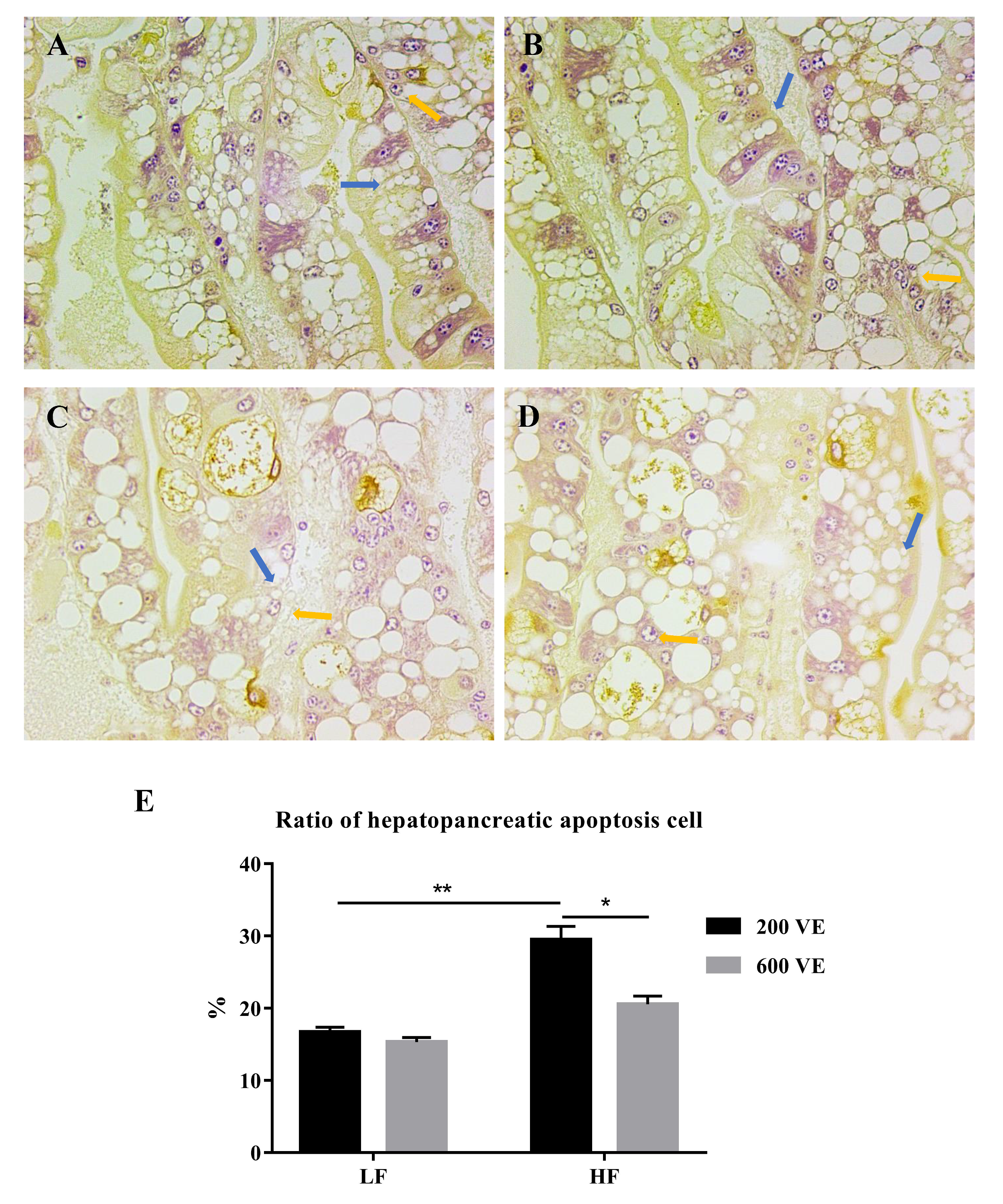
3.3. Hepatopancreas Ultrastructure and Apoptosis

The prawn hepatopancreas ultrastructure pictures are presented in Figure 3. The prawns fed the LF diets exhibited normal ultrastructures with round and clear nuclei; fewer lipid droplets were visible. The 600 VE group exhibited increased endoplasmic reticulum compared with the 200 VE group. Nevertheless, the prawns fed the HF diet showed hypertrophied lipid droplets in the cytoplasm, and lipophagy was observed. A total of 600 VE in the HF diet led to alleviated hepatic lipid accumulation with fewer droplets surrounding the centered nucleus compared with the 200 VE group.
The apoptosis of the hepatopancreatic cells is shown in Figure 4. The cells with stained brown nuclei were considered in a state of apoptosis and were counted to calculate the hepatopancreatic apoptosis cell ratio. Three paraffin sections were used for the TUNEL assay in each group. In the prawns fed LF diets, apoptotic cells were about 16–17% of the total hepatocytes (Figure 4A,B). In the hepatopancreases from the prawns fed the HF/200 VE diet, the apoptotic cells were about 30% of the total hepatocytes, which was significantly higher than in those fed LF/200 VE diet (p < 0.05) (Figure 4C). The 600 VE addition significantly decreased the ratio of apoptosis cells compared with 200 VE in the HF groups (p < 0.05) (Figure 4D).

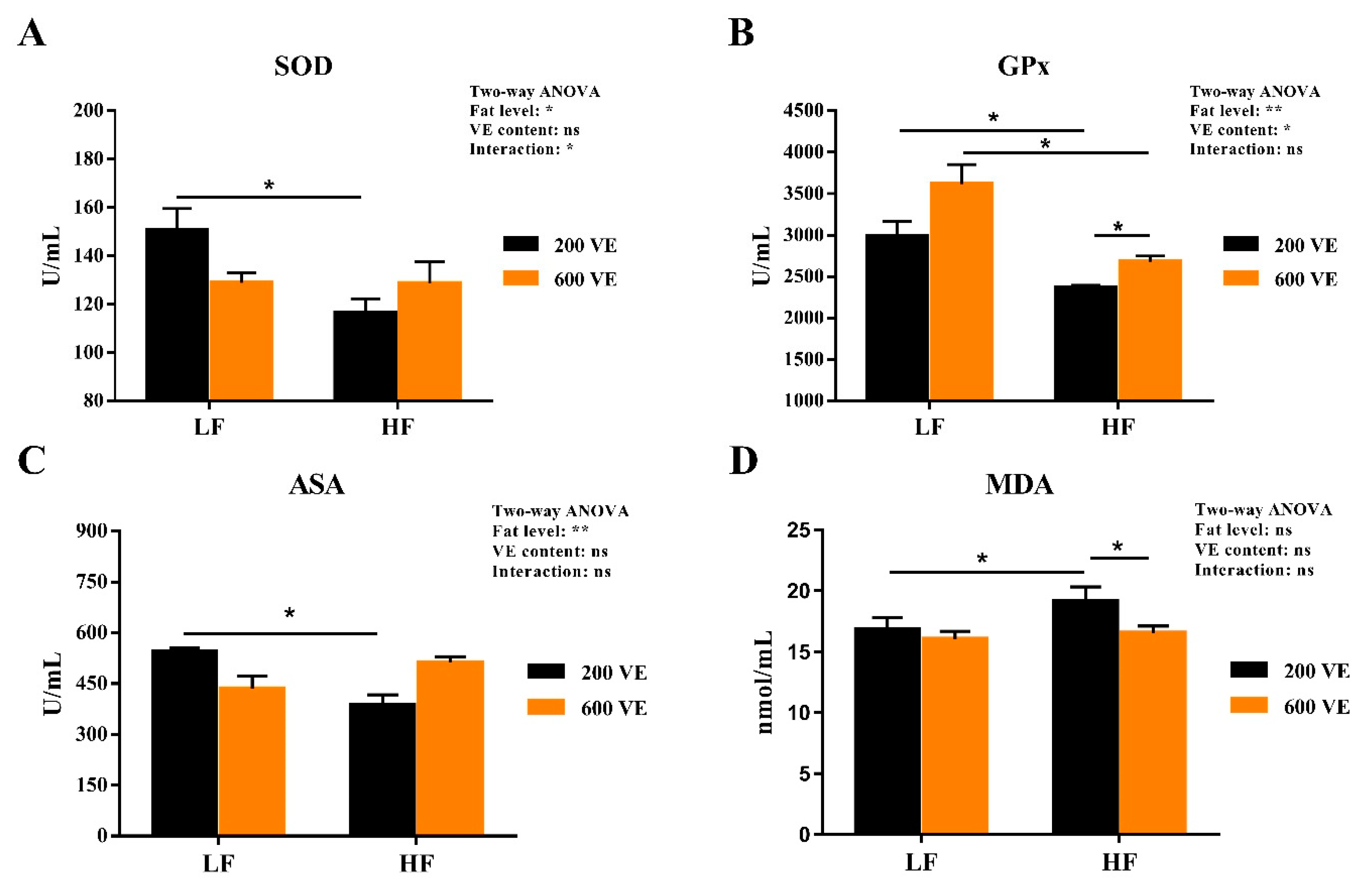
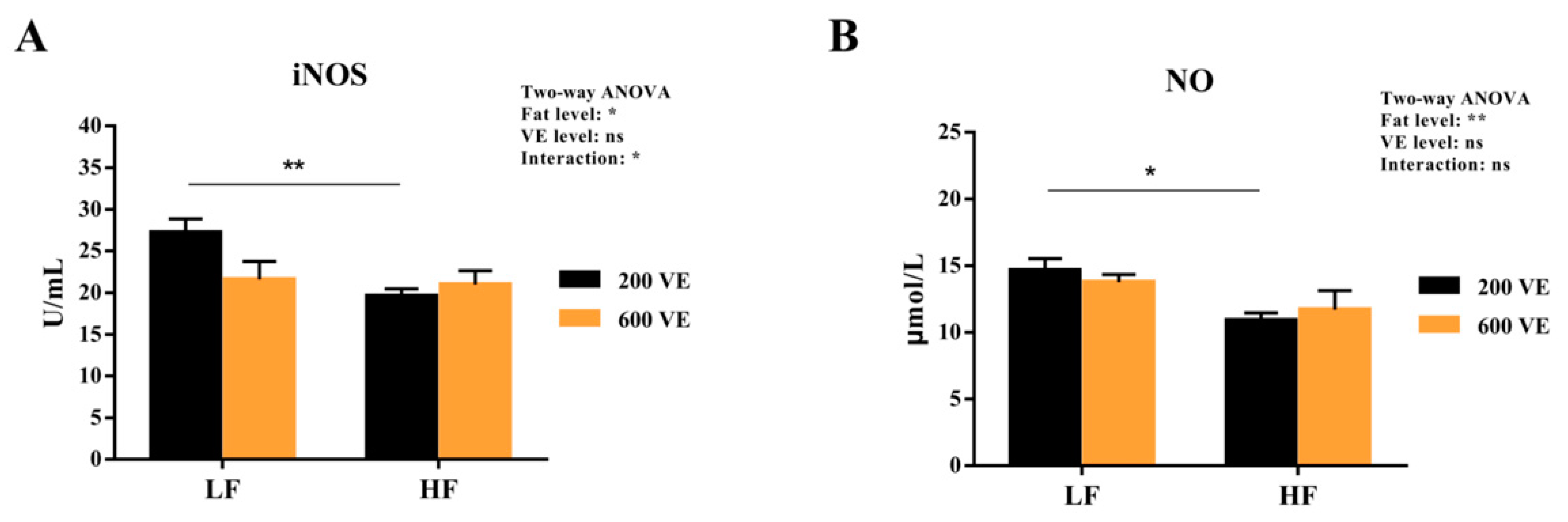
3.4. Hemolymph Antioxidant Capacity

The hemolymph antioxidant indexes are shown in Figure 5. SOD, GPX, and ASA were significantly affected by dietary fat levels (p < 0.05), GPx was significantly affected by dietary VE levels, and SOD was significantly affected by the interaction of dietary fat and VE (p < 0.05). In prawns fed LF diets, dietary levels of VE had no significant effect on the levels of SOD, GPx, ASA, and MDA (p < 0.05). In the prawns fed 200VE diets, the SOD, GPx, and ASA activities were significantly reduced when the dietary fat level was increased, while the MDA content was significantly increased (p < 0.05). In the prawns fed HF diets, the GPx activity was significantly increased and the MDA level was significantly decreased when the VE level was increased (p < 0.05).
The hemolymph iNOS activity and NO content are shown in Figure 6. The iNOS and NO were significantly affected by dietary fat levels (p < 0.05) and iNOS was significantly affected by the interaction of dietary fat and VE (p < 0.05). In the prawns fed 200 VE diets, the iNOS and NO levels were significantly reduced when the dietary fat level was increased (p < 0.05). In prawns fed the same fat-level diets, VE levels showed no significant difference in iNOS activity or NO content (p < 0.05).

3.5. Hepatopancreas NF-κB Signal Pathway Expression

The hemolymph gene expression of the NF-κB signal pathway expression is shown in Figure 7. Imd, relish, toll, and dorsal expression were significantly affected by dietary fat levels (p < 0.05), Imd, relish, dorsal expression was significantly affected by dietary VE levels (p < 0.05), and the interaction of dietary fat and VE showed no significant difference in NF-κB signal pathway expression (p > 0.05). In the prawns fed LF diets, dietary 600 VE significantly inhibited the expression of imd and dorsal compared with 200 VE (p < 0.05). In the prawns fed 200 VE diets, dietary HF significantly inhibited the expression of imd, relish, toll, and dorsal compared with LF (p < 0.05). In the prawns fed HF diets, dietary 600 VE significantly inhibited imd and relish expression compared with 200VE (p < 0.05). No significant difference was observed in the expression of toll or dorsal (p > 0.05).

3.6. In Vivo Knock-Down of Relish and Dorsal by RNA Interference

A relish and dorsal knock-down experiment was performed to further characterize the role of NF-κB in the antioxidative process induced by dietary HF. As shown in Figure 8A,B, relish and dorsal expression reduced significantly at 4d–7d post-injection of relish- and dorsal-specific dsRNA (p < 0.05). After a two-week in vivo knock-down of relish and dorsal by RNA interference, the expression of dorsal was suppressed by the dorsal dsRNA injection (p < 0.05) (Figure 8C), and the expression of relish was suppressed by the relish dsRNA injection (p < 0.05) (Figure 8D).

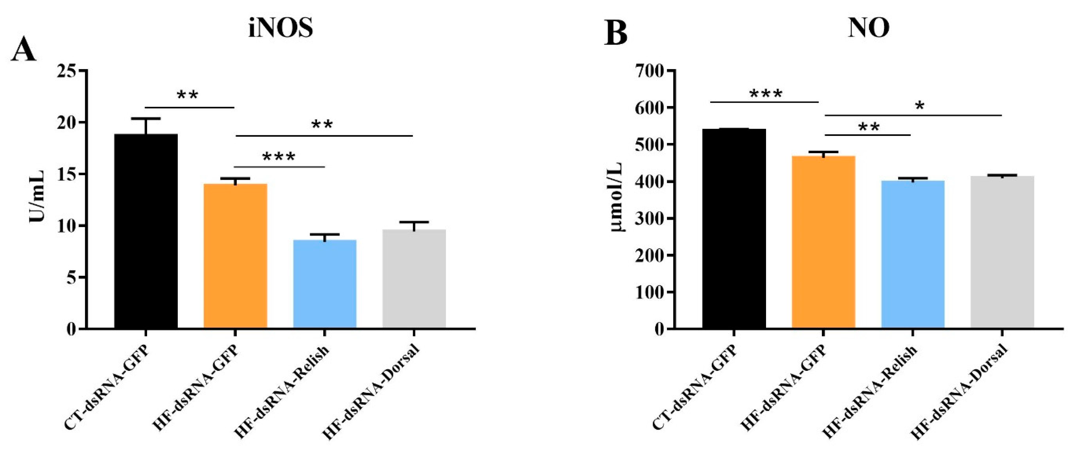
3.7. Hemolymph Antioxidant Capacity after NF-κB Suppression

As shown in Figure 9, the iNOS activity and NO content in the hemolymph of M. rosenbergii fed with the HF diet significantly decreased (p < 0.05). A further decline in the iNOS and NO levels was observed when the expression of relish and dorsal was suppressed (p < 0.05). Hemolymph ASA activity and MDA content are showed in Figure 10. Compared with the control group, the HF group’s MDA content significantly increased and its ASA activity significantly decreased (p < 0.05). In the prawns fed HF diet, relish and dorsal suppression further increased the MDA content and decreased the ASA activity (p < 0.05).

4. Discussion

Physiological conditions limit carnivorous fish’s demand for and utilization of fat. The long-term intake of high amounts of fat will accumulate in the body, leading to obstacles in fat transport, affecting body tissue fat, and reducing body fat deposition and fat metabolism by regulating lipid metabolism. Improving fish health has become one of the feasible means. In this study, the high-fat diet reduced the growth performance and survival rate of M. rosenbergii. Similar results were also observed in spotted seabass (Lateolabrax maculatus) [29], Pacific white shrimp (Litopenaeus vannamei) [14], largemouth bass (Micropterus salmoides) [30], and blunt snout bream (Megalobrama amblycephala) [31]. The reason for this might be the damaged antioxidant capacity, stress and disease resistance induced by dietary high fat. The previous studies pointed out that a high-fat diet could induce aberrant hepatic lipid secretion, reduce mortality rates and worsen the adverse effects of antibiotics by activating oxidant stress and endoplasmic reticulum stress [32,33,34]. This is also supported by the fact that high fat reduced the survival rate of M. rosenbergii in the present study. VE supplementation alleviated the growth inhibition induced by high fat levels in the present study, which was also confirmed in turbot (Scophthalmus maximus) [3]. Previous studies found that VE could alleviate high-fat-diet-induced hepatic oxidative stress and hypoimmunity to keep health and growth, while VE deficiency inhibited fat metabolism and induced lipid peroxidation [3,35]. The above results suggested that VE may be beneficial for maintaining the growth and health of prawns by reducing lipid metabolism disorder and the oxidative stress induced by high-fat diets.
Haemolymph biochemical indicators are important in diagnosing the health of aquatic animals. ALT and AST are sensitive biomarkers for hepatopancreas injury, and the TC and TG content can reflect lipid metabolism. In this study, the increase in the hemolymph ALT and AST activities of M. rosenbergii fed with a high-fat diet indicated the impaired function and metabolism of the hepatopancreas. Similar results were also observed in blunt snout bream [27] and grass carp (Ctenopharyngodon idella) [36]. The increase in hemolymph TC and TG in high fat group also indicated lipid metabolic disturbances and disorders because high serum TG and TC are significant risk factors related to fatty liver that can result in oxidative stress and decrease disease resistance. The present study found that extra dietary VE reduced the hemolymph levels of TG and TC, as well as ALT and AST activities in prawns fed an HF diet. The previous study demonstrated that the appropriate amounts of dietary VE improve the metabolic health of the liver in grass carp juveniles [37]. This was further supported by the fact that the VE requirement of aquatic animals increases as the fat content of feed increases [38]. These findings suggested that extra VE supplementation may contribute to the hepatopancreas health benefits of M. rosenbergii.
The hepatopancreas is the main organ responsible for the absorption and storage of the ingested substances. In the present study, the HF diet caused excessive lipid droplet accumulation in the hepatopancreas, which was also widely reported in other aquatic species [27,29,39]. In addition, the high-fat diet also induced hepatopancreas lipophagy and apoptosis. Lipophagy is the selective autophagy of lipid droplets degraded by lysosomes, which is associated with the regulation of lipid metabolism [40]. Existing evidence shows that high-fat diets can induce endoplasmic reticulum stress, autophagy, and apoptosis [41]. These pathologies may also cause the abnormal secretion of lipoproteins and lipid peroxidation, leading to a vicious circle. The results obtained here are still highly significant for human beings, although there are huge differences in the physiological structure of aquatic and terrestrial animals. In mice, high-fat diets could induce dyslipidemia and non-alcoholic fatty liver disease (NAFLD) [42,43]. Nutritional liver disease in shrimp has not yet been clearly defined, but the apoptosis and steatosis of the hepatopancreas found in prawns fed high levels of fat in the present study were similar to the NAFLD symptoms in humans. Nevertheless, these deformities can be relieved by extra VE supplementation. According to previous studies, VE can protect cells against lipid peroxidation and attenuate hepatic steatosis and mitochondrial damage [44]. VE could ameliorate apoptosis and autophagy induced by stress and disease in mammals [45,46,47]. Thus, based on histological and TUNEL apoptosis observation, the lipid-lowering effect of VE may be attributed to the attenuation of apoptosis and autophagy induced by high-fat diets. Similar results were also found in rats [48], indicating that M. rosenbergii can serve as a model for developing drugs for NAFLD.
Oxidative stress damage refers to the excess reactive oxygen species (ROS) production stimulated by factors such as the environment and nutrition, which breaks the redox equilibrium and ultimately leads to cell and tissue damage. In the present study, the HF diet reduced hemolymph ASA activity and increased MDA content. Similar results were also observed in largemouth bass (Micropterus salmoides) [49] and M. amblycephala [8]. This indicates that excess fat might induce oxidative stress damage and lipid peroxidation as MDA is the main component of lipid peroxide, which damages cell structures and functions [3]. In addition, the antioxidant enzyme system composed of SOD and GPx can remove excessive free radicals, reducing the damage by lipid peroxidation. The inhibition of the SOD and GPx activities in the HF group further supported the notion that excess fat induced oxidative stress. Nevertheless, VE improved GPx activities and reduced MDA content in the prawns fed the high-fat diet. This might due to the fact that VE has a significant effect of enhancing specific immunity and anti-oxidants by undergoing a redox reaction with the oxygen-containing groups of ROS to block peroxide production [50]. The ameliorative effect of VE on oxidative stress and hypoimmunity induced by a high-fat diet and oxidized oil also supported these results [3,51]. NO generated by iNOS could inhibit lipid peroxidation, which relies on its ability to eliminate oxygen free radicals [52]. The inhibition of hemolymph iNOS and NO in prawns fed with high fat also indicated the oxidative damage induced by excess fat, which is supported by the results in mussels [53] and L. vannamei [54]. However, VE showed no significant effect on iNOS and NO. As a non-enzymatic oxidant, VE halts lipid peroxidation by donating its phenolic hydrogen to peroxyl radicals, forming tocopherol radicals [55]. This may partly explain the reason of the invalidation of VE on iNOS and NO levels.
In general, the activation of transcription factors NF-κB is an indispensable step for increasing iNOS activity, which subsequently induces the release of NO [56]. In the present study, the expression of imd-relish and toll-doral, two major homologs of the components of NF-κB signal pathways, were inhibited by dietary high fat. It is speculated that both the imd-relish and the toll-doral signal pathway were suppressed by oxidative stress, which further reduced the production of NO, forming a vicious circle. A previous study also supported the notion that oxidative stress induced by high fat could inhibit the expression of the toll signal pathway [3]. However, VE only decreased imd-relish expression in the HF group. The regulation of VE in high-fat diets might attribute to the production of anti-microbial peptides as relish translocates into the nucleus to activate the expression of antibacterial peptide genes after stimulation [57]. To further verify the effect of the regulation of NF-κB on oxidative stress induced by high levels of fat, relish and dorsal expression were knocked down, respectively. The levels of iNOS and NO were suppressed, while MDA content increased and ASA activity decreased after relish and dorsal knock-down. The results above suggested that dietary high fat induced oxidative stress via the suppression of the NF-κB/NO signal pathway. Some research supported the notion that the NF-κB signal pathway also plays a crucial regulatory role in NAFLD induced by high-fat diets [58,59]. In vertebrates, NF-κB is activated by high-fat diets to induce inflammation. Nevertheless, crustaceans lack inflammatory cytokines. The different regulation of NF-κB in crustaceans and vertebrates still requires further in-depth studies.

5. Conclusions

The present study investigated the growth performance and oxidative status of M. rosenbergii fed different fat and VE levels, and revealed the molecular regulatory mechanism in response to a high-fat diet. In the present study, VE ameliorated the growth retardation and oxidative stress induced by a high-fat diet. A putative mechanism that could explain our results is shown in Figure 11. The high-fat diet caused excessive lipid deposition in M. rosenbergii, which further induced hepatopancreas lipophagy, apoptosis, and lipid peroxidation, resulting in oxidative stress damage. The toll-dorsal and imd-relish signaling pathways were inhibited by the high fat levels, which mediated pro-oxidant targets (iNOS and NO) and antioxidant targets (SOD and GPx) for the negative feedback regulation of oxidative stress. Dietary VE supplementation combined with the high-fat diet alleviated hepatopancreas oxidative stress and apoptosis. The reason for this may be related to the antioxidant properties of VE and the activation of the antioxidant enzyme system.

Author Contributions

The author contributions were as follows: C.S. contributed in the areas of experimental design, sampling, data analysis, and write-up; B.L., Q.Z. and X.Z. contributed to the experimental design and manuscript review; F.S. and M.L. contributed to feeding and cultivating of experimental prawns, sampling, and statistics and X.X. contributed to feed production, feeding of prawns, and sampling. All authors have read and agreed to the published version of the manuscript.

Funding

This work is supported by the Project of National Key R&D Program of China (2019YFD0900200), the National Natural Science Foundation of China (32002404), the Natural Science Foundation of Jiangsu Province for Youths (BK20180172), China Agriculture Research System of MOF and MARA (CARS-48), Central Public-interest Scientific Institution Basal Research Fund, CAFS (2020TD59). All the funders had no role in the design, analysis or writing of this article.

Institutional Review Board Statement

The care and use of animals followed Animal Research Institute Committee guidelines of Nanjing Agricultural University, China. This study has been approved by the Committee of the Animal Research Institute of Nanjing Agricultural University, China (SYXK (Su) 2019-0136, approved 21 May 2017).

Informed Consent Statement

Not applicable.

Data Availability Statement

Data is contained within the article.

Acknowledgments

The authors would acknowledge Zhejiang Southern Taihu Lake freshwater aquatic seed industry CO. LTD for the contribution of juvenile prawns and the administration in the feeding trial.

Conflicts of Interest

The authors declare no conflict of interest.

References

  1. Méndez-Martínez, Y.; García-Guerrero, M.U.; Arcos-Ortega, F.G.; Martínez-Córdova, L.R.; Yamasaki-Granados, S.; Pérez-Rodríguez, J.C.; Cortés-Jacinto, E. Effect of different ratios of dietary protein-energy on growth, body proximal composition, digestive enzyme activity, and hepatopancreas histology in Macrobrachium americanum (Bate, 1868) prawn juveniles. Aquaculture 2018, 485, 1–11. [Google Scholar] [CrossRef]
  2. Zhang, N.N.; Ma, Q.Q.; Fan, W.J.; Xing, Q.; Zhao, Y.L.; Chen, L.Q.; Ye, J.Y.; Zhang, M.L.; Du, Z.Y. Effects of the dietary protein to energy ratio on growth, feed utilization and body composition in Macrobrachium nipponense. Aquac. Nutr. 2017, 23, 313–321. [Google Scholar] [CrossRef]
  3. Jia, Y.; Jing, Q.; Niu, H.; Huang, B. Ameliorative effect of vitamin E on hepatic oxidative stress and hypoimmunity induced by high-fat diet in turbot (Scophthalmus maximus). Fish Shellfish Immunol. 2017, 67, 634–642. [Google Scholar] [CrossRef]
  4. Lu, K.; Wang, L.; Zhang, D.; Liu, W.; Xu, W. Berberine attenuates oxidative stress and hepatocytes apoptosis via protecting mitochondria in blunt snout bream Megalobrama amblycephala fed high-fat diets. Fish Physiol. Biochem. 2017, 43, 65–76. [Google Scholar] [CrossRef] [PubMed]
  5. Li, Y.; Fan, B.; Huang, Y.; Wu, D.; Zhang, M.; Zhao, Y. Effects of dietary vitamin E on reproductive performance and antioxidant capacity of Macrobrachium nipponense female shrimp. Aquac. Nutr. 2018, 24, 1698–1708. [Google Scholar] [CrossRef]
  6. Griboff, J.; Morales, D.; Bertrand, L.; Bonansea, R.I.; Monferrán, M.V.; Asis, R.; Wunderlin, D.A.; Amé, M.V. Oxidative stress response induced by atrazine in Palaemonetes argentinus: The protective effect of vitamin E. Ecotoxicol. Environ. Saf. 2014, 108, 1–8. [Google Scholar] [CrossRef]
  7. Mourente, G.; Dıaz-Salvago, E.; Bell, J.G.; Tocher, D.R. Increased activities of hepatic antioxidant defence enzymes in juvenile gilthead sea bream (Sparus aurata L.) fed dietary oxidised oil: Attenuation by dietary vitamin E. Aquaculture 2002, 214, 343–361. [Google Scholar] [CrossRef]
  8. Lu, K.L.; Xu, W.N.; Liu, W.B.; Wang, L.N.; Zhang, C.N.; Li, X.F. Association of mitochondrial dysfunction with oxidative stress and immune suppression in blunt snout bream Megalobrama amblycephala fed a high-fat diet. J. Aquat. Anim. Health 2014, 26, 100–112. [Google Scholar] [CrossRef] [PubMed]
  9. Essick, E.E.; Sam, F. Oxidative stress and autophagy in cardiac disease, neurological disorders, aging and cancer. Oxid. Med. Cell. Longev. 2010, 3, 168–177. [Google Scholar] [CrossRef] [Green Version]
  10. Jiang, S.; Du, P.; An, L.; Yuan, G.; Sun, Z. Anti-diabetic effect of Coptis Chinensis polysaccharide in high-fat diet with STZ-induced diabetic mice. Int. J. Biol. Macromol. 2013, 55, 118–122. [Google Scholar] [CrossRef] [PubMed]
  11. Furukawa, S.; Fujita, T.; Shimabukuro, M.; Iwaki, M.; Yamada, Y.; Nakajima, Y.; Nakayama, O.; Makishima, M.; Matsuda, M.; Shimomura, I. Increased oxidative stress in obesity and its impact on metabolic syndrome. J. Clin. Investig. 2004, 114, 1752–1761. [Google Scholar] [CrossRef] [PubMed]
  12. Cherif, S.; Fendri, A.; Miled, N.; Trabelsi, H.; Mejdoub, H.; Gargouri, Y. Crab digestive lipase acting at high temperature: Purification and biochemical characterization. Biochimie 2007, 89, 1012–1018. [Google Scholar] [CrossRef] [PubMed]
  13. Council, N.R. Nutrient Requirements of Fish and Shrimp; National Academies Press: Pittsburgh, PA, USA, 2011. [Google Scholar]
  14. Xu, C.; Li, E.; Liu, Y.; Wang, S.; Wang, X.; Chen, K.; Qin, J.G.; Chen, L. Effect of dietary lipid level on growth, lipid metabolism and health status of the Pacific white shrimp Litopenaeus vannamei at two salinities. Aquac. Nutr. 2018, 24, 204–214. [Google Scholar] [CrossRef]
  15. Zhao, J.; Wen, X.; Li, S.; Zhu, D.; Li, Y. Effects of dietary lipid levels on growth, feed utilization, body composition and antioxidants of juvenile mud crab Scylla paramamosain (Estampador). Aquaculture 2015, 435, 200–206. [Google Scholar] [CrossRef]
  16. D’Ignazio, L.; Bandarra, D.; Rocha, S. NF-κB and HIF crosstalk in immune responses. FEBS J. 2016, 283, 413–424. [Google Scholar] [CrossRef] [Green Version]
  17. Meyerovich, K.; Fukaya, M.; Terra, L.F.; Ortis, F.; Eizirik, D.L.; Cardozo, A.K. The non-canonical NF-κB pathway is induced by cytokines in pancreatic beta cells and contributes to cell death and proinflammatory responses in vitro. Diabetologia 2016, 59, 512–521. [Google Scholar] [CrossRef] [Green Version]
  18. Jiang, Y.S.; Wu, X.Z. Characterization of a Rel\NF-κB homologue in a gastropod abalone, Haliotis diversicolor supertexta. Dev. Comp. Immunol. 2007, 31, 121–131. [Google Scholar] [CrossRef]
  19. Li, F.; Wang, L.; Zhang, H.; Zheng, P.; Zhao, J.; Qiu, L.; Zhang, Y.; Song, L. Molecular cloning and expression of a Relish gene in Chinese mitten crab Eriocheir sinensis. Int. J. Immunogenet. 2010, 37, 499–508. [Google Scholar] [CrossRef]
  20. Morgan, M.J.; Liu, Z.G. Crosstalk of reactive oxygen species and NF-κB signaling. Cell Res. 2011, 21, 103–115. [Google Scholar] [CrossRef] [Green Version]
  21. Arockiaraj, J.; Avin, F.A.; Vanaraja, P.; Easwvaran, S.; Singh, A.; Othman, R.Y.; Bhassu, S. Immune role of MrNFκBI-α, an IκB family member characterized in prawn M. rosenbergii. Fish Shellfish Immunol. 2012, 33, 619–625. [Google Scholar] [CrossRef]
  22. Huang, X.; Wang, W.; Ren, Q. Dorsal transcription factor is involved in regulating expression of crustin genes during white spot syndrome virus infection. Dev. Comp. Immunol. 2016, 63, 18–26. [Google Scholar] [CrossRef] [PubMed]
  23. Srisuk, C.; Longyant, S.; Senapin, S.; Sithigorngul, P.; Chaivisuthangkura, P. Molecular cloning and characterization of a Toll receptor gene from Macrobrachium rosenbergii. Fish Shellfish Immunol. 2014, 36, 552–562. [Google Scholar] [CrossRef]
  24. Shi, Y.R. Study on Sequences Analysis and Function of IMD and Relish in IMD Signal Pathway in Macrobrachium rosenbergii. Master’s Thesis, Nanjing Normal University, Nanjing, China, 2016. [Google Scholar]
  25. Fishery Bureau of Ministry of Agriculture of the People’s Republic of China. China Fishery Statistics Yearbook 2021; China Agriculture Press: Beijing, China, 2021.
  26. Paul, P.; Rahman, M.A.; Mer, M.; Hossain, M.; Islam, M.S.; Mondal, S.; Haq, M.M. Effect of stocking density on the growth and production of freshwater prawn (Macrobrachium rosenbergii). Int. J. Fish. Aquac. Sci. 2016, 6, 77–86. [Google Scholar]
  27. Lu, K.L.; Xu, W.N.; Li, J.Y.; Li, X.F.; Huang, G.Q.; Liu, W.B. Alterations of liver histology and blood biochemistry in blunt snout bream Megalobrama amblycephala fed high-fat diets. Fish. Sci. 2013, 79, 661–671. [Google Scholar] [CrossRef]
  28. Wangari, M.R.; Gao, Q.; Sun, C.; Liu, B.; Song, C.; Tadese, D.A.; Zhou, Q.; Zhang, H.; Liu, B. Effect of dietary Clostridium butyricum and different feeding patterns on growth performance, antioxidant and immune capacity in freshwater prawn (Macrobrachium rosenbergii). Aquac. Res. 2021, 52, 12–22. [Google Scholar] [CrossRef]
  29. Dong, Y.Z.; Xia, T.; Lin, J.B.; Wang, L.; Song, K.; Zhang, C.X. Quercetin attenuates high-fat diet-induced excessive fat deposition of spotted seabass (Lateolabrax maculatus) through the regulatory for mitochondria and endoplasmic reticulum. Front. Mar. Sci. 2021, 8, 746811. [Google Scholar] [CrossRef]
  30. Yin, P.; Xie, S.; Zhuang, Z.; He, X.; Tang, X.; Tian, L.; Liu, Y.; Niu, J. Dietary supplementation of bile acid attenuate adverse effects of high-fat diet on growth performance, antioxidant ability, lipid accumulation and intestinal health in juvenile largemouth bass (Micropterus salmoides). Aquaculture 2021, 531, 735864. [Google Scholar] [CrossRef]
  31. Zhou, W.; Rahimnejad, S.; Lu, K.; Wang, L.; Liu, W. Effects of berberine on growth, liver histology, and expression of lipid-related genes in blunt snout bream (Megalobrama amblycephala) fed high-fat diets. Fish Physiol. Biochem. 2019, 45, 83–91. [Google Scholar] [CrossRef]
  32. Limbu, S.M.; Ma, Q.; Zhang, M.L.; Du, Z.Y. High fat diet worsens the adverse effects of antibiotic on intestinal health in juvenile Nile tilapia (Oreochromis niloticus). Sci. Total Environ. 2019, 680, 169–180. [Google Scholar] [CrossRef]
  33. Sink, T.D.; Lochmann, R.T.; Goodwin, A.E.; Marecaux, E. Mortality rates in golden shiners fed high-fat diets with or without a dairy-yeast prebiotic before challenge with Flavobacterium columnare. N. Am. J. Aquac. 2007, 69, 305–308. [Google Scholar] [CrossRef]
  34. Cao, X.F.; Dai, Y.J.; Liu, M.Y.; Yuan, X.Y.; Wang, C.C.; Huang, Y.Y.; Liu, W.B.; Jiang, G.Z. High-fat diet induces aberrant hepatic lipid secretion in blunt snout bream by activating endoplasmic reticulum stress-associated IRE1/XBP1 pathway. Biochim. Biophys. Acta Mol. Cell Biol. Lipids 2019, 1864, 213–223. [Google Scholar] [CrossRef] [PubMed]
  35. Qiang, J.; Wasipe, A.; He, J.; Tao, Y.F.; Xu, P.; Bao, J.W.; Chen, D.J.; Zhu, J.H. Dietary vitamin E deficiency inhibits fat metabolism, antioxidant capacity, and immune regulation of inflammatory response in genetically improved farmed tilapia (GIFT, Oreochromis niloticus) fingerlings following Streptococcus iniae infection. Fish Shellfish Immunol. 2019, 92, 395–404. [Google Scholar] [CrossRef] [PubMed]
  36. Liu, G.; Yu, H.; Wang, C.; Li, P.; Liu, S.; Zhang, X.; Zhang, C.; Qi, M.; Ji, H. Nano-selenium supplements in high-fat diets relieve hepatopancreas injury and improve survival of grass carp Ctenopharyngodon Idella by reducing lipid deposition. Aquaculture 2021, 538, 736580. [Google Scholar] [CrossRef]
  37. Li, J.; Liang, X.F.; Tan, Q.; Yuan, X.; Liu, L.; Zhou, Y.; Li, B. Effects of vitamin E on growth performance and antioxidant status in juvenile grass carp Ctenopharyngodon idellus. Aquaculture 2014, 430, 21–27. [Google Scholar] [CrossRef]
  38. Lin, Y.H.; Shiau, S.Y. Dietary vitamin E requirement of grouper, Epinephelus malabaricus, at two lipid levels, and their effects on immune responses. Aquaculture 2005, 248, 235–244. [Google Scholar] [CrossRef]
  39. Wang, L.; Xu, B.; Sagada, G.; Ng, W.K.; Chen, K.; Zhang, J.; Shao, Q. Dietary berberine regulates lipid metabolism in muscle and liver of black sea bream (Acanthopagrus schlegelii) fed normal or high-lipid diets. Br. J. Nutr. 2021, 125, 481–493. [Google Scholar] [CrossRef]
  40. Liu, C.; Liao, J.Z.; Li, P.Y. Traditional Chinese herbal extracts inducing autophagy as a novel approach in therapy of nonalcoholic fatty liver disease. World J. Gastroenterol. 2017, 23, 1964–1973. [Google Scholar] [CrossRef]
  41. Ling, S.C.; Wu, K.; Zhang, D.G.; Luo, Z. Endoplasmic reticulum stress-mediated autophagy and apoptosis alleviate dietary Fat-induced triglyceride accumulation in the intestine and in isolated intestinal epithelial cells of yellow catfish. J. Nutr. 2019, 149, 1732–1741. [Google Scholar] [CrossRef]
  42. Uetake, Y.; Ikeda, H.; Irie, R.; Tejima, K.; Matsui, H.; Ogura, S.; Wang, H.; Mu, S.; Hirohama, D.; Ando, K.; et al. High-salt in addition to high-fat diet may enhance inflammation and fibrosis in liver steatosis induced by oxidative stress and dyslipidemia in mice. Lipids Health Dis. 2015, 14, 6. [Google Scholar] [CrossRef] [Green Version]
  43. Sarna, L.K.; Sid, V.; Wang, P.; Siow, Y.L.; House, J.D.; Karmin, O. Tyrosol attenuates high fat diet-induced hepatic oxidative stress: Potential involvement of cystathionine β-synthase and cystathionine γ-lyase. Lipids 2016, 51, 583–590. [Google Scholar] [CrossRef]
  44. Rasool, M.; Malik, A.; Saleem, S.; Ashraf, M.A.B.; Khan, A.Q.; Waquar, S.; Zahid, A.; Shaheen, S.; Abu-Elmagd, M.; Gauthaman, K.; et al. Role of oxidative stress and the identification of biomarkers associated with thyroid dysfunction in schizophrenics. Front. Pharmacol. 2021, 12, 646287. [Google Scholar] [CrossRef] [PubMed]
  45. Kaur, S.; Bansal, M.P. Protective role of dietary-supplemented selenium and vitamin E in heat-induced apoptosis and oxidative stress in mice testes. Andrologia 2015, 47, 1109–1119. [Google Scholar] [CrossRef] [PubMed]
  46. Khorramabadi, K.M.; Talebi, A.R.; Sarcheshmeh, A.A.; Mirjalili, A. Protective effect of vitamin E on oxidative stress and sperm apoptosis in diabetic mice. Int. J. Reprod. Biomed. 2019, 17, 127–134. [Google Scholar] [CrossRef] [PubMed]
  47. Zhao, Y.; Zhang, W.; Jia, Q.; Feng, Z.; Guo, J.; Han, X.; Liu, Y.; Shang, H.; Wang, Y.; Liu, W.J. High dose vitamin E attenuates diabetic nephropathy via alleviation of autophagic stress. Front. Physiol. 2019, 10, 1939. [Google Scholar] [CrossRef] [PubMed] [Green Version]
  48. Asrullah, M.; Arsanti Lestari, L.; Helmyati, S.; Farmawati, A. Effect supplementation of mung bean sprouts (Phaseolus radiatus L.) and vitamin E in rats fed high fat diet. KnE Life Sci. 2019, 4, 36–46. [Google Scholar] [CrossRef] [Green Version]
  49. Xie, S.; Yin, P.; Tian, L.; Yu, Y.; Liu, Y.; Niu, J. Dietary supplementation of astaxanthin improved the growth performance, antioxidant ability and immune response of juvenile largemouth bass (Micropterus salmoides) fed high-fat diet. Mar. Drugs 2020, 18, 642. [Google Scholar] [CrossRef] [PubMed]
  50. Cheng, C.H.; Guo, Z.X.; Wang, A.L. Growth performance and protective effect of vitamin E on oxidative stress pufferfish (Takifugu obscurus) following by ammonia stress. Fish Physiol. Biochem. 2018, 44, 735–745. [Google Scholar] [CrossRef] [PubMed]
  51. Gao, J.; Koshio, S.; Ishikawa, M.; Yokoyama, S.; Mamauag, R.E.P.; Han, Y. Effects of dietary oxidized fish oil with vitamin E supplementation on growth performance and reduction of lipid peroxidation in tissues and blood of red sea bream Pagrus major. Aquaculture 2012, 356–357, 73–79. [Google Scholar] [CrossRef]
  52. Sun, C.; Liu, B.; Zhou, Q.; Xiong, Z.; Shan, F.; Zhang, H. Response of Macrobrachium rosenbergii to vegetable oils replacing dietary fish oil: Insights from antioxidant defense. Front. Physiol. 2020, 11, 218. [Google Scholar] [CrossRef] [Green Version]
  53. Belavgeni, A.; Dailianis, S. The role of phosphatidylinositol-3-OH-kinase (PI3-kinase) and respiratory burst enzymes in the [omim][BF4]-mediated toxic mode of action in mussel hemocytes. Fish Shellfish Immunol. 2017, 68, 144–153. [Google Scholar] [CrossRef]
  54. Duan, Y.; Dong, H.; Wang, Y.; Li, H.; Liu, Q.; Zhang, Y.; Zhang, J. Intestine oxidative stress and immune response to sulfide stress in Pacific white shrimp Litopenaeus vannamei. Fish Shellfish Immunol. 2017, 63, 201–207. [Google Scholar] [CrossRef] [PubMed]
  55. Carocho, M.; Ferreira, I.C.F.R. A review on antioxidants, prooxidants and related controversy: Natural and synthetic compounds, screening and analysis methodologies and future perspectives. Food Chem. Toxicol. 2013, 51, 15–25. [Google Scholar] [CrossRef] [PubMed]
  56. Pautz, A.; Art, J.; Hahn, S.; Nowag, S.; Voss, C.; Kleinert, H. Regulation of the expression of inducible nitric oxide synthase. Nitric Oxide Biol. Chem. 2010, 23, 75–93. [Google Scholar] [CrossRef]
  57. Shi, Y.R.; Jin, M.; Ma, F.T.; Huang, Y.; Huang, X.; Feng, J.L.; Zhao, L.L.; Chen, Y.H.; Ren, Q. Involvement of relish gene from Macrobrachium rosenbergii in the expression of anti-microbial peptides. Dev. Comp. Immunol. 2015, 52, 236–244. [Google Scholar] [CrossRef] [PubMed]
  58. Zhao, W.; Yan, Y.; Xiao, Z.; Wang, M.; Xu, M.; Wang, Z.; Wang, Y.; Zhuang, Z.; Yang, D.; Chen, G.; et al. Bicyclol ameliorates nonalcoholic fatty liver disease in mice via inhibiting MAPKs and NF-κB signaling pathways. Biomed. Pharmacother. 2021, 141, 111874. [Google Scholar] [CrossRef]
  59. Liu, P.; Wu, P.; Yang, B.; Wang, T.; Li, J.; Song, X.; Sun, W. Kaempferol prevents the progression from simple steatosis to non-alcoholic steatohepatitis by inhibiting the NF-κB pathway in oleic acid-induced HepG2 cells and high-fat diet-induced rats. J. Funct. Foods 2021, 85, 104655. [Google Scholar] [CrossRef]
Figure 1. Growth performance and feed utilization of Macrobrachium rosenbergii fed with different dietary fat and VE levels. (A) survival rate (SR, %) of M. rosenbergii fed with different dietary fat and VE levels; (B) Weight gain rate (WGR, %) of M. rosenbergii fed with different dietary fat and VE levels; (C) specific growth rate (SGR, %/day) of M. rosenbergii fed with different dietary fat and VE levels; (D) feed conversion ratio (FCR) of M. rosenbergii fed with different dietary fat and VE levels. Values are means ± SEM. ***: p < 0.001, **: p < 0.01, *: p < 0.05. WGR = (final weight − initial weight) × 100/initial weight; SGR = (Ln final weight − Ln initial weight) × 100/day; FCR = feed consumption (g)/weight gain (g); SR = final number of prawns/initial number of prawns.
Antioxidants 11 00228 g001
Figure 2. Hemolymph biochemistry parameters of Macrobrachium rosenbergii fed with different dietary fat and VE levels. (A) hemolymph alanine aminotransferase (ALT) activity of M. rosenbergii fed with different dietary fat and VE levels; (B) hemolymph aspartate aminotransferase (AST) activity of M. rosenbergii fed with different dietary fat and VE levels; (C) hemolymph total cholesterol (TC) content of M. rosenbergii fed with different dietary fat and VE levels; (D) hemolymph total triglycerides (TG) content of M. rosenbergii fed with different dietary fat and VE levels. Values are means ± SEM. ***: p < 0.001, **: p < 0.01, *: p < 0.05, ns: p < 0.05.
Antioxidants 11 00228 g002
Figure 3. Transmission electron microscope images of Macrobrachium rosenbergii hepatocyte ultrastructure (2500×) fed with different dietary fat and VE levels. (A) prawns fed diet including 9% fat and 200 mg/kg VE; (B) prawns fed diet including 9% fat and 600 mg/kg VE; (C) prawns fed diet including 13% fat and 200 mg/kg VE; (D) prawns fed diet including 13% fat and 600 mg/kg VE. N: nucleus; LD: lipid droplet; ER, endoplasmic reticulum; LP, lipophagy.
Antioxidants 11 00228 g003
Figure 4. Hepatocyte apoptosis of Macrobrachium rosenbergii fed with different dietary fat and VE levels. (A) prawns fed diet including 9% fat and 200 mg/kg VE; (B) prawns fed diet including 9% fat and 600 mg/kg VE; (C) prawns fed diet including 13% fat and 200 mg/kg VE; (D) prawns fed diet including 13% fat and 600 mg/kg VE; (E) ratio of hepatopancreatic apoptosis cells (n = 3). Blue arrow: normal cells; yellow arrow: apoptotic cells. **: p < 0.01, *: p < 0.05.
Antioxidants 11 00228 g004
Figure 5. Hemolymph oxidative status of Macrobrachium rosenbergii fed with different dietary fat and VE levels. (A) hemolymph superoxide dismutase (SOD) activity of M. rosenbergii fed with different dietary fat and VE levels; (B) hemolymph glutathione peroxidase (GPx) activity of M. rosenbergii fed with different dietary fat and VE levels; (C) hemolymph anti-superoxide anion (ASA) activity of M. rosenbergii fed with different dietary fat and VE levels; (D) hemolymph malonaldehyde (MDA) content of M. rosenbergii fed with different dietary fat and VE levels. Values are means ± SEM. **: p < 0.01, *: p < 0.05, ns: p < 0.05.
Antioxidants 11 00228 g005
Figure 6. Hemolymph iNOS activity and NO content of Macrobrachium rosenbergii fed with different dietary fat and VE levels. (A) hemolymph inducible nitric oxide synthase (iNOS) activity of M. rosenbergii fed with different dietary fat and VE levels; (B) hemolymph nitric oxide (NO) content of M. rosenbergii fed with different dietary fat and VE levels. Values are means ± SEM. **: p < 0.01, *: p < 0.05, ns: p < 0.05.
Antioxidants 11 00228 g006
Figure 7. Relative expression of NF-κB signal pathway in hepatopancreas. (A) relative expression of imd in hepatopancreas; (B) relative expression of relish in hepatopancreas; (C) relative expression of toll in hepatopancreas; (D) relative expression of dorsal in hepatopancreas. Values are means ± SEM. ***: p < 0.001, **: p < 0.01, *: p < 0.05, ns: p < 0.05.
Antioxidants 11 00228 g007
Figure 8. Knock-down of relish and dorsal in vivo expression by dsRNA-mediated RNA interference. (A) Time-course study of dorsal expression in hepatopancreas after RNA interference; (B) Time-course study of relish expression in hepatopancreas after RNA interference; (C) dorsal expression in M. rosenbergii fed with high-fat diet after RNA interference; (D) relish expression in M. rosenbergii fed with high-fat diet after RNA interference. Values are means ± SEM. **: p < 0.01, *: p < 0.05.
Antioxidants 11 00228 g008
Figure 9. Hemolymph iNOS activity and NO content of Macrobrachium rosenbergii after knock-down by dsRNA-mediated RNA interference. (A) Hemolymph inducible nitric oxide synthase (iNOS) activity of M. rosenbergii after RNA interference; (B) Hemolymph nitric oxide (NO) content of M. rosenbergii after RNA interference. Values are means ± SEM. ***: p < 0.001, **: p < 0.01, *: p < 0.05.
Antioxidants 11 00228 g009
Figure 10. Hemolymph ASA activity and MDA content of Macrobrachium rosenbergii after knock-down by dsRNA-mediated RNA interference. (A) Hemolymph anti-superoxide anion (ASA) activity of M. rosenbergii after RNA interference; (B) Hemolymph malonaldehyde (MDA) content of M. rosenbergii after RNA interference. Values are means ± SEM. **: p < 0.01, *: p < 0.05.
Antioxidants 11 00228 g010
Figure 11. NF-κB/NO signaling mechanism in response to dietary high fat and VE. The red arrows indicate the effects of high fat; the green arrows indicate the effects of VE.
Antioxidants 11 00228 g011
Table 1. Formulation and proximate composition of the experimental diets.
LF/200VELF/600VEHF/200VEHF/600VE
Ingredients%
Fish meal 122.0022.0022.0022.00
Casein 224.00 24.0024.0024.00
Gelatin 16.006.006.006.00
α-starch 320.0020.0020.0020.00
Dextrin 35.005.005.005.00
Fish oil 16.006.0010.0010.00
Soybean oil 10.000.000.000.00
Rapeseed oil 10.000.000.000.00
Microcystalline cellulose 45.005.001.001.00
Carboxymethyl cellulose 43.003.003.003.00
Bentonite 11.531.491.531.49
Soybean lecithin (50%) 12.002.002.002.00
Cholesterol 10.300.300.300.30
Ecdysone (2%) 50.100.100.100.10
DMPT 50.050.050.050.05
Choline chloride 51.001.001.001.00
Vitamin premix 51.001.001.001.00
Vitamin E 10.020.060.020.06
Mineral premix 51.001.001.001.00
Calcium dihydrogen phosphate 12.002.002.002.00
Proximate composition %
Crude protein40.9040.9040.9040.90
Crude lipid9.589.5813.2813.28
Gross energy (MJ/kg)15.8015.8017.3817.38
Notes: 1 Obtained from Wuxi Tongwei feedstuffs Co., Ltd., Wuxi, China; 2 obtained from Hulunbeier Sanyuan Milk Co., Ltd., Inner Mongolia, China; 3 obtained from Yinhe Dextrin Co., Ltd., Zhengzhou, China; 4 obtained from Yifeng Food Additives Co., Ltd., Shanghai, China; 5 obtained from Jiangsu Fuyuda Food Products Co., Ltd., Yangzhou, China.
Table 2. Sequences of the primers used in the study.
GeneGenBank Acc. No.Primer Sequences (5′–3′)Length (bp)PurposeReference
T7-relishKR827675.1TAATACGACTCACTATAGGGATCATCATGAGGCGGAAAAG40RNAi
TAATACGACTCACTATAGGGTGGCATGTAGGTGAAATCCA40
T7-dorsalKX219631.1TAATACGACTCACTATAGGGCAAGTGTTCCTCGAAGGCTC40RNAi
TAATACGACTCACTATAGGGAACTTCACCAATTTGTCCGC40
relishKR827675.1GATGAGCCTTCAGTGCCAGA20RT-qPCR
CCAGGTGACGCCATGTATCA20
dorsalKX219631.1TCAGTAGCGACACCATGCAG20RT-qPCR
CGAGCCTTCGAGGAACACTT20
imd CGACCACATTCTCCTCCTCCC21RT-qPCR[24]
TTCAGTGCATCCACGTCCCTC21
tollKX610955.1TTCGTGACTTGTCGGCTCTC20RT-qPCR
GCAGTTGTTGAAGGCATCGG20
β-actinAY651918.2TCCGTAAGGACCTGTATGCC20RT-qPCR
TCGGGAGGTGCGATGATTTT20
Publisher’s Note: MDPI stays neutral with regard to jurisdictional claims in published maps and institutional affiliations.

