Article

Transposon-Directed Insertion-Site Sequencing Reveals Glycolysis Gene gpmA as Part of the H2O2 Defense Mechanisms in Escherichia coli

1
Department of Pathology and Immunology, Faculty of Medicine, University of Geneva, 1211 Geneva, Switzerland
2
Institute for Molecular Bioscience, University of Queensland, St. Lucia, Brisbane, QLD 4072, Australia
3
READS Unit, Faculty of Medicine, University of Geneva, 1211 Geneva, Switzerland
4
Genomic Research Laboratory, Infectious Diseases Service, University Hospitals of Geneva, University Medical Center, Michel-Servet 1, 1211 Geneva, Switzerland
*
Author to whom correspondence should be addressed.
Antioxidants 2022, 11(10), 2053; https://doi.org/10.3390/antiox11102053
Submission received: 30 September 2022 / Revised: 13 October 2022 / Accepted: 14 October 2022 / Published: 18 October 2022
(This article belongs to the Special Issue A Lesson from Microorganisms: How to Counteract Oxidative Stress)

Abstract

:
Hydrogen peroxide (H2O2) is a common effector of defense mechanisms against pathogenic infections. However, bacterial factors involved in H2O2 tolerance remain unclear. Here we used transposon-directed insertion-site sequencing (TraDIS), a technique allowing the screening of the whole genome, to identify genes implicated in H2O2 tolerance in Escherichia coli. Our TraDIS analysis identified 10 mutants with fitness defect upon H2O2 exposure, among which previously H2O2-associated genes (oxyR, dps, dksA, rpoS, hfq and polA) and other genes with no known association with H2O2 tolerance in E. coli (corA, rbsR, nhaA and gpmA). This is the first description of the impact of gpmA, a gene involved in glycolysis, on the susceptibility of E. coli to H2O2. Indeed, confirmatory experiments showed that the deletion of gpmA led to a specific hypersensitivity to H2O2 comparable to the deletion of the major H2O2 scavenger gene katG. This hypersensitivity was not due to an alteration of catalase function and was independent of the carbon source or the presence of oxygen. Transcription of gpmA was upregulated under H2O2 exposure, highlighting its role under oxidative stress. In summary, our TraDIS approach identified gpmA as a member of the oxidative stress defense mechanism in E. coli.

1. Introduction

Escherichia coli is a Gram-negative facultative anaerobic bacterium. It is a frequent member of the normal human microbiota but can also be a pathogen causing food poisoning, urinary tract infection and even septic shock [1]. The burden of diarrheal infections by pathogenic strains of E. coli is immense; in 79 low-income countries alone, more than 200 million episodes of childhood diarrhea due to E. coli and Shigella occur each year [2]. In high-income countries, E. coli is the primary cause of blood stream infections, accounting for 27% of the documented bacteremia episodes [3]. The emergence of antibiotic resistance in Gram-negative bacteria is also concerning and a recent study of 203 countries identified E. coli as the leading pathogen for deaths associated with antimicrobial resistance in 2019 [4].
Reactive oxygen species (ROS), and more specifically hydrogen peroxide (H2O2), have a strong impact on bacterial pathogenesis. Millimolar H2O2 can be produced by certain strains of Lactobacilli of the human normal microbiota [5]. H2O2 production prevents the colonization by pathogens of the urinary tract [6]. Similarly, H2O2 is produced by phagocytes during the oxidative burst, a necessary step for the killing of pathogens [7]. The effect of H2O2 on bacteria has been partially studied, but a complete picture of how H2O2 affects bacteria and the bacterial response has not been elucidated for any bacterial species. Previous studies on H2O2 tolerance, using DNA microarrays and RNA-seq, identified genes regulated under H2O2 exposure [8,9,10] in E. coli. These studies permitted a better understanding of the regulation of numerous genes and pathways affected by H2O2 exposure. In particular, OxyR, a specific H2O2-responsive transcription factor, and SoxR which senses oxidative stress and nitric oxide, have been identified as playing an important role in resistance to H2O2 [9,11]. OxyR senses hydrogen peroxide through the oxidation of its cysteine residues, which orchestrate a conformational change allowing it to regulate the expression of 38 genes [9,12]. The iron–sulfur cluster of SoxR is oxidized by redox cycling compounds or superoxide, leading to the activation of the transcription factor which regulates the expression of 11 genes, which includes SoxS, another transcription factor that further regulates 34 genes [9,13]. However, transcriptional analyses do not identify genes required for survival in oxidative conditions. Diverse mutagenesis techniques were used to identify the genes involved in E. coli survival under H2O2 exposure, but only a limited number of genes were identified each time [14,15,16].
The combination of transposon mutagenesis and high-throughput sequencing is a powerful technique that allows interrogation of the whole genome and represents a new standard for global functional genomic studies [17]. Here, we performed transposon-directed insertion-site sequencing (TraDIS) [18] to identify genes implicated in tolerance to exogenous H2O2 exposure. A similar approach was used on Salmonella Typhimurium to identify genes implicated in H2O2 tolerance, deepening the understanding of how the bacteria survive oxidative burst [19,20]. The results of our study highlighted the role of gpmA, which encodes a phosphoglycerate mutase, an enzyme of the glycolysis, under H2O2 exposure. This is the first study identifying gpmA as a factor of H2O2 tolerance in E. coli.

2. Materials and Methods

2.1. Bacterial Strains, Media and Growth Conditions

All bacterial strains and plasmid used in this study are documented in Table 1. E. coli strains were cultured at 37 °C in Luria–Bertani (LB) (Becton & Dickinson, Basel, Switzerland) broth or on Luria–Bertani Agar (Becton & Dickinson). H2O2 35% w/w (Acros Organics, VWR Life Science, Nyon, Switzerland) was added at the indicated final concentration. LB was supplemented with 0.4% glucose, 0.4% glycerol, 0.4% sodium acetate, 0.4% sodium citrate, 50 mM sodium nitrate (Sigma-Aldrich, St. Louis, MI, USA, Merck and Cie, Schaffhausen, Switzerland) where indicated. Minimal medium M9 plates, constituted by M9 salts (VWR Life Science), 0.1 mM CaCl2 (Sigma-Aldrich), 0.2 mM MgSO4 (Sigma-Aldrich), 1.5% (w/v) agar (Carl Roth, Arlesheim, Switzerland), were used when indicated. Antibiotics were used when indicated at the following concentrations: ampicillin 100 μg/mL (10044, Sigma-Aldrich), kanamycin 50 μg/mL (PanReac AppliChem, VWR, Switzerland). For anaerobic assays, bacteria were grown in deoxygenated LB with the corresponding antibiotic and every step was performed under anaerobic condition (Coy Laboratory Products, Labgene scientific SA, Châtel-Saint-Denis, Switzerland). Overnight cultures and agar plates were grown overnight at 40 °C in anaerobic chamber.
The TraDIS screen was performed using a library of transposon mutants previously generated in E. coli strain BW25113 [18]. The E. coli strain MG1655 is referred in this paper as the wild-type (WT). The strain BEFB02 with oxyR deletion was a kind gift from B. Ezraty. Other gene deletions were obtained from the Keio collection [21] and transduced in a MG1655 background by P1 transduction.

2.2. TraDIS

The TraDIS library was thawed and diluted in 50 mL of LB broth to reach an OD595 of 0.02 (approximatively 8 × 10⁸ CFU). H2O2 was added to H2O2-treated samples to reach a concentration of 2.5 mM whereas pure medium was added in untreated controls. The experiment was performed in duplicates. Bacteria were grown at 37 °C in aerating conditions (250 mL flask, shaking 250 rpm) until an OD595 = 1.
Bacteria were collected and the DNA was extracted using a DNeasy Blood & Tissue Kit (Qiagen) according to the manufacturer’s instructions. Samples were prepared for sequencing as described previously [18]. Briefly, genomic DNA was fragmented by ultrasonication, fragments were end-repaired using the NEBNext Ultra I kit (New England Biolabs, Notting Hill, Australia) and transposon fragments enriched by PCR using primers specific for the transposon and adapter. Samples were quantified by qPCR using the NEBNext Library Quant Kit for Illumina kit (New England Biolabs) according to the manufacturer’s instructions and sequenced using an Illumina MiSeq with 150-cycle v3 cartridges.
The TraDIS data were analyzed using Bio::TraDIS pipeline [24] with the following parameters: 50 reads per gene as minimal threshold and 5% trim at each side of gene to avoid the consideration of meaningless transposon insertions that can occur within gene extremities. Sequencing reads were mapped to the E. coli BW25113 reference genome (accession: CP009273.1) downloaded from NCBI. Sequencing reads are available at the European Nucleotide Archive (ENA) at EMBL-EBI under accession number PRJEB56340 (https://www.ebi.ac.uk/ena/browser/view/PRJEB56340, accessed on 13 October 2022). Processed data are available for viewing at our online browser: https://tradis-vault.qfab.org/.

2.3. P1 Transduction

Strains from the Keio library were grown with 50 mg/mL kanamycin. The deletions of genes of interest from the corresponding Keio library mutant were transduced in E. coli MG1655 using phage P1 as previously described [25]. P1 phage was a kind donation from G. Panis (University of Geneva). The deleted mutants were verified using PCR with appropriate gene-specific primers (Supplementary Table S1).

2.4. H2O2 Susceptibiliy Assessed by Disk Diffusion Assay

To assess the susceptibility to H2O2 and other oxidants, we used disk diffusion assay as previously described [10]. Briefly, an overnight culture of bacteria was diluted in LB to McFarland 0.5 using a Densimat (bioMérieux, Marcy-l’Étoile, France) and LB agar plates were inoculated using a sterile cotton swab. Sterile cellulose disks (5 mm diameter) were placed on the plate and 10 μL of 1 M H2O2 diluted in sterile water was added to the center of the disk. Other oxidants were used at the following concentrations: methylhydroquinone (Sigma-Aldrich) MHQ 0.5 M in water; methyl viologen dichloride hydrate, also called paraquat, (Sigma-Aldrich) PQ 1 M in water; diamide (Sigma-Aldrich) DI 0.2 M in water; menadione (Sigma-Aldrich) K3 360 mM in DMSO; cumene hydroperoxide (Sigma-Aldrich) CHP 0.25 M in DMSO; sodium hypochlorite (Sigma-Aldrich) NaOCl 5%; ciprofloxacin (Sigma-Aldrich) CIP 0.5 µg/µL in water; ampicillin AMP 1µg/µL in water.
Plates were incubated at 37 °C for 18 h and the diameter of inhibition was measured in mm. The area of inhibition was calculated as: [diameter of inhibition/2]2 × 3.14. To compare the effect of different oxidants, data were normalized as following: [area of inhibition of the interested mutant] × 100/[area of inhibition of the WT].

2.5. H2O2 Survival Assay

For survival assay, the susceptibility of E. coli to H2O2 was tested in liquid medium. Briefly, overnight cultures were diluted to 2 × 10⁷ CFU/mL in 10 mL LB. 1 mL of H2O2 diluted in LB was added to the bacterial suspension to reach a final concentration of 2.5 mM. The corresponding control received 1 mL LB without H2O2. Bacteria were grown at 37 °C, 180 rpm. At indicated time points, 20 μL of each sample were serially diluted in LB by 10-fold dilutions. 10 μL of each dilution were spotted on LB agar supplemented with 100 U/mL of bovine liver catalase (Sigma-Aldrich). Plates were incubated overnight at 37 °C. Percent survival was calculated as [CFU from H2O2-treated sample/CFU from untreated sample] × 100.

2.6. Expression Levels Assessed by qRT-PCR

Overnight cultures were diluted in 10 mL of LB to OD595 0.02. These fresh cultures were grown at 37 °C, 180 rpm for 2 h to reach exponential phase. Bacterial suspension was divided in 2 mL samples, and 200 μL of H2O2 diluted in LB was added to reach the final concentrations indicated in the figures. The same volume of LB was added in the corresponding control conditions. Samples were incubated at 37 °C for 10 min. Subsequently, 1 mL was stabilized with 2 mL RNAprotect Bacteria Reagent (Qiagen, Hombrechtikon, Switzerland). RNA was purified using RNeasy Plus Mini Kit (Qiagen) according to the manufacturer instructions with on-column DNA digestion by RNase-Free DNase Set (Qiagen).
Quantitative PCR (qRT-PCR) was performed on RNA samples as previously described [10]. Briefly, the cDNA was produced by reverse-transcribing 500 ng of total RNA using a mix of random hexamers and oligo d(T) primers and Primescript reverse transcriptase enzyme (Takara Bio, Saint Germain-en-Laye, France). The efficiency of each pair of primers was tested with four serial dilutions of cDNA. Oligonucleotides are indicated in Table 2. PCR reactions (10 µL volume) contained 1:20 diluted cDNA, 2 × Power SYBR Green Master Mix (Thermo Fisher, Fisher Scientific AG, Reinach. Switzerland), and 300 nM of forward and reverse primers. PCRs were performed on a SDS 7900 HT instrument (Thermo Fisher) with the following parameters: 50 °C for 2 min, 95 °C for 10 min, and 45 cycles of 95 °C for 15 s, 60 °C for 1 min. Each reaction was performed in three replicates on 384-well plate. Raw Ct values obtained with SDS 2.2 (Thermo Fisher) were imported into Excel and normalization factors were calculated using the GeNorm method as described by Vandesompele et al. [26]. The absence of residual genomic DNA in RNA samples was verified by performing PCR reactions without RTase with the primer pair gyrB_N. Significance was assessed by one-way ANOVA with ad hoc Tukey’s multiple comparisons test.

2.7. H2O2 Degradation Mesurements by Amplex Red

Overnight cultures were diluted in LB to McFarland 1.0 using a Densimat (bioMérieux) and further diluted 10 fold in fresh LB. 10 mL were grown in a Falcon 50 at 37 °C for 2 h to reach exponential phase of growth. Pellets were washed with DPBS (Gibco Thermo Fisher) and resuspended to reach OD595 = 0.1 in DPBS. 1 mL of H2O2 diluted in sterile water was added to 10 mL of bacterial suspension for a final concentration of 1 mM of H2O2. At indicated time points, 10 μL were taken from each sample and diluted 1:200 in DPBS; 100 μL of each sample were transferred into a 96-well black plate with clear bottom (Corning). Amplex Red (Thermo Fisher) was used to detect H2O2 according to manufacturer’s instructions. Briefly, 100 μL of Amplex Red mix was added to each well for a final concentration of 27.5 μM Amplex Red and 0.1 UI/mL horseradish peroxidase (Sigma-Aldrich). The plate was incubated for 10 min at 37 °C and the fluorescence (excitation 535 nm, detection 595 nm) was read in a Spectramax Paradigm (Molecular Devices, Wokingham, UK). A H2O2 calibration curve was generated by 1:2 serial dilutions of H2O2 in DPBS (from 0.11 mM to 1.07 × 10⁻⁴ mM) and used to calculate the H2O2 concentration of the samples by linear regression.

2.8. Complementation of gpmA

The E. coli MG1655 gpmA gene with its native promoter was amplified from genomic DNA using KOD DNA polymerase (Toyobo) and the primers in Table 2 (gene ID Ecocyc database: EG11699). The single amino acid replacement of the 11th histidine by an alanine (gpmA His11Ala) was obtained by overlap PCR using primers described in Table 2. The pWSK29 plasmid [23] was a kind gift from M. Roch (Geneva University). The plasmid and the PCR products were digested with the restriction enzymes EcoRI and KpnI (Thermo Fisher) and were gel-purified using QIAquick gel cleanup kit (Qiagen). T4 ligase (New England Biolabs) was used for the ligations and the ligation products (pWSK29 with either gpmA or gpmA His11Ala) were transformed in TOP10 Chemically Competent E. coli (C404010, Thermo Fisher). The coding region of the two cloned plasmids was verified by Sanger sequencing. Plasmids were electroporated in either WT or ΔgpmA strains.
The complemented ΔgpmA strains (pWSK29 with either gpmA or gpmA His11Ala) were compared to the WT and the ΔgpmA strains harboring the pWSK29 (empty) plasmid on LB agar plates containing ampicillin 100 μg/mL.

2.9. Software

Artemis v.18.1.0 for Windows (Wellcome Sanger Institute, Cambridge, UK) was used to visualize the TraDIS data [28]. Graphpad Prism v.9.4.1 for Windows (GraphPad Software, San Diego, CA, USA) was used for data processing, graph plotting and statistical analysis. Inkscape v.1.1.1 for Windows was used for image editing (https://inkscape.org). Representation on metabolic map of previously acquired RNA-seq data was performed using the metabolism tool of Ecocyc [29,30]. All TraDIS data can be visualized at http://tradis-vault.qfab.org/.

3. Results

3.1. TraDIS Was Performed under Sublethal H2O2 Exposure

To determine the optimal dose of H2O2 to apply for the TraDIS experiment, we tested different concentrations of H2O2 on the E.coli strain BW25113, the strain used to generate the TraDIS library [18] (Figure 1A). The application of 2.5 mM H2O2 increased the lag phase by 70 min, whereas 5 mM or more resulted in complete absence of bacterial growth. The growth rate of bacteria during the exponential phase (between OD 0.2 and 1.6) was identical when treated with 2.5 mM compared to no treatment.
We performed the TraDIS experiment in similar conditions with 2.5 mM H2O2. The H2O2-treated condition reached OD = 1 approximatively 140 min after untreated controls. We used the genome browser Artemis to observe the insertion site of the transposons (Figure 1B). To analyze the comparative fitness of each gene under both conditions, we performed fitness analysis with the Bio::TraDIS pipeline.
In TraDIS and other Tn-seq techniques, the fitness of each gene deletion is assessed by sequencing. Mutants that are less fit in a given condition will be outcompeted and therefore less abundant, which is approximately measured by insertion frequency. To ensure the relevance of our data, we scrutinized the TraDIS data for an impact on oxyR. The transcription factor OxyR is a well-described H2O2 sensor that regulates E. coli antioxidant response and deletion of the gene has been shown to increase sensitivity against oxidative stress [31]. As expected, the frequency of insertions was significantly reduced after exposure to H2O2 indicating oxyR mutants are less fit than the wild type in the presence of H2O2 (Figure 1C). Using the values derived for oxyR as a threshold, we identified nine genes that displayed higher fold-change values suggesting of a role for each of these genes in H2O2 tolerance (Table 3). Several genes were already described in the oxidative stress response.
No transposon insertion was significantly overrepresented in the H2O2 condition, suggesting that no gene deletion is protective against H2O2 in these conditions. This analysis considered only transposon insertions inside the coding regions of genes. Transposons disrupting promoters or altering the expression of genes such as polar effect were not considered by this analysis.

3.2. H2O2 Susceptibility of Single-Gene Deletion Identified by TraDIS

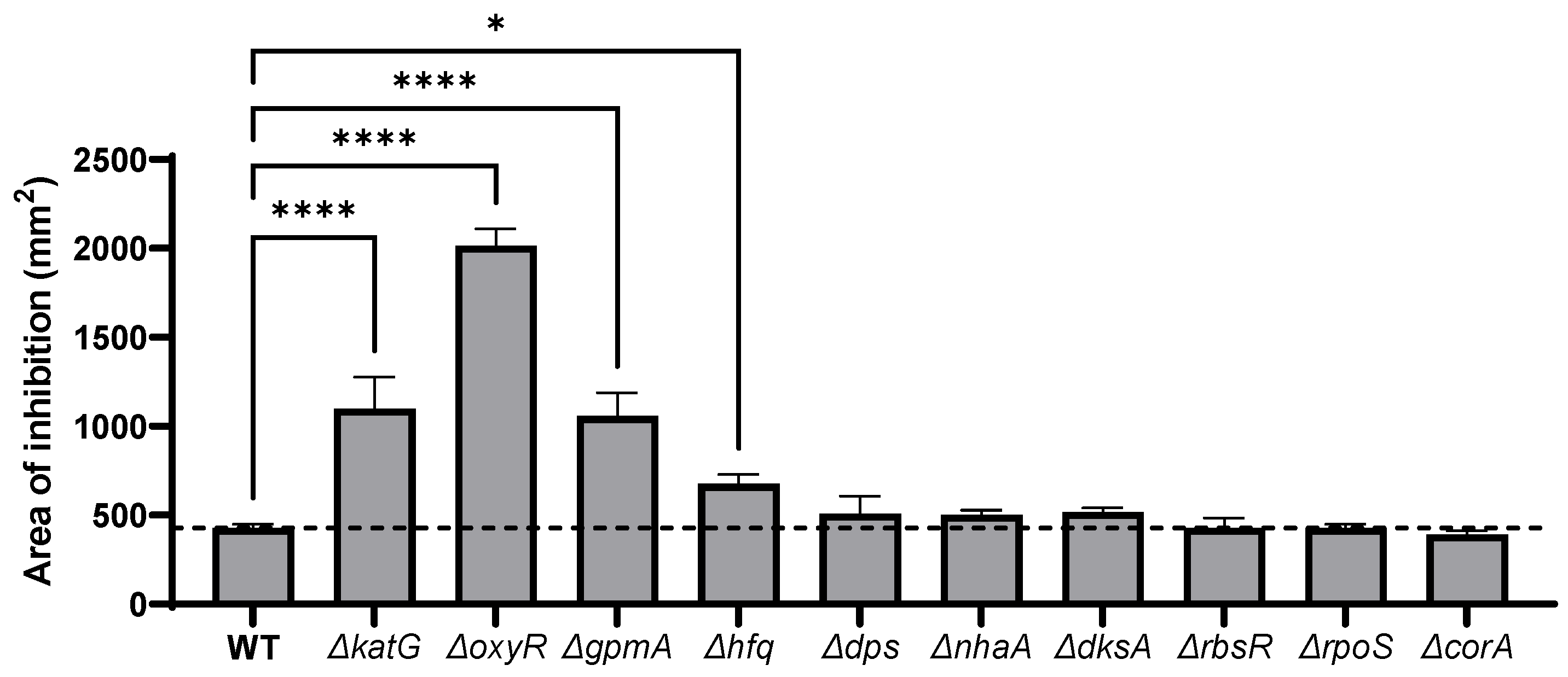
Single-deletion mutants of genes identified by the TraDIS experiments were tested against H2O2 to evaluate the sensitivity of each mutant. To ensure the absence of undesired mutations, cognate E. coli strain MG1655 mutants were created by P1 phage transduction of the relevant mutations from the Keio collection [21]. The susceptibility to H2O2 of the single-gene deletion mutants was assessed by disk diffusion assay. The mutant deleted for the catalase katG, known as the principal H2O2 scavenger at high concentration [32], was used as positive control. As expected, the deletion of oxyR led to a dramatic increase of the inhibition diameter generated by H2O2 (Figure 2). The deletion of gpmA increased the sensitivity to H2O2 to the same extent as the katG deletion. Similarly, loss of hfq also increased significantly the sensitivity to H2O2. Other genetic deletions did not significantly alter the H2O2 susceptibility in the disk diffusion assay.

3.3. ΔgpmA Mutant Was More Sensitive to H2O2 but Not to Other Oxidants

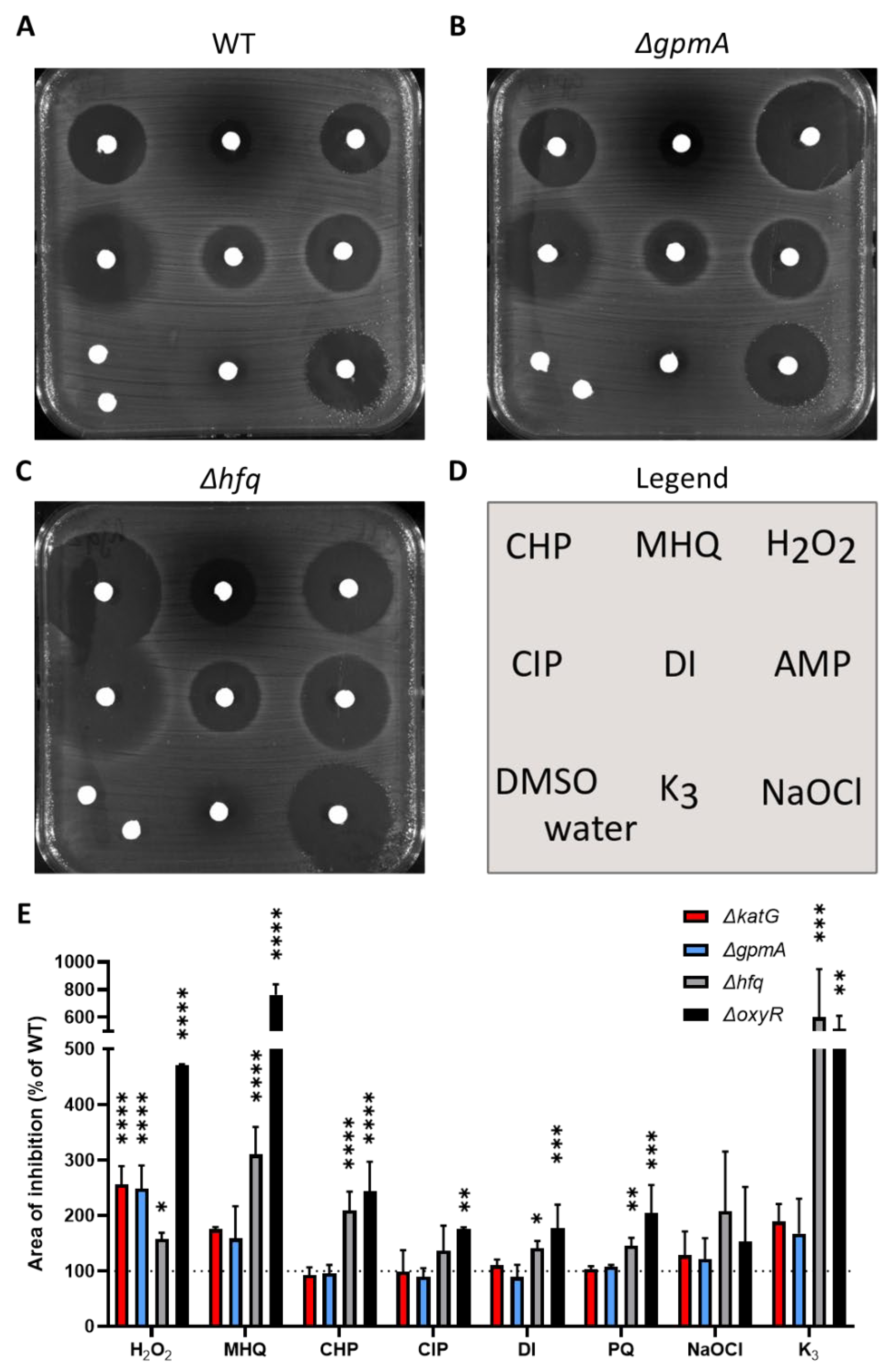
Single-deletion mutants were tested against other oxidants by disk diffusion assay (Figure 3). The deletion of oxyR and hfq led to an increase of the inhibition area of a wide range of oxidants (Figure 3C,E). The deletion of gpmA led to a hypersensitivity to H2O2 but not to other oxidants or antibiotics (Figure 3B,E). This pattern was highly similar to the sensitivity of the ΔkatG mutant used as positive control (Figure 2E).
The deletion mutants of the other genes identified by TraDIS were also tested against these oxidants (Supplementary Materials Figure S1). The ΔdksA mutant was more sensitive to methylhydroquinone, cumene peroxide, diamide and ciprofloxacin, and the ΔnhaA mutant was slightly more sensitive to diamide and ciprofloxacin. This suggests that these mutants, despite no increased sensitivity to H2O2 in these conditions, were more sensitive to other oxidative stresses. Other mutants did not display significant differences compared to WT.

3.4. gpmA Is Upregulated by H2O2 Exposure

In a previous study, we performed a RNA-seq analysis of E. coli BW25113 after a 10 min exposure to a sublethal concentration (2.5 mM) of H2O2 [10]. Among the ten genes identified by TraDIS, only two genes, dps and gpmA, were significantly dysregulated by H2O2 (Figure 4A). In these settings, gpmA was upregulated over fourfold. As gpmA is part of the glycolysis reaction in E. coli, we extracted the transcriptomic data for the glycolysis and the TCA cycle (Supplementary Materials Figure S2). Other enzymes from the glycolysis (pgi, pfkAB, fbaAB, pgk) were also upregulated, suggesting an increased activity of glycolysis following exposure to H2O2.
We confirmed the impact of H2O2 on gpmA expression in the MG1655 strain used in this study by qRT-PCR. The gpmA RNA was upregulated following sublethal exposure of H2O2 in a dose-dependent manner (Figure 4C). Induction of gpmA expression was less impressive than the catalase katG, a known H2O2-responsive gene (Figure 4B).

3.5. Catalase Activity Is Not Involved in the Increased Sensitivity of ΔgpmA to H2O2

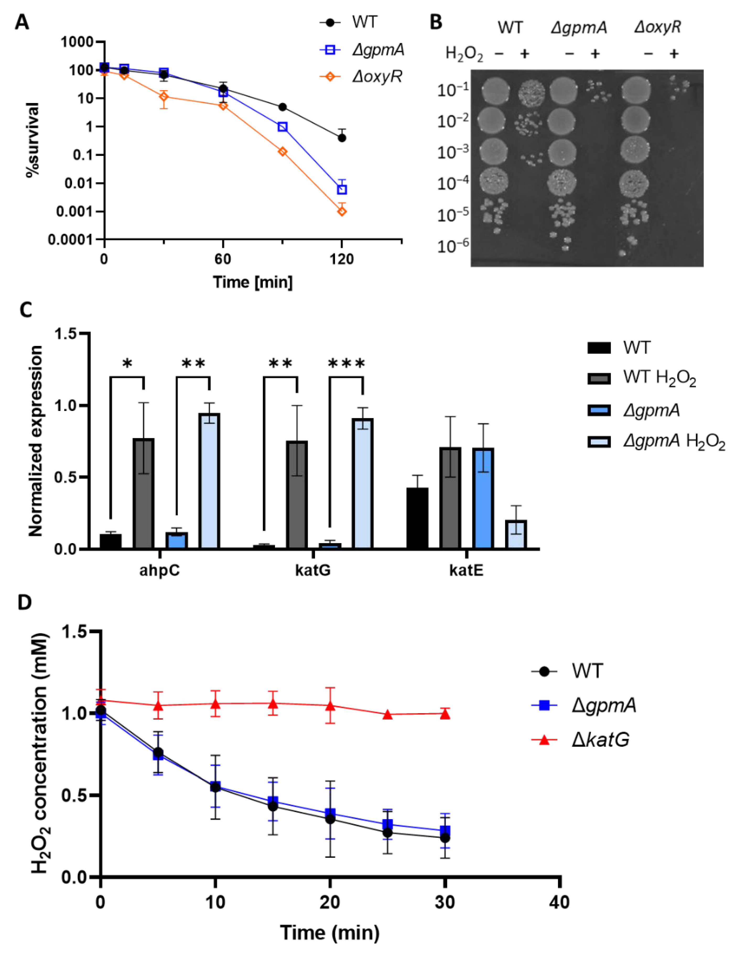
As the ΔgpmA mutant displayed a similar sensitivity to oxidants compared to the ΔkatG mutant (Figure 3E), we measured catalase expression and activity in the presence of H2O2. The ΔgpmA mutant did not exhibit a growth defect compared to the WT in liquid LB (Supplementary Figure S3), so we first assessed the sensitivity of the gpmA mutant to H2O2 in liquid LB medium by counting surviving bacteria after H2O2 exposure (Figure 5A). Two hours after the addition of 2.5 mM H2O2, a 100-fold difference in the number of surviving bacteria in the gpmA and the oxyR mutant compared to the WT was observed (Figure 5B).
We measured the expression levels of the three enzymes of E. coli that are known to degrade H2O2, the alkyl hydroperoxide reductase encoded by ahpC, the catalase/hydroperoxidase HPI encoded by katG and the catalase HPII encoded by katE. We compared the WT and the ΔgpmA strain, in presence or in absence of H2O2 (Figure 5C). There was no significant difference in the expression of the three genes between the WT and the ΔgpmA strain. Upregulation of ahpC and katG was observed after the addition of 2.5 mM of H2O2 in both strains. There was no significant difference in katE expression after H2O2 exposure, which was expected as it is not regulated by OxyR but by RpoS and upregulated during the stationary phase of bacterial growth [33].
To test if the catalase activity was affected by the deletion of gpmA, we measured the degradation of 1 mM of H2O2 of the WT and the ΔgpmA using the H2O2-sensitive probe Amplex Red. There was no difference in H2O2 degradation between the WT and the ΔgpmA strains. The ΔkatG strain, which is defective for the main H2O2 scavenger at high concentration, was unable to degrade H2O2. Altogether, this suggests that the higher sensitivity of the ΔgpmA to H2O2 is independent of catalase activity.

3.6. Other Carbon Sources Cannot Compensate the H2O2 Hypersensitivity of ΔgpmA Mutant

In LB medium, amino acids are the main source of carbon and there is virtually no glucose [34]. We wondered if the supplementation with metabolites entering the central metabolism at different levels could affect the H2O2 sensitivity of the ΔgpmA mutant. The addition of alternative carbon source did not significantly modify the H2O2 susceptibility of the WT or the ΔgpmA mutant (Figure 6A). We also tested H2O2 sensitivity in M9 minimal media with these metabolites as the only carbon source. There was no difference in the sensitivity of the WT in M9 + glucose compared to LB + glucose. The WT strain was slightly more sensitive in M9 + acetate compared to M9 + glucose. The ΔgpmA strain displayed a higher sensitivity in the M9 conditions compared to LB conditions. M9 plates with citrate as the sole source of carbon led to limited growth even after 48 h and were therefore not measurable. Addition of 0.5% pyruvate led to a complete disappearance of the zone of inhibition (data not shown) probably because pyruvate reacts with H2O2 to produce CO2, acetate and water [35].
In Salmonella Typhimurium, the ΔgpmA mutant was more susceptible to H2O2 than the WT in aerobic conditions, but not in anaerobic conditions, and the addition of the electron acceptor nitrate restored the hypersusceptibility of ΔgpmA [20]. We tested the H2O2 susceptibility of E. coli WT and ΔgpmA in anaerobic conditions. Interestingly, it appeared that the WT was slightly more sensitive to H2O2 in anaerobic conditions than in aerobic conditions suggesting that the exposure to oxygen protect in part against H2O2 damage. However, the ΔgpmA mutant did not display any difference in H2O2 sensitivity between anaerobic and aerobic conditions and the difference between the ΔgpmA mutant and the WT was maintained in anaerobic conditions. As E. coli is also able to use other electron acceptors than oxygen for respiration, we tested the addition of sodium nitrate in anaerobic conditions, but this did not change the area of inhibition induced by H2O2 compared to the anaerobic condition without nitrate (data not shown).
Altogether, we explored a potential impact of factors affecting glycolysis following H2O2 exposure, but we did not observe significant changes in conditions of low oxygen or using different carbon sources.

3.7. The Function of gpmA Is Necessary for H2O2 Tolerance

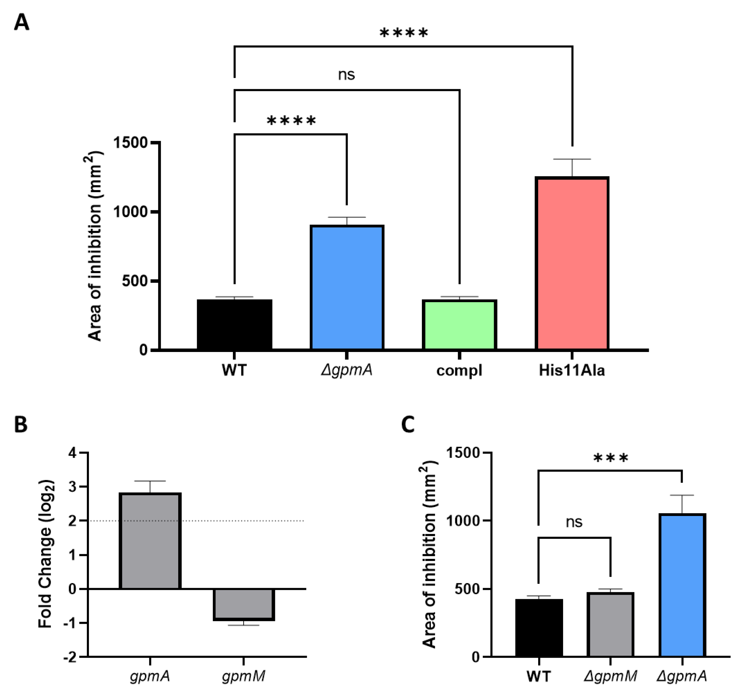
The ΔgpmA was complemented by native gpmA gene including its natural promoter using the low copy plasmid pWSK29. The complemented strain displayed similar H2O2 susceptibility than the WT strain (Figure 7A). A mutation previously described as to be necessary for the function of gpmA, namely the substitution of the histidine 11 residue by an alanine [36], resulted in restauration of the hypersensitivity to H2O2. These data suggest that the function of gpmA is necessary to reach the WT levels of tolerance against H2O2.
E. coli possess a secondphosphoglycerate mutase encoded by gpmM, which presents no sequence similarity with gpmA [37]. Contrary to gpmA, the expression level of gpmM was slightly downregulated after the addition of H2O2 in a previous RNA-seq dataset (Figure 7B). This suggests that following exposure to H2O2, gpmA represents the principal form of phophoglygerate mutase. We tested the ΔgpmM mutant for H2O2 sensitivity. Contrary to gpmA, the deletion of gpmM did not increase the sensitivity to H2O2 (Figure 7C), suggesting a possible alternative function of gpmA in conditions of H2O2 exposure.

4. Discussion

The production of H2O2 by phagocytes from the human immune system and by Lactobacilli species of the normal microbiota are essential for the prevention of colonization from various opportunistic pathogens. Although H2O2 effects on bacteria have been studied for years, the mechanisms by which H2O2 exerts its antimicrobial activity is still incompletely understood [14,16].
Our TraDIS analysis identified 10 mutants with fitness defect upon H2O2 exposure, implicating a role for these genes under H2O2-induced oxidative stress. Only three of the ten genes, oxyR, gpmA and hfq, showed a significantly higher susceptibility to H2O2 when knocked-out. This could be due to the differences in the settings between the TraDIS experiment and the disk diffusion assay. For example, the DNA-binding protein encoded by dps protects DNA from H2O2 damage through iron sequestration and this defense is more important in stationary phase of growth [38]. However stationary phase cultures of each knockout was treated with H2O2 in liquid medium, their respective growth was not different compared to the WT, except for the ΔoxyR strain (data not shown). The majority of genes we identified by TraDIS (oxyR, dps, rpoS, dksA, hfq, polA) have already been reported to respond to oxidative stress in E. coli. The transcription factor OxyR is a well described sensor of H2O2, which regulates an extensive and coordinated antioxidant transcriptional response [9,39]. The RNA polymerase subunit RpoS regulates the general stress response and was previously described to be activated by oxidative stress [40], and the deletion of this gene increases sensitivity to H2O2 [41]. The RNA polymerase accessory protein DksA senses oxidative stress through its cysteine residues and participates to the transcriptional response against oxidative stress [42]. Hfq, a RNA-binding protein that affects many cellular processes influences both the small RNA OxyS and the translation of rpoS described above in E. coli [43,44]. The DNA polymerase I encoded by polA is implicated in DNA repair and non-functional PolA increases H2O2-sensitivity [45,46]. The polA, rbsR, dps, oxyR, corA, rpoS genes were also identified in a similar experiment performed previously on Salmonella enterica serovar Typhimurium under sublethal H2O2 exposure [19]. The dksA and nhaA mutants, despite showing no increase in sensitivity to H2O2, were slightly more sensitive to other oxidants than WT using disk diffusion assay. Other validation experiments, such as competition assay with WT under H2O2 stress, might better reflect the TraDIS experimental conditions.
On the other hand, several genes previously identified in the literature as necessary for H2O2 tolerance were not identified by this TraDIS experiment. For example, xthA, whose deletion mutant is more sensitive to H2O2 [47], displayed a decreased fitness in H2O2 condition but did not reach the threshold of significance. An explanation could lie in the fact that unlike antibiotics, H2O2 is rapidly degraded by bacteria. The duration of the exposure to H2O2 performed for the TraDIS may have been insufficient to identify all genes implicated in H2O2 tolerance. Secondly, the stress was applied against pooled mutants in liquid where other mutants could provide cross-protection for susceptible mutants. For example, the catalase KatG, which is known to protect against H2O2, was not identified by TraDIS, probably because of this phenomenon. This was also the case in a previous study that used Tn-seq with H2O2 in Salmonella Typhimurium, where none of the catalase genes were identified [19]. Thus, our TraDIS data only identified those mutants that showed fitness defects despite cross-protection and inherent H2O2 degradation.
The TraDIS experiment also identified genes that, to our knowledge, were not previously associated with E. coli H2O2 sensitivity. The magnesium ion transporter encoded by corA had been shown to be more sensitive to lactoperoxidase–thiocyanate stress but not to H2O2 [48]. rbsR controls the transcription of the operon involved in ribose catabolism and transport and the salvage pathway of purine nucleotide synthesis [49]. corA and rbsR were also identified in a similar Tn-seq experiment using H2O2 on Salmonella enterica serovar Typhimurium [19]. The Na+:H+ antiporter nhaA is implicated in other stress responses against sodium ion, pH homeostasis and in maintaining antibiotic tolerance under starvation [50]. The glycolysis enzyme gpmA has been previously identified by a Tn-seq experiment following H2O2 exposure in the Gram-negative bacteria Salmonella enterica serovar Typhimurium [20].
When tested with diverse oxidants that damage bacteria through different modes of action, ΔgpmA was specifically more sensitive to H2O2, like the ΔkatG strain. However, it was not through a differential expression of H2O2-scavenging genes or a decreased catalase activity of the strain, suggesting a different mode of action. Moreover, the upregulation of gpmA by sublethal exposure of H2O2 suggests the importance of gpmA in H2O2 tolerance. Under oxidative stress, some enzymes of the central metabolism have been shown to be upregulated. The glucose-6-phosphate isomerase encoded by pgi have been shown to be regulated by the oxidative stress sensitive regulators SoxRS [51]. Similarly, in the TCA cycle, the aconitase acnA and the fumarase fumC are regulated by SoxRS and are upregulated under H2O2 exposure [52,53]. The hypersensitivity to H2O2 of ΔgpmA mutant could be complemented with the low-copy plasmid pWSK29 expressing the WT gpmA gene under its native promoter but not if the histidine 11 was mutated to an alanine. This strongly suggests that the function of gpmA affects E. coli tolerance to H2O2.
Surprisingly, addition of other metabolites or the absence of oxygen did not abolish the difference in H2O2 sensitivity between the WT and the ΔgpmA mutant. These data contrast with previous work on Salmonella enterica serovar Typhimurium, where other metabolites entering metabolism downstream of gpmA reaction (for a scheme of glycolysis, see Supplementary Figure S2) could complement the increased sensitivity of a ΔgpmA mutant and where anoxic environment abolished the difference of H2O2 susceptibility between WT and ΔgpmA mutant [20]. In the same study, metabolomics approach showed that H2O2 exposure led to an increase of glycolysis and fermentation that was important in Salmonella H2O2 tolerance. This contrasts with previous metabolomics analysis on E. coli after H2O2 treatment which reported a decrease of metabolites related to glycolysis and TCA cycle, changes that were common to other stress conditions such as heat shock and cold stress [54]. Altogether, this suggests a different metabolic adaptation to H2O2 stress between E. coli and Salmonella and a difference of gpmA function. More research is needed to better understand the mechanisms of gpmA effects in H2O2 tolerance in E. coli and in other organisms.
Contrary to vertebrates that only possess one phosphoglycerate mutase, some eubacteria, among which relevant pathogens including E. coli, encode two enzymes that display no sequence similarity [55]. The 2,3-bisphosphoglycerate-dependent phosphoglycerate mutase (or dPGM), encoded by gpmA is common to bacteria and vertebrates, whereas 2,3-bisphosphoglycerate-independent phosphoglycerate mutase (or iPGM) encoded by gpmM is shared by bacteria and higher plants. As the double deletion of gpmA and gpmM have been suspected non-viable in E. coli, the glycolysis function is assumed by gpmM in the gpmA-deleted strain and vice versa [56]. The deletion of E. coli gpmM did not affect H2O2 sensitivity, suggesting that only gpmA function has a role under H2O2 exposure. This led to the hypothesis that gpmM could be damaged by H2O2 and its function is replaced by gpmA under H2O2 exposure. This happens for other enzymes of the TCA cycle, the aconitase and the fumarase, where oxidative-resistant isoforms (acnA, fumC) replace oxidative-sensitive isoforms (acnB, fumA, fumB), after H2O2 exposure [52,53]. Cysteine residues can be more prone to oxidation by H2O2 than other amino acids [57]. GpmM possesses two cysteine residues, which can result in H2O2-induced damage from oxidation of these residues. As GpmA does not possess cysteine residues, it could be more resistant to H2O2 than GpmM. The cysteine residues of GpmM are not implicated in active sites described in current models (Ecocyc, Uniprot). While Cys397 seems buried and is not conserved in Gram-positive bacteria, Cys424 seems to be more accessible on the protein models and is present in both Gram-negative (P. aeruginosa, Salmonella enterica, K. pneumoniae) and Gram-positive bacteria (S. aureus, B. subtilis). Additional studies are needed to evaluate their potential implication in oxidative stress susceptibility.

5. Conclusions

This work was aimed at expanding the knowledge of which genes are implicated in H2O2 tolerance. The main finding of this study was that a functional gpmA gene is required for tolerance to H2O2. This is the first time that gpmA was highlighted as an important contributor to the E. coli tolerance to H2O2, and it links defense against oxidative stress to central metabolism.

Supplementary Materials

The following are available online at https://www.mdpi.com/article/10.3390/antiox11102053/s1, Table S1: Primers used to validate the gene replacement by the kanamycin cassette from the Keio collection, Figure S1: Sensitivity of the deletion mutants of the TraDIS exposed to various oxidants, Figure S2: Schematic diagram of H2O2-induced transcriptional changes of glycolysis and TCA cycle, Figure S3: The deletion of gpmA did not affect bacterial growth.

Author Contributions

Conceptualization, M.R., V.J., K.-H.K. and P.F.; methodology, E.C.A.G., K.P. and M.R.; software, E.C.A.G. and K.P.; validation, K.P.; formal analysis, E.C.A.G. and M.R.; investigation, M.R., E.C.A.G. and K.P.; resources, E.C.A.G., K.P. and I.R.H.; data curation, E.C.A.G., K.P. and M.R.; writing—original draft preparation, M.R. and V.J.; writing—review and editing, P.F.; visualization, M.R.; supervision, K.-H.K., I.R.H. and P.F.; project administration, V.J., K.-H.K., I.R.H. and P.F.; funding acquisition, K.-H.K. All authors have read and agreed to the published version of the manuscript.

Funding

This study was granted by Swiss National Science Foundation to Karl-Heinz Krause (Funding number 31003A-179478). The TraDIS work was supported by University of Queensland funding to Ian Henderson.

Institutional Review Board Statement

Not applicable.

Informed Consent Statement

Not applicable.

Data Availability Statement

The data for this study have been deposited in the European Nucleotide Archive (ENA) at EMBL-EBI under accession number PRJEB56340 (https://www.ebi.ac.uk/ena/browser/view/PRJEB56340). Processed data are available for viewing at our online browser: https://tradis-vault.qfab.org/.

Acknowledgments

The authors gratefully acknowledge Benjamin Ezraty for the donation of the MG1655 ΔoxyR::CmR strain; Mélanie Roch for the donation of the pWSK29 plasmid; Gaël Panis for the donation of the P1 phage; Roberto Sierra, Mélanie Roch, Gaël Panis, Clement David, Aleksander Czauderna, Simone Becattini and the collaborators of the genomics platform iGE3 of the faculty of Medicine of the University of Geneva: Didier Chollet and Mylène Docquier for technical support.

Conflicts of Interest

The authors declare no conflict of interest.

References

  1. Dunne, K.A.; Chaudhuri, R.R.; Rossiter, A.E.; Beriotto, I.; Browning, D.F.; Squire, D.; Cunningham, A.F.; Cole, J.A.; Loman, N.; Henderson, I.R. Sequencing a Piece of History: Complete Genome Sequence of the Original Escherichia Coli Strain. Microb. Genom. 2017, 3, mgen000106. [Google Scholar] [CrossRef]
  2. Anderson, J.D.; Bagamian, K.H.; Muhib, F.; Amaya, M.P.; Laytner, L.A.; Wierzba, T.; Rheingans, R. Burden of Enterotoxigenic Escherichia Coli and Shigella Non-Fatal Diarrhoeal Infections in 79 Low-Income and Lower Middle-Income Countries: A Modelling Analysis. Lancet Glob. Health 2019, 7, e321–e330. [Google Scholar] [CrossRef] [Green Version]
  3. Bonten, M.; Johnson, J.R.; van den Biggelaar, A.H.J.; Georgalis, L.; Geurtsen, J.; de Palacios, P.I.; Gravenstein, S.; Verstraeten, T.; Hermans, P.; Poolman, J.T. Epidemiology of Escherichia Coli Bacteremia: A Systematic Literature Review. Clin. Infect. Dis. 2021, 72, 1211–1219. [Google Scholar] [CrossRef]
  4. Murray, C.J.; Ikuta, K.S.; Sharara, F.; Swetschinski, L.; Aguilar, G.R.; Gray, A.; Han, C.; Bisignano, C.; Rao, P.; Wool, E.; et al. Global Burden of Bacterial Antimicrobial Resistance in 2019: A Systematic Analysis. Lancet 2022, 399, 629–655. [Google Scholar] [CrossRef]
  5. Pridmore, R.D.; Pittet, A.-C.; Praplan, F.; Cavadini, C. Hydrogen Peroxide Production by Lactobacillus Johnsonii NCC 533 and Its Role in Anti-Salmonella Activity. FEMS Microbiol. Lett. 2008, 283, 210–215. [Google Scholar] [CrossRef] [Green Version]
  6. Gupta, K.; Stapleton, A.E.; Hooton, T.M.; Roberts, P.L.; Fennell, C.L.; Stamm, W.E. Inverse Association of H2O2-Producing Lactobacilli and Vaginal Escherichia Coli Colonization in Women with Recurrent Urinary Tract Infections. J. Infect. Dis. 1998, 178, 446–450. [Google Scholar] [CrossRef] [Green Version]
  7. Winterbourn, C.C.; Kettle, A.J. Redox Reactions and Microbial Killing in the Neutrophil Phagosome. Antioxid. Redox Signal. 2013, 18, 642–660. [Google Scholar] [CrossRef]
  8. Zheng, M.; Wang, X.; Templeton, L.J.; Smulski, D.R.; LaRossa, R.A.; Storz, G. DNA Microarray-Mediated Transcriptional Profiling of the Escherichia Coli Response to Hydrogen Peroxide. J. Bacteriol. 2001, 183, 4562–4570. [Google Scholar] [CrossRef] [Green Version]
  9. Seo, S.W.; Kim, D.; Szubin, R.; Palsson, B.O. Genome-Wide Reconstruction of OxyR and SoxRS Transcriptional Regulatory Networks under Oxidative Stress in Escherichia Coli K-12 MG1655. Cell Rep. 2015, 12, 1289–1299. [Google Scholar] [CrossRef] [Green Version]
  10. Roth, M.; Jaquet, V.; Lemeille, S.; Bonetti, E.-J.; Cambet, Y.; François, P.; Krause, K.-H. Transcriptomic Analysis of E. Coli after Exposure to a Sublethal Concentration of Hydrogen Peroxide Revealed a Coordinated Up-Regulation of the Cysteine Biosynthesis Pathway. Antioxidants 2022, 11, 655. [Google Scholar] [CrossRef]
  11. Kobayashi, K.; Fujikawa, M.; Kozawa, T. Oxidative Stress Sensing by the Iron–Sulfur Cluster in the Transcription Factor, SoxR. J. Inorg. Biochem. 2014, 133, 87–91. [Google Scholar] [CrossRef]
  12. Choi, H.-J.; Kim, S.-J.; Mukhopadhyay, P.; Cho, S.; Woo, J.-R.; Storz, G.; Ryu, S.-E. Structural Basis of the Redox Switch in the OxyR Transcription Factor. Cell 2001, 105, 103–113. [Google Scholar] [CrossRef] [Green Version]
  13. Imlay, J.A. Transcription Factors That Defend Bacteria against Reactive Oxygen Species. Annu. Rev. Microbiol. 2015, 69, 93–108. [Google Scholar] [CrossRef] [Green Version]
  14. Farr, S.B.; D’Ari, R.; Touati, D. Oxygen-Dependent Mutagenesis in Escherichia Coli Lacking Superoxide Dismutase. Proc. Natl. Acad. Sci. USA 1986, 83, 8268–8272. [Google Scholar] [CrossRef] [Green Version]
  15. Mukhopadhyay, S.; Schellhorn, H.E. Identification and Characterization of Hydrogen Peroxide-Sensitive Mutants of Escherichia Coli: Genes That Require OxyR for Expression. J. Bacteriol. 1997, 179, 330–338. [Google Scholar] [CrossRef] [Green Version]
  16. Rodríguez-Rojas, A.; Kim, J.J.; Johnston, P.R.; Makarova, O.; Eravci, M.; Weise, C.; Hengge, R.; Rolff, J. Non-Lethal Exposure to H2O2 Boosts Bacterial Survival and Evolvability against Oxidative Stress. PLoS Genet. 2020, 16, e1008649. [Google Scholar] [CrossRef] [Green Version]
  17. Chao, M.C.; Abel, S.; Davis, B.M.; Waldor, M.K. The Design and Analysis of Transposon-Insertion Sequencing Experiments. Nat. Rev. Microbiol. 2016, 14, 119–128. [Google Scholar] [CrossRef] [Green Version]
  18. Goodall, E.C.A.; Robinson, A.; Johnston, I.G.; Jabbari, S.; Turner, K.A.; Cunningham, A.F.; Lund, P.A.; Cole, J.A.; Henderson, I.R. The Essential Genome of Escherichia Coli K-12. mBio 2018, 9, e02096-17. [Google Scholar] [CrossRef] [Green Version]
  19. Karash, S.; Liyanage, R.; Qassab, A.; Lay, J.O.; Kwon, Y.M. A Comprehensive Assessment of the Genetic Determinants in Salmonella Typhimurium for Resistance to Hydrogen Peroxide Using Proteogenomics. Sci. Rep. 2017, 7, 17073. [Google Scholar] [CrossRef] [Green Version]
  20. Chakraborty, S.; Liu, L.; Fitzsimmons, L.; Porwollik, S.; Kim, J.-S.; Desai, P.; McClelland, M.; Vazquez-Torres, A. Glycolytic Reprograming in Salmonella Counters NOX2-Mediated Dissipation of ΔpH. Nat. Commun. 2020, 11, 1783. [Google Scholar] [CrossRef]
  21. Baba, T.; Ara, T.; Hasegawa, M.; Takai, Y.; Okumura, Y.; Baba, M.; Datsenko, K.A.; Tomita, M.; Wanner, B.L.; Mori, H. Construction of Escherichia Coli K-12 in-Frame, Single-Gene Knockout Mutants: The Keio Collection. Mol. Syst. Biol. 2006, 2, 2006.0008. [Google Scholar] [CrossRef] [Green Version]
  22. Ezraty, B.; Vergnes, A.; Banzhaf, M.; Duverger, Y.; Huguenot, A.; Brochado, A.R.; Su, S.-Y.; Espinosa, L.; Loiseau, L.; Py, B.; et al. Fe-S Cluster Biosynthesis Controls Uptake of Aminoglycosides in a ROS-Less Death Pathway. Science 2013, 340, 1583–1587. [Google Scholar] [CrossRef]
  23. Wang, R.F.; Kushner, S.R. Construction of Versatile Low-Copy-Number Vectors for Cloning, Sequencing and Gene Expression in Escherichia Coli. Gene 1991, 100, 195–199. [Google Scholar] [CrossRef]
  24. Barquist, L.; Mayho, M.; Cummins, C.; Cain, A.K.; Boinett, C.J.; Page, A.J.; Langridge, G.C.; Quail, M.A.; Keane, J.A.; Parkhill, J. The TraDIS Toolkit: Sequencing and Analysis for Dense Transposon Mutant Libraries. Bioinformatics 2016, 32, 1109–1111. [Google Scholar] [CrossRef] [Green Version]
  25. Thomason, L.C.; Costantino, N.; Court, D.L.E. Coli Genome Manipulation by P1 Transduction. Curr. Protoc. Mol. Biol. 2007, 1, 1.17.1–1.17.8. [Google Scholar] [CrossRef]
  26. Vandesompele, J.; De Preter, K.; Pattyn, F.; Poppe, B.; Van Roy, N.; De Paepe, A.; Speleman, F. Accurate Normalization of Real-Time Quantitative RT-PCR Data by Geometric Averaging of Multiple Internal Control Genes. Genome Biol. 2002, 3, research0034.1. [Google Scholar] [CrossRef] [Green Version]
  27. Rocha, D.J.P.G.; Castro, T.L.P.; Aguiar, E.R.G.R.; Pacheco, L.G.C. Gene Expression Analysis in Bacteria by RT-QPCR. In Quantitative Real-Time PCR: Methods and Protocols; Biassoni, R., Raso, A., Eds.; Methods in Molecular Biology; Springer: New York, NY, USA, 2020; pp. 119–137. ISBN 978-1-4939-9833-3. [Google Scholar]
  28. Carver, T.; Harris, S.R.; Berriman, M.; Parkhill, J.; McQuillan, J.A. Artemis: An Integrated Platform for Visualization and Analysis of High-Throughput Sequence-Based Experimental Data. Bioinformatics 2012, 28, 464–469. [Google Scholar] [CrossRef] [Green Version]
  29. Keseler, I.M.; Collado-Vides, J.; Santos-Zavaleta, A.; Peralta-Gil, M.; Gama-Castro, S.; Muñiz-Rascado, L.; Bonavides-Martinez, C.; Paley, S.; Krummenacker, M.; Altman, T.; et al. EcoCyc: A Comprehensive Database of Escherichia Coli Biology. Nucleic Acids Res. 2011, 39, D583–D590. [Google Scholar] [CrossRef]
  30. Keseler, I.M.; Gama-Castro, S.; Mackie, A.; Billington, R.; Bonavides-Martínez, C.; Caspi, R.; Kothari, A.; Krummenacker, M.; Midford, P.E.; Muñiz-Rascado, L.; et al. The EcoCyc Database in 2021. Front. Microbiol. 2021, 12, 711077. [Google Scholar] [CrossRef]
  31. Christman, M.F.; Morgan, R.W.; Jacobson, F.S.; Ames, B.N. Positive Control of a Regulon for Defenses against Oxidative Stress and Some Heat-Shock Proteins in Salmonella Typhimurium. Cell 1985, 41, 753–762. [Google Scholar] [CrossRef]
  32. Seaver, L.C.; Imlay, J.A. Hydrogen Peroxide Fluxes and Compartmentalization inside Growing Escherichia Coli. J. Bacteriol. 2001, 183, 7182–7189. [Google Scholar] [CrossRef] [Green Version]
  33. Schellhorn, H.E.; Hassan, H.M. Transcriptional Regulation of KatE in Escherichia Coli K-12. J. Bacteriol. 1988, 170, 4286–4292. [Google Scholar] [CrossRef] [Green Version]
  34. Sezonov, G.; Joseleau-Petit, D.; D’Ari, R. Escherichia Coli Physiology in Luria-Bertani Broth. J. Bacteriol. 2007, 189, 8746–8749. [Google Scholar] [CrossRef] [Green Version]
  35. Guarino, V.A.; Oldham, W.M.; Loscalzo, J.; Zhang, Y.-Y. Reaction Rate of Pyruvate and Hydrogen Peroxide: Assessing Antioxidant Capacity of Pyruvate under Biological Conditions. Sci. Rep. 2019, 9, 19568. [Google Scholar] [CrossRef] [Green Version]
  36. Bond, C.S.; White, M.F.; Hunter, W.N. High Resolution Structure of the Phosphohistidine-Activated Form of Escherichia Coli Cofactor-Dependent Phosphoglycerate Mutase*. J. Biol. Chem. 2001, 276, 3247–3253. [Google Scholar] [CrossRef] [Green Version]
  37. Fraser, H.I.; Kvaratskhelia, M.; White, M.F. The Two Analogous Phosphoglycerate Mutases of Escherichia Coli. FEBS Lett. 1999, 455, 344–348. [Google Scholar] [CrossRef] [Green Version]
  38. Martinez, A.; Kolter, R. Protection of DNA during Oxidative Stress by the Nonspecific DNA-Binding Protein Dps. J. Bacteriol. 1997, 179, 5188–5194. [Google Scholar] [CrossRef] [Green Version]
  39. Sen, A.; Imlay, J.A. How Microbes Defend Themselves From Incoming Hydrogen Peroxide. Front. Immunol. 2021, 12, 1104. [Google Scholar] [CrossRef]
  40. Lange, R.; Hengge-Aronis, R. Identification of a Central Regulator of Stationary-Phase Gene Expression in Escherichia Coli. Mol. Microbiol. 1991, 5, 49–59. [Google Scholar] [CrossRef]
  41. Loewen, P.C.; Triggs, B.L. Genetic Mapping of KatF, a Locus That with KatE Affects the Synthesis of a Second Catalase Species in Escherichia Coli. J. Bacteriol. 1984, 160, 668–675. [Google Scholar] [CrossRef]
  42. Henard, C.A.; Tapscott, T.; Crawford, M.A.; Husain, M.; Doulias, P.-T.; Porwollik, S.; Liu, L.; McClelland, M.; Ischiropoulos, H.; Vázquez-Torres, A. The 4-Cysteine Zinc-Finger Motif of the RNA Polymerase Regulator DksA Serves as a Thiol Switch for Sensing Oxidative and Nitrosative Stress. Mol. Microbiol. 2014, 91, 790–804. [Google Scholar] [CrossRef] [Green Version]
  43. Zhang, A.; Wassarman, K.M.; Ortega, J.; Steven, A.C.; Storz, G. The Sm-like Hfq Protein Increases OxyS RNA Interaction with Target MRNAs. Mol. Cell 2002, 9, 11–22. [Google Scholar] [CrossRef]
  44. Muffler, A.; Fischer, D.; Hengge-Aronis, R. The RNA-Binding Protein HF-I, Known as a Host Factor for Phage Qbeta RNA Replication, Is Essential for RpoS Translation in Escherichia Coli. Genes Dev. 1996, 10, 1143–1151. [Google Scholar] [CrossRef] [Green Version]
  45. Liu, Y.; Imlay, J.A. Cell Death from Antibiotics without the Involvement of Reactive Oxygen Species. Science 2013, 339, 1210–1213. [Google Scholar] [CrossRef] [Green Version]
  46. Imlay, J.A.; Linn, S. Mutagenesis and Stress Responses Induced in Escherichia Coli by Hydrogen Peroxide. J. Bacteriol. 1987, 169, 2967–2976. [Google Scholar] [CrossRef] [Green Version]
  47. Demple, B.; Halbrook, J.; Linn, S. Escherichia Coli Xth Mutants Are Hypersensitive to Hydrogen Peroxide. J. Bacteriol. 1983, 153, 1079–1082. [Google Scholar] [CrossRef] [Green Version]
  48. Sermon, J.; Wevers, E.M.-R.P.; Jansen, L.; De Spiegeleer, P.; Vanoirbeek, K.; Aertsen, A.; Michiels, C.W. CorA Affects Tolerance of Escherichia Coli and Salmonella Enterica Serovar Typhimurium to the Lactoperoxidase Enzyme System but Not to Other Forms of Oxidative Stress. Appl. Environ. Microbiol. 2005, 71, 6515–6523. [Google Scholar] [CrossRef] [Green Version]
  49. Shimada, T.; Kori, A.; Ishihama, A. Involvement of the Ribose Operon Repressor RbsR in Regulation of Purine Nucleotide Synthesis in Escherichia Coli. FEMS Microbiol. Lett. 2013, 344, 159–165. [Google Scholar] [CrossRef] [Green Version]
  50. Wan, Y.; Wang, M.; Chan, E.W.C.; Chen, S. Membrane Transporters of the Major Facilitator Superfamily Are Essential for Long-Term Maintenance of Phenotypic Tolerance to Multiple Antibiotics in E. Coli. Microbiol. Spectr. 2021, 9, e0184621. [Google Scholar] [CrossRef]
  51. Rungrassamee, W.; Liu, X.; Pomposiello, P.J. Activation of Glucose Transport under Oxidative Stress in Escherichia Coli. Arch. Microbiol. 2008, 190, 41–49. [Google Scholar] [CrossRef]
  52. Varghese, S.; Tang, Y.; Imlay, J.A. Contrasting Sensitivities of Escherichia Coli Aconitases A and B to Oxidation and Iron Depletion. J. Bacteriol. 2003, 185, 221–230. [Google Scholar] [CrossRef] [Green Version]
  53. Park, S.J.; Gunsalus, R.P. Oxygen, Iron, Carbon, and Superoxide Control of the Fumarase FumA and FumC Genes of Escherichia Coli: Role of the ArcA, Fnr, and SoxR Gene Products. J. Bacteriol. 1995, 177, 6255–6262. [Google Scholar] [CrossRef] [Green Version]
  54. Jozefczuk, S.; Klie, S.; Catchpole, G.; Szymanski, J.; Cuadros-Inostroza, A.; Steinhauser, D.; Selbig, J.; Willmitzer, L. Metabolomic and Transcriptomic Stress Response of Escherichia Coli. Mol. Syst. Biol. 2010, 6, 364. [Google Scholar] [CrossRef]
  55. Radin, J.N.; Kelliher, J.L.; Solórzano, P.K.P.; Grim, K.P.; Ramezanifard, R.; Slauch, J.M.; Kehl-Fie, T.E. Metal-Independent Variants of Phosphoglycerate Mutase Promote Resistance to Nutritional Immunity and Retention of Glycolysis during Infection. PLoS Pathog. 2019, 15, e1007971. [Google Scholar] [CrossRef] [Green Version]
  56. Foster, J.M.; Davis, P.J.; Raverdy, S.; Sibley, M.H.; Raleigh, E.A.; Kumar, S.; Carlow, C.K.S. Evolution of Bacterial Phosphoglycerate Mutases: Non-Homologous Isofunctional Enzymes Undergoing Gene Losses, Gains and Lateral Transfers. PLoS ONE 2010, 5, e13576. [Google Scholar] [CrossRef]
  57. Imlay, J.A. The Molecular Mechanisms and Physiological Consequences of Oxidative Stress: Lessons from a Model Bacterium. Nat. Rev. Microbiol. 2013, 11, 443–454. [Google Scholar] [CrossRef]
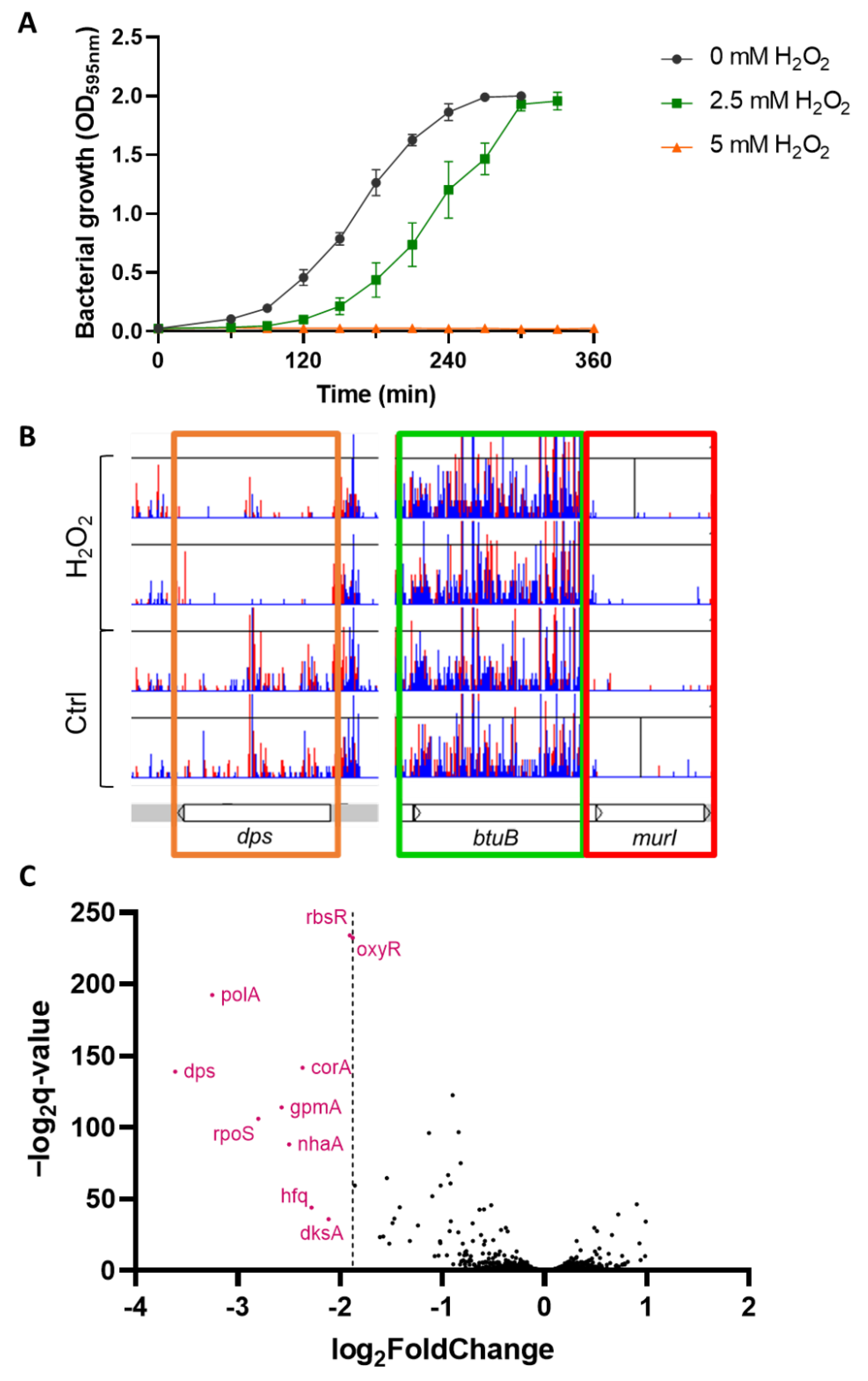
Figure 1. Transposon-directed insertion sequencing (TraDIS) screen of E.coli BW25113 under sublethal concentration of H2O2 (A) Bacterial growth of E. coli BW25113 over time with 2.5, 5 mM H2O2 and untreated control. The concentration of 2.5 mM was chosen for TraDIS experiment as it displayed a high reproducibility and a delay of approximatively 70 min compared to control (mean +/− SD, N = 3); (B) Visualization of the TraDIS data using Artemis where each sample is represented by a histogram depicting the localization (X-axis) and the frequency (Y-axis) of transposon insertion sites (in red are antisense insertions, in blue are same sense insertions). Representative examples of essential gene (murI in red box), non-essential genes (btuB, in green box) and genes with a reduced fitness in the H2O2 condition (dps in orange box), (N = 2) (C) Fitness analysis of the TraDIS data, H2O2-treated condition compared to control. Each dot represents a gene, X-axis represents the difference in number of insertions in the H2O2 condition compared to control, Y-axis represents the statistical significance. Nine genes (in pink) displayed a significant and more extreme change than the H2O2-sensor gene oxyR.
Antioxidants 11 02053 g001
Figure 2. Sensitivity to H2O2 of genes identified by TraDIS. The sensitivity to 10 μL of 1 M H2O2 of each single-gene deletion mutant identified by the TraDIS was assessed by disk diffusion assay. The coding regions of E. coli MG1655 (WT) were replaced by the kanamycin cassette of the corresponding mutant originated from the Keio collection using the phage P1 to ensure the absence of undesired mutation. Data were analyzed by one-way ANOVA with Tukey test for multiple comparison and *, **** correspond to p < 0.05 and 0.0001 respectively (mean +/− SD, N = 3).
Antioxidants 11 02053 g002
Figure 3. Sensitivity of the ΔoxyR, ΔgpmA and Δhfq mutants exposed to various oxidants. (A) WT, (B) ΔgpmA, (C) Δhfq. (D) Oxidants applied on each disk (CHP: cumene hydroperoxide, MHQ: methylhydroquinone, H2O2: hydrogen peroxide, CIP: ciprofloxacin, DI: diamide, AMP: ampicillin, K3: menadione, NaOCl: sodium hypochlorite, DMSO: dimethylsulfoxide). (E) Quantification of the area of inhibition normalized to WT for each oxidant. One-way ANOVA with Tukey multiple comparison was performed separately for each oxidant on the area of inhibition of the WT, ΔkatG and the 9 mutants identified by TraDIS (Figure S1). The significance of the difference with the WT is represented on the normalized data by stars (mean +/− SD, N = 3). *, **, ***, **** correspond to p < 0.05, 0.01, 0.001, and 0.0001, respectively.
Antioxidants 11 02053 g003
Figure 4. gpmA expression is upregulated following exposure to H2O2. (A) Differential expression of the 10 genes identified by TraDIS 10 min after exposition to 2.5 mM H2O2 compared to no treatment (mean +/− SD, N = 4). Data originated from previously performed RNA-seq (deposited on ENA with the accession number: PRJEB51098) [10]. (B,C) Levels of expression of katG and gpmA, respectively, in the strain MG1655 under increasing concentration of H2O2, assessed by qRT-PCR, ** corresponds to p < 0.01. (SEM +/− SD, N = 3).
Antioxidants 11 02053 g004
Figure 5. WT and gpmA displayed no difference in catalase expression and activity. (A) Survival of 2 × 10⁷ cells of WT (black), ΔgpmA (blue) and ΔoxyR (orange) over time after an exposure to 2.5 mM H2O2 in liquid LB (mean +/− SD, N = 3). (B) Representative image of the survival 2h after H2O2 treatment. Each spot represents 10 μL at the given dilution factor. (C) Expression levels of ahpC, katG and katE in WT (black) or ΔgpmA (blue) 10 min after the addition of 2.5 mM H2O2 or corresponding control (mean +/− SEM, N = 3). Data were analyzed by Welch T-test, and *, **, *** correspond to p < 0.05, 0.01, 0.001, respectively. (D) Degradation of 1 mM H2O2 over time by WT (black), ΔgpmA (blue) and ΔkatG (red) assessed by Amplex Red (mean +/− SD, N = 3).
Antioxidants 11 02053 g005
Figure 6. The difference in H2O2 tolerance between WT and ΔgpmA mutant is not affected by the addition of other carbon sources or the absence of oxygen. (A) Area of inhibition assessed by disk diffusion assay of WT and ΔgpmA strain on LB and M9 minimal medium complemented with diverse carbon source. GLU: glucose, GLY: glycerol, ACE: acetate, CIT: citrate (mean +/− SD, N = 3). (B) Area of inhibition of the WT and ΔgpmA strain under aerobic and anaerobic conditions (mean +/− SD, N = 3). Data in (A,B) were analyzed by one-way ANOVA with Tukey test for multiple comparison, and *, ***, **** correspond to p < 0.05, 0.001, 0.0001 respectively.
Antioxidants 11 02053 g006
Figure 7. The function of gpmA, but not gpmM, is necessary to reach WT level of H2O2 tolerance. (A) Sensitivity to H2O2 assessed by disk diffusion assay for the WT with the empty plasmid, the ΔgpmA mutant with the empty plasmid, the ΔgpmA mutant with the plasmid encoding the native sequence of gpmA (compl) and the ΔgpmA mutant complemented with the plasmid encoding gpmA with the replacement of the histidine 11 by an alanine (His11Ala). All tests were performed in LB + ampicillin. Data were analyzed by one-way ANOVA with Tukey test for multiple comparison and **** correspond to p < 0.0001 (mean +/− SD, N = 3). (B) Differential expression of gpmA and gpmM 10 min after exposition to 2.5 mM H2O2 compared to no treatment (mean +/− SD, N = 4). Data from previously performed RNA-seq (deposited on ENA with the accession number: PRJEB51098) [10]. (C) Sensitivity to H2O2 assessed by disk diffusion assay of WT, ΔgpmA and ΔgpmM mutants. Data were analyzed by Welch t-test and *** correspond to p < 0.001 (mean +/− SD, N = 3).
Antioxidants 11 02053 g007
Table 1. Bacterial strains and plasmid used in this study.
NameGenotypeSource or Reference
BW25113F-, ∆(araD-araB)567, ∆lacZ4787(::rrnB-3), rph-1, ∆(rhaD-rhaB)568, hsdR514CGSC 1 [21]
MG1655F-, λ, rph-1CGSC 1
BEFB02MG1655, ΔoxyR::Cmr[22]
JW3914BW25113, ΔkatG::kan[21]
ΔkatGMG1655, ΔkatG::kanThis study
JW0738BW25113, ΔgpmA::kan[21]
ΔgpmAMG1655, ΔgpmA::kanThis study
JW4130BW25113, Δhfq::kan[21]
ΔhfqMG1655, Δhfq::kanThis study
JW0797BW25113, Δdps::kan[21]
ΔhfqMG1655, Δdps::kanThis study
JW3789BW25113, ΔcorA::kan[21]
ΔhfqMG1655, ΔcorA::kanThis study
JW5437BW25113, ΔrpoS::kan[21]
ΔrpoSMG1655, ΔrpoS::kanThis study
JW3732BW25113, ΔrbsR::kan[21]
ΔrbsRMG1655, ΔrbsR::kanThis study
JW0141BW25113, ΔdksA::kan[21]
ΔdksAMG1655, ΔdksA::kanThis study
JW0018BW25113, ΔnhaA::kan[21]
ΔnhaAMG1655, ΔnhaA::kanThis study
JW3587BW25113, ΔgpmM::kan[21]
ΔgpmMMG1655, ΔgpmM::kanThis study
pWSK29AmpR[23]
1 E. coli Genetic Stock Center.
Table 2. Primers used in this study.
NameSequenceGene Accession ID EcocycEfficiency
(RT-qPCR Primers)
Reference
RT-qPCR primers
gyrB_N_qPCR_FGTCCTGAAAGGGCTGGATGEG104241.89 (89.37%)[27]
gyrB_N_qPCR_RCGAATACCATGTGGTG-CAGA
gyrB_V_qPCR_FGAAATTCTCCTCCCAGACCAEG104241.83 (82.56%)[27]
gyrB_V_qPCR_RGCAGTTCGTTCATCTGCTGT
katG_qPCR_FGGGCCGACCTGTTTATCCTCEG105111.92 (92.09%)[10]
katG_qPCR_RATCCAGATCCGGTTCCCAGA
gpmA_qPCR_FAGCCATGCCTGATCCAGTTCEG116992.00 (100.45%)This study
gpmA_qPCR_RTTTCACCGGTTGGTACGACG
hfq_qPCR_FCTACTGTTGTCCCGTCTCGCEG104382.01 (101.14%)This study
hfq_qPCR_RTCGGTTTCTTCGCTGTCCTG
ahpC_qPCR_FTGCGACCTTCGTTGTTGACCEG113842.00 (100.23%)This study
ahpC_qPCR_RCGGAGCCAGAGTTGCTTCAC
katE_qPCR_FTCCGGAATACGAACTGGGCTEG105092.08 (108.44%)This study
katE_qPCR_RATTTTGCCGACACGCTGAAC
Cloning primers for gpmA (EG11699)
pWSK_gpmA_KpnI_R GGGGTACCCCGACGTTTACTTCGCTTTACCCTG This study
pWSK_EcoRI_gpmA_FGGAATTCCATCACCAGCAAACACCGAC This study
gpmA_His11Ala_F CTGGTTCTGGTTCGTGCGGGCGAAAGTCAG This study
gpmA_His11Ala_R CTGACTTTCGCCCGCACGAACCAGAACCAG This study
Table 3. Genes underrepresented in the H2O2 condition of the TraDIS experiment. The gene function and the fold change compared to control are detailed.
Gene NameFunctionLog2 FCq Value
corAmagnesium/nickel/cobalt transporter−2.372.11 × 10−43
dksAtranscriptional regulator of rRNA transcription, DnaK suppressor protein−2.111.56 × 10−11
dpsFe-binding and storage protein; stress-inducible DNA-binding protein−3.611.37 × 10−42
gpmAphosphoglyceromutase 1−2.574.52 × 10−35
hfqglobal sRNA chaperone; HF-I, host factor for RNA phage Q beta replication−2.285.73 × 10−14
nhaAsodium-proton antiporter−2.502.71 × 10−27
oxyRoxidative and nitrosative stress transcriptional regulator−1.881.17 × 10−70
polAfused DNA polymerase I 5′->3′ polymerase/3′->5′ exonuclease/5’->3’ exonuclease−3.251.29 × 10−58
rbsRtranscriptional repressor of ribose metabolism−1.903.27 × 10−71
rpoSRNA polymerase, sigma S (sigma 38) factor−2.801.11 × 10−32
Publisher’s Note: MDPI stays neutral with regard to jurisdictional claims in published maps and institutional affiliations.