Article

Exploring the Chemical Profiles and Biological Values of Two Spondias Species (S. dulcis and S. mombin): Valuable Sources of Bioactive Natural Products

by
Kouadio Ibrahime Sinan
1,
Gokhan Zengin
1,*,
Dimitrina Zheleva-Dimitrova
2,
Reneta Gevrenova
2,
Marie Carene Nancy Picot-Allain
3,
Stefano Dall’Acqua
4,*,
Tapan Behl
5,
Bey Hing Goh
6,7,
Patrick Tang Siah Ying
8 and
Mohamad Fawzi Mahomoodally
3,*
1
Biochemistry and Physiology Research Laboratory, Department of Biology, Science Faculty, Selcuk University, Konya 42130, Turkey
2
Department of Pharmacognosy, Faculty of Pharmacy, Medical University—Sofia, 2 Dunav Str., 1000 Sofia, Bulgaria
3
Department of Health Sciences, Faculty of Medicine and Health Sciences, University of Mauritius, Réduit 80837, Mauritius
4
Department of Pharmaceutical and Pharmacological Sciences, University of Padova, Via Marzolo 5, 35131 Padova, Italy
5
Chitkara College of Pharmacy, Chitkara University, Punjab 140401, India
6
Biofunctional Molecule Exploratory (BMEX) Research Group, School of Pharmacy, Monash University Malaysia, Bandar Sunway 47500, Malaysia
7
College of Pharmaceutical Sciences, Zhejiang University, Hangzhou 310058, China
8
Chemical Engineering Discipline, School of Engineering, Monash University, Selangor 47500, Malaysia
*
Authors to whom correspondence should be addressed.
Antioxidants 2021, 10(11), 1771; https://doi.org/10.3390/antiox10111771
Submission received: 12 October 2021 / Revised: 31 October 2021 / Accepted: 3 November 2021 / Published: 5 November 2021

Abstract

:
Spondias species have been used in traditional medicine for different human ailments. In this study, the effect of different solvents (ethyl acetate, methanol, and water) and extraction methods (infusion, maceration, and Soxhlet extraction) on the enzyme inhibitory activity against acetylcholinesterase, butyrylcholinesterase, tyrosinase, α-amylase, α-glucosidase, and antioxidant properties of S. mombin and S. dulcis leaves and stem bark were evaluated. Ultra-high-performance liquid chromatography–high resolution mass spectrometry (UHPLC-HRMS) yield in the identification and/or annotation of 98 compounds showing that the main secondary metabolites of the plant are gallic and ellagic acids and their derivatives, ellagitannins, hydroxybenzoic, hydroxycinnamic, acylquinic acids and flavonols, flavanones, and flavanonols. The leaves infusion of both Spondias species showed highest inhibition against acetylcholinesterase (AChE) (10.10 and 10.45 mg galantamine equivalent (GALAE)/g, for S. dulcis and S. mombin, respectively). The ethyl acetate extracts of the stem bark of S. mombin and S. dulcis actively inhibited α-glucosidase. Methanolic extracts of the leaves and stem bark exhibited highest tyrosinase inhibitory action. Antioxidant activity and higher levels of phenolics were observed for the methanolic extracts of Spondias. The results suggested that the Spondias species could be considered as natural phyto-therapeutic agents in medicinal and cosmeceutical applications.

1. Introduction

The use of plants as sources of drugs and bioactive compounds have been attracting considerable scientific attention over the past decades, considering not only the well-known medicinal species but also plants used in traditional medicines of specific countries and part of the world and different edible plants. In particular the study and use of traditionally used plants has been fueled by the recognition of the importance of medicinal plants in the health care system as well as the increased public interest for the use of natural products. In the quest for novel beneficial compounds from plants, ethnobotanical documentation can provide the basis for pharmacological studies. Furthermore, the study of food plants can be of great interest because they contain many classes of bioactive constituents, as polyphenols terpenes, limonoids, carotenoids, each presenting significant biological activities [1], furthermore exploration of food plants may be attractive due to the expected lower toxicity compared to some medicinal species.
For the past decade, the enzyme inhibition has been one of the most popular topics in medical and pharmaceutical applications. In these applications, several enzymes, have been selected as targets that play a key role in the pathologies of some diseases, including Alzheimer’s disease, diabetes mellitus, or obesity. For example, inhibiting acetylcholinesterase increases acetylcholine levels in the synaptic cleavage, so this fact could help to enhance cognitive functions in Alzheimer’s disease [2]. In diabetes mellitus patients, controlling blood glucose levels after a high-carbohydrate diet is the most important treatment strategy. In this sense, the inhibition of amylase and glucosidase is a cornerstone of the strategy and this mechanism could control postprandial glucose level after the diet [3]. Tyrosinase is a key enzyme in the synthesis of melanin and thus inhibiting tyrosinase may help to skin disorders and hyperpigmentation problems [4]. Taken together, the discovery and development of new and safer enzyme inhibitors can help treat the above diseases.
Spondias genus, belonging to the Anacardiaceae family, comprises of 18 species distributed in tropical, subtropical, and temperate regions of the globe [5]. Sameh et al. (2018) published a comprehensive review reporting the multiple biological activities of different Spondias species [6]. Some species are known for their edible fruits, for example Yellow mombin (Spondias mombin L.) a tropical fruit that present high levels of potassium, magnesium, phosphorus, and copper, as well as rich in polyphenols and carotenoids especially Zeinoxanthin and β-criptoxanthin [7]. As a member of the Spondias genus, S. mombin is a deciduous erected tree that grows up to 15–20 m with a trunk measuring 60–75 cm wide. The plant is commonly found in tropical America and has also been naturalized in parts of Africa and Asia [8]. The leaves and young leaves of S. mombin are used in traditional cuisine. The young leaves are cooked as greens and the green fruits pickled in vinegar while ripe fruits and are used to make wine [9].
In ethnomedicine, decoction of S. mombin crushed leaves with lemon is used as anthelminthic and against gallbladder stones; an infusion made from S. mombin flowers and leaves is used to relieve stomach ache, biliousness, cystitis, urethritis, eye, and throat inflammations; a decoction of the young leaves is used against diarrhea and dysentery by Belizeans [10]. S. mombin has been used to manage prostatitis and herpes labialis, intestinal disorders, diabetes, typhoid fever, and as an abortifacient [9,11]. Recent studies conducted on zebrafish showed that the hydroethanolic extract of S. mombin leaves was associated with the anxiolytic and antidepressant activities [12]. S. mombin leaves have also been reported to possess antioxidant, anti-inflammatory, hepatoprotective, sedative, antiepileptic, antipsychotic, anti-α-amylase, and antileishmanial activity effects and was found to ameliorate gastric ulceration via oxidative and proton pump inhibition [8,11,13,14].
S. dulcis (S. cytherea) produce fruits that are called Otaheite apple or Golden apple [15], but also known as ambarella, cajarana, and cassemango. The ripe fruit can be used for juices and jam beverages; the fruit is also consumed raw. Young leaves are used as seasoning or cooked as a vegetable while the mature leaves are used in salads. Cambodian folk populations have been using S. dulcis bark against diarrhea; the fruits of S. dulcis were used against itchiness, internal ulceration, sore throat, skin inflammation, to enhance sight, and treat eye infections [16,17]. Shawkat et al. (2013) previously reported the antimicrobial, antioxidant, and thrombolytic activity of S. dulcis fruits and leaves [17].
Although, S. dulcis and S. mombin are widely spread and have been extensively used in both as food and as traditional medicine for the management of multiple ailments, limited data have been reported on their biological properties and the chemical characterization of the Spondias species [18,19]. This study was therefore designed to focus on the comparative assessment of the leaves and stem barks extracts obtained with different solvents (water, ethyl acetate, and methanol) and different techniques (infusion, maceration, and Soxhlet extraction) from S. dulcis and S. mombin with regard to biological properties (antioxidant and enzyme inhibitory effects) and chemical characterization.

2. Materials and Methods

2.1. Plant Material and Preparation of Extracts

The Spondias species were obtained in a field study in Côte d’Ivoire (Gbêkê region, Brobo city, Prikro village) during summer 2019. The plant samples were authenticated by botanist Dr. Kouadio Bene. The plant samples were deposited at the Selcuk University, Department of Biology, Kenya. Stem barks and leaves were carefully separated, and they were dried in an air ventilated condition for 10 days without sunshine. For extracts preparation, infusion, Soxhlet (SOX), and maceration (MAC) were used. The details for the performed extractions are given in the supplemental material. Methanol and ethyl acetate were removed by using rotary evaporator, while infusion was lyophilized. All dried extracts were stored at +4 °C until further uses.

2.2. Chemicals and Reagents

Gallic (3), ellagic (38), protocatechuic (45), neochlorogenic (48), caffeic (49), chlorogenic (50), syringic acids (53), myricitrin (59), isoquercitrin (60), hyperoside (64), kaempferol 3-O-glucoside (67), quercitrin (68), isorhamnetin 3-O-glucoside (72), quercetin (77), myricetin (78), kaempferol (80), (+)-catechin (82), and prunin (87) were obtained from Extrasynthese (Genay, France). All reagents and solvents were of analytical grade.

2.3. Profile of Bioactive Compounds

Total phenolic content (TPC), total flavonoid content (TFC), total flavanol (TFvL), and total phenolic acid (TPaC) contents of Spondia extracts were determined as previously described [20,21]. The results were expressed as equivalent of gallic acid (GAE) for TPC, rutin (RE) for TFC, caffeic acid (CAE) for TpaC, and catechin (CE) for TFvL.

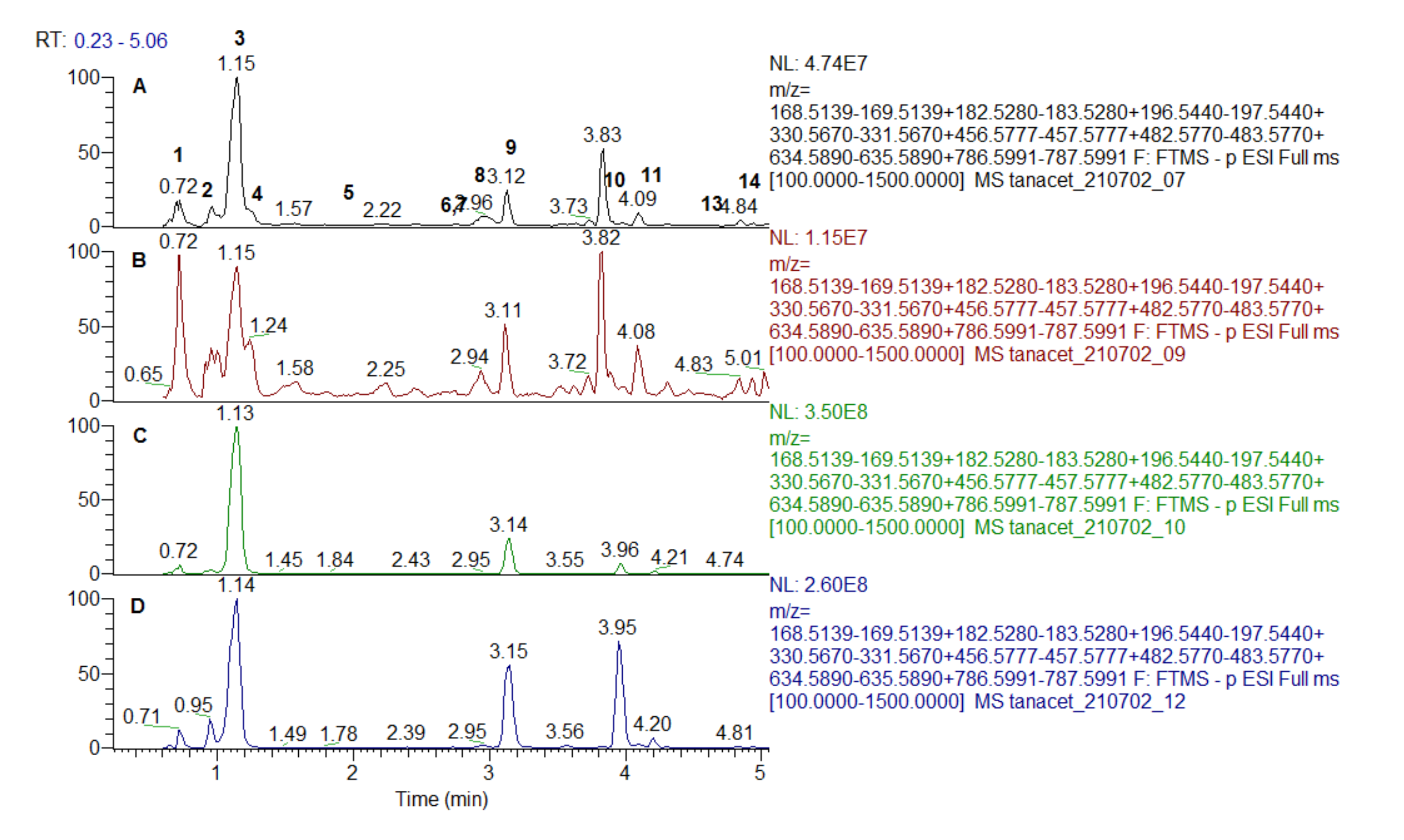
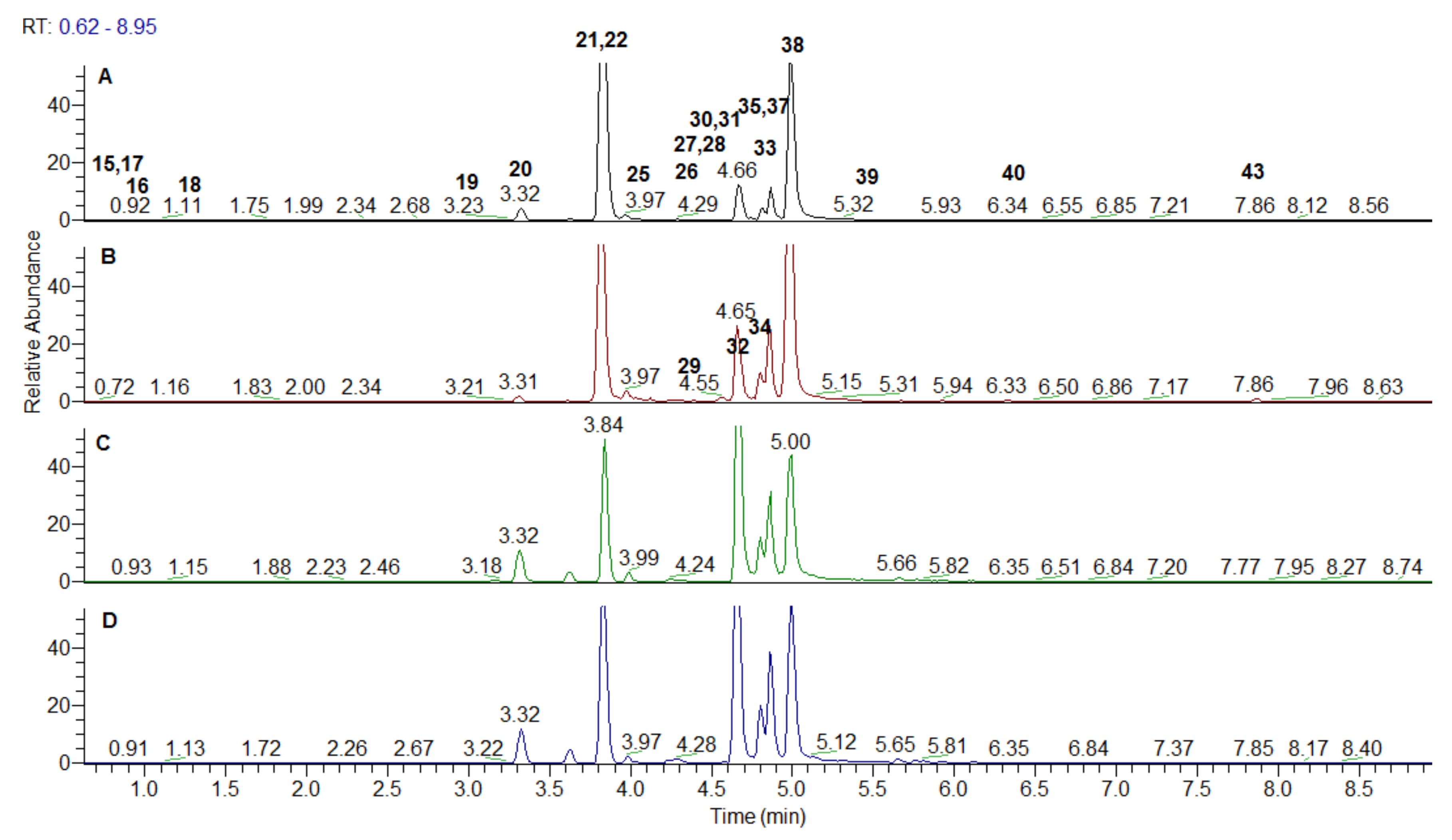
2.4. Chromatographic Separation

Separation was achieved on an UHPLC system Dionex Ultimate 3000RSLC (ThermoFisher Scientific, Inc., Waltham, MA, USA) with reversed phase column Kromasil Eternity XT C18 (1.8 µm, 2.1 × 100 mm) column maintained at 40 °C. The binary mobile phase consisted of A: 0.1% formic acid in water and B: 0.1% formic acid in acetonitrile. The run time was 33 min. The following gradient was utilized: the mobile phase was held at 5% B for 1 min, gradually turned to 30% B over 19 min, increased gradually to 50% B over 5 min, increased gradually to 70% B over 5 min, and finally increased gradually to 95% over 3 min. The system was then turned to the initial condition of 5% B and equilibrated over 4 min. The flow rate and the injection volume were set to 300 µL/min and 1 µL, respectively. The effluents were connected on-line with a Q Exactive Plus Orbitrap mass spectrometer where the compounds were detected [22].

2.5. Mass Chromatography Conditions

Mass analyses were carried out on a Q Exactive Plus mass spectrometer (ThermoFisher Scientific, Inc.) equipped with a heated electrospray ionization (HESI-II) probe (ThermoScientific). The tune parameters were as follows: spray voltage 3.5 kV; sheath gas flow rate 38; auxiliary gas flow rate 12; spare gas flow rate 0; capillary temperature 320 °C; probe heater temperature 320 °C; and S-lens RF level 50. Acquisition was acquired at Full-scan MS and Data Dependent-MS2 modes. Full-scan spectra over the m/z range 100 to 1500 were acquired in negative ionization mode at a resolution of 70,000. Other instrument parameters for Full MS mode were set as follows: AGC target 3e6, maximum ion time 100 ms, number of scan ranges 1. For DD-MS2 mode, instrument parameters were as follows: microscans 1, resolution 17,500, AGC target 1e5, maximum ion time 50 ms, MSX count 1, isolation window 2.0 m/z, stepped collision energy (NCE) 20, 40, and 70 eV. Data acquisition and processing were carried out with Xcalibur 4.2 software (ThermoScientific) [22].

2.6. Determination of Antioxidant and Enzyme Inhibitory Effects

The antioxidant activity of Spondias samples was determined by 1,1-diphenyl-2-picryhydrazyl (DPPH) and 2,2′-azino-bis(3-ethylbenzothiazoline-6-sulfonic acid) (ABTS) radical scavenging, cupric reducing antioxidant capacity (CUPRAC), ferric reducing antioxidant power (FRAP), metal chelating (MCA), and phosphomolybdenum (PBD) assays [23,24]. DPPH, ABTS, CUPRAC, and FRAP activities were expressed as mg trolox equivalents (TE)/g extract. In MCA, the data were presented as mg EDTA equivalents (EDTAE)/g extract, whereas in PBD assay, the results were provided as mmol TE/g extract. Acetylcholinesterase (AChE) and butyrylcholinesterase (BChE) inhibition was assessed as described in Uysal et al. [23] and Grochowski et al. [24] and, expressed as mg galantamine equivalents (GALAE)/g extract, while amylase and glucosidase inhibition [23,24] was reported as mg acarbose equivalents (ACAE)/g extract. Tyrosinase inhibition was also expressed as kojic acid equivalents (KAE)/g extract [23,24].

2.7. Statistical Analysis

Statistical analyses were performed under Xlstat v 2020 and R v 3.6.2 software, respectively. Outcomes were done as mean ± standard deviation and statistical difference was determined by one-way analysis of variance (ANOVA). At p < 0.05 level, Turkey’s post hoc test was done for the sample’s comparisons. Then, both total chemical contents and bioactivities datasets were scaled, centered, and submitted to the principal component analysis (PCA). Subsequently, Cluster Image Map (CIM) analysis was performed on the result of PCA. Similarly, Cluster Image Map (CIM) analysis was done on the relative content of identified chemical compounds by using the peak area data matrix derived from UHPLC-MS analysis. Before analysis, the data matrix was logarithmic transformed. Both Cluster Image Map analysis were based on “Ward” and “Euclidean” as linkage rule and distance, respectively.

3. Results and Discussion

Based on the analytical parameters (retention times, MS and MS/MS accurate masses, fragmentation patterns and comparison with reference standards and literature data), 98 metabolites including gallic and ellagic acids and derivatives, ellagitannins, hydroxybenzoic, hydroxycinnamic, acylquinic acids and derivatives, flavonols, flavanones, flavanonols, flavan-3-ols, and others are unambiguously identified. A list of compounds is reported in Table 1. The fragmentation of the identified metabolites is given in supplemental material (Table S1).

3.1. Gallic Acid and Galloyl Derivatives

Extracted ion chromatogram of the galloyl derivatives are depicted in Figure 1 and Figure 2. MS/MS fragmentation pathway of gallic acid (GA) (3) and galloyl derivatives yielded characteristic ions at m/z 169.013 [GA-H], 151.003 [GA-H-H2O], 125.023 [GA-H-CO2], and 107.012 [GA-H-H2O-CO2] (Table 1).
MS/MS spectra of three isobaric compounds (1, 2, and 4) with [M-H] at m/z 331.067 were acquired (Table 1). Compounds 1 and 2 afforded fragment ions resulting from hexose cross cleavages based on the loss of –CHOH: 0,4Hex (−60 Da) at m/z 271.046, 0,3Hex (−90 Da) at m/z 241.035 and 0,2Hex (−120 Da) at m/z 211.024. Similar to the MS/MS pattern of caffeoyl hexoses, they were ascribed as isomeric galloyl-hexoses (sugar esters), while 4 was tentatively identified as gallic acid-O-hexoside [22]. In the same manner, 5 and 8 were assigned to digalloyl hexoside and digalloyl hexose. Regarding 11 and 13, three galloyl residues were evidenced by the transitions 635.089 → 483.079 [M-H-galloyl], 483.079 → 313.057 [M-H-galloyl-H2O], and 313.057→169.013 [GA-H] (Table 1). Tetragalloyl hexoside (14) was witnessed by the subsequent losses of galloyl residues at m/z 465.065 [M-H-2galloyl-H2O] and 295.046 [M-H-3galloyl-2H2O]. Herein, we report for the first time galloyl-, digalloyl-, and trigalloyl hexosides in both studied species.
Compounds 6 and 7 shared the same [M-H] at m/z 495.078 (Table 1). They were tentatively assigned to digalloylquinic acids as indicated by the common abundant MS/MS ion at m/z 343.067 derived from the loss of a galloyl residue and the prominent ions at m/z 191.055 (deprotonated quinic acid) and “dehydrated” quinic acid at m/z 173.045 [25].
Methylgallate (9) and dimetylgallate (12) were deduced from the typical loss of methyl radical (•CH3) (−15 Da) at m/z 168.005 (9) and consequent losses of methyl radicals at m/z 182.0211 and 166.998 (12) (Table 1). In (-) ESI-MS/MS spectrum 10 ([M-H] at m/z 457.078) yielded fragment ions at m/z 305.067 [M-H-galloyl], 287.056 [M-H-GA], and 169.013 [GA-H] (a base peak) indicating a galloyl derivative. On the other hand, epigallocatechin was observed at m/z 287.056 (C15H21O6) together with a series of fragment ions at m/z 269.045 [M-H-GA-H2O], 261.076 [M-H-GA-CO], 243.066 [M-H-GA-CO2], and 219.066 [M-H-GA-CO-CO2]. Thus, 10 was tentatively identified as epigallocatechin gallate previously reported in S. tuberosa [26].

3.2. Ellagic Acid Derivatives and Ellagitannins

Spondias ellagitannins include monomeric HHDP (hexahydroxydiphenoyl) unit-bearing ellagitannins (15–19, 21, 24, 27, and 35) and dimeric derivatives 31 and 36 (Table 1, Figure 3 and Figure 4). All of them could be referred to Okuda’s type II ellagitannins [27], while 22 and 29 possesses dehydrohexahydroxydiphenoyl (DHHDP) unit, being Okuda’s type III tannin (dehydroellagotannin) [28]. Main criteria in the peak annotation of the ellagitannins were the neutral mass losses of 152 Da (galloyl moiety), 170 Da (gallic acid), 302 Da (HHDP), 332 Da (galloyl hexose), and 482 Da (HHDP hexose) as was reported in the literature [29,30]. Afterwards, the fragmentation patterns yielded the diagnostic fragment ion at m/z 300.999 resulting from the spontaneous lactonization of HHDP into ellagic acid (EA) and m/z 169.013 consistent with a galloyl residue [29]. MS/MS fragmentation pathway of EA (38) afforded characteristic ions corresponding of the neutral losses of CO and CO2 at m/z 257.010 [EA-H-CO2], 245.009 [EA-H-2CO], 217.013 [EA-H-3CO], and concomitant losses at m/z 229.013 [EA-H-CO-CO2], 201.019 [EA-H-2CO-CO2], 185.023 [EA-H-2CO-2CO2], 173.023 [EA-H-3CO-CO2], 145.028 [EA-H-4CO-CO2], 129.033 [EA-H-3CO-2CO2], and 117.033 [EA-H-5CO-CO2] (Table 1). Compound 38 was unambiguously identified by comparison with reference standard.
MS/MS spectra of the isobaric pair 15 and 16 with [M-H]at m/z 481.062 were acquired in both Spondias species (Table 1). The typical fragment ion at m/z 300.999 resulted from the loss of 180 Da (Hex + H2O) indicating HHDP-hexoside.
Four isobars 17–19 and 21 shared the same [M-H]at m/z 633.073; the abundant fragment ion at m/z 300.999 suggested the loss of 332 Da and galloyl-hexose moiety (Table 1). Accordingly, the aforementioned compounds were ascribed as galloyl-HHDP-hexoside isomers. This assumption was in line with the presence of monomeric ellagitannin corilagin previously annotated in S. mombrin and S. tuberosa [26]. In the same way, digalloyl-HHDP-hexoside isomers 24, 27 and 35 at m/z 785.084 [M-H] were evidenced at by the characteristic losses of 484 Da (152 + 152 + 162 + 18) consistent with two galloyl and hexosyl residues. Di-HHDP-hexoside (36) ([M-H] at m/z 783.075) was found in S. dulcis leaves witnessed by the loss of HHDP-hexose (482 Da) at m/z 300.999. Compound 31 from S. mombrin leaves was deduced from the transitions 953.090 → 463.168 ([M-H-HHDP-H2O-GA] and 463.168 → 300.999 ([EA-H]] (Table 1). It was assigned to galloyl-bisHHDP-hexoside [26]. Type III ellagitannin galloyl-HHDP-DHHDP-hexoside (22) was evidenced by the fragment ions at m/z 463.052 [M-H-C21H12O14], 343.009 and 300.999 suggesting the concomitant loss of DHHDP and gallic acid (−488 Da), subsequent cross-ring cleavage of hexose unit (0,2X, −120 Da) and EA, respectively. Compound 22 was ascribed to geraniin, while additional galloyl residue was found in 29 (galloyl-geraniin) (Table 1). Both compounds 22 and 29 were previously isolated from S. mombrin leaves and stems [31]. Based on the prominent ions at m/z 315.011 [M-H-Hex] and m/z 299.9905 [M-H-Hex-CH3], compounds 28 and 37 were assigned as methylellagic acid-O-hexoside isomers (Table 1). In the same manner, the tentative structure of dimethylellagic acid-hexoside was proposed for the isobaric compounds 39 and 41.
Methylellagic (40) and dimetylallagic acid (43) were annotated in both species, while trimethylellagic acid (42) was evidenced only in dulcis leaves. In addition, EA-deohyhexoside (33) was commonly found in the studied extracts, while EA-hexoside/pentoside (25, 32, 33) were evidenced only in the S. mombrin leaves methanol extract.
Compound 20 ([M-H] at m/z 291.045, C13H7O8, 0.755 ppm) gave and a base peak at m/z 247.025 and a series of low abundant ions at 219.030 [M-H-CO2-CO], 191.034 [M-H-CO2-2CO], 163.039 [M-H-CO2-3CO], 135.044 [M-H-CO2-4CO], and 107.049 [M-H-CO2-5CO] (Table 1). Based on the comparison with date from the literature, 20 was annotated as brevifolin carboxylic acid [26,30]. Concerning 26 ([M-H] at m/z 247.025), there was mass difference of 44 Da with 20 indicating that the associated molecule missed a carboxyl group; the MS/MS spectrum matched that of 20 (Table 1). Accordingly, 26 was annotated as brevifolin. Compound 30 ([M-H] at m/z 305.031) afforded a prominent ion at m/z 273.004 ([M-H-CH3OH] (a base peak) suggesting a methyl ester of 20—methylbrevifolin carboxylate.

3.3. Hydroxybenzoic, Hydroxycinnamic, Acylquinic Acids and Derivatives

Based on the retention times, accurate masses, MS/MS fragmentation patterns, and comparison with reference standards and literature data, six hydroxybenzoic (44–46, 51, 53, and 54), two hydroxycinnamic (47 and 49), and three acylquinic (48, 50, and 52) acids and derivatives were unambiguously identified (Table 1) [22].

3.4. Flavonols

The flavonol aglycones quercetin (77), myricetin (78), and kaempferol (80) were identified by comparison with the retention times in UHPLC-HRMS and MS/MS fragmentation fingerprints of reference standards (Table 1).
In general, MS/MS fragmentation pathways of flavonoid glycosides demonstrated neutral mass losses of 162.053, 146.058, 132.042, and 176.033 Da consistent with hexose, deoxyhexose, pentose, and hexuronic acid, supported by the Retro-Diels-Alder (RDA) cleavages of the flavonoid skeleton [22]. Compounds 55, 56, 60, 63, 64, 65, 68, and 70 showed a product ion at m/z 301.035, together with RDA ions at m/z 151.002 [1,3A], 121.027 [1,2B], and 107.012 [0,4A], suggesting the presence of the quercetin core [32]. The 3-glycosylation position of 60, 64, and 68 was determined by the low abundant Y0- ion (m/z 301.035) compared with the radical aglycone ion [Y0-H] (m/z 300.027), as typical MS/MS fragmentation behavior of flavonoid 3-O-glycosides, instead of 7-O-glycosides [32]. Accordingly, and based on the comparison to the reference standards, 60, 64, and 68 were identified as isoquercitrin, hyperoside, and quercitrin. Regarding 63 and 70, a base peak of the radical aglycone ion at m/z 300.027, allowed for the annotation of quercetin 4′-O-glycosilation. Based on the neutral losses of 176.033 and 162.053, 63 and 70 could be related to quercetin 4′-O-hexuronide and quercetin 4′-O-hexoside, respectively (Table 1). Compounds 55 [M-H] at m/z 625.141 and 56 [M-H] at m/z 595.130 corresponded to dihexoside and hexoside-pentoside of quercetin, respectively. The absence of interglycosidic bonds cleavages revealed a 3-O-position of diglycoside chains [33]. In the same manner, due to the fragment ions at m/z 285.039, 284.032, 151.003, and 107.012, compounds 62, 67, 69, 73, and 75 were ascribed as kaempferol-O-glycosides. Diagnostic ions at m/z 317.029 [Y0] and 316.022 [Y0-H], supported by RDA ions at m/z 137.023 [1,2B], 164.010 [1,3B-H], and 178.997 [1,2A] could associate 58, 59, 61, 71, and 74 to myricetin derivatives (Table 1). Regarding 66 with [M-H] at m/z 477.103, consequent losses of deoxyhexose and one •CH3 at m/z 331.046, and 316.022 suggested methylmyricetin-O-deoxyhexoside. The methoxyl group could be in ring B, deduced from the ion at m/z 136.014 (1,2B-•CH3). Similar to the aforementioned flavonol, 72 possessed a methoxyl group in a ring B deduced from the prominent ion at m/z 300.027 arising from loss of methyl radical from the deprotonated aglycon ion [M-H-Glc-•CH3] and fragments at m/z 151.002 and 107.012 assigned to A-ring (Table 1). Thus, 72 was identified as isorhamnetin 3-O-glucoside confirmed by a comparison with reference standard. In contrast to 72, peaks 76 and 79 produced A-ring fragments at m/z 165.018 [1,3A] and 121.028 [0,4A], retaining the methoxyl group in ring A. Accordingly, 76 and 79 were related to rhamnetin 3-O-hexoside and rhamnetin O-deoxyhexoside [34] (Table 1).

3.5. Flavanones, Flavanonols and Flavan 3-ols

Compound 82, [M-H] at m/z 289.071 gave a fragment ions at m/z 271.060 [M-H-H2O], 245.081 [M-H-CO2], 205.050, 203.070 (cleavage of the A-ring of flavan-3-ol), 179.033 (cleavage of B-ring), and RDA ions at m/z 151.038 [1,3B], 137.022 [1,3A], and 109.028 [0,4A + 2H] [35]. Based on the comparison to a reference standard, 72 was unambiguously identified as (+)-epicatechin. Compounds 81, 88, and 90 demonstrated similar fragmentation patterns. Compound 81 differs from 72 by one hexose (165.053 Da) and was assigned as (epi)catechin-O-hexoside. Compounds 88 and 90 revealed a fragment ion at m/z 169.013, corresponding to a galloyl residue and were tentatively ascribed to (epi)catechin-gallate (Table 1).
Three isobars 83, 84, and 86 shared the same deprotonated molecular ion at m/z 465.103. MS/MS spectra of compounds produced fragment ion at m/z 304.054 [M-H-Hex], corresponding to taxifolin O-hexosides. Prominent fragment ions at m/z 259.061 [Agl-H-CO2], 241.050 [Agl-H-CO2-H2O], and RDA ions at m/z 178.997 [1,2A], 151.002 [1,3A], 149.023 [1.3B], and 125.023 [1,4A] confirmed the presence of taxifolin [36]. Based on the ratio between deprotonated molecular ion and fragments at m/z 304.054 [YO-] and 303.051 [YO-H], the position of glycosylation of taxifolin can be distinguished (Table 1). The flavanon aglycon naringenin (93) ([M-H] at m/z 271.061) was witnessed by RDA ions at m/z 151.002 [1,3A], 119.048 [1,3B], and 107.012 [0,4A] [33]. Compounds 87 and 91 ([M-H] at m/z 433.114) differed from 93 by 162.05 Da, corresponding to hexosides of naringenin. Compound 87 was unambiguously identified as naringenin 7-O-glycoside, deduced from the base peak at m/z 271.061 [Agl-H] and comparison with reference standard. Regarding 91, a series of fragment ions at m/z 343.082 [M-H-90], 283.062 [M-H-150], and a base peak at m/z 313.072 [M-H-120] were indicative for naringenin 8-C-hexoside [37]. In the same manner 94 could be related to flavanon aglycon pinocembrin, and 92 to its O-hexoside, while 89 was ascribed to eriodictiol 7-O-hexoside [33].

3.6. Others

Compound 95 [M-H]- at m/z yielded a precursor ion at m/z 341.108 (C12H22O11) together with the fragment ions at m/z 179.055 ([M-H-Hex] and 89.022 ([M-H-Hex-90] suggesting disaccharide sucrose, previously found in Spondias species [26]. Regarding 98, a base peak at m/z 151.038, together with prominent fragment ions at m/z 136.015 and 121.028 are in accordance with the furofuran lignan pinoresinol [38]. In the MS/MS spectrum of 97, the main fragment ions were observed at m/z 341.066 and 217.013, which were similar to those of cinchonain Ib. The ion at m/z 341.066 was produced from the neutral loss of C6H6O2 (110 Da), which confirmed the existence of a dihydroxyphenyl group. The ion at m/z 217.013 was generated from the neutral losses of C6H6O2 and C7H8O2 (234 Da), produced from the elimination of dihydroxytoluene. Thus, compounds 97 was related to dihydroxylphenylpropanoid-substituted catechin cinchonains Ib [39].

3.7. Enzyme Inhibitory Properties

Enzyme inhibition is one of the most attractive topics in medical and pharmaceutical research. For example, the inhibition of acetylcholinesterase is closely related to the alleviation of the observed symptoms of Alzheimer’s disease. As another example, the inhibition of α-amylase and α-glucosidase is the basis of oral anti-diabetic agents for the treatment of diabetes type II. Tyrosinase is also considered to be the main target in the control of skin disorders and hyperpigmentation problems. In this context, we selected the enzymes (cholinesterases, α-amylase, and α-glucosidase and tyrosinase) to determine inhibitory properties of the tested Spondia extracts. From Table 2, it was noted that the stem bark maceration-EA of S. dulcis and the leaves infusion and Soxhlet-MeOH as well as the stem bark maceration-EA (no stir) of S. mombin (10.33, 10.45, 10.37, and 10.31 mg GALAE/g, respectively) showed highest inhibition against acetylcholinesterase. The pathogenesis of Alzheimer’s disease is complex and the mechanisms are poorly understood [40], but scientific evidences have attested of the role of cholinesterase enzymes. Acetylcholinesterase inhibition has been advocated in the management of Alzheimer’s disease, since this enzyme is directly linked to the hydrolysis of the neurotransmitter, acetylcholine, at the synaptic cleft. Moreover, isoquercitrin, previously identified in S. mombin hydroethanolic extract [12] and in other Spondias species, including S. tuberosa, was reported to modulate acetylcholinesterase activity, as well as β- and γ-secretase and Aβ aggregation [41]. Clinical studies have revealed that another cholinesterase enzyme, butyrylcholinesterase, was also implicated in the exacerbation of the health condition of Alzheimer’s disease patients [42,43,44]. These facts support the quest for therapeutic candidates possessing butyrylcholinesterase inhibitory activity. Data presented in Table 2, showed that the different extracts of the leaves and stem bark of the selected Spondias species exhibited variable inhibitory action against butyrylcholinesterase, with the highest anti-BChE value exhibited by leaves maceration-MeOH (not stir) of S. mombin. However, it was observed that the studied extracts displayed more prominent inhibitory action against acetylcholinesterase compared to butyrylcholinesterase. A previous study carried out by Elufioye et al. (2017) demonstrated the inhibitory action of S. mombin leaves methanolic extract on acetylcholinesterase and butyrylcholinesterase, with special focus on the inhibitory action of botulin, campesterol, and phytol, isolated from S. mombin leaves [45]. The oxytocic ability of S. mombin leaves extracts has been reported, thereby validating its traditional use as abortifacient [45]. In addition, in vivo study has demonstrated that S. mombin leaves extract could affect gonadotrophin secretion by the pituitary gland in male Wistar rats [46], advocating the ability of S. mombin leaves extract to cross the blood–brain barrier (BBB). The BBB shields the brain from harmful toxins and pathogens, but this protective characteristic is also a hurdle to the entry of therapeutic agents. One of the main reasons why around 98% of newly developed drug candidates fail clinical trial is their inability/poor ability to cross the BBB [47]. Therefore, assessing the potential of new compounds to cross the BBB is crucial. On the other hand, as far as our literature search could establish, this study can be regarded as the first attempt to report the in vitro cholinesterase inhibitory activity of S. dulcis leaves and stem bark.
An increasing number of scientific reports describe the relationship between Alzheimer’s disease and diabetes type II. In fact, substantial epidemiological evidence suggest that Alzheimer’s disease and diabetes type II are intertwined conditions [48,49,50]. Kandimalla and colleagues [51] coined the term “type 3 diabetes” to emphasize on the shared molecular and cellular features related to Alzheimer’s disease and diabetes type II. S. mombin has been used in traditional medicine to manage diabetes and was also previously reported to inhibit α-amylase [9]. Nevertheless, it is worth mentioning that novel therapeutic strategies for the management of diabetes type II encompass lower inhibition of α-amylase and stronger α-glucosidase inhibition. It has been postulated that the simultaneous inhibition of these carbohydrate hydrolyzing enzymes would result in abnormal bacterial fermentation of undigested carbohydrates in the colon, causing abdominal distention, flatulence, meteorism, and possibly diarrhea [52]. Results gathered from this study showed that both Spondias species were poor inhibitors of α-amylase (Table 2). On the other hand, several extracts of S. mombin and S. dulcis leaves and stem bark actively inhibited α-glucosidase. In contrast no activity was recorded for 11 extracts. S. dulcis fruit ethanol extract was reported to inhibit α-glucosidase activity [53].
The ability of the Spondias extracts to inhibit tyrosinase activity was assessed and presented in Table 2. In general, methanolic extracts of the leaves and stem bark of the selected Spondias species exhibited highest tyrosinase inhibitory action. However, the strongest anti-tyrosinase activity was recorded by stem bark maceration-MeOH (no stir) extract of S. mombin. The quest for new tyrosinase inhibitors has mainly been driven by the side effects, namely, contact dermatitis, which is accompanied by rashes, irritation, itchiness, inflamed skin, and pain, caused by currently used depigmenting agents, such as kojic acid [54]. Moreover, the increased public demand for naturally derived products has also been a major factor boosting the search for novel agents. Oyasowo et al. (2018), recently reported the inhibitory action of S. mombin methanolic root bark extract against tyrosinase [55]. The application of S. mombin in the formulation of a cosmetic product having depigmenting as well as anti-aging and anti-radical action has also been documented. Data gathered from the present study demonstrated that S. dulcis also showed potent tyrosinase inhibitory activity. It is worth highlighting that the extracts obtained by infusion of the leaves showed no activity against tyrosinase.

3.8. Antioxidant Properties
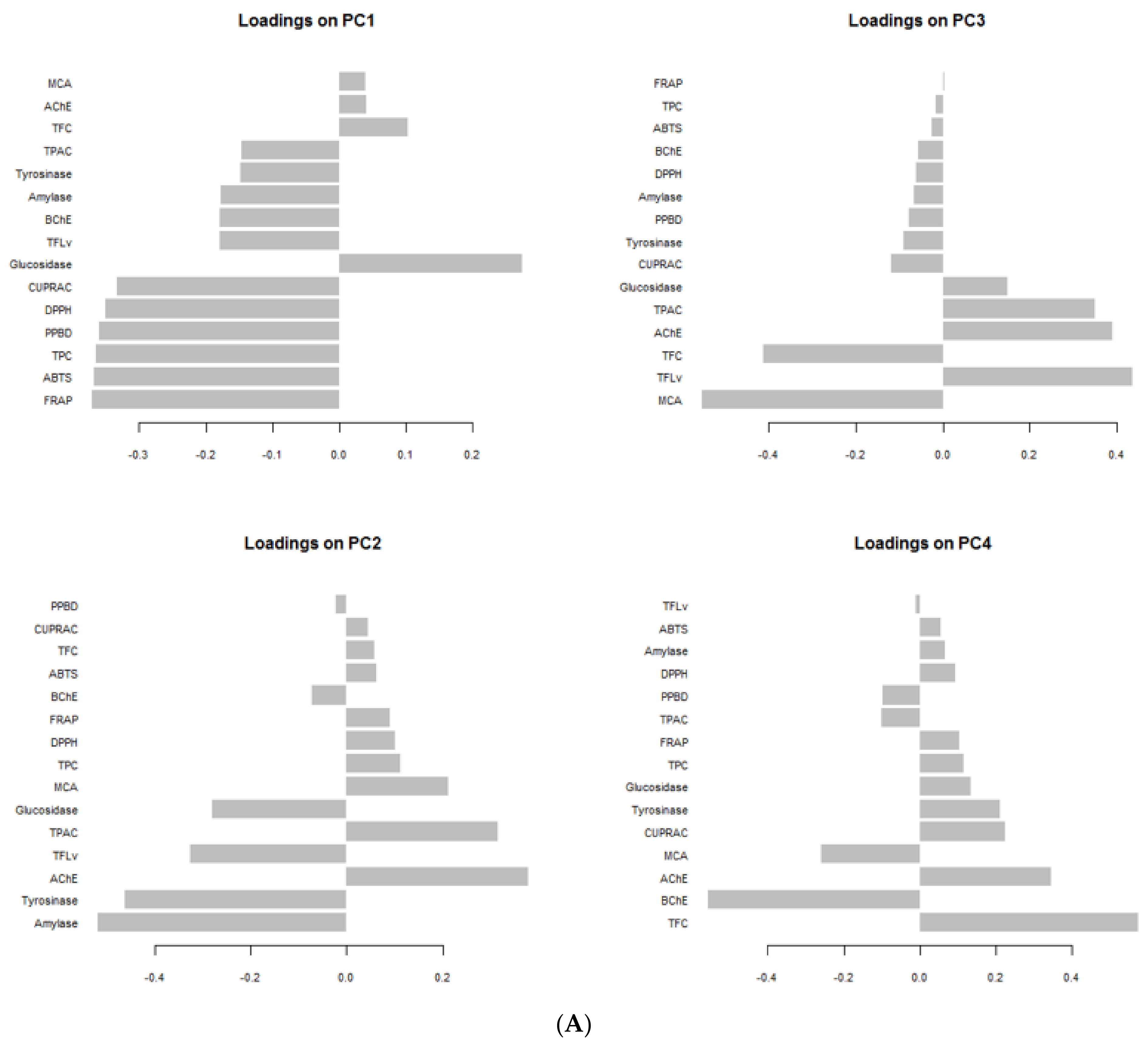
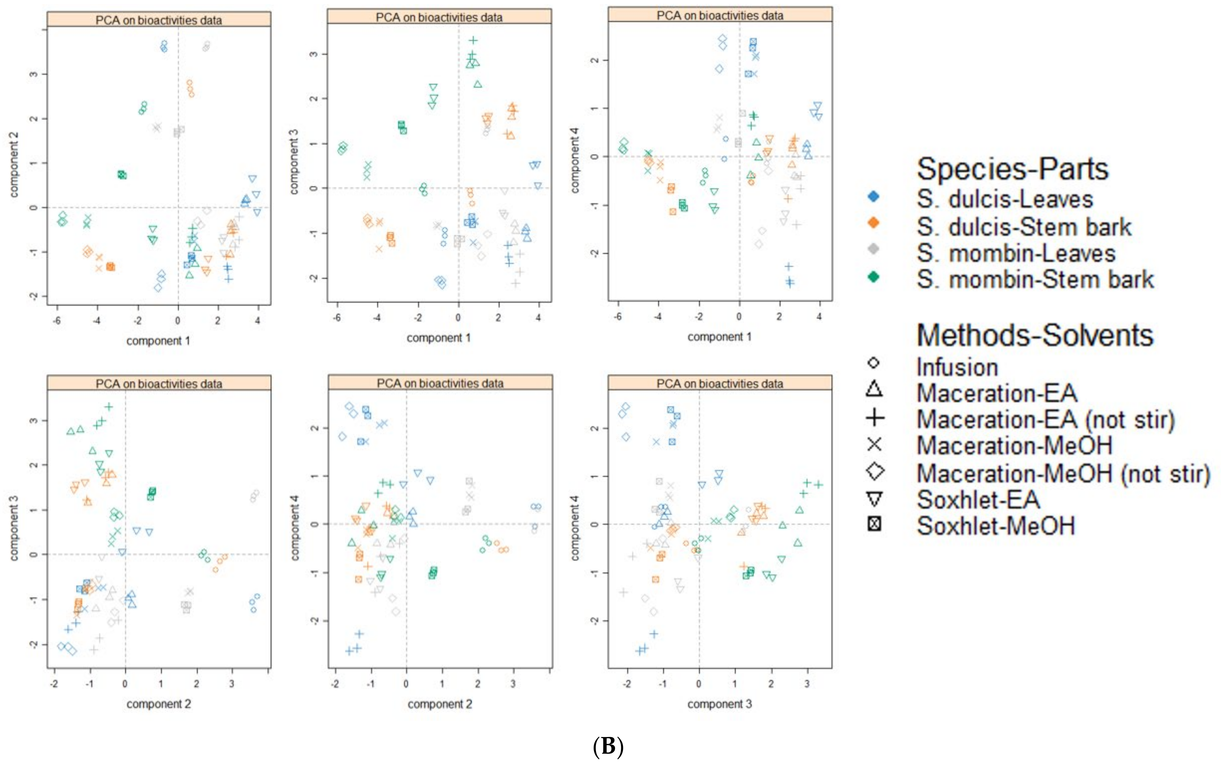
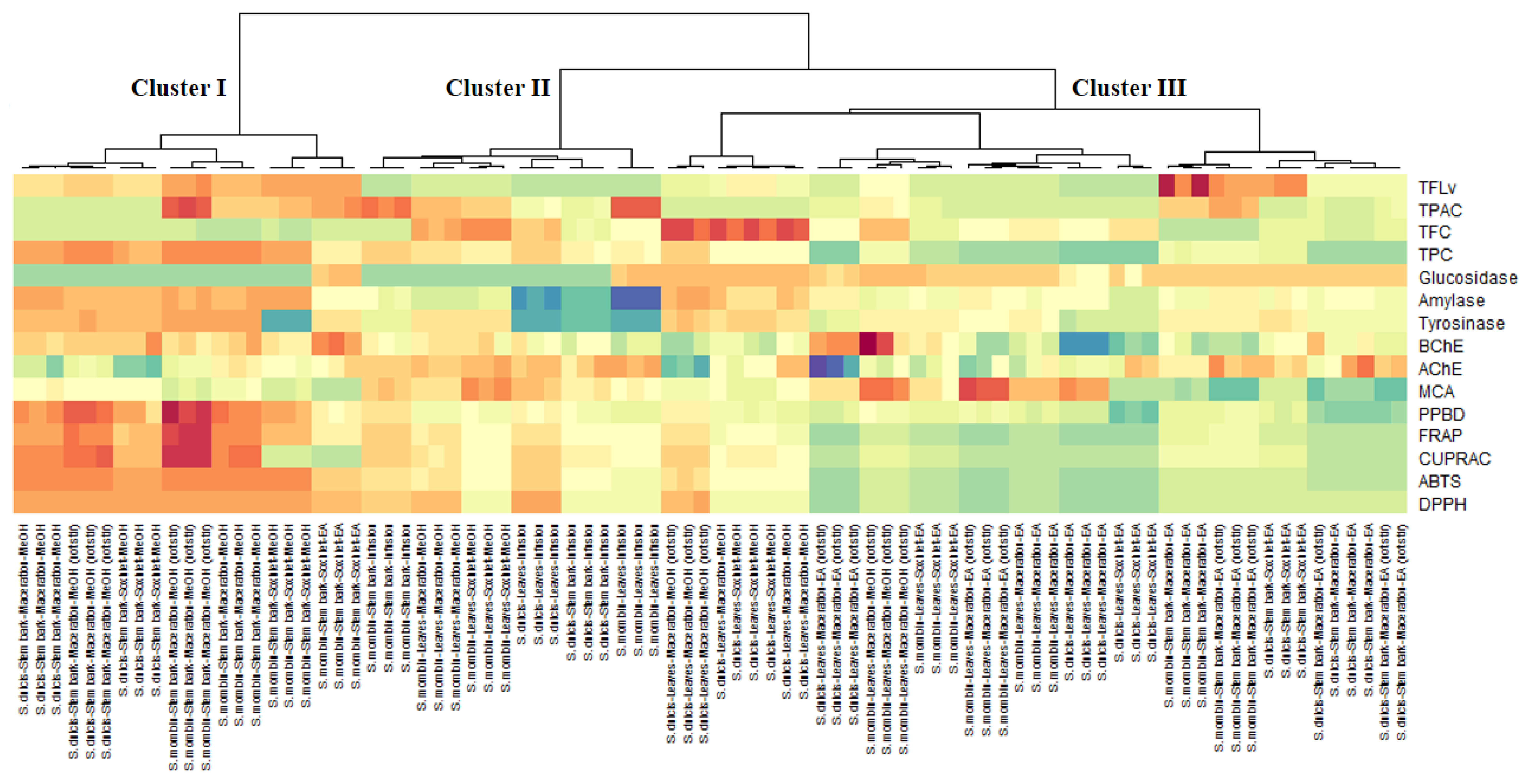
The role of oxidative stress in the pathogenesis of diabetes type II, Alzheimer’s disease, and skin hyperpigmentation conditions has been extensively documented [56,57,58]. In the present study, a number of assays were used to provide a comprehensive understanding of the antioxidant potential of S. mombin and S. dulcis leaves and stem bark extracts. Moreover, since many studies have supported the link between total bioactive compounds profile and the antioxidant potential of herbal extracts, the total phenolic, flavonoid, phenolic acid, and flavonol contents of Spondias species extracts was assessed [8,11,17]. The antioxidant along with the hepatoprotective effect of S. mombin leaf and stem bark methanolic extract has been previously established in vivo. Another group of researchers reported the antioxidant properties of S. dulcis fruits and leaves methanolic extracts. From Table 3, methanolic extracts of the selected Spondias species possessed highest level of phenolic compounds. The stem bark extracts of both species possessed higher phenolic content compared to the leaves’ extracts. Moreover, stem bark maceration-MeOH (no stir) of both species as well as and stem bark-MeOH of S mombin had stronger TPC (240.24; 244.28, and 245.50 mg GAE/g, respectively). The opposite was noted for the flavonoid content, highest TFC was recorded for leaves extracts. In addition, methanolic extracts of S. dulcis leaves (40.71–43.41 mg RE/g) showed higher TFC. In most cases, the phenolic acid content was higher in extracts obtained by infusion, however, the higher amount was achieved by S. mombin infusion leaves infusion and methanol stem bark obtained by maceration without stirring. Phenolic acids solubility in water and organic solvents such as ethyl acetate and methanol was evaluated Vilas-Boas and colleagues (2018) who reported that extraction temperature and solvent nature will affect the solubility of phenolic acids differently [59]. The highest flavonol content was recorded from S. mombin stem bark ethyl acetate extract. Assessment of the total antioxidant capacity of the extracts revealed that extracts showing highest TPC showed highest antioxidant activity. This finding corroborates with a number of studies which have also reported the positive correlation between total antioxidant capacity and TPC. The radical scavenging, reducing power, and metal chelating properties of S. mombin and S. dulcis leaves and stem bark extracts were summarized in Table 4. Widely used DPPH and ABTS radical scavenging assays showed that in general the methanolic extracts were the most active radical scavengers. Similar trend was observed for the CUPRAC and FRAP assays. The stem bark extracts showed higher radical scavenging and reducing capacity compared to the leaves’ extracts. S. mombin ethyl acetate leaves extract obtained by maceration without stirring showed highest metal chelating potential. Findings gathered on the antioxidant potential of the selected Spondias species revealed that these species have potential as antioxidant agent with special emphasis on the higher activity of the stem bark.

3.9. Data Mining

Furthermore, using the total chemical compounds and bioactivities datasets, principal component analysis was performed to uncover the variation among the extracts of Spondias species. Primarily, the small number of principal component apprehended most of the variation in the data were selected with reference to Kaiser rule [60]. Hence, four dimensions which possessed an eigenvalue higher than 1 and 80% of the variability were retained (Table S2). The bioactivities describing each principal component can be identify by referring to Figure 5A. The first component (PC1) predominantly represented the variation in antioxidant, TPC, and anti-glucosidase, the second component (PC2) largely depicted the variation in anti-amylase, anti-tyrosinase, anti-AChE, TFvL, and TPac, the third component (PC3) referred to variation in MCA, TFvl, and TFC while the fourth component (PC4) was mainly determined by TFC and anti-BChE. Regarding the distribution of the samples on the score plot (Figure 5B) deriving from the four principal components, there was some variability, however, it was not possible to identify clearly the different homogeneous groups. Therefore, a heat-map was created to classify the samples and also to identify the total bioactive compounds and biological activities characterizing each obtained clusters. As shown in Figure 6 three major clusters was obtained. Overall, the samples being in cluster I were found to have the highest biological activities, in particular the antioxidant properties, anti-tyrosinase, anti-amylase and anti-BChE activities. Well over the maximum amount of TPC and TFvl were achieved by the members of this clusters. Interestingly, this clusters were composed exclusively of the stem bark extracts of both species the majority of which were obtained using the methanol as solvent. This finding revealed that the stem bark of both species is more active than the leaves. It also suggested that the stem bark is richer in polar polyphenols. Sinan et al. [61] reported variation of antioxidant and enzyme inhibitory activities between the leaves and stem bark of U. togoensis and C. procera. Many authors argued that the concentration of molecules in plants tissues and/or parts vary considerably in terms of its ontogenetic development, the activity of some enzymes and under the impact of soil nutrient and environmental conditions [61,62]. Thereby, the biological activity of the extract of any plant being tightly bound to quantity of quality of molecules it contains, the ideal part should be chosen to derive the full potential of said plant. Furthermore, as evidenced that the stem bark of both species contained highest level of phenolic compounds and since the solubility of phenolic compounds depend on the present and position of –OH groups and the molecular size and the length of constituent hydrocarbon chains [63], it would be reasonable to use an intermediate polar solvent for the future investigations on these two species. The effectiveness of polar solvent like methanol is due to its intermediate polarity that allows it to easily dissolve low molecular weight having protonatable functional groups, i.e., OH [64]. However, considering the toxicity of methanol solvent for human, it would be preferred to evaluate other non-toxic solvent, i.e., ethanol, which possess polarity close to that of methanol.
The consolidation of the 12 samples according to the amount of identified secondary metabolites was shown in Figure 7. As can be seen, the samples can be separated in four main cluster. Samples included in the cluster I (Spondias mombin leaves-infusion and S. mombin-leaves-MAC (no stir)) were richer in many compounds following by that of the cluster 3 (1-Spondias dulcis-leaves-Infusion and 3-S. dulcis-leaves-MAC (no stir)-MEOH) and cluster 2 (S. mombin-stem bark-Infusion; S. mombin-stem bark-MAC (no stir)-EA, and S. mombin-stem bark-MAC (no stir)-MEOH). The samples constituting the cluster 4 were lower in the majority of identified compounds. Moreover, some extracts were, individually, remarkably rich in many compounds. Illustratively, Spondias mombin leaves-infusion was rich in galloyl-geraniin (S29), ellagic acid- pentoside isomer (S32), ellagic acid-pentoside (S34), caffeic acid O-hexoside (S47), and kaempferol-3-O-dihexoside (S57). Similarly, ellagic acid deoxyhexoside (S33) was abundant in Spondias dulcis-leaves-Infusion while 3-S. cythera-leaves-MAC (no stir)-MEOH was rich in di HHDP-hexoside (S36), myricetin 7-O-deoxyhexoside (S71), and naringenin 8-C-hexoside (S91).

4. Conclusions

Data presented in this study highlight the antioxidant and enzyme inhibitory potential of S. mombin and S. dulcis. It was also observed that the type of extraction solvent affects the extraction of bioactive secondary metabolites which subsequently determine the biological activity of the herbal extract. Extracts of S. mombin and S. dulcis leaves and stem bark obtained by organic solvent showed potent tyrosinase, acetylcholinesterase, and α-glucosidase inhibition while relatively low inhibition was observed against butyrylcholinesterase and α-amylase. It was also noted that methanol was a good extracting solvent of phenolics, which was in turn associated with high radical scavenging and reducing properties. Ellagic and gallic acid derivatives, and ellagitannins together with flavonols and flavanones could be associated with the prospective biological activity of stem bark extracts. The selected Spondias species could be considered as valuable sources of bioactive compounds in the pharmaceutical, cosmeceutical, and nutraceutical applications.

Supplementary Materials

The following are available online at https://www.mdpi.com/article/10.3390/antiox10111771/s1, Table S1. Eigen values and percentage of explained variance of each principal component (PC).

Author Contributions

Conceptualization, K.I.S., G.Z. and S.D.; methodology, K.I.S., G.Z., D.Z.-D. and R.G.; software, M.C.N.P.-A. and T.B.; validation, S.D., B.H.G. and P.T.S.Y.; formal analysis, G.Z., D.Z.-D. and R.G.; investigation, G.Z. and M.F.M.; resources, K.I.S.; data curation, G.Z.; writing—original draft preparation, K.I.S., G.Z., D.Z.-D., R.G. and M.F.M.; writing—review and editing, T.B., B.H.G. and P.T.S.Y.; visualization, K.I.S.; supervision, G.Z.; project administration, S.D.; funding acquisition, S.D. All authors have read and agreed to the published version of the manuscript.

Funding

This research received no external funding.

Institutional Review Board Statement

Not applicable.

Informed Consent Statement

Not applicable.

Data Availability Statement

Data is contained within the article and Supplementary Materials.

Conflicts of Interest

The authors declare no conflict of interest.

References

  1. Samtiya, M.; Aluko, R.E.; Dhewa, T.; Moreno-Rojas, J.M. Potential Health Benefits of Plant Food-Derived Bioactive Components: An Overview. Foods 2021, 10, 839. [Google Scholar] [CrossRef] [PubMed]
  2. Marucci, G.; Buccioni, M.; Ben, D.D.; Lambertucci, C.; Volpini, R.; Amenta, F. Efficacy of acetylcholinesterase inhibitors in Alzheimer’s disease. Neuropharmacology 2021, 190, 108352. [Google Scholar] [CrossRef] [PubMed]
  3. Papoutsis, K.; Zhang, J.; Bowyer, M.C.; Brunton, N.; Gibney, E.R.; Lyng, J. Fruit, vegetables, and mushrooms for the preparation of extracts with α-amylase and α-glucosidase inhibition properties: A review. Food Chem. 2021, 338, 128119. [Google Scholar] [CrossRef] [PubMed]
  4. Mukherjee, P.K.; Biswas, R.; Sharma, A.; Banerjee, S.; Biswas, S.; Katiyar, C.K. Validation of medicinal herbs for anti-tyrosinase potential. J. Herb. Med. 2018, 14, 1–16. [Google Scholar] [CrossRef]
  5. Sameh, S.; Al-Sayed, E.; Labib, R.M.; Singab, A.N.B. Comparative metabolic profiling of essential oils from Spondias pinnata (Linn. F.) Kurz and characterization of their antibacterial activities. Ind. Crops Prod. 2019, 137, 468–474. [Google Scholar] [CrossRef]
  6. Sameh, S.; Al-Sayed, E.; Labib, R.M.; Singab, A.N. Genus Spondias: A Phytochemical and Pharmacological Review. Evid.-Based Complementary Altern. Med. 2018, 2018, 5382904. [Google Scholar] [CrossRef] [Green Version]
  7. Tiburski, J.H.; Rosenthal, A.; Deliza, R.; de Oliveira Godoy, R.L.; Pacheco, S. Nutritional properties of yellow mombin (Spondias mombin L.) pulp. Food Res. Int. 2011, 44, 2326–2331. [Google Scholar] [CrossRef] [Green Version]
  8. Nwidu, L.L.; Elmorsy, E.; Oboma, Y.I.; Carter, W.G. Hepatoprotective and antioxidant activities of Spondias mombin leaf and stem extracts against carbon tetrachloride-induced hepatotoxicity. J. Taibah Univ. Med. Sci. 2018, 13, 262–271. [Google Scholar] [CrossRef]
  9. Fred-Jaiyesimi, A.; Kio, A.; Richard, W. α-Amylase inhibitory effect of 3β-olean-12-en-3-yl (9Z)-hexadec-9-enoate isolated from Spondias mombin leaf. Food Chem. 2009, 116, 285–288. [Google Scholar] [CrossRef]
  10. Ishola, I.O.; Ikuomola, B.O.; Adeyemi, O.O. Protective role of Spondias mombin leaf and Cola acuminata seed extracts against scopolamineinduced cognitive dysfunction. Alex. J. Med. 2018, 54, 27–39. [Google Scholar]
  11. Cabral, B.; Siqueira, E.; Bitencourt, M.A.; Lima, M.C.; Lima, A.K.; Ortmann, C.F.; Chaves, V.C.; Fernandes-Pedrosa, M.F.; Rocha, H.A.; Scortecci, K.C. Phytochemical study and anti-inflammatory and antioxidant potential of Spondias mombin leaves. Rev. Bras. Farmacogn. 2016, 26, 304–311. [Google Scholar] [CrossRef] [Green Version]
  12. dos Santos Sampaio, T.I.; de Melo, N.C.; de Freitas Paiva, B.T.; da Silva Aleluia, G.A.; da Silva Neto, F.L.P.; da Silva, H.R.; Keita, H.; Cruz, R.A.S.; Sánchez-Ortiz, B.L.; Pineda-Peña, E.A.; et al. Leaves of Spondias mombin L. a traditional anxiolytic and antidepressant: Pharmacological evaluation on zebrafish (Danio rerio). J. Ethnopharmacol. 2018, 224, 563–578. [Google Scholar] [CrossRef]
  13. Ayoka, A.O.; Akomolafe, R.O.; Iwalewa, E.O.; Akanmu, M.A.; Ukponmwan, O.E. Sedative, antiepileptic and antipsychotic effects of Spondias mombin L. (Anacardiaceae) in mice and rats. J. Ethnopharmacol. 2006, 103, 166–175. [Google Scholar] [CrossRef]
  14. Accioly, M.P.; Bevilaqua, C.M.L.; Rondon, F.C.; de Morais, S.M.; Machado, L.K.; Almeida, C.A.; de Andrade Jr, H.F.; Cardoso, R.P. Leishmanicidal activity in vitro of Musa paradisiaca L. and Spondias mombin L. fractions. Vet. Parasitol. 2012, 187, 79–84. [Google Scholar] [CrossRef]
  15. Franquin, S.; Marcelin, O.; Aurore, G.; Reynes, M.; Brillouet, J.-M. Physicochemical characterisation of the mature-green Golden apple (Spondias cytherea Sonnerat). Fruits 2005, 60, 203–210. [Google Scholar] [CrossRef]
  16. Sarker, M.; Nimmi, I.; Kawsar, M.H. Preliminary screening of six popular fruits of Bangladesh for in vitro IgM production and proliferation of splenocytes. Bangladesh Pharm J. 2012, 15, 31–37. [Google Scholar]
  17. Islam, S.M.A.; Ahmed, K.T.; Manik, M.K.; Wahid, M.A.; Kamal, C.S.I. A comparative study of the antioxidant, antimicrobial, cytotoxic and thrombolytic potential of the fruits and leaves of Spondias dulcis. Asian Pac. J. Trop Biomed. 2013, 3, 682–691. [Google Scholar] [CrossRef] [Green Version]
  18. Koubala, B.; Mbome, L.; Kansci, G.; Mbiapo, F.T.; Crepeau, M.-J.; Thibault, J.-F.; Ralet, M.-C. Physicochemical properties of pectins from ambarella peels (Spondias cytherea) obtained using different extraction conditions. Food Chem. 2008, 106, 1202–1207. [Google Scholar] [CrossRef]
  19. Koubala, B.B.; Kansci, G.; Ralet, M.-C. Ambarella—Spondias cytherea. In Exotic Fruits; Rodrigues, S., de Oliveira Silva, E., de Brito, E.S., Eds.; Academic Press: ondon, UK, 2018; pp. 15–22. [Google Scholar]
  20. Vladimir-Knežević, S.; Blažeković, B.; Bival Štefan, M.; Alegro, A.; Kőszegi, T.; Petrik, J. Antioxidant activities and polyphenolic contents of three selected Micromeria species from Croatia. Molecules 2011, 16, 1454–1470. [Google Scholar] [CrossRef] [Green Version]
  21. Zengin, G.; Aktumsek, A. Investigation of antioxidant potentials of solvent extracts from different anatomical parts of Asphodeline anatolica E. Tuzlaci: An endemic plant to Turkey. Afr. J. Tradit. Complementary Altern. Med. 2014, 11, 481–488. [Google Scholar] [CrossRef] [Green Version]
  22. Gevrenova, R.; Zheleva-Dimitrova, D.; Balabanova, V.; Voynikov, Y.; Sinan, K.I.; Mahomoodally, M.F.; Zengin, G. Integrated phytochemistry, bio-functional potential and multivariate analysis of Tanacetum macrophyllum (Waldst. & Kit.) Sch. Bip. and Telekia speciosa (Schreb.) Baumg.(Asteraceae). Ind. Crops Prod. 2020, 155, 112817. [Google Scholar]
  23. Uysal, S.; Zengin, G.; Locatelli, M.; Bahadori, M.B.; Mocan, A.; Bellagamba, G.; De Luca, E.; Mollica, A.; Aktumsek, A. Cytotoxic and enzyme inhibitory potential of two Potentilla species (P. speciosa L. and P. reptans Willd.) and their chemical composition. Front. Pharmacol. 2017, 8, 290. [Google Scholar] [CrossRef]
  24. Grochowski, D.M.; Uysal, S.; Aktumsek, A.; Granica, S.; Zengin, G.; Ceylan, R.; Locatelli, M.; Tomczyk, M. In vitro enzyme inhibitory properties, antioxidant activities, and phytochemical profile of Potentilla thuringiaca. Phytochem. Lett. 2017, 20, 365–372. [Google Scholar] [CrossRef]
  25. Sayed, E.A.; Martiskainen, O.; Sinkkonen, J.; Pihlaja, K.; Ayoub, N.; Singab, A.-E.N.; El-Azizi, M. Chemical composition and bioactivity of Pleiogynium timorense (Anacardiaceae). Nat. Prod. Commun. 2010, 5, 1934578X1000500410. [Google Scholar] [CrossRef] [Green Version]
  26. Guedes, J.A.C.; Santiago, Y.G.; dos Reis Luz, L.; Silva, M.F.S.; Ramires, C.M.C.; de Lima, M.A.C.; de Oliveira, V.R.; do Ó Pessoa, C.; Canuto, K.M.; de Brito, E.S. Comparative analyses of metabolic fingerprint integrated with cytotoxic activity and in silico approaches of the leaves extract of Spondias mombin L. and Spondias tuberosa Arr. Cam. from Northeast, Brazil. Phytochem. Lett. 2020, 40, 26–36. [Google Scholar] [CrossRef]
  27. Okuda, T.; Yoshida, T.; Hatano, T. Correlation of oxidative transformations of hydrolyzable tannins and plant evolution. Phytochemistry 2000, 55, 513–529. [Google Scholar] [CrossRef]
  28. Cheng, H.S.; Ton, S.H.; Kadir, K.A. Ellagitannin geraniin: A review of the natural sources, biosynthesis, pharmacokinetics and biological effects. Phytochem. Rev. 2017, 16, 159–193. [Google Scholar] [CrossRef]
  29. Yisimayili, Z.; Abdulla, R.; Tian, Q.; Wang, Y.; Chen, M.; Sun, Z.; Li, Z.; Liu, F.; Aisa, H.A.; Huang, C. A comprehensive study of pomegranate flowers polyphenols and metabolites in rat biological samples by high-performance liquid chromatography quadrupole time-of-flight mass spectrometry. J. Chromatogr. A 2019, 1604, 460472. [Google Scholar] [CrossRef]
  30. Zhu, M.; Dong, X.; Guo, M. Phenolic profiling of Duchesnea indica combining macroporous resin chromatography (MRC) with HPLC-ESI-MS/MS and ESI-IT-MS. Molecules 2015, 20, 22463–22475. [Google Scholar] [CrossRef] [Green Version]
  31. Corthout, J.; Pieters, L.; Claeys, M.; Berghe, D.V.; Vlietinck, A. Antiviral ellagitannins from Spondias mombin. Phytochemistry 1991, 30, 1129–1130. [Google Scholar] [CrossRef]
  32. Raffaella, P.; Acquavia, M.A.; Cataldi, T.R.; Alberto, O.; Donatella, C.; Bufo, S.A.; Laura, S.; Rosanna, C.; Guerrieri, A.; Giuliana, B. Profiling of quercetin glycosides and acyl glycosides in sun-dried peperoni di Senise peppers (Capsicum annuum L.) by a combination of LC-ESI (-)-MS/MS and polarity prediction in reversed-phase separations. Anal. Bioanal. Chem. 2020, 412, 3005–3015. [Google Scholar]
  33. de Rijke, E.; Out, P.; Niessen, W.M.; Ariese, F.; Gooijer, C.; Udo, A.T. Analytical separation and detection methods for flavonoids. J. Chromatogr. A 2006, 1112, 31–63. [Google Scholar] [CrossRef] [PubMed]
  34. Justesen, U. Collision-induced fragmentation of deprotonated methoxylated flavonoids, obtained by electrospray ionization mass spectrometry. J. Mass Spectrom. 2001, 36, 169–178. [Google Scholar] [CrossRef] [PubMed]
  35. Yuzuak, S.; Ballington, J.; Xie, D.-Y. HPLC-qTOF-MS/MS-based profiling of flavan-3-ols and dimeric proanthocyanidins in berries of two muscadine grape hybrids FLH 13-11 and FLH 17-66. Metabolites 2018, 8, 57. [Google Scholar] [CrossRef] [PubMed] [Green Version]
  36. Razgonova, M.; Zakharenko, A.; Pikula, K.; Manakov, Y.; Ercisli, S.; Derbush, I.; Kislin, E.; Seryodkin, I.; Sabitov, A.; Kalenik, T. LC-MS/MS Screening of Phenolic Compounds in Wild and Cultivated Grapes Vitis amurensis Rupr. Molecules 2021, 26, 3650. [Google Scholar] [CrossRef] [PubMed]
  37. Zheleva-Dimitrova, D.; Gevrenova, R.; Zaharieva, M.M.; Najdenski, H.; Ruseva, S.; Lozanov, V.; Balabanova, V.; Yagi, S.; Momekov, G.; Mitev, V. HPLC-UV and LC–MS analyses of acylquinic acids in Geigeria alata (DC) Oliv. & Hiern. and their contribution to antioxidant and antimicrobial capacity. Phytochem. Anal. 2017, 28, 176–184. [Google Scholar]
  38. Eklund, P.C.; Backman, M.J.; Kronberg, L.Å.; Smeds, A.I.; Sjöholm, R.E. Identification of lignans by liquid chromatography-electrospray ionization ion-trap mass spectrometry. J. Mass Spectrom. 2008, 43, 97–107. [Google Scholar] [CrossRef]
  39. Gu, W.-Y.; Li, N.; Leung, E.L.-H.; Zhou, H.; Luo, G.-A.; Liu, L.; Wu, J.-L. Metabolites software-assisted flavonoid hunting in plants using ultra-high performance liquid chromatography-quadrupole-time of flight mass spectrometry. Molecules 2015, 20, 3955–3971. [Google Scholar] [CrossRef] [Green Version]
  40. Santos, C.Y.; Snyder, P.J.; Wu, W.-C.; Zhang, M.; Echeverria, A.; Alber, J. Pathophysiologic relationship between Alzheimer’s disease, cerebrovascular disease, and cardiovascular risk: A review and synthesis. Alzheimer’s Dement. Diagn. Assess. Dis. Monit. 2017, 7, 69–87. [Google Scholar] [CrossRef] [Green Version]
  41. Carmona, V.; Martín-Aragón, S.; Goldberg, J.; Schubert, D.; Bermejo-Bescós, P. Several targets involved in Alzheimer’s disease amyloidogenesis are affected by morin and isoquercitrin. Nutr. Neurosci. 2020, 23, 575–590. [Google Scholar] [CrossRef]
  42. Mushtaq, G.; Greig, N.H.; Khan, J.A.; Kamal, M.A. Status of acetylcholinesterase and butyrylcholinesterase in Alzheimer’s disease and type 2 diabetes mellitus. CNS Neurol. Disord. Drug Targets 2014, 13, 1432–1439. [Google Scholar] [CrossRef] [PubMed]
  43. Sokolow, S.; Li, X.; Chen, L.; Taylor, K.D.; Rotter, J.I.; Rissman, R.A.; Aisen, P.S.; Apostolova, L.G. Deleterious Effect of Butyrylcholinesterase K-Variant in Donepezil Treatment of Mild Cognitive Impairment. J. Alzheimers Dis. 2017, 56, 229–237. [Google Scholar] [CrossRef] [PubMed] [Green Version]
  44. Mesulam, M.-M. Butyrylcholinesterase in Alzheimer’s disease. In Alzheimer Disease; Springer, Birkhäuser: Boston, MA, USA, 1994; pp. 79–83. [Google Scholar]
  45. Elufioye, T.O.; Obuotor, E.M.; Agbedahunsi, J.M.; Adesanya, S.A. Anticholinesterase constituents from the leaves of Spondias mombin L. (Anacardiaceae). Biol. Targets Ther. 2017, 11, 107. [Google Scholar] [CrossRef] [PubMed] [Green Version]
  46. Asuquo, O.; Ekanem, T.; Udoh, P.; Mesembe, O. Antigonadotrophic Effect of Spondias Mombin Leaf Extract In Male Wistar Rats. J. Biol. Agric. Healthc. 2012, 2, 14–17. [Google Scholar]
  47. Bhuvanendran, S.; Hanapi, N.A.; Ahemad, N.; Othman, I.; Yusof, S.R.; Shaikh, M.F. Embelin, a Potent Molecule for Alzheimer’s Disease: A Proof of Concept From Blood-Brain Barrier Permeability, Acetylcholinesterase Inhibition and Molecular Docking Studies. Front. Neurosci. 2019, 13, 495. [Google Scholar] [CrossRef] [Green Version]
  48. Strachan, M.W.; Deary, I.J.; Ewing, F.M.; Frier, B.M. Is type II diabetes associated with an increased risk of cognitive dysfunction? A critical review of published studies. Diabetes Care 1997, 20, 438–445. [Google Scholar] [CrossRef]
  49. Stewart, R.; Liolitsa, D. Type 2 diabetes mellitus, cognitive impairment and dementia. Diabet Med. 1999, 16, 93–112. [Google Scholar] [CrossRef]
  50. Yang, Y.; Song, W. Molecular links between Alzheimer’s disease and diabetes mellitus. Neuroscience 2013, 250, 140–150. [Google Scholar] [CrossRef]
  51. Kandimalla, R.; Thirumala, V.; Reddy, P.H. Is Alzheimer’s disease a Type 3 Diabetes? A critical appraisal. Biochim. Biophys. Acta (BBA)-Mol. Basis Dis. 2017, 1863, 1078–1089. [Google Scholar] [CrossRef]
  52. Dalar, A.; Konczak, I. Phenolic contents, antioxidant capacities and inhibitory activities against key metabolic syndrome relevant enzymes of herbal teas from Eastern Anatolia. Ind. Crops Prod. 2013, 44, 383–390. [Google Scholar] [CrossRef]
  53. Julfikar Hossain, S.; Tsujiyama, I.; Takasugi, M.; Biswas, R.; Aoshima, H. Total phenolic content, antioxidative, antiamylase, anti-glucosidase, and antihistamine release activities of Bangladeshi fruits. Food Sci. Technol. Res. 2008, 14, 261–268. [Google Scholar] [CrossRef] [Green Version]
  54. Saeedi, M.; Eslamifar, M.; Khezri, K. Kojic acid applications in cosmetic and pharmaceutical preparations. Biomed. Pharmacother. 2019, 110, 582–593. [Google Scholar] [CrossRef]
  55. Fadare, O.; Obuotor, E.M. Estimation of the kinetic parameters of the inhibition of tyrosinase by an extract of S. mombin (root bark) and the investigation of likely interactions of composite phytochemicals using molecular docking calculations. Am. J. Pharmacol. Sci. 2018, 6, 13–18. [Google Scholar]
  56. Ionescu-Tucker, A.; Cotman, C.W. Emerging roles of oxidative stress in brain aging and Alzheimer’s disease. Neurobiol. Aging 2021, 107, 86–95. [Google Scholar] [CrossRef]
  57. Halim, M.; Halim, A. The effects of inflammation, aging and oxidative stress on the pathogenesis of diabetes mellitus (type 2 diabetes). Diabetes Metab. Syndr. Clin. Res. Rev. 2019, 13, 1165–1172. [Google Scholar] [CrossRef]
  58. Gu, Y.; Han, J.; Jiang, C.; Zhang, Y. Biomarkers, oxidative stress and autophagy in skin aging. Ageing Res. Rev. 2020, 59, 101036. [Google Scholar] [CrossRef]
  59. Vilas-Boas, S.M.; Brandão, P.; Martins, M.A.; Silva, L.P.; Schreiner, T.B.; Fernandes, L.; Ferreira, O.; Pinho, S.P. Solubility and solid phase studies of isomeric phenolic acids in pure solvents. J. Mol. Liq. 2018, 272, 1048–1057. [Google Scholar] [CrossRef] [Green Version]
  60. Zwick, W.R.; Velicer, W.F. Comparison of five rules for determining the number of components to retain. Psychol. Bull. 1986, 99, 432. [Google Scholar] [CrossRef]
  61. Sinan, K.I.; Ferrarese, I.; Aktumsek, A.; Peron, G.; Glamocilja, J.; Sokovic, M.; Nenadić, M.; Dall’Acqua, S.; Zengin, G. NMR and LC-MSn coupled with pharmacological network analysis for the assessment of phytochemical content and biopharmaceutical potential of Carapa procera extracts. J. Pharm. Biomed. Anal. 2021, 114184. [Google Scholar] [CrossRef]
  62. Ghasemzadeh, A.; Nasiri, A.; Jaafar, H.Z.; Baghdadi, A.; Ahmad, I. Changes in phytochemical synthesis, chalcone synthase activity and pharmaceutical qualities of Sabah snake grass (Clinacanthus nutans L.) in relation to plant age. Molecules 2014, 19, 17632–17648. [Google Scholar] [CrossRef]
  63. Iloki-Assanga, S.B.; Lewis-Luján, L.M.; Lara-Espinoza, C.L.; Gil-Salido, A.A.; Fernandez-Angulo, D.; Rubio-Pino, J.L.; Haines, D.D. Solvent effects on phytochemical constituent profiles and antioxidant activities, using four different extraction formulations for analysis of Bucida buceras L. and Phoradendron Californicum. BMC Res. Notes 2015, 8, 1–14. [Google Scholar] [CrossRef] [Green Version]
  64. Felhi, S.; Daoud, A.; Hajlaoui, H.; Mnafgui, K.; Gharsallah, N.; Kadri, A. Solvent extraction effects on phytochemical constituents profiles, antioxidant and antimicrobial activities and functional group analysis of Ecballium elaterium seeds and peels fruits. Food Sci. Technol. 2017, 37, 483–492. [Google Scholar] [CrossRef] [Green Version]
Figure 1. Extracted ion chromatogram of gallic acid and galloyl derivatives from Spondias dulcis extracts: (A)—leaves water extracts; (B)—leaves methanol extract; (C)—stem bark water extract; (D)—stem bark methanol extract.
Antioxidants 10 01771 g001
Figure 2. Extracted ion chromatogram of gallic acid and galloyl derivatives from Spondias mombrin extracts: (A)—leaves water extracts; (B)—leaves methanol extract; (C)—stem bark water extract; (D)—stem bark methanol extract.
Antioxidants 10 01771 g002
Figure 3. Extracted ion chromatogram of ellagitannins and ellagic acid derivatives from Spondias dulcis extracts: (A)—leaves water extracts; (B)—leaves methanol extract; (C)—stem bark water extract; (D)—stem bark methanol extract.
Antioxidants 10 01771 g003
Figure 4. Extracted ion chromatogram of ellagitannins and ellagic acid derivatives from Spondias mombrin extracts: (A)—leaves water extracts; (B)—leaves methanol extract; (C)—stem bark water extract; (D)—stem bark methanol extract.
Antioxidants 10 01771 g004
Figure 5. Principal component analysis (PCA) showing the variability of the total chemical composition, antioxidant and anti-enzymatic activity of Spondias species. (A) Loading plots displaying the relationship between the total chemical composition, antioxidant and anti-enzymatic activity and the four significant PCs. (B) Score plots showing the distribution of the samples in the six 2-dimension plans obtained from the four significant PCs.
Antioxidants 10 01771 g005aAntioxidants 10 01771 g005b
Figure 6. Cluster image map (CIM) analysis of the total chemical composition, antioxidant and anti-enzymatic activity of Spondias species (Red color: high concentration or bioactivity, Blue color: low concentration or bioactivity).
Antioxidants 10 01771 g006
Figure 7. Cluster image map (CIM) analysis of the chemical composition data of Spondias species (Red color: high concentration, Blue color: low concentration). Extract 1-Spondias dulcis-leaves-Infusion, Extract 2-S. dulcis-leaves-MAC (no stir)-EA; Extract 3-S. dulcis-leaves-MAC (no stir)-MEOH; Extract 4-S. dulcis-stem bark-infusion; Extract 5-S. dulcis-stem bark-MAC (no stir)-EA; Extract 6-S. dulcis-stem bark-MAC (no stir)-MEOH; Extract 7-Spondias mombin leaves-infusion; Extract 8- S. mombin leaves-MAC (no stir) EA; Extract 9-S. mombin-leaves-MAC (no stir)- MEOH; Extract 10-S. mombin-stem bark-Infusion; Extract 11-S. mombin-stem bark-MAC (no stir)-EA; Extract 12-S. mombin-stem bark-MAC (no stir)-MEOH.). For compounds numbers refer to Table 1.
Antioxidants 10 01771 g007
Table 1. Specialized metabolites in Spondias extracts assayed by UHPLC-ESI-MS/MS.
NoIdentified/Tentatively Annotated CompoundMolecular FormulaExact Mass
[M-H]
Distribution
Gallic acid and galloyl derivatives
1.galloyl hexoseC13H16O10331.06711,2,3,4,5,6,7,8,9,10,11,12
2.galloyl hexose isomerC13H16O10331.06711,3,4,5,6,7,8,9,10,11,12
3.gallic acid *C7H6O5169.01421,2,3,4,5,6,7,8,9,10,11,12
4.gallic acid hexosideC13H16O10331.06711,3,7,8,9,10,11,12
5.digalloyl hexosideC20H20O14483.07801,3,6,7,9,10,11,12
6.digalloylquinic acidC21H20O14495.07761,3,4,5,6,7
7.digalloylquinic acid isomerC21H20O14495.07901,3,4,5,6
8.digalloyl hexoseC20H20O14483.07801,3,5,6,7,8,9,10,11,12
9.methylgallateC8H8O5183.02891,2,3,4,5,6,7,8,9,10,11,12
10.epigallocatechin-O-gallateC22H18O11457.07761,2,3,4,5,6,7,8,9,10,11,12
11.trigalloyl hexosideC27H24O18635.08906,7,8,9,10,11,12
12.dimethylgallateC9H10O5197.04551,2,3
13.trigalloyl hexoside isomerC27H24O18635.08901,3,4,6,7,9,10,11,12
14.tetragalloyl hexosideC34H28O22787.09991,3,4,6,7,9,10,11,12
Ellagic acid derivatives and ellagitannins
15.HHDP-hexosideC20H18O14481.06241,3,7,9,12
16.HHDP-hexoside isomerC20H18O14
isomer
481.06241,3,7,9,10,12
17.galloyl-HHDP-hexosideC27H22O18633.07331,3,8,9
18.galloyl-HHDP-hexoside isomerC27H22O18633.07331,3,7,9
19.galloyl-HHDP-hexoside isomerC27H22O18633.07331,3,5,6,7,9,10,12
20.brevifolin carboxylic acidC13H8O8291.01491,3,4,6,7,9,10
21.galloyl-HHDP-hexoside isomerC27H22O18633.07331,3,4,6,7,8,9,10,11,12
22.geraniinC41H28O27951.07453,7,10,12
23.valoneic acid-dilactoneC21H10O13469.00491,3,6
24.digalloyl-HHDP-hexosideC34H26O22785.08431,3,5,6
25.ellagic acid-hexosideC20H16O13463.05187,8,9
26.brevifolinC12H8O6247.02451,3,7,9,12
27.digalloyl-HHDP-hexoside isomerC34H26O22785.08437,9,10,11,12
28.methylellagic acid-O-hexosideC21H18O13477.06751,3,4,5,6,7,9,10
29.galloyl-geraniinC48H32O311103.08559
30.methyl brevifolin carboxylateC14H10O8305.03041,3,7,9
31.galloyl-bisHHDP-hexosideC41H30O27953.09027,9
32.ellagic acid- pentoside isomerC19H14O12433.04129
33.ellagic acid deoxyhexosideC20H16O12447.05711,3,7,8,9,10,11,12
34.ellagic acid- pentosideC19H14O12433.04129
35.digalloyl-HHDP-hexoside isomerC34H26O22785.08437,9,10,11,12
36.di HHDP-hexosideC34H24O22783.06861,3
37.methylellagic acid O-hexosideC21H18O13477.06751,3,4,5,6,7,9,10
38.ellagic acid *C14H6O8300.99911,2,3,4,5,6,7,8,9,10,11,12
39.dimethylellagic acid O-hexosideC22H20O13491.08311,2,3,4,5,6,7
40.methylellagic acidC15H8O8315.01461,3,6,7,9,12
41.dimethylellagic acid O-hexosideC22H20O13491.08311,3,4,5,6
42.trimethylellagic acidC17H12O8343.04591,3
43.dimethylellagic acidC16H10O8329.03031,3,7,9
Hydroxybenzoic, hydroxycinnamic, acylquinic acids and derivatives
44.protocatechuic acid O-hexosideC13H16O9315.07231,3,6,7,9,10,11,12
45.protocatechuic acid *C7H6O4153.01811,2,3,4,5,6,7,8,9,10,11,12
46.syringic acid O-hexosideC15H20O10359.09561,2,3,6,7,9,10
47.caffeic acid O-hexosideC15H18O9341.08839
48.neochlorogenic acid *C16H18O9353.08787,8,9,10,11,12
49.caffeic acid *C9H8O4179.03428,9
50.chlorogenic acid *C16H18O9353.08777,8,9
51.vanillic acid O-hexosideC14H18O9329.08881,3,4,5,6,7,10,11,12
52.4-caffeoylquinic aidC16H18O9353.08807,8
53.syringic acid *C9H10O5197.04471,3,12
54.salicylic acidC7H6O3137.02311,2,3,4,5,6,7,8,9,10,11,12
Flavonols
55.quercetin 3-O-dihexosideC27H30O17625.14157,8,9
56.quercetin 3-O-hexoside-O-pentosideC26H28O16595.13071,7,8,9
57.kaempferol 3-O-dihexosideC27H30O16609.14369
58.myricetin 3-O-pentosideC20H18O12449.07271,3,5,7
59.myricitrin (myricetin 3-O-rhamnoside) *C21H20O12463.08821,3,4,5,6
60.isoquercitrin (quercetin 3-O-glucoside) *C21H20O12463.08837,8,9,10,12
61.myricetin 3-O-hexoside-7-O-deoxyhexosideC27H30O17625.14181,3
62.kaempferol 3-O-pentosyl-hexosideC26H28O15579.13597,8,9
63.quercetin 4′-O-hexuronideC21H18O13477.06801
64.hyperoside (quercetin 3-O-galactoside) *C21H20O12463.08855,7,8,9,12
65.quercetin-O-pentosideC20H18O11433.07761,3,7,8,9,10,11
66.methylmyricetin-O-deoxyhexosideC22H22O12477.10381,3
67.kaempferol 3-O-glucoside *C21H20O11447.09331,3,5,6,7,8,9
68.quercitrin (quercetin 3-O-rhamnoside) *C21H20O11447.09331,6,7,9
69.kaempferol 7-O-glucosideC21H20O11447.09417,9,10,11,12
70.quercetin 4′-O-hexosideC21H20O12463.08927,9,11
71.myricetin 7-O-deoxyhexosideC21H20O12463.08853,5,6
72.isorhamnetin 3-O-glucoside *C22H22O12477.104211,12
73.kaempferol-O-pentosideC20H18O10417.08281,3,6,7,9
74.myricitrin-O-gallateC28H24O16615.09991,3,5,6
75.kaempferol 3-O-deoxyhexosideC21H20O10431.09851,3,4,5,6
76.rhamnetin 3-O-hexosideC22H22O12477.10355,6,9
77.Quercetin *C15H10O7301.03471,3,5,7,9,11,12
78.Myricetin *C15H10O8317.03011,3,6,7,9,11,12
79.rhamnetin 3-O-deoxyhexosideC22H22O11461.10903,4,5,6
80.Kaempferol *C15H10O6285.04077,9
Flavanones, flavanonols and flavan-3-ols
81.(epi)catechin O-hexosideC21H24O11451.12416,7,9,10,11,12
82.(+)-catechin *C15H14O6289.07171,3,4,5,6,7,9,10,11,12
83.taxifolin 3-O-hexosideC21H22O12465.10391,3,7,9,10,11,12
84.taxifolin 7-O-hexosideC21H22O12465.10371,7,9,10,11,12
85.(epi)catechin-gallateC22H18O10441.08289,11,12
86.taxifolin 4′-O-hexosideC21H22O12465.10371,7,9,11,12
87.naringenin 7-O-glucoside (prunin) *C21H22O10433.11401,3,4,5,6,9,10,11,12
88.(epi)catechin-gallate isomer IC22H18O10441.08269,10,11,12
89.eriodictiol 7-O-hexosideC21H22O11449.10903,6,7,9,10,11,12
90.(epi)catechin-gallate isomer IIC22H18O10441.08261,4,6,9,10,11,12
91.naringenin 8-C-hexosideC21H22O10433.11443
92.pinocembrin-O-hexosideC21H22O9417.11959,10,11,12
93.naringeninC15H12O5271.06143,5,6,7,9,11,12
94.pinocembrinC15H12O4255.06649,11,12
Others
95.sucroseC12H22O11341.10881,2,3,4,5,6,7,8,9,10,11,12
96.tuberonic acid-hexosideC18H28O9387.16571,3
97.cinchonain IC24H20O9451.10377,8,9
98.pinoresinol (Eklund et al., 2008)C20H22O6357.13451,3,9
*-compare to reference standard; 1-Spondias dulcis-leaves-Infusion, 2-S. dulcis-leaves-MAC (no stir)-EA; 3-S. dulcis-leaves-MAC (no stir)-MEOH; 4-S. dulcis-stem bark-infusion; 5-S. dulcis-stem bark-MAC (no stir)-EA; 6-S. dulcis-stem bark-MAC (no stir)-MEOH; 7-Spondias mombin leaves-infusion; 8-S. mombin leaves-MAC (no stir) EA; 9-S. mombin-leaves-MAC (no stir)-MEOH; 10-S. mombin-stem bark-Infusion; 11-S. mombin-stem bark-MAC (no stir)-EA; 12-S. mombin-stem bark-MAC (no stir)-MEOH.
Table 2. Enzyme inhibitory effects of the tested extracts.
SpeciesPartsMethods-SolventsAChE Inhibition
(mg GALAE/g)
BChE Inhibition
(mg GALAE/g)
Tyrosinase Inhibition
(mg KAE/g)
Amylase Inhibition
(mmol ACAE/g)
Glucosidase Inhibition
(mmol ACAE/g)
Spondias dulcisLeavesInfusion10.10 ± 0.11 ab3.83 ± 0.48 cdefgna0.25 ± 0.04 nna
Maceration-EA8.60 ± 0.23 bcdefna82.27 ± 8.84 n0.71 ± 0.01 efghi11.07 ± 0.21 d
Maceration-MeOH9.79 ± 0.79 abcd3.44 ± 0.25 cdefg165.44 ± 2.39 d0.88 ± 0.03 c17.37 ± 0.11 a
Maceration-EA (not stir)5.77 ± 0.66 h6.21 ± 0.18 ab94.17 ± 5.97 m0.76 ± 0.01 de14.73 ± 0.74 bc
Maceration-MeOH (not stir)6.89 ± 0.21 gh1.98 ± 0.44 ghi184.49 ± 2.51 c0.97 ± 0.03 a17.70 ± 0.04 a
Soxhlet-EA9.51 ± 0.99 abcd1.21 ± 0.44 hi79.88 ± 2.02 n0.54 ± 0.01 l14.27 ± 2.96 c
Soxhlet-MeOH8.82 ± 0.25 abcde2.55 ± 0.72 efgh178.97 ± 1.28 c0.80 ± 0.01 d17.61 ± 0.03 a
Stem barksInfusion9.95 ± 0.53 abcd3.36 ± 0.68 cdefgh15.72 ± 1.90 o0.32 ± 0.01 mna
Maceration-EA10.33 ± 1.09 a3.62 ± 0.39 cdefg119.85 ± 1.47 k0.70 ± 0.01 efghi16.88 ± 0.02 a
Maceration-MeOH7.81 ± 0.62 efg4.83 ± 0.14 abcd197.72 ± 1.43 b0.99 ± 0.01 ana
Maceration-EA (not stir)9.58 ± 0.77 abcd3.68 ± 1.49 cdefg116.15 ± 3.31 k0.67 ± 0.01 hij16.94 ± 0.11 a
Maceration-MeOH (not stir)8.39 ± 0.19 cdefg5.09 ± 0.08 abc201.48 ± 1.17 ab0.94 ± 0.01 abcna
Soxhlet-EA9.98 ± 0.23 abc3.55 ± 0.37 cdefg155.95 ± 3.27 efg0.71 ± 0.04 efgh16.91 ± 0.03 a
Soxhlet-MeOH7.00 ± 0.14 fgh5.39 ± 0.72 abc198.93 ± 0.85 ab0.90 ± 0.01 bcna
Spondias mombinLeavesInfusion10.45 ± 0.16 a2.54 ± 0.56 efghna0.13 ± 0.01 o17.12 ± 0.13 a
Maceration-EA8.66 ± 0.77 bcde2.88 ± 0.52 defgh140.14 ± 2.50 i0.69 ± 0.04 fghi16.20 ± 0.13 ab
Maceration-MeOH10.01 ± 0.13 abc4.48 ± 0.21 bcde159.58 ± 1.37 def0.55 ± 0.01 lna
Maceration-EA (not stir)7.77 ± 0.97 efg2.17 ± 0.87 fgh129.52 ± 1.19 j0.73 ± 0.02 efg16.23 ± 0.50 ab
Maceration-MeOH (not stir)8.59 ± 0.45 bcdef6.89 ± 1.95 a153.70 ± 0.62 fgh0.62 ± 0.01 jk17.41 ± 0.19 a
Soxhlet-EA8.33 ± 0.65 defg4.15 ± 0.44 bcdef147.24 ± 1.93 hi0.68 ± 0.02 fghij16.79 ± 0.40 a
Soxhlet-MeOH10.37 ± 0.07 a4.32 ± 0.94 bcde156.41 ± 1.05 efg0.57 ± 0.01 klna
Stem barksInfusion9.93 ± 0.04 abcd3.71 ± 0.34 cdefg104.08 ± 1.11 l0.65 ± 0.04 ijna
Maceration-EA9.27 ± 0.03 abcde2.89 ± 1.03 defgh150.72 ± 1.82 gh0.68 ± 0.03 ghij16.96 ± 0.04 a
Maceration-MeOH9.34 ± 0.21 abcde5.26 ± 0.57 abc203.72 ± 1.02 ab0.94 ± 0.03 abcna
Maceration-EA (not stir)10.31 ± 0.47 a1.97 ± 0.19 ghi146.82 ± 0.19 hi0.74 ± 0.01 def16.82 ± 0.03 a
Maceration-MeOH (not stir)8.59 ± 0.12 bcdef4.51 ± 0.30 bcde207.00 ± 0.73 a0.95 ± 0.01 abna
Soxhlet-EA9.39 ± 0.39 abcde6.28 ± 0.33 ab163.23 ± 2.54 de0.72 ± 0.01 efgh17.16 ± 0.03 a
Soxhlet-MeOH9.02 ± 0.14 abcde4.36 ± 0.09 bcdena0.96 ± 0.01 abna
Values are reported as mean ± S.D. GALAE: Galantamine equivalent; KAE: Kojic acid equivalent; ACAE: Acarbose equivalent; nd: not detected. Different letters (ao) indicate significant differences in the tested extracts (p < 0.05).
Table 3. Total bioactive compounds (phenolic (TPC), flavonoid (TFC), phenolic acid (TPAC), and flavonol (TFlv) and total antioxidant capacity (by phosphomolybdenum assay) of the tested extracts.
SpeciesPartsMethods-SolventsTPC (mg GAE/g)TFC (mg RE/g)TPAC (mg CAE/g)TFlv (mg CE/g)Phosphomolybdenum (mmol TE/g)
Spondias dulcisLeavesInfusion179.89 ± 0.14 ef25.30 ± 0.59 d6.91 ± 0.74 h0.46 ± 0.01 k4.89 ± 0.14 efg
Maceration-EA29.27 ± 0.91 qr13.80 ± 0.49 gnd1.82 ± 0.06 jk3.33 ± 0.14 k
Maceration-MeOH134.39 ± 1.96 j43.41 ± 1.08 and7.35 ± 0.04 gh3.94 ± 0.12 hijk
Maceration-EA (not stir)32.60 ± 0.32 pq13.21 ± 0.59 g2.63 ± 0.22 ij2.33 ± 0.02 jk3.49 ± 0.14 ijk
Maceration-MeOH (not stir)182.53 ± 1.15 e43.11 ± 3.28 and4.85 ± 0.05 hij4.98 ± 0.02 ef
Soxhlet-EA25.85 ± 0.97 r21.26 ± 0.26 end1.22 ± 0.01 jk1.75 ± 0.12 l
Soxhlet-MeOH136.96 ± 1.86 ij40.71 ± 3.63 and9.48 ± 0.01 fg3.87 ± 0.25 hijk
Stem barksInfusion143.60 ± 0.90 h8.53 ± 0.43 h4.28 ± 0.52 i1.32 ± 0.01 jk4.27 ± 0.09 fgh
Maceration-EA47.28 ± 1.02 o4.42 ± 0.22 ijknd7.48 ± 0.05 gh2.16 ± 0.08 l
Maceration-MeOH230.08 ± 3.14 b2.30 ± 0.20 klnd12.52 ± 0.17 def6.82 ± 0.28 cd
Maceration-EA (not stir)44.19 ± 0.93 o5.85 ± 0.08 hi3.77 ± 0.81 ij6.24 ± 0.13 ghi2.31 ± 0.11 l
Maceration-MeOH (not stir)240.24 ± 1.08 a2.40 ± 0.09 klnd14.27 ± 0.07 cde7.77 ± 0.18 b
Soxhlet-EA92.69 ± 0.99 m6.47 ± 0.23 hind18.95 ± 0.47 b3.43 ± 0.27 jk
Soxhlet-MeOH216.16 ± 1.00 c2.47 ± 0.07 jklnd11.66 ± 0.02 ef6.24 ± 0.70 d
Spondias mombinLeavesInfusion143.12 ± 1.00 hi12.14 ± 0 11 g27.16 ± 0.64 a0.60 ± 0.01 k3.65 ± 0.14 hijk
Maceration-EA46.37 ± 0.41 o17.06 ± 0.33 fnd2.81 ± 0.02 ijk3.52 ± 0.29 hijk
Maceration-MeOH174.93 ± 1.00 f29.02 ± 0.69 c16.87 ± 0.51 d2.09 ± 0.02 jk4.18 ± 0.08 ghij
Maceration-EA (not stir)36.97 ± 0.98 p13.51 ± 0.34 gnd2.23 ± 0.03 jk3.85 ± 0.18 hijk
Maceration-MeOH (not stir)112.14 ± 0.24 l27.51 ± 0.12 cd9.64 ± 0.56 g8.78 ± 0.08 fg4.26 ± 0.21 fghi
Soxhlet-EA59.54 ± 0.24 n5.58 ± 0.37 hij2.43 ± 0.77 j3.04 ± 0.01 ijk3.76 ± 0.18 hijk
Soxhlet-MeOH151.20 ± 1.55 g33.16 ± 0.15 b14.76 ± 0.07 ef3.31 ± 0.03 ijk4.29 ± 0.13 fgh
Stem barksInfusion189.85 ± 1.25 d3.39 ± 0.09 ijkl23.25 ± 1.18 b0.85 ± 0.02 k5.34 ± 0.09 e
Maceration-EA107.43 ± 1.35 l1.21 ± 0.14 l14.50 ± 0.91 f25.83 ± 6.22 a3.71 ± 0.07 hijk
Maceration-MeOH245.50 ± 3.20 a1.41 ± 0.11 kl15.22 ± 0.23 def15.48 ± 0.29 bcd7.20 ± 0.30 bc
Maceration-EA (not stir)119.57 ± 4.83 k1.26 ± 0.04 kl19.32 ± 1.40 c18.50 ± 0.39 b4.05 ± 0.22 hijk
Maceration-MeOH (not stir)244.28 ± 5.42 a1.77 ± 0.21 kl27.54 ± 0.73 a18.13 ± 0.43 b8.76 ± 0.49 a
Soxhlet-EA156.46 ± 1.60 g1.94 ± 0.04 kl20.12 ± 0.93 c18.03 ± 0.41 bc4.89 ± 0.18 efg
Soxhlet-MeOH228.18 ± 2.31 b1.55 ± 0.07 kl16.44 ± 0.29 de17.68 ± 0.05 bc6.50 ± 0.34 cd
Values are reported as mean ± S.D. GAE: Gallic acid equivalent; RE: Rutin equivalent; CAE: Caffeic acid equivalent; CE: Catechin equivalent: TE: Trolox equivalent; nd: not detected. Different letters (aq) indicate significant differences in the tested extracts (p < 0.05).
Table 4. Antioxidant properties of the tested extracts.
SpeciesPartsMethods-SolventsDPPH (mg TE/g)ABTS (mg TE/g)CUPRAC (mg TE/g)FRAP (mg TE/g)Metal Chelating (mg EDTAE/g)
Spondias dulcisLeavesInfusion619.35 ± 1.10 b1115.63 ± 3.70 f1109.52 ± 7.96 f687.23 ± 5.19 f30.97 ± 2.15 def
Maceration-EA17.82 ± 2.41 jk29.11 ± 6.59 p104.60 ± 1.36 q37.64 ± 0.64 o34.78 ± 1.64 cd
Maceration-MeOH259.85 ± 0.14 e658.05 ± 0.53 j733.72 ± 22.82 j405.48 ± 4.73 j19.78 ± 0.31 lm
Maceration-EA (not stir)16.23 ± 1.92 k21.21 ± 0.17 p122.46 ± 2.02 q45.69 ± 1.21 no25.67 ± 0.67 ghi
Maceration-MeOH (not stir)609.71 ± 1.58 b1076.92 ± 6.96 g1107.96 ± 16.31 f632.18 ± 5.99 g17.58 ± 0.97 mn
Soxhlet-EA28.51 ± 2.58 j15.98 ± 4.21 p93.71 ± 3.86 q47.77 ± 1.24 no10.02 ± 0.37 pq
Soxhlet-MeOH261.69 ± 0.25 e662.37 ± 0.81 j884.09 ± 12.38 h502.83 ± 5.24 i14.64 ± 1.08 no
Stem barksInfusion252.51 ± 0.37 e660.88 ± 0.86 j810.96 ± 7.10 i494.47 ± 7.75 i24.68 ± 0.83 hijk
Maceration-EA117.97 ± 1.11 gh180.04 ± 1.64 m204.32 ± 4.55 o97.41 ± 3.58 mn5.77 ± 0.56 r
Maceration-MeOH657.31 ± 0.45 a1601.11 ± 10.38 b1567.07 ± 27.85 d954.81 ± 28.93 c21.45 ± 2.88 jklm
Maceration-EA (not stir)107.54 ± 0.81 h156.72 ± 2.25 mn186.75 ± 2.23 o96.84 ± 1.94 mnna
Maceration-MeOH (not stir)656.25 ± 0.54 a1657.40 ± 1.72 a1787.56 ± 13.10 b1065.65 ± 23.89 b20.80 ± 0.67 klm
Soxhlet-EA131.03 ± 0.13 f330.61 ± 0.12 l409.34 ± 13.29 m218.78 ± 13.82 l11.89 ± 0.56 opq
Soxhlet-MeOH657.27 ± 0.59 a1479.83 ± 7.59 d1309.52 ± 12.49 e814.43 ± 27.99 d20.41 ± 1.40 lm
Spondias mombinLeavesInfusion254.63 ± 2.05 e628.26 ± 4.35 k664.65 ± 4.24 k490.70 ± 2.10 i21.86 ± 0.63 ijkl
Maceration-EA59.66 ± 2.18 i89.25 ± 4.67 o178.94 ± 3.24 op71.95 ± 2.74 mno31.71 ± 0.94 de
Maceration-MeOH570.90 ± 14.85 c944.48 ± 12.25 i955.54 ± 29.11 g613.45 ± 24.53 gh27.27 ± 0.40 fgh
Maceration-EA (not stir)24.61 ± 1.25 jk21.50 ± 2.48 p137.66 ± 6.38 pq52.94 ± 0.56 no45.19 ± 0.93 a
Maceration-MeOH (not stir)125.91 ± 0.06 fg329.50 ± 0.28 l443.61 ± 33.81 m225.48 ± 29.90 l40.40 ± 1.59 b
Soxhlet-EA65.04 ± 1.99 i133.21 ± 7.15 n261.95 ± 3.73 n112.77 ± 1.64 m25.09 ± 2.85 hij
Soxhlet-MeOH258.87 ± 0.20 e658.11 ± 0.21 j776.86 ± 12.30 ij466.31 ± 4.46 i36.54 ± 1.01 bc
Stem barksInfusion528.64 ± 2.15 d1009.25 ± 9.40 h1118.43 ± 6.39 f749.16 ± 3.86 e29.51 ± 1.14 efg
Maceration-EA130.13 ± 0.22 f331.05 ± 0.27 l527.95 ± 3.27 l299.55 ± 2.14 k8.46 ± 1.68 qr
Maceration-MeOH657.82 ± 0.32 a1584.71 ± 24.46 bc1672.05 ± 20.01 c1030.53 ± 22.80 b18.49 ± 1.56 lmn
Maceration-EA (not stir)131.46 ± 0.40 f331.55 ± 0.14 l627.77 ± 23.13 k375.28 ± 10.25 jna
Maceration-MeOH (not stir)660.19 ± 1.18 a1659.38 ± 1.31 a2123.67 ± 28.84 a1379.24 ± 35.00 a12.80 ± 1.14 op
Soxhlet-EA560.05 ± 10.56 c1219.18 ± 6.47 e180.16 ± 5.12 op572.17 ± 37.58 h9.26 ± 0.61 pqr
Soxhlet-MeOH656.01 ± 0.63 a1566.78 ± 22.64 c285.80 ± 4.55 n845.72 ± 16.88 d13.05 ± 1.04 op
Values are reported as mean ± S.D. GAE: TE: Trolox equivalent; EDTAE: EDTA equivalent. Different letters (aq) indicate significant differences in the tested extracts (p < 0.05).
Publisher’s Note: MDPI stays neutral with regard to jurisdictional claims in published maps and institutional affiliations.

