Review

The Interplay of Oxidative Stress, Mitochondrial Dysfunction, and Neuroinflammation in Autism Spectrum Disorder: Behavioral Implications and Therapeutic Strategies

1
LSU Health, Neuroscience Center of Excellence, School of Medicine, New Orleans, LA 70112, USA
2
School of Pharmacy, Jamia Hamdard University, New Delhi 110062, India
*
Author to whom correspondence should be addressed.
Brain Sci. 2025, 15(8), 853; https://doi.org/10.3390/brainsci15080853
Submission received: 19 July 2025 / Revised: 5 August 2025 / Accepted: 8 August 2025 / Published: 11 August 2025

Abstract

Autism spectrum disorder (ASD) deals with several symptoms, including language and speech impairment and developmental delays. The main brain regions affected could be the prefrontal cortex (PFC) or the temporal lobe. The detrimental features could include oxidative stress, mitochondrial dysfunction, and neuroinflammation. Most often, these phenomena are interrelated and can lead to one another, creating a vicious cycle. They also influence the regulation of certain genes involved in the pathogenesis of ASD or related behavior. In the brain regions prone to these detrimental features, a cascade of free radicals, inflammatory cytokines, and mitochondrial energy disruptions is initiated. These actions during the prenatal or developmental stage of the child potentially lead to ASD symptomatic features, such as social isolation, communication difficulty, speech and language impairment, cognitive dysfunction, and intellectual disability. The more recent theories, including genetics, epigenetics, and the gut–brain axis, have been demonstrated to play a greater role in ASD pathology, often being associated with the more common ones as mentioned above. We also introduced some of the neurological disorders possessing shared genetic and behavioral traits with ASD. Many genes playing a role in ASD-like features and their potential targeted drugs were explained briefly. However, there are limited therapeutic options, and molecular pathways related to this disorder are less explored. Currently, researchers and therapists are racing to uncover a concrete remedy. This review also provides a brief outline of potential antioxidant, mitochondrial, and anti-inflammatory therapies. We finally included some novel strategies to diagnose and manage autistic pathology and symptoms.

1. Introduction

Autism spectrum disorder (ASD) encompasses a range of medically diagnosed disorders that are said to result from impaired social interactions and communication, as well as the repetitive performance of certain behaviors and interests. Children with autism commonly experience multiple speech and language impairments, which are one of the greatest barriers faced, greatly affecting learning, sociability, and quality of life [1]. Many interacting pathways contribute to these communication deficits in ASD, such as genetic, environmental, and biological mechanisms [2]. Recently, it has been shown that oxidative stress is one of the most relevant biological mechanisms that may affect the pathophysiology of ASD or its subtypes [3,4,5]. The oxidative stress mechanism relates to an imbalance between reactive free radical molecules that can damage cells and the antioxidant systems of the organism. This mechanism can lead to oxidative injury in cells and tissues, such as neurons, affecting neurodevelopmental processes [6]. Children with ASD are found to have greater levels of oxidative stress markers, such as lipid and DNA damage biomarkers, when compared with neurotypical individuals. This signifies that these children are more prone to an enhanced state of neurodevelopmental complications in comparison to neurotypical children [7]. ASD-related speech and language areas for the typical speech, motor, and language centers, which are of great use for communication and sociability, involve many brain regions such as the right temporal, right parahippocampal gyrus, ventral striatum, limbic areas, putamen, and entorhinal cortex [8]. These regions, when exposed to oxidative stress, can affect the ASD brain regions. Oxidative stress has been an apparent issue in the pathogenesis of ASD, especially in compromised neuronal signaling, mitochondrial dysfunction, and neurogenesis. These neurobiological imbalances may directly interfere with key areas of the brain that are concerned with speech and language processing, such as the prefrontal cortex (PFC) and the temporal lobes. According to empirical data, the disruption of neurotransmitter homeostasis, synaptic plasticity, and maintenance of mitochondrial energy output, which are central to neurodevelopmental activities, including communication and language acquisition, are diminished by increased oxidative stress [3,9]. The usefulness of oxidative stress as a diagnosis and treatment target is also supported by the levels of related markers such as elevated levels of malondialdehyde (MDA) and 8-hydroxy-2′-deoxyguanosine (8-OHdG) and reduced glutathione (GSH) levels in patients with ASD [10,11]. Early studies of specific antioxidant treatment have shown beneficial effects on oxidative damage and cognitive and behavioral outcomes in ASD children, with only limited consistency in positive clinical effects, hence needing further thorough investigations. The significance of this discovery is the importance of further research into oxidative stress as not only a biochemical indicator, but rather, as a mechanism pathway that leads to language and communication deficits in autism.
Oxidative stress has been reported to generate abnormalities in neuronal signaling, mitochondria, and new cell generation in the brain [12], which are important elements for proper communication and language learning [13]. Analyzing oxidative stress as a reason for language and speech conditions in children with autism can help define biological markers for easy diagnosis of the condition and a way to help treat the condition. For example, using antioxidants would help alleviate the negative effects caused by oxidative stress in aiding healthy brain development. This review paper deals with the problems and importance of oxidative stress in children with autism, in particular, its influence on speech and language development. This work aims to sensitize parents, administrators, and other stakeholders about oxidative stress as an associated factor in speech and language problems in children with ASD and emphasizes that more research should be performed in this area. Furthermore, a highly related phenomenon, mitochondrial dysfunction, will also be elaborated in the context of ASD-related oxidative stress. Additionally, peripheral and central-generated inflammation can also be a contributing factor in ASD pathophysiology. Moreover, our review collectively presents the role of these detrimental features (oxidative stress, mitochondrial dysfunction, etc.) in connection with autism, where they are interconnected rather than having individual roles. Moreover, speech and language impairments are also one of the main focuses of this review (Figure 1). Speech impairment is one of the features in autistic children, other than intellectual and social distortions. Additionally, the phenomena of oxidative stress, mitochondrial dysfunction, and neuroinflammation are more notoriously associated with neurodegenerative diseases such as Alzheimer’s disease and Parkinson’s disease rather than with neurodevelopmental disorders such as autism, which has not been as widely researched as compared to the neurodegenerative diseases in the context of these features.

2. Oxidative Stress in Autism Spectrum Disorder

Numerous investigations that have been conducted in the last ten years across the globe have been performed to investigate oxidative stress and its correlation with the changes in biochemistry and molecular structure of an organism, as an important characteristic of ASD. ELISA, LC-MS/MS, and various biochemical assays have significantly shown an increase in oxidative stress factors such as 8-Hydroxy-deoxyguanosine, bisphenol A, 8-isoprostane, oxidized glutathione, and lipid peroxides in the cell lines of autistic groups as compared to control groups [14,15] and decreased levels of antioxidants such as glutathione, vitamins E, C, and A, etc. [16]. There has been a significant rise in mitochondrial dysfunction in the frontal lobe, temporal lobe, and cerebellum that suggests that lactate, pyruvate, creatine kinase, coenzyme Q10, and mitochondrial enzymes are directly related to energy metabolism dysfunction [9,17]. There is also evidence of immune dysregulation and persistent inflammation observed as a rise in cytokines (such as TGFβ2), HSP70, inflammatory proteins, and lipid mediators including cysteinyl leukotrienes in the serum samples of ASD patients [18,19]. Accompanied by the changes in fatty acid profiles, there has been a decrease in the activity of pyruvate kinase and hexokinase, with an increased concentration of the S-adenosylmethionine pathway members, causing systemic dysfunction [20]. Environmental and epigenetic factors such as bisphenol A exposure and alterations in gene expression that concern antioxidant processes, including SOD2 and GCLM alleles, point to their involvement in the pathophysiology of ASD [21,22]. In addition to that, several markers of oxidative stress, such as 10-F4t-NeuroP, 3-chlorotyrosine, and thioredoxin-related enzymes in ASD subjects’ plasma, have also been reported rather consistently [23]. These findings point in the direction of future studies developing site-specific antioxidant therapies, examining the mechanistic relations underlying oxidative stress and manifestations of ASD, and establishing biomarkers for early detection and individualized treatment.
Due to the imbalance between ROS and antioxidant systems, various tissues of the body can be damaged, including the brain [24]. Pivotal studies have suggested that autistic subjects experience increased oxidative stress markers, including lipid peroxidation products and protein carbonyls, along with DNA oxidation markers. If such an oxidative state exists at greater levels in brain areas of the PFC and temporal lobes concerning language and speech functioning, then it makes social and communication norms more difficult [10]. Increased oxidative stress can interfere with the function of mitochondria and neurotransmitters, which are both important in processing and learning language [25]. There is emerging literature demonstrating the benefits of antioxidant therapies in alleviating oxidative stress and enhancing the behavior of children diagnosed with ASD [26]. However, results are inconsistent, and more work is still needed. In people with ASD, considerable impairment of antioxidant systems has been demonstrated, wherein there is a decrease in glutathione levels, a key antioxidant that wards off oxidative stress to the cell. Glutathione is crucial in detoxifying ROS, in the maintenance of cellular integrity, and in the amelioration of neurodevelopmental processes. Research has shown that people with ASD generally show lower concentrations of reduced glutathione, which is the active form of glutathione that helps to mitigate stress, and the GSH/GSSG ratio in the cerebellum region of the brain indicates oxidative stress and weak protection at the cellular level [27]. The deficit of this glutathione discloses an insufficient ability to compensate for oxidative stress in the brain, something that could explain some neurodevelopmental features characteristic of autism. Oxidative stress is a vital component related to the development of ASD, which can alter the functioning of the central nervous system (CNS) and the gastrointestinal tract (GIT). This takes place with an elevation in lipid, protein, and DNA oxidation, along with the presence of inflammation and mitochondrial injury. These steps are linked with environmental risk factors, genetic predisposition, and lower antioxidant capacity. Studies show that oxidative stress markers are heightened in patients suffering from ASD, which suggests that oxidative stress may be one of the culprits of the pathophysiology of ASD, especially in terms of neuroinflammation hyperactivation, abnormal immune responses, and destruction of brain tissues.
Oxidative stress develops, as a rule, from the excessive production of ROS or, rather, the inability to remove these highly damaging intermediates and oxidative damage. Specific biomarkers referring to the exacerbation of oxidative stress related to ASD have been identified, which are discussed as follows.

2.1. Malondialdehyde (MDA)

Free radical attacks on lipids cause lipid peroxidation, which destroys cell membranes and produces MDA as a result. MDA levels often rise during oxidative stress, as one might expect. Research indicates that individuals with ASD have higher MDA concentrations in their urine and blood, suggesting increased oxidative stress and lipid peroxidation throughout their bodies [28,29].

2.2. 8-Hydroxy-2′-deoxyguanosine (8-OHdG)

8-Hydroxy-2-deoxyguanosine (8-OHdG) is one of the most developed oxidative DNA damage biomarkers, which results from the reaction between ROS and guanine bases of the DNA. An increased level of 8-OHdG identifies an increase in oxidative stress in the microenvironment of cells, thus demonstrating the level along with the degree of oxidation of DNA and the following genotoxic burden in the given tissues. Over the recent past, increased levels of 8-OHdG have also been reported in central (brain) and peripheral (blood/urine) tissue samples of ASD patients compared with those of their neurotypical peers. These results imply chronic oxidative damage to DNA in ASD, an occurrence that can trigger poor neuronal functioning, abnormal neurodevelopment, and the behavioral alterations commonly witnessed in ASD patients [11,30]. Moreover, the observation of excess 8-OHdG in decorated SUGs of the peripheral blood can possibly be used as a non-invasive measure of access to oxidative stress conditions in ASD. Moreover, the biomarker enhances the argument that systemic oxidative imbalance may have effects on the neural and peripheral tissues, which confirms the interdependence of the ASD pathophysiology by oxidative stress, mitochondrial dysfunction, and immune dysregulation [31,32].

2.3. Protein Carbonyls

The presence of protein carbonyls in biological systems has been linked to protein oxidation, as these compounds are created during the reaction of proteins with reactive carbonyl species. More recently, elevated levels of protein carbonyls have been noted in the brains of patients suffering from ASD, lending credence to the hypothesis that oxidative stress leads to protein degradation in these individuals [33,34].

2.4. Glutathione (GSH/GSSH)

Glutathione is a thiol-containing molecule, the primary and one of the most crucial antioxidants in the brain, helping to balance the oxidative stress system by countering it. The reduced glutathione (GSH) and oxidized glutathione (GSSG) ratio is an essential indicator of cellular oxidative damage. It also acts through working with glutathione reductase and peroxidase to maintain redox equilibrium. The postmortem brain samples of autistic subjects have demonstrated a reduced glutathione redox capacity in the cerebellum, signifying an elevated oxidative stress level affecting ASD patients [11,16,28].
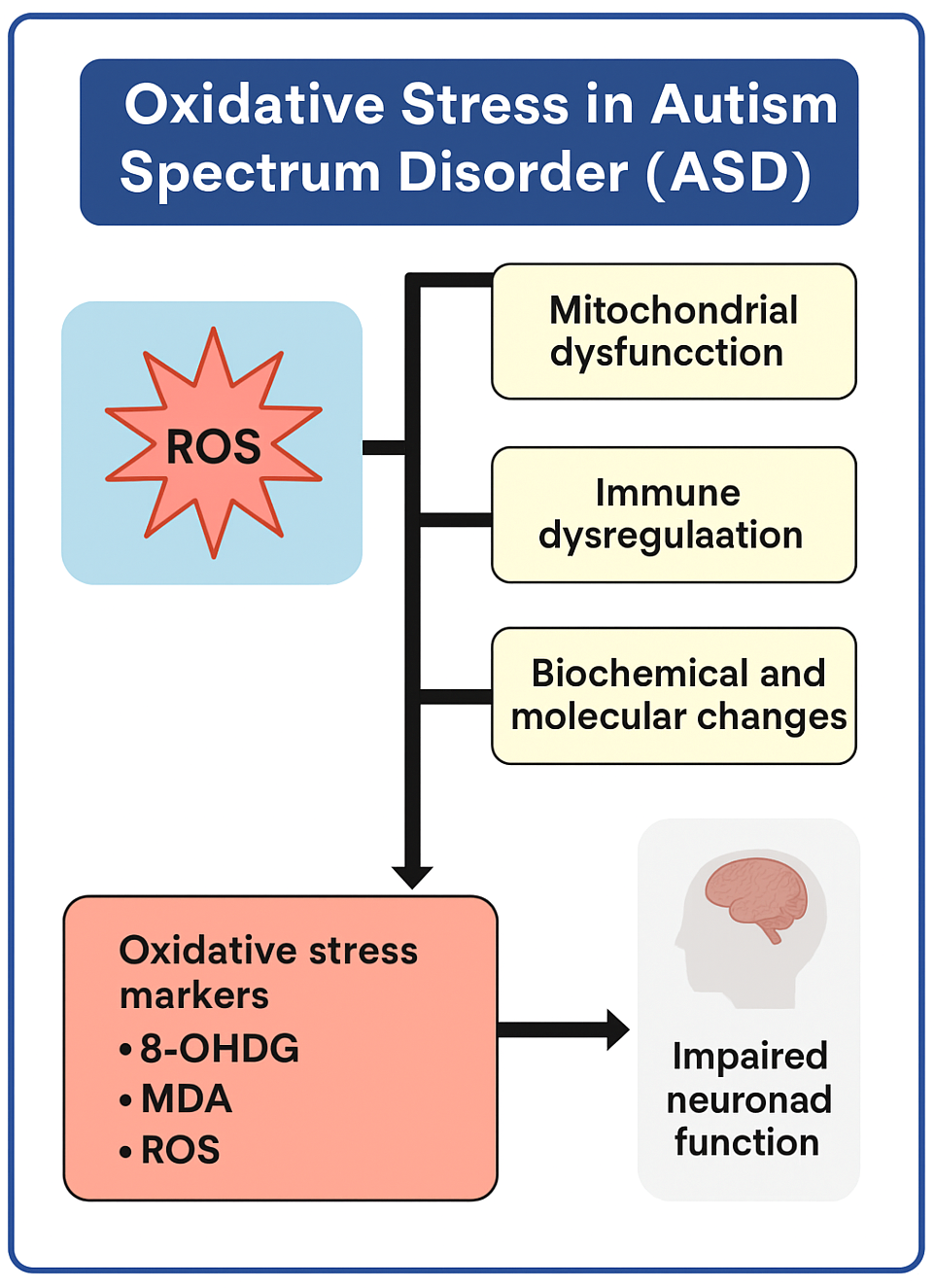
ASD, as a neurodevelopmental disorder, exhibits a wide range of phenotypic manifestations and a multifactorial nature. In its pathogenesis, an indispensable role of oxidative stress is anticipated according to empirical and theoretical evidence. Oxidative stress is a fundamental cellular process, and it entails the involvement of ROS. An increase in the levels of ROS, especially in mitochondria, disturbs organelle homeostasis and triggers the dysregulation of neurotransmitter metabolism, among other metabolic processes. These changes, which follow in these systems, along with immune dysregulation, lead to widespread biochemical and molecular illnesses. Interestingly, the biomarkers of oxidative stress, such as 8-hydroxy-2′-deoxyguanosine (8-OHdG), malondialdehyde (MDA), and ROS itself, are directly related to disturbances in neuronal functioning, which is very consistent with the cognitive impairment, speech problems, and behavioral abnormalities found in ASD (Figure 2).

3. Impact of Oxidative Stress on Speech and Language Development

Oxidative stress can affect the PFC and the temporal lobe, which are important for speech and language processing. If these areas are affected, then understanding and processing language become harder.
When the brain’s defense systems, such as the blood–brain barrier, meninges, and neuroimmune system in the form of microglia, are overstretched or compromised, it causes the release of free radicals, leading to oxidative stress. This can result in serious damage to vital areas such as the PFC, hippocampus, and amygdala. This damage can propagate neurodegenerative conditions such as Alzheimer’s disease and Parkinson’s disease alongside other neuropsychiatric and neurodevelopmental disorders such as depression, anxiety, schizophrenia, and autism by impairing cognition, memory, intellectual, social, and emotional regulation [35,36]. Oxidative damage is worsened by chronic stress and causes severe damage to the brain’s plasticity. Antioxidant defense systems such as superoxide dismutase, catalase, and glutathione help upregulate the Nrf2-ARE pathway. This upregulation leads to the activation of cellular defense mechanisms [37]. Because of these factors, treatments that target oxidative stress, such as antioxidants, are essential for preserving brain health. The impact of oxidative stress on the PFC is a reduction in the dendritic branching and connectivity, which ultimately leads to cognitive, emotional, and behavioral deficits present in autism. The existing reports do not explicitly point out the temporal lobe, but they do note oxidative damage in the hippocampus, which is associated with learning and memory, that may interfere with language functions in autism. Heightened levels of ROS, neuroinflammation, and the depletion of glutathione fuel these deficits, which indicate the importance of the efficacy of antioxidant therapies toward oxidative stress-induced brain dysfunctions in autism [38,39]. The temporal lobe and PFC, key regions for cognition, emotional control, and language, are compromised by oxidative stress. In ASD, oxidative alterations to the PFC lead to the disruption of a person’s executive functions, which include decision-making, impulse control, attention, social interaction, and spontaneous flexibility in adjusting to novel circumstances. Mitochondrial dysfunction, coupled with irregular glutamate signaling, further exacerbates the impairment of emotion regulation and behavioral flexibility, which are fundamental characteristics of ASD. Moreover, oxidative stress within the temporal lobe, including Wernicke’s area, compromises the ability to comprehend speech and hinders one’s capacity for verbal expression, resulting in speech delays and impaired auditory language processing [40]. These disabilities are frequently associated with sensory processing disorders as oxidative stress impacts hearing ability, making it challenging for people suffering from autism to filter, comprehend speech, and interpret surrounding sounds accurately [11]. Studies suggest that people with ASD have an increased concentration of ROS and a decreased reserve of antioxidants, thereby being easily susceptible to neuronal damage in certain vital areas of the brain. Hence, oxidative stress might be the reason for the prominent features of autism, such as language impairment, problems with social communication, and rigidity of thought. Some reports indicate that administering antioxidant medications may reverse the process of oxidative damage and subsequently improve the ability to process language, control higher mental functions, and regulate the senses in children with ASD [41].

4. Mitochondrial Dysfunction in ASD-Related Neurological Disorders

Mitochondria are crucial for producing energy within the cell. ASD is characterized by mitochondrial dysfunction that leads to decreased ATP production and an increase in ROS and reactive nitrogen species (RNS). This dysfunction originates from abnormalities in the electron transport chain (ETC) components and disturbances in calcium homeostasis, which increase oxidative stress and programmed cell death in neurons [42]. Neurodevelopmental processes, particularly those crucial for speech and language, may be impacted by oxidative stress and mitochondrial dysfunction. Increased oxidative stress in ASD children is shown by the significant reduction of glutathione (GSH) coupled with increased ROS, which has been correlated with communication deficits, specifically speech and language skills. This may result from injury to Broca’s and Wernicke’s areas or nonspecific developmental retardation due to oxidative injury of the neural tissues. In a paper published by Siddiqui et al., the authors have explored the increasing evidence of the link between ASD, a neurodevelopmental disorder that exhibits social communication deficits and repetitive behaviors, and abnormalities of the mitochondria [43]. Research has shown that individuals with ASD tend to be less physically active and to be prone to compromised energy metabolism and energy utilization when compared to the rest of the healthy population. This imbalance in energy metabolism is particularly reflected in the brain’s frontal and cerebellum regions, where there is diminished activity of the mitochondrial ETC complexes, as mitochondria are the epicenter of energy generation and are highly influenced by the individual’s physical activity. Because of that, there is a low output of ATP. Further, analyses of gene expression profiling have indicated the downregulation of the genes controlling energy metabolism within the mitochondria [44]. Moreover, there is increased oxidative stress, which indicates higher levels of cellular injury due to the excess ROS. In addition to that, there is evidence of altered mitochondrial DNA (mtDNA) copy number mutations and deletion in ASD patients, which suggests possible genetic factors responsible for the mitochondrial deficits [45]. Nevertheless, all these facts together have not fully elucidated the issue due to methodological inconsistencies and limited sample sizes, which make it unclear whether mitochondrial dysfunction is the cause or consequence of ASD. Expanding the sample size and utilizing advanced in vivo imaging modalities, such as near-infrared spectroscopy, would greatly enhance the understanding of the mitochondria’s role in the development of the disorder, as well as possible treatment strategies.
The complex issue of mitochondrial defects has become a key component of ASD. Research shows that approximately five percent of people suffering from ASD are considered to have met the criteria for mitochondrial disease (MD), which is much more than the general population’s prevalence of 0.01%. Even beyond the diagnostic criteria set for MD, a considerable number of individuals with ASD display biochemical evidence of mitochondrial damage, such as increased lactate and pyruvate levels, lowered carnitine, and ETC impairment, all of which indicate altered bioenergetics [46]. Clinically, ASD patients suffering from mitochondrial damage often have developmental regression, seizures, motor delays, and intestinal problems that point to the fact that mitochondrial damage affects both the brain and the body systems in people with ASD [9]. Neuroimaging as well as post-mortem research substantiate this link by showing that there is a perturbation in oxidative metabolism, decreased production of ATP, and increased oxidative stress in the ASD brain. Although genetic mutations explain 21% of ASD cases with MD, environmental factors, especially immune dysregulation and oxidative damage, are now seen more as contributing to secondary mitochondrial dysfunction. Additionally, in vitro studies show increased excretion of ROS and lowered antioxidant abilities among people with ASD, pointing to the increased vulnerability of mitochondria. Even though there is no concrete or definite mitochondrial-targeted therapy yet available, there are preliminary indications that affected individuals may benefit from carnitine, coenzyme Q10, and B vitamins, which enhance mitochondrial activity, thus improving symptoms. Further details are provided in Section 7. Further studies are needed to determine whether mitochondrial impairment leads to primary pathology in ASD or is simply a secondary effect, and what could be the most important elements of intervention using mitochondrial targets.
Moreover, mitochondrial dysfunction in conjunction with oxidative stress plays a significant role in various ASD-related neurological disorders in terms of its genetic impact and autism-associated developmental and cognitive symptoms (Table 1). Hence, some diseases involving mitochondrial dysfunction have been shown to correlate with ASD in terms of pathology and symptoms. For example, Smith–Lemli–Opitz Syndrome (SLOS) is a developmental disorder caused by a mutation in 7-dehydrocholesterol (7DHC), which leads to reduced cholesterol synthesis and energy production. This can impair proper brain development and may result in intellectual disability, a common symptom in ASD [47]. In addition, Helsmoortel–Van der Aa Syndrome (HVDAS) is considered a very rare neurodevelopmental disorder caused by an ANDP mutation, with possible manifestations such as developmental and intellectual delays, resembling ASD features. A small percentage of ASD patients (0.17%) also show a mutation in the ANDP gene [48]. Likewise, DiGeorge Syndrome (DGS) involves loss of function in the SLC25A gene, leading to mitochondrial dysfunction and energy imbalance. This disruption can slow down learning during childhood [49]. Similarly, the gene UBE3A, which is widely expressed in mitochondria and involved in brain development and synaptic plasticity, is linked to Angelman syndrome (AS). This creates an association between AS and ASD. Malfunction of this gene can hinder learning during neurodevelopmental stages [50]. Concerning ASD, Fragile X Syndrome (FXS) is another intellectual developmental disorder characterized by mutations in mitochondrial genes, including FMR1, which presents with neurodevelopmental and psychiatric challenges [51]. Rett syndrome, an X-linked neurodevelopmental disorder involving a dysfunctional MECP2 gene, is also commonly associated with ASD [52]. Cornelia de Lange Syndrome (CdLS) also exhibits autism-related features, such as impairments in verbal communication and a genetic abnormality involving TRMT61A protein mislocalization, ultimately affecting normal mitochondrial function [53]. Lastly, autism has also been linked to mutations or deletions in the SHANK3 gene, which have been reported in Phelan–McDermid Syndrome (PMS). These mutations are responsible for synaptic dysfunction in mitochondria [54].

5. Mitochondrial Dysfunction and Speech and Language Development

Disruption in speech and language development may occur due to mitochondrial dysfunction. Evidence shows that certain children with ASD and behavioral deficits in communication skills have low GSH levels alongside high ROS in mitochondria. This likely results from damage to the neural tissue processing language, which involves Broca’s and Wernicke’s areas, or generalized oxidative neural mitochondrial damage resulting in developmental delays. The genes associated with speech and language development can be upregulated or downregulated upon encountering oxidative stress and mitochondrial dysfunction. The generation of free radicals in the speech centers mentioned above could contribute to the degeneration or atrophy of these areas. This could result in severe impairment in the learning process of language or speech maintenance. Oxidative free radicals and inflammatory cytokines resulting from mitochondrial dysfunction also impact cognitive decline while verbal processing, thereby enhancing the brain’s vulnerability to ASD symptoms. Furthermore, dysfunctional mitochondria in these brain regions are responsible for the impaired or imbalanced generation of required energy for the propagation of vocal speech and language acquisition. Pregnant mothers consuming drugs or a diet rich in antioxidants have been reported to produce offspring with a better capacity to learn language from adults during the developmental period of sensory and motor learning of language [30,55].

6. Neuroinflammation in Autism Spectrum Disorder

Inflammation is a phenomenon common to both peripheral and central systems; however, when its epicenter is the brain, then it is regarded as neuroinflammation. Simultaneously, peripheral inflammation too often leads to neuroinflammatory conditions. Out of these, ASD has also been linked to this detrimental feature. This was reported to be associated with upregulated proinflammatory cytokines (IL-1, IL-6, IL-8, IL-12, IL-18, TNF, etc.) and downregulated anti-inflammatory cytokines (e.g., IL-4, IL-10, IL-13, TGF, etc.) [56]. These cytokines have the potential to cross the blood–brain barrier (BBB). The major brain regions in ASD prone to neuroinflammation are the cerebral cortex, hippocampus, amygdala, cerebellum, and white matter [57]. When these brain regions become overwhelmed by the mentioned inflammatory cytokines, then social, cognitive, language, and speech impairments come into play. This targeted action leads to a distorted lifestyle of ASD patients and a huge burden on caregivers. Other factors contributing to ASD-related neuroinflammation include microglial activation, autoimmune reaction, and genetic predisposition [58]. Even though these additional factors are indirectly related, they still play a significant role in comorbidity in ASD patients.
Furthermore, chronic neuroinflammation is seen in postmortem tissues of ASD patients’ brains, with microglial activation and high levels of proinflammatory cytokines reflecting a marker of persistent immune reaction [59].

6.1. Maternal Immune Activation-Mediated Neuroinflammation

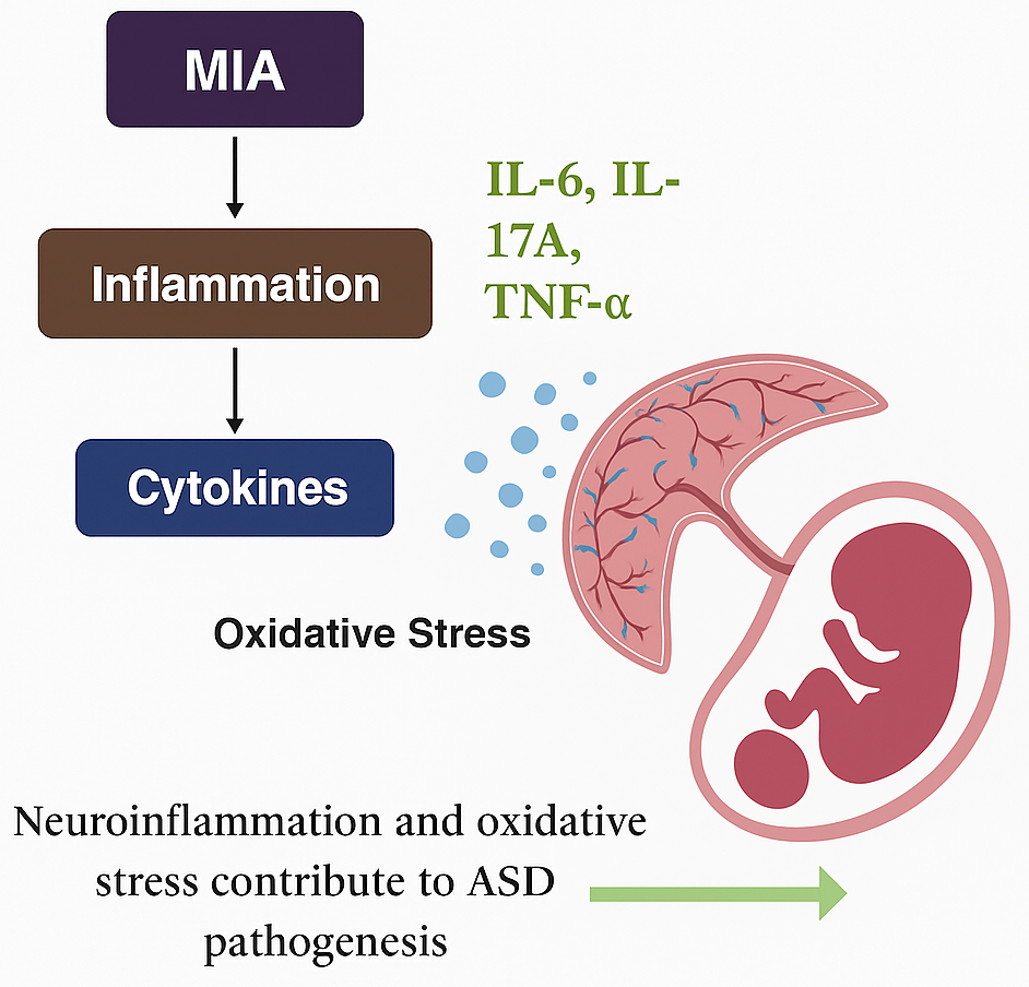
Maternal immune activation (MIA) is an environmental factor that influences the risk of ASD and occurs when a mother develops an immune response during pregnancy because of an infection, autoimmune disease, or chronic inflammation [60]. This response triggers the secretion of proinflammatory cytokines, including IL-6, IL-17A, and TNF-α, which can cross the placenta and disrupt fetal brain development [61]. MIA can disrupt neurogenesis, synaptic growth, and neurotransmitter balance and would result in ASD-like traits [62] (Figure 3). Social emulation, repetitive behaviors, and abnormalities of sensation are the most frequent types of such behaviors. In addition, prenatal brain oxidative stress, which serves as one of the principal etiological factors of autism, is associated with significant mitochondrial damage that further worsens the neuronal assault and amplifies the central neural circuit’s destruction [63]. Emerging evidence from studies conducted on both animals and humans indicates that inflammatory processes caused by maternal immune activation can potentially be modified in a way that reduces the chances of suffering from ASD or its symptoms. Experimental studies on animals reveal that the overstimulation of proinflammatory cytokines and their receptors at critical stages of fetal brain formation can damage the cortical structure and result in permanent behavioral disturbances such as those exhibited by patients with ASD.

6.2. Microglial Activation

Microglia are resident immune cells of the brain, responsible for defending from brain infections and clearing cellular waste. Upon activation, they also trigger the release of inflammatory mediators, paving the way to neuroinflammation. Every case of ASD has a common manifestation of neuroinflammation. This also involves the brain microglial cells, leading to chronic inflammation and oxidative damage. In this regard, microglial activation leads to oxidative stress via the NADPH oxidase (NOX) enzyme, more precisely the NOX2 isoform being overexpressed on microglial cells, facilitating the production of the superoxide free radical [64]. The heightened oxidative stress can further lead to microglia-mediated neuroinflammation and vice versa, propagating the cycle to continue. Such processes are regarded as neurotoxicity and are detrimental to developing brains [65].
Microglia also express GABA receptors, and hence, dysregulated expression of the receptors results in abnormal cortical development and the underdevelopment of GABAergic neurons, which are crucial for inhibitory neurotransmission, and, ultimately, an imbalance in excitatory/inhibitory signaling [66]. These activities can be a potential contributor to the pathogenesis of ASD.

7. Antioxidant, Mitochondrial, and Anti-Inflammatory Therapy

Antioxidants such as polyphenols and carotenoids have been widely investigated preclinically; however, translation to clinical levels is minimal. Exogenous antioxidants are often a good replacement when endogenous ones have been depleted due to any reason, for example, mitochondrial dysfunction or a dysregulated immune system. Antioxidant therapy could also be used as adjuvant therapy or monotherapy when required. However, these plant-derived therapies, including polyphenols, carotenoids, etc., have not been widely tested for their toxicity, thereby limiting their use clinically. On the other hand, the dose at which these preclinically tested substances show efficacy might not necessarily elicit the same response. These drawbacks emphasize the further investigations of antioxidant substances at in vitro and in vivo levels. Some of the extensively recognized naturally occurring antioxidants include vitamins, flavonoids, anthocyanins, carotenoids, etc. [27,67].
Some other substances, such as thiol antioxidants (N-acetyl cysteine), free radical scavengers (vitamin C), sulforaphane, and coenzyme Q10, also seem to reduce tissue oxidation, inflammation, and complex autism symptoms [68]. Here, vitamin C acts by donating an electron to neutralize free radicals and also helps in initiating another antioxidant, vitamin E. Coenzyme Q10 is a well-known, naturally produced biochemical cofactor in the human body, having a role in antioxidant mechanisms, and is also available as a supplement when there is a deficiency. Furthermore, antioxidants affect several signaling pathways. For example, resveratrol targets NADPH oxidase to reduce ROS generation. In the category of carotenoids, beta-carotene, lutein, and lycopene are found in fruits and vegetables; hence, consumption by children might slow down the ASD-related oxidative stress. Dry fruits such as walnuts and almonds are also high sources of antioxidants in the form of omega-3 fatty acids. Other polyphenols including curcumin, ellagic acid, gallic acid, ferulic acid, and vanillic acid can also be investigated and formulated for their antioxidant action against autism [67].
Additionally, in the context of mitochondria, mitoquinone mesylate (mitoQ) and 2,2,6,6-Tetramethylpiperidine 1-oxyl (mitoTEMPO) have been reported to act as mitochondria-targeted antioxidants. Here, mitoQ functions by scavenging ROS; on the other hand, mitoTEMPO is an SOD-mimicking compound, thereby reducing the overall oxidative damage [69]. Interestingly, peptides have been designed to navigate proteins or other therapeutic molecules towards the mitochondria to fix the mitochondrial dysfunction. Examples of mitochondria-targeting peptides include Szeto–Schiller peptides, self-assembled peptide nanomaterials, mitochondria-penetrating peptides, etc. [70,71].
In terms of anti-inflammatory agents to be repurposed or to potentially be used for ASD, there is a long list, but some of the important ones are corticosteroids such as prednisolone, NSAIDs including celecoxib, etc. Tetracycline, like minocycline, has also shown promising immunoregulatory and anti-inflammatory actions [72]. Alternatively, several natural anti-inflammatory agents have shown effects at the preclinical stage and can be extended to the clinical level. For example, resveratrol, luteolin, and quercetin have been investigated to reduce proinflammatory cytokines in animal models of ASD. Some of the clinical studies have also shown that GSH, Vitamin C, and Palmitoylethanolamide can alleviate inflammatory conditions in autistic patients [73]. Furthermore, immune-regulated improvement in inflammatory conditions through intravenous immunoglobulins and B-cell targeted therapy with rituximab has also been beneficial to some extent. Additional research on other inflammatory signal blocking agents, such as anakinra (IL-1β inhibitor), and inflammasome blockers, such as colchicine, etc., will open the door for further potential anti-inflammatory ASD therapies [74].
Of note, IL-6 or IL-17A blocking during pregnancy has been shown to have a therapeutic benefit in reducing such effects through experimental intervention. In one notable study, Choi and collaborators (2016) demonstrated that the administration of neutralizing IL-17A monoclonal antibodies in pregnant mice exposed to MIA effectively prevented cortical malformations and limited the appearance of ASD-like behavior in the offspring [62]. Similarly, IL-6 signaling blockade has proven to be able to reduce behavioral deficits and neurodevelopmental abnormalities caused by prenatal inflammation [63]. For example, some interventions such as anti-inflammatory celecoxib and immunoregulatory prednisolone during pregnancy have been shown to block IL-17A and IL-6 cytokines, which seems to diminish the chances of the child exhibiting ASD-like behaviors [72].
These findings show that inflammation- and oxidation-focused therapies, including anti-inflammatory agents, probiotics, and nutritional antioxidants, can be used during pregnancy to potentially decrease the risk of ASD and support better development in affected children.

8. Environmental Factors in Autism Spectrum Disorder

Environmental sources have also been reported to trigger ASD-related pathophysiology. The exposure to these environmental culprits could be either to the pregnant mother or the newly born infant. Either way, it can adversely impact the neurodevelopmental process of the child. Environmental exposures in the form of heavy metals such as mercury, arsenic, and lead; pollution with particulate matter; and even maternal factors, can enhance ROS production, which together build oxidative stress and negatively affect early brain development, which is critical for language development [75]. Additionally, certain pesticides, maternal obesity, immune dysregulation, and diabetes mellitus can also predispose the fetus to etiological factors of ASD [76]. Moreover, infections and certain medications including antidepressants and antiepileptic drugs consumed during pregnancy can produce hazardous results to a child’s brain if they exhibit neurotoxicity [77]. Nutritional deficiency of certain essential minerals and vitamins can lead to severe consequences, such as diminished progression of the normal neurodevelopmental process. There is growing evidence that oxidative stress, mitochondrial dysfunction, and immune dysregulation are key components in the environmentally generated pathophysiology of ASD. Studies show that ASD patients exposed to a deleterious environment have increased chances of oxidative stress leading to damage to lipids, proteins, and DNA in important parts of the brain responsible for speech, social behavior, memory, and motor activities [78]. It is widely known that the loss of function of the ETC, changes in mitochondrial gene expression, and reduced ATP synthesis occur with neurodevelopmental disabilities when mothers or children are exposed to harmful surroundings. Together, these abnormalities point to an underlying structure for ASD that may arise from preexisting environmentally induced factors. Although the specific causal mechanisms are unknown, this collection of literature amplifies the need for deliberately focused therapies to reverse the damage sustained by the mitochondria, decrease oxidative injury, and alter the level of inflammation in the brain to improve the condition of individuals with ASD.

9. Gut–Brain Axis in Autism Spectrum Disorder

The gut microbiome has been reported to impact brain health to cause many neurological disorders, including ASD. The gut bacteria have the potential to release neurotransmitters and hence influence brain function [79]. These microorganisms also influence the immune system and inflammatory process, thereby worsening the patient’s capacity to defend against the harmfulness of the bacteria. Furthermore, dietary imbalance can also destroy the flora of the gut, and even useful bacteria may lose their abundance [80]. This could aggravate the brain’s protective mechanisms, and hence, more complications with dysbiosis will arise. These disturbances could appear even in the form of oxidative stress as well, as there will be energy metabolism dysregulation propagated by mitochondrial dysfunction of the gut. Children with immunodeficiency and those who often suffer from diarrhea may lose essential nutrients and microbiome and, hence, are more prone to gut-related brain dysfunction and even downregulation of the genes responsible for social communication and speech development [81]. Hence, proper maintenance of a diet moderately cooked and loaded with essential nutrients is necessary for the smooth functioning of the gut–brain axis.
In addition to food, certain drugs can also hurt the gut flora, resulting in repetitive or restrictive behavior in children. Probiotics, prebiotics, and synbiotics can have a positive influence; on the other hand, antibiotics can initiate side effects. Interestingly, ASD patients taking medications for symptomatic relief should be cautious in preventing drug interactions and drug-induced microbial flora deteriorations. Additionally, the maternal gut flora can also influence the offspring depending on the placental permeability of drugs or foods [82].
In a recent report published in Nature Neuroscience, a temporal and bidirectional relationship between microbiome composition and ASD phenotypes was found. These patterns were depicted in age-matched and sex-matched cohorts through dietary profiles, metabolomics, and inflammatory cytokines [83]. This further strengthened the concept of the gut–brain axis role in ASD symptoms.
Connecting the gut relations with brain oxidative stress, mitochondrial dysfunction, and neuroinflammation, it has long been hypothesized that an unbalanced diet or dysbiosis may have potential contributions to generating oxidative stress in the brain. High-fat diets rich in cholesterol and LDL are the risky ingredients that play a role in brain oxidative damage and immune-dysregulated neuroinflammation. These factors suggest a strong gut–brain axis role in ASD.

10. Genetics and Epigenetics in Autism Spectrum Disorder

The early stage of brain development is highly influenced by environmental factors, which determine the expression levels of genes responsible for speech, language, communication, intellectual ability, and social behavior. These changes in the gene parameters are regarded as epigenetics and can be considered a novel trend and target in ASD research. Histone modifications with histone deacetylases and DNA methylation of genes with methyltransferases can lead to the development of autistic traits and social isolation behavior.
In the context of the present review, oxidative stress and proinflammatory cytokines have been noted to alter histone acetylation and DNA methylation, predisposing to the genetic changes associated with ASD-related pathology. Hence, these epigenetic changes can be reliable pathways to target ASD with the purpose of therapeutic interventions. Furthermore, epigenetic alterations in mitochondrial DNA have been investigated to have a greater influence on initiating the ASD-related dysfunction of energy metabolism. This can delay or halt the energy production necessary for the normal functioning of the brain in intellectual tasks such as focus, attention, thinking, and even the communication and development of speech and language centers in the brain.
Furthermore, an opinion piece has discussed gene polymorphisms resulting from dietary variation. This variation could be in the form of polymorphisms in the methylenetetrahydrofolate reductase (MTHFR) C677T gene in ASD children who lacked folic acid fortification in their diet. Additionally, microRNA dysregulation, transgenerational inheritance, increased expression of Tet methylcytosine dioxygenases, hypomethylation in the MAP8KIP3 or NALP1L5 gene, and downregulation of DNA methyltransferase 1 were also found to be associated with autism-related disorders [84].
Several other genes that play a role in the pathogenesis of ASD include SHANK3, MECP2, YTHDF, FRMPD4, FOXP2, PTCHD, HOX, CHD8, etc. These genes have been widely reported to play roles in neurodevelopmental features. For example, FOXP2 has a role in speech and language development, where it is downregulated in children with ASD speech difficulty. Similarly, either the upregulation or downregulation of these genes can have a role in features such as nerve cell communication, intellectual development, neurotransmitter release, and others [85,86,87].
Polymorphisms of genes that are part of antioxidant pathways, including glutathione peroxidase (GPx) and superoxide dismutase (SOD), are just a couple of genetic factors that make some people more susceptible to oxidative stress [68].

11. Recent Improvements in the Evaluation and Management of Autism Spectrum Disorders: Diagnosis and Treatment

ASD refers to a wide range of neurodevelopmental disorders defined by deficits in social interactions, communication skills, and other sensory behaviors. Increased efforts in biomarker discovery may allow for the more accurate sub-typing of ASDs with specific targeted interventions (Table 2).
Table 2. Gene mutations and genetic targets involved in ASD-related behavioral and neurological symptoms and possible treatment options.
Gene FunctionGenetic Variant/Mutation TypeBehavioral ChangesNeurological EffectsMolecular and Cellular AlterationsPossible Treatments
Dup 15q11-q13
[88]
Chromosome 15 portion duplicationReduced social interaction, fewer vocalizations, developmental delaysRigid behavior patternsImpaired serotonin signaling, enhanced spine densityBehavioral and speech therapy, no specific gene manipulation therapy
TBR11
[89]
Missense mutationReduced social interaction, altered food preference learning, intellectual disabilityRigid behavior, learning difficultiesAbnormal axonal links in amygdala,
reduced NMDAR activity
Lithium chloride, D-cycloserine rescues synaptic function in Tbr1 mutant mice
FMR1
[90]
Trinucleotide repeat expansionReduced social interaction, fewer vocalizationsRepetitive movements, hypersensitivity to sound, learning issuesDisrupted neural connectivity, increased mGluR activity, and replaced synaptic plasticityPirenperone reduces hyperactivity in Fmr1 KO mice
SHANK2
[91]
Frameshift mutationReduced social behaviors, hyperactivityRepetitive movements, cognitive difficultiesImpaired excitatory, synaptic dysfunctionDownstream of SHANK2 such as NMDA receptor and ERK pathway can be targeted
SHANK3
[92]
Frameshift mutationReduced social behaviorsExcessive grooming, anxietyInsufficient striatal activation and decreased AMPAR functionIGF1 enhances long-term potentiation and motor function in SHANK-deficient mouse model
SCN1A
[93]
Missense mutationReduced social behaviorsSeizures, learning deficits, grooming repetitionReduced activity of inhibitory interneuronsClonazepam improves neurobehavioral activities by targeting the Scn1 gene in PFC
CNTNAP2
[94]
DeletionReduced social behaviors, intellectual slowness, fewer vocalizationsHyperactivity, repetitive grooming, seizuresReduced interneuron population, disrupted neuronal migrationRisperidone and Oxytocin alleviate repetitive behavior in Cntnap2 −/− mice
TSC1, TSC2
[95]
Loss-of-function mutationReduced social interaction, increased vocalizationsEnhanced repetitive behaviors, coordination difficulties, cognitive impairmentEnlarged brain size, overactive mTOR signaling, impaired autophagyRapamycin targets mTOR (overactivated by Tsc1 and tsc2) and hence decreases ASD neuropathology
PTEN
[96]
Frameshift mutationReduced social interactionLearning impairment, seizures, anxietyAbnormal neuronal growth, PI3K pathway overactivationRapamycin improves social and stereotypic behavior in Pten KO mice
NLGN3
[97]
Point mutationReduced social interaction, fewer vocalizationsIncreased motor activityIncreased mTOR/Akt activation, impaired GABAergic transmission in striatal neurons, synaptic dysfunctionThere are no specific drugs to target Nlgn3, but rapamycin can be tested
NRXN1A
[98]
Exonic deletionReduced social behaviors, aggressionMotor learning issues, sensory processing deficits, spatial learning deficitsLowered glutamate transmission, decreased synaptic densityNo specific drugs, but extensive research is ongoing to target Nrxn1
MECP2
[99]
Loss-of-function mutationReduced social interactionRepetitive movements, motor deficits, seizuresSynaptic dysfunction, increased microglia activation,
BDNF activation
IGF1, Clenbuterol, Fingolimod can enhance neuronal plasticity by targeting Mecp2
CX3CR1
[100]
DeletionReduced social interactionImpairment in learning and memory, anxietyImpaired synaptic pruning, overactive microglia, neuroinflammationMicroglial modulators, E6130 as anti-inflammatory, and AZD8797 to inhibit CX3CR1
CHD8
[101]
Loss-of-function mutationSocial deficit, repetitive behaviorRepetitive grooming, increased brain size, learning impairmentDisrupted neuronal differentiation, abnormal cortical developmentFluoxetine partially restores neurogenesis in CHD8-ablated mice
TCF4
[102]
Point mutationRestlessnessAbnormal neuronal migration and excitabilityImpaired neuronal plasticity, altered brain connectivityNicradipine improves learning, memory, and restlessness in TCF4 +/− mice 
EIF4E KO
[103]
Missense mutationReduced sociabilitySelf-grooming, contextual fear memoryDysregulated protein synthesis, abnormal synaptic functionMnk inhibitors dephosphorylate EIF4E to downregulate it
It is widely accepted that ASD stems from multiple causes, both of genetic as well as environmental origin. Newer research suggests the contribution of monogenic mutations (e.g., SHANK3, FMR1, MECP2) and polygenic interactions that result in synaptic dysfunction and other neurodevelopmental insults [104]. These genetic challenges could have possible links to the mentioned features of ROS, impaired energy metabolism, and inflammatory cytokines. Genetic analysis of ASD-specific genes allows the identification of risk-associated variants. Hence, the early detection and selective targeting of these genes for their upregulation or downregulation might be a significant step towards the evaluation and management of ASD. Maternal infections, toxin exposure during prenatal stages, and inadequate nutrition are other factors that aggravate the risk of ASD, indicating the gene–environment interaction [105]. Therefore, confinement and avoidance of the mother during pregnancy and even after the birth of the child from these environmental hazards is necessary to reduce the chances of the child being prone to ASD pathology. Furthermore, the development and refinement of technologies geared towards diagnosis have made quantitative improvements in the accuracy of identifying patients with conditions and implementing relevant therapies in those patients. Commonly used neuroimaging techniques including fMRI, DTI, and PET scans aid in the diagnosis of structural and functional abnormalities within the brain tissues of an ASD patient [106]. These approaches to depict brain regions specific for intellectual, communication, and vocal centers are crucial in uncovering brain atrophy, apoptosis, the deposition of certain plaques, or changes in symmetry impacted by phenomena such as oxidative stress, mitochondrial dysfunction, and neuroinflammation. All these outcomes could be contributing to the determination of ASD-related pathology and possible treatments to halt or reverse it. Moreover, AI-based screening and eye-tracking devices are other non-invasive modes of assisting in the early steps of ASD identification, enabling faster therapeutic actions to be taken [107]. Until recently, both behavioral interventions, including Applied Behavior Analysis (ABA) and social skills training, and their challenging techniques have always been the gold standard therapies for ASD [108]. Still, antipsychotic medications, notably risperidone and aripiprazole, are also commonly used to treat the violent and disruptive behavior patients may have [109]. Additionally, transcranial magnetic stimulation (TMS) and transcranial direct current stimulation (tDCS) are notable attempts at treating the abnormal neural circuits if distorted in ASD subjects [110]. Furthermore, therapies that focus on biofeedback try to improve self-regulation in patients suffering from ASD. There are also new virtual reality and AI-powered cognitive therapies that are more tailored to the patient’s needs. Diet and nutrition have recently become a focus as to what could prove useful in treating ASD. Research has shown promising effects on gluten- and casein-free diets, omega-3 supplementation, and targeted gut microbiome therapies to reduce symptoms related to ASD [111]. Many researchers have started paying attention to the role of the gut in the brain and vice versa, resulting in studies focused on probiotics, prebiotics, and fecal microbiota administration or ingestion as possible supplementary therapies for ASD [112]. Precision medicine, gene editing technologies (CRISPR-Cas9), and stem cell-based prospects have the potential for precision treatment and are where future research will be directed [113]. Additionally, more needs to be accomplished to encourage international cooperation, early intervention access, and public education to correct the imbalance in ASD diagnosis and treatment.

Author Contributions

Conceptualization, A.A.; investigation, S.B.R. and A.A.; resources, S.B.R.; writing—original draft preparation, S.B.R. and A.A.; writing—review and editing, A.A.; visualization, A.A.; supervision, A.A. All authors have read and agreed to the published version of the manuscript.

Funding

This review received no external funding.

Institutional Review Board Statement

Not applicable.

Informed Consent Statement

Not applicable.

Data Availability Statement

No new data were created or analyzed in this study.

Acknowledgments

We acknowledge LSU Health and Jamia Hamdard for infrastructure.

Conflicts of Interest

The authors declare no conflicts of interest.

Abbreviations

ASD: Autism Spectrum Disorder; PFC: Prefrontal Cortex; ROS: Reactive Oxygen Species; ETC: Electron Transport Chain; MDA: Malondialdehyde; 8-OHdG: 8-Hydroxy-2′-deoxyguanosine; GSH: Reduced Glutathione; GSSG: Oxidized Glutathione; CNS: Central Nervous System; GIT: Gastrointestinal Tract; mtDNA: Mitochondrial DNA; MIA: Maternal Immune Activation; IL: Interleukin; TNF-α: Tumor Necrosis Factor-alpha; BBB: Blood–Brain Barrier; TGF-β: Transforming Growth Factor-beta; HSP70: Heat Shock Protein 70; ATP: Adenosine Triphosphate; SOD: Superoxide Dismutase; GCLM: Glutamate-Cysteine Ligase Modifier Subunit; NAC: N-Acetyl Cysteine; DTI: Diffusion Tensor Imaging; fMRI: Functional Magnetic Resonance Imaging; PET: Positron Emission Tomography; TMS: Transcranial Magnetic Stimulation; tDCS: Transcranial Direct Current Stimulation; ABA: Applied Behavior Analysis; FMR1: Fragile X Mental Retardation 1 Gene; SHANK3: SH3 and Multiple Ankyrin Repeat Domains 3; MECP2: Methyl-CpG Binding Protein 2; CHD8: Chromodomain Helicase DNA Binding Protein 8; PTEN: Phosphatase and Tensin Homolog; SCN1A: Sodium Voltage-Gated Channel Alpha Subunit 1; CNTNAP2: Contactin Associated Protein-Like 2; NLGN3: Neuroligin 3; TSC1/2: Tuberous Sclerosis Complex 1 and 2; BDNF: Brain-Derived Neurotrophic Factor; IGF-1: Insulin-like Growth Factor 1; CRISPR: Clustered Regularly Interspaced Short Palindromic Repeats.

References

  1. St Amant, H.G.; Schrager, S.M.; Peña-Ricardo, C.; Williams, M.E.; Vanderbilt, D.L. Language barriers impact access to services for children with autism spectrum disorders. J. Autism Dev. Disord. 2018, 48, 333–340. [Google Scholar] [CrossRef]
  2. Deth, R.; Muratore, C.; Benzecry, J.; Power-Charnitsky, V.-A.; Waly, M. How environmental and genetic factors combine to cause autism: A redox/methylation hypothesis. Neurotoxicology 2008, 29, 190–201. [Google Scholar] [CrossRef] [PubMed]
  3. Bjørklund, G.; Meguid, N.A.; El-Bana, M.A.; Tinkov, A.A.; Saad, K.; Dadar, M.; Hemimi, M.; Skalny, A.V.; Hosnedlová, B.; Kizek, R.; et al. Oxidative stress in autism spectrum disorder. Frye 2020, 57, 2314–2332. [Google Scholar] [CrossRef] [PubMed]
  4. Kuźniar-Pałka, A. The Role of Oxidative Stress in Autism Spectrum Disorder Pathophysiology, Diagnosis and Treatment. Biomedicines 2025, 13, 388. [Google Scholar] [CrossRef] [PubMed]
  5. Curpan, A.S.; Luca, A.-C.; Ciobica, A.; Mosca, L. Potential novel therapies for neurodevelopmental diseases targeting oxidative stress. Oxidative Med. Cell. Longev. 2021, 2021, 6640206. [Google Scholar] [CrossRef]
  6. Manivasagam, T.; Arunadevi, S.; Essa, M.M.; SaravanaBabu, C.; Borah, A.; Thenmozhi, A.J.; Qoronfleh, M.W. Role of oxidative stress and antioxidants in autism. Pers. Food Interv. Ther. Autism Spectr. Disord. Manag. 2020, 24, 193–206. [Google Scholar]
  7. Membrino, V.; Di Paolo, A.; Alia, S.; Papiri, G.; Vignini, A. The Role of Oxidative Stress in Autism Spectrum Disorder: A Narrative Literature Review. Oxygen 2023, 3, 34–44. [Google Scholar] [CrossRef]
  8. Del Casale, A.; Ferracuti, S.; Alcibiade, A.; Simone, S.; Modesti, M.N.; Pompili, M. Neuroanatomical correlates of autism spectrum disorders: A meta-analysis of structural magnetic resonance imaging (MRI) studies. Psychiatry Res. Neuroimaging 2022, 325, 111516. [Google Scholar] [CrossRef]
  9. Frye, R.E. Mitochondrial dysfunction in autism spectrum disorder: Unique abnormalities and targeted treatments. In Seminars in Pediatric Neurology; WB Saunders: Philadelphia, PA, USA, 2020; Volume 35, p. 100829. [Google Scholar]
  10. Liu, X.; Lin, J.; Zhang, H.; Khan, N.U.; Zhang, J.; Tang, X.; Cao, X.; Shen, L. Oxidative stress in autism spectrum disorder—Current progress of mechanisms and biomarkers. Front. Psychiatry 2022, 13, 813304. [Google Scholar] [CrossRef]
  11. Rose, S.; Melnyk, S.; Pavliv, O.; Bai, S.; Nick, T.G.; E Frye, R.; James, S.J. Evidence of oxidative damage and inflammation associated with low glutathione redox status in the autism brain. Transl. Psychiatry 2012, 2, 134. [Google Scholar] [CrossRef]
  12. Yui, K.; Sato, A.; Imataka, G. Mitochondrial dysfunction and its relationship with mTOR signaling and oxidative damage in autism spectrum disorders. Mini Rev. Med. Chem. 2015, 15, 373–389. [Google Scholar] [CrossRef] [PubMed]
  13. E Frye, R.; DeLaTorre, R.; Taylor, H.; Slattery, J.; Melnyk, S.; Chowdhury, N.; James, S.J. Redox metabolism abnormalities in autistic children associated with mitochondrial disease. Transl. Psychiatry 2013, 3, 273. [Google Scholar] [CrossRef] [PubMed]
  14. Steffensen, I.-L.; Dirven, H.; Couderq, S.; David, A.; D’cruz, S.C.; Fernández, M.F.; Mustieles, V.; Rodríguez-Carrillo, A.; Hofer, T. Bisphenols and oxidative stress biomarkers—Associations found in human studies, evaluation of methods used, and strengths and weaknesses of the biomarkers. Int. J. Environ. Res. Public Health 2020, 17, 3609. [Google Scholar] [CrossRef] [PubMed]
  15. Kaur, K.; Chauhan, V.; Gu, F.; Chauhan, A. Bisphenol A induces oxidative stress and mitochondrial dysfunction in lymphoblasts from children with autism and unaffected siblings. Free Radic. Biol. Med. 2014, 76, 25–33. [Google Scholar] [CrossRef]
  16. Bjørklund, G.; Tinkov, A.A.; Hosnedlová, B.; Kizek, R.; Ajsuvakova, O.P.; Chirumbolo, S.; Skalnaya, M.G.; Peana, M.; Dadar, M.; El-Ansary, A.; et al. The role of glutathione redox imbalance in autism spectrum disorder: A review. Free Radic. Biol. Med. 2020, 160, 149–162. [Google Scholar] [CrossRef]
  17. Bjørklund, G.; Waly, M.I.; Al-Farsi, Y.; Saad, K.; Dadar, M.; Rahman, M.M.; Elhoufey, A.; Chirumbolo, S.; Jóźwik-Pruska, J.; Kałużna-Czaplińska, J. The role of vitamins in autism spectrum disorder: What do we know? J. Mol. Neurosci. 2019, 67, 373–387. [Google Scholar] [CrossRef]
  18. Rossignol, D.A.; Frye, R.E. Mitochondrial dysfunction in autism spectrum disorders: A systematic review and meta-analysis. Mol. Psychiatry 2012, 17, 290–314. [Google Scholar] [CrossRef]
  19. Habib, R.; Noureen, N.; Nadeem, N. Decoding Common Features of Neurodegenerative Disorders: From Differentially Expressed Genes to Pathways. Curr. Genom. 2018, 19, 300–312. [Google Scholar] [CrossRef]
  20. Tsilioni, I.; Taliou, A.; Francis, K.; Theoharides, T.C. Children with autism spectrum disorders, who improved with a luteolin-containing dietary formulation, show reduced serum levels of TNF and IL-6. Transl. Psychiatry 2015, 5, 647. [Google Scholar] [CrossRef]
  21. Castora, F.J. Mitochondrial function and abnormalities implicated in the pathogenesis of ASD. Prog. Neuro-Psychopharmacol. Biol. Psychiatry 2019, 92, 83–108. [Google Scholar] [CrossRef]
  22. Costa, L.G.; Chang, Y.C.; Cole, T.B. Developmental neurotoxicity of traffic-related air pollution: Focus on autism. Curr. Environ. Health Rep. 2017, 4, 156–165. [Google Scholar] [CrossRef] [PubMed]
  23. Zhang, Y.; Hodgson, N.W.; Trivedi, M.S.; Abdolmaleky, H.M.; Fournier, M.; Cuenod, M.; Do, K.Q.; Deth, R.C.; Bauer, J.A. Decreased brain levels of vitamin B12 in aging, autism and schizophrenia. PLoS ONE 2016, 11, e0146797. [Google Scholar] [CrossRef] [PubMed]
  24. Signorini, C.; De Felice, C.; Durand, T.; Galano, J.M.; Oger, C.; Leoncini, S.; Ciccoli, L.; Carone, M.; Ulivelli, M.; Manna, C.; et al. Relevance of 4-F4t-neuroprostane and 10-F4t-neuroprostane to neurological diseases. Free Radic. Biol. Med. 2018, 115, 278–287. [Google Scholar] [CrossRef] [PubMed]
  25. Ansari, M.A.; Scheff, S.W. Oxidative stress in the progression of Alzheimer disease in the frontal cortex. J. Neuropathol. Exp. Neurol. 2010, 69, 155–167. [Google Scholar] [CrossRef]
  26. Hollis, F.; Kanellopoulos, A.K.; Bagni, C. Kanellopoulos, and Claudia Bagni. Mitochondrial dysfunction in Autism Spectrum Disorder: Clinical features and perspectives. Curr. Opin. Neurobiol. 2017, 45, 178–187. [Google Scholar] [CrossRef]
  27. Liu, Y.; Yang, Z.; Du, Y.; Shi, S.; Cheng, Y. Antioxidant interventions in autism spectrum disorders: A meta-analysis. Prog. Neuro-Psychopharmacol. Biol. Psychiatry 2022, 113, 110476. [Google Scholar] [CrossRef]
  28. Gu, F.; Chauhan, V.; Chauhan, A. Impaired synthesis and antioxidant defense of glutathione in the cerebellum of autistic subjects: Alterations in the activities and protein expression of glutathione-related enzymes. Free Radic. Biol. Med. 2013, 65, 488–496. [Google Scholar] [CrossRef]
  29. González-Fraguela, M.E.; Hung, M.-L.D.; Vera, H.; Maragoto, C.; Noris, E.; Blanco, L.; Galvizu, R.; Robinson, M. Oxidative stress markers in children with autism spectrum disorders. Br. J. Med. Med. Res. 2013, 3, 307–317. [Google Scholar] [CrossRef]
  30. Chauhan, A.; Chauhan, V. Oxidative stress in autism. Pathophysiology 2006, 13, 171–181. [Google Scholar] [CrossRef]
  31. Yui, K.; Imataka, G.; Sasaki, H.; Shiroki, R. The role of lipid peroxidation in individuals with autism spectrum disorders. Metab. Brain Dis. 2020, 35, 1101–1108. [Google Scholar] [CrossRef]
  32. Zaky, E.A.; Elhameed, S.A.A.; Ismail, S.M.; Eldamer, N.M.; Abdelaziz, A.W. Analysis of urinary 8-hydroxy-2-deoxyguanosine as a biomarker of oxidative DNA damage in pediatric children with autism spectrum disorder. Res. Autism Spectr. Disord. 2023, 102, 102129. [Google Scholar] [CrossRef]
  33. Ming, X.; Stein, T.P.; Brimacombe, M.; Johnson, W.G.; Lambert, G.H.; Wagner, G.C. Increased excretion of a lipid peroxidation biomarker in autism. Prostaglandins Leukot. Essent. Fat. Acids 2005, 73, 379–384. [Google Scholar] [CrossRef]
  34. Feng, C.; Chen, Y.; Pan, J.; Yang, A.; Niu, L.; Min, J.; Meng, X.; Liao, L.; Zhang, K.; Shen, L. Redox proteomic identification of carbonylated proteins in autism plasma: Insight into oxidative stress and its related biomarkers in autism. Clin. Proteom. 2017, 14, 2. [Google Scholar] [CrossRef]
  35. Yenkoyan, K.; Harutyunyan, H.; Harutyunyan, A. A certain role of SOD/CAT imbalance in pathogenesis of autism spectrum disorders. Free Radic. Biol. Med. 2018, 123, 85–95. [Google Scholar] [CrossRef]
  36. Sajdel-Sulkowska, E.M.; Xu, M.; McGinnis, W.; Koibuchi, N. Brain region-specific changes in oxidative stress and neurotrophin levels in autism spectrum disorders (ASD). Cerebellum 2011, 10, 43–48. [Google Scholar] [CrossRef] [PubMed]
  37. Li, M.; Long, C.; Yang, L. Hippocampal-prefrontal circuit and disrupted functional connectivity in psychiatric and neurodegenerative disorders. BioMed Res. Int. 2015, 2015, 810548. [Google Scholar] [CrossRef] [PubMed]
  38. Nadeem, A.; Ahmad, S.F.; Al-Ayadhi, L.Y.; Attia, S.M.; Al-Harbi, N.O.; Alzahrani, K.S.; Bakheet, S.A. Differential regulation of Nrf2 is linked to elevated inflammation and nitrative stress in monocytes of children with autism. Psychoneuroendocrinology 2020, 113, 104554. [Google Scholar] [CrossRef] [PubMed]
  39. Modafferi, S.; Lupo, G.; Tomasello, M.; Rampulla, F.; Ontario, M.; Scuto, M.; Salinaro, A.T.; Arcidiacono, A.; Anfuso, C.D.; Legmouz, M.; et al. Antioxidants, hormetic nutrition, and autism. Curr. Neuropharmacol. 2024, 22, 1156–1168. [Google Scholar] [CrossRef]
  40. Penna, E.; Pizzella, A.; Cimmino, F.; Trinchese, G.; Cavaliere, G.; Catapano, A.; Allocca, I.; Chun, J.T.; Campanozzi, A.; Messina, G.; et al. Neurodevelopmental disorders: Effect of high-fat diet on synaptic plasticity and mitochondrial functions. Brain Sci. 2020, 10, 805. [Google Scholar] [CrossRef]
  41. McKenna, K.; Prasad, S.; Cooper, J.; King, A.M.; Shahzeidi, S.; Mittal, J.; Zalta, M.; Mittal, R.; Eshraghi, A.A. Incidence of Otolaryngological Manifestations in Individuals with Autism Spectrum Disorder: A Special Focus on Auditory Disorders. Audiol. Res. 2024, 14, 35–61. [Google Scholar] [CrossRef]
  42. Khaliulin, I.; Hamoudi, W.; Amal, H. The multifaceted role of mitochondria in autism spectrum disorder. Mol. Psychiatry 2024, 30, 629–650. [Google Scholar] [CrossRef]
  43. Siddiqui, M.F.; Elwell, C.; Johnson, M.H. Mitochondrial dysfunction in autism spectrum disorders. Autism-Open Access 2016, 6, 1000190. [Google Scholar] [CrossRef]
  44. Oyarzábal, A.; Musokhranova, U.; Barros, L.F.; García-Cazorla, A. Energy metabolism in childhood neurodevelopmental disorders. EBioMedicine 2021, 69, 103474. [Google Scholar] [CrossRef]
  45. Varga, N.Á.; Pentelényi, K.; Balicza, P.; Gézsi, A.; Reményi, V.; Hársfalvi, V.; Bencsik, R.; Illés, A.; Prekop, C.; Molnár, M.J. Mitochondrial dysfunction and autism: Comprehensive genetic analyses of children with autism and mtDNA deletion. Behav. Brain Funct. 2018, 14, 4. [Google Scholar] [CrossRef]
  46. Kępka, A.; Ochocińska, A.; Chojnowska, S.; Borzym-Kluczyk, M.; Skorupa, E.; Knaś, M.; Waszkiewicz, N. Potential role of L-carnitine in autism spectrum disorder. J. Clin. Med. 2021, 10, 1202. [Google Scholar] [CrossRef] [PubMed]
  47. Chang, S.; Ren, G.; Steiner, R.D.; Merkens, L.; Roullet, J.B.; Korade, Z.; DiMuzio, P.J.; Tulenko, T.N. Elevated autophagy and mitochondrial dysfunction in the Smith–Lemli–Opitz syndrome. Mol. Genet. Metab. Rep. 2014, 1, 431–442. [Google Scholar] [CrossRef] [PubMed]
  48. Alishvandi, A.; Hanaei, S. Helsmoortel-Van Der Aa Syndrome (HVDAS). In Genetic Syndromes: A Comprehensive Reference Guide; Springer Nature: Cham, Switzerland, 2025; pp. 1–6. [Google Scholar]
  49. Kasprzyk-Pawelec, A.; Tan, M.; Rahhal, R.; McIntosh, A.; Fernandez, H.R.; Mosaoa, R.M.; Jiang, L.; Pearson, G.W.; Glasgow, E.; Vockley, J.; et al. Inactivation of the SLC25A1 gene during embryogenesis induces a unique senescence program controlled by p53. Cell Death Differ. 2024, 32, 818–836. [Google Scholar] [CrossRef] [PubMed]
  50. Gupta, P.K.; Feuermann, Y.; Kaphzan, H. Dynamic shift in localization of UBE3A across developmental stages in an Angelman syndrome mouse model. Neurobiol. Dis. 2025, 210, 106912. [Google Scholar] [CrossRef]
  51. Caron, C.; McCullagh, E.A.; Bertolin, G. Sex-specific loss of mitochondrial membrane integrity in the auditory brainstem of a mouse model of Fragile X Syndrome. Open Biol. 2025, 15, 240384. [Google Scholar] [CrossRef]
  52. Mietto, M.; Montanari, S.; Falzarano, M.S.; Manzati, E.; Rimessi, P.; Fabris, M.; Selvatici, R.; Gualandi, F.; Neri, M.; Fortunato, F.; et al. MECP2 mRNA Profile in Brain Tissues from a Rett Syndrome Patient and Three Human Controls: Mutated Allele Preferential Transcription and In Situ RNA Mapping. Biomolecules 2025, 15, 687. [Google Scholar] [CrossRef]
  53. Uwibambe, E.; Yalcouyé, A.; Aboagye, E.T.; Xhakaza, L.; Popel, K.; Dukuze, N.; Bharadwaj, T.; de Kock, C.; Schrauwen, I.; Leal, S.M.; et al. Exome sequencing revealed a novel homozygous variant in TRMT61 A in a multiplex family with atypical Cornelia de Lange Syndrome from Rwanda. BMC Med. Genom. 2025, 18, 85. [Google Scholar] [CrossRef] [PubMed]
  54. Giona, F.; Beretta, S.; Zippo, A.; Stefanoni, A.; Tomasoni, Z.; Vicidomini, C.; Ponzoni, L.; Sala, M.; Jones, C.K.; Conn, P.J.; et al. Shank3 modulates Rpl3 expression and protein synthesis via mGlu5: Implications for Phelan McDermid syndrome. Mol. Psychiatry 2025, 30, 3599–3614. [Google Scholar] [CrossRef] [PubMed]
  55. Rossignol, D.A.; Frye, R.E. Evidence linking oxidative stress, mitochondrial dysfunction, and inflammation in the brain of individuals with autism. Front. Physiol. 2014, 5, 150. [Google Scholar] [CrossRef] [PubMed]
  56. Nadeem, A.; Ahmad, S.F.; Al-Harbi, N.O.; Al-Ayadhi, L.Y.; Sarawi, W.; Attia, S.M.; Bakheet, S.A.; Alqarni, S.A.; Ali, N.; AsSobeai, H.M. Imbalance in pro-inflammatory and anti-inflammatory cytokines milieu in B cells of children with autism. Mol. Immunol. 2022, 141, 297–304. [Google Scholar] [CrossRef]
  57. Rexrode, L.E.; Hartley, J.; Showmaker, K.C.; Challagundla, L.; Vandewege, M.W.; Martin, B.E.; Blair, E.; Bollavarapu, R.; Antonyraj, R.B.; Hilton, K.; et al. Molecular profiling of the hippocampus of children with autism spectrum disorder. Mol. Psychiatry 2024, 29, 1968–1979. [Google Scholar] [CrossRef]
  58. Hughes, H.K.; RJMoreno Ashwood, P. Innate immune dysfunction and neuroinflammation in autism spectrum disorder (ASD). Brain Behav. Immun. 2023, 108, 245–254. [Google Scholar] [CrossRef]
  59. Saghazadeh, A.; Ataeinia, B.; Keynejad, K.; Abdolalizadeh, A.; Hirbod-Mobarakeh, A.; Rezaei, N. A meta-analysis of pro-inflammatory cytokines in autism spectrum disorders: Effects of age, gender, and latitude. J. Psychiatr. Res. 2019, 115, 90–102. [Google Scholar] [CrossRef]
  60. Gilbert, J.; Man, H.Y. Fundamental elements in autism: From neurogenesis and neurite growth to synaptic plasticity. Front. Cell. Neurosci. 2017, 11, 359. [Google Scholar] [CrossRef]
  61. Rose, S.; Niyazov, D.M.; Rossignol, D.A.; Goldenthal, M.; Kahler, S.G.; Frye, R.E. Clinical and molecular characteristics of mitochondrial dysfunction in autism spectrum disorder. Mol. Diagn. Ther. 2018, 22, 571–593. [Google Scholar] [CrossRef]
  62. Choi, G.B.; Yim, Y.S.; Wong, H.; Kim, S.; Kim, H.; Kim, S.V.; Hoeffer, C.A.; Littman, D.R.; Huh, J.R. The maternal interleukin-17a pathway in mice promotes autism-like phenotypes in offspring. Science 2016, 351, 933–939. [Google Scholar] [CrossRef]
  63. Smith, S.E.P.; Li, J.; Garbett, K.; Mirnics, K.; Patterson, P.H. Maternal immune activation alters fetal brain development through interleukin-6. J. Neurosci. 2007, 27, 10695–10702. [Google Scholar] [CrossRef]
  64. Bordt, E.A.; Polster, B.M. NADPH oxidase-and mitochondria-derived reactive oxygen species in proinflammatory microglial activation: A bipartisan affair? Free Radic. Biol. Med. 2014, 76, 34–46. [Google Scholar] [CrossRef]
  65. Bilbo, S.D.; Block, C.L.; Bolton, J.L.; Hanamsagar, R.; Tran, P.K. Beyond infection-Maternal immune activation by environmental factors, microglial development, and relevance for autism spectrum disorders. Exp. Neurol. 2018, 299, 241–251. [Google Scholar] [CrossRef]
  66. Di, J.; Li, J.; O’hAra, B.; Alberts, I.; Xiong, L.; Li, J.; Li, X. The role of GABAergic neural circuits in the pathogenesis of autism spectrum disorder. Int. J. Dev. Neurosci. 2020, 80, 73–85. [Google Scholar] [CrossRef]
  67. Pangrazzi, L.; Balasco, L.; Bozzi, Y. Natural antioxidants: A novel therapeutic approach to autism spectrum disorders? Antioxidants 2020, 9, 1186. [Google Scholar] [CrossRef]
  68. Frustaci, A.; Neri, M.; Cesario, A.; Adams, J.B.; Domenici, E.; Bernardina, B.D.; Bonassi, S. Oxidative stress-related biomarkers in autism: Systematic review and meta-analyses. Free Radic. Biol. Med. 2012, 52, 2128–2141. [Google Scholar] [CrossRef] [PubMed]
  69. Liang, Z.; Zhang, N.; Wang, X.; Zhang, J.; Li, K.; Lei, T. Epothilone B inactivation of Sirtuin1 promotes mitochondrial reactive oxygen species to induce dysfunction and ferroptosis of Schwann cells. Eur. J. Pharm. Sci. 2023, 181, 106350. [Google Scholar] [CrossRef] [PubMed]
  70. Kim, S.; Nam, H.Y.; Lee, J.; Seo, J. Mitochondrion-targeting peptides and peptidomimetics: Recent progress and design principles. Biochemistry 2019, 59, 270–284. [Google Scholar] [CrossRef]
  71. Luo, Z.; Gao, Y.; Duan, Z.; Yi, Y.; Wang, H. Mitochondria-targeted self-assembly of peptide-based nanomaterials. Front. Bioeng. Biotechnol. 2021, 9, 782234. [Google Scholar] [CrossRef] [PubMed]
  72. rteaga-Henríquez, G.; Gisbert, L.; Ramos-Quiroga, J.A. Immunoregulatory and/or anti-inflammatory agents for the management of core and associated symptoms in individuals with autism spectrum disorder: A narrative review of randomized, placebo-controlled trials. CNS Drugs 2023, 37, 215–229. [Google Scholar] [CrossRef] [PubMed]
  73. Singh, R.; Kisku, A.; Kungumaraj, H.; Nagaraj, V.; Pal, A.; Kumar, S.; Sulakhiya, K. Autism spectrum disorders: A recent update on targeting inflammatory pathways with natural anti-inflammatory agents. Biomedicines 2023, 11, 115. [Google Scholar] [CrossRef] [PubMed]
  74. Jyonouchi, H. Autism spectrum disorder and a possible role of anti-inflammatory treatments: Experience in the pediatric allergy/immunology clinic. Front. Psychiatry 2024, 15, 1333717. [Google Scholar] [CrossRef]
  75. Bellinger, D.C.; Needleman, H.L. Prenatal and early postnatal exposure to lead: Developmental effects, correlates, and implications. Int. J. Ment. Health 1985, 14, 78–111. [Google Scholar] [CrossRef]
  76. Yu, X.; Mostafijur Rahman, M.; Carter, S.A.; Lin, J.C.; Zhuang, Z.; Chow, T.; Lurmann, F.W.; Kleeman, M.J.; Martinez, M.P.; van Donkelaar, A.; et al. Prenatal air pollution, maternal immune activation, and autism spectrum disorder. Environ. Int. 2023, 179, 108148. [Google Scholar] [CrossRef]
  77. Sato, A.; Kotajima-Murakami, H.; Tanaka, M.; Katoh, Y.; Ikeda, K. Influence of prenatal drug exposure, maternal inflammation, and parental aging on the development of autism spectrum disorder. Front. Psychiatry 2022, 13, 821455. [Google Scholar] [CrossRef]
  78. Kern, J.K.; A Geier, D.; Sykes, L.K.; Geier, M.R. Evidence of neurodegeneration in autism spectrum disorder. Transl. Neurodegener. 2013, 2, 17. [Google Scholar] [CrossRef]
  79. Yu, Y.; Zhao, F. Microbiota-gut-brain axis in autism spectrum disorder. J. Genet. Genom. 2021, 48, 755–762. [Google Scholar] [CrossRef]
  80. Młynarska, E.; Barszcz, E.; Budny, E.; Gajewska, A.; Kopeć, K.; Wasiak, J.; Rysz, J.; Franczyk, B. The Gut–Brain–Microbiota Connection and Its Role in Autism Spectrum Disorders. Nutrients 2025, 17, 1135. [Google Scholar] [CrossRef]
  81. Al-Beltagi, M.; Saeed, N.K.; Bediwy, A.S.; Elbeltagi, R.; Alhawamdeh, R. Role of gastrointestinal health in managing children with autism spectrum disorder. World J. Clin. Pediatr. 2023, 12, 171. [Google Scholar] [CrossRef]
  82. Gonçalves, C.L.; Doifode, T.; Rezende, V.L.; Costa, M.A.; Rhoads, J.M.; Soutullo, C.A. The many faces of microbiota-gut-brain axis in autism spectrum disorder. Life Sci. 2024, 337, 122357. [Google Scholar] [CrossRef]
  83. Morton, J.T.; Jin, D.-M.; Mills, R.H.; Shao, Y.; Rahman, G.; McDonald, D.; Zhu, Q.; Balaban, M.; Jiang, Y.; Cantrell, K.; et al. Multi-level analysis of the gut–brain axis shows autism spectrum disorder-associated molecular and microbial profiles. Nat. Neurosci. 2023, 26, 1208–1217. [Google Scholar] [CrossRef]
  84. Eshraghi, A.A.; Liu, G.; Kay, S.-I.S.; Eshraghi, R.S.; Mittal, J.; Moshiree, B.; Mittal, R. Epigenetics and autism spectrum disorder: Is there a correlation? Front. Cell. Neurosci. 2018, 12, 78. [Google Scholar] [CrossRef]
  85. Oztenekecioglu, B.; Mavis, M.; Osum, M.; Kalkan, R. Genetic and epigenetic alterations in autism spectrum disorder. Glob. Med. Genet. 2021, 8, 144–148. [Google Scholar] [CrossRef] [PubMed]
  86. Magielski, J.H.; Ruggiero, S.M.; Xian, J.; Parthasarathy, S.; Galer, P.D.; Ganesan, S.; Back, A.; McKee, J.L.; McSalley, I.; Gonzalez, A.K.; et al. The clinical and genetic spectrum of paediatric speech and language disorders. Brain 2025, 148, 663–674. [Google Scholar] [CrossRef] [PubMed]
  87. Pan, H.; Zhu, F.; Chen, K.; Zhang, Y. Genetic analysis of a child with severe intellectual disability caused by a novel variant in the FERM domain of the FRMPD4 protein. J. Genet. 2024, 103, 14. [Google Scholar] [CrossRef] [PubMed]
  88. Fink, J.J.; Schreiner, J.D.; Bloom, J.E.; James, J.; Baker, D.S.; Robinson, T.M.; Lieberman, R.; Loew, L.M.; Chamberlain, S.J.; Levine, E.S. Hyperexcitable phenotypes in induced pluripotent stem cell–derived neurons from patients with 15q11-q13 duplication syndrome, a genetic form of autism. Biol. Psychiatry 2021, 90, 756–765. [Google Scholar] [CrossRef]
  89. Hsu, T.-T.; Wang, C.-Y.; Hsueh, P.-P. Tbr1 autism mouse model displays altered structural and functional amygdalar connectivity and abnormal whole-brain synchronization. bioRxiv 2023, 14, 548970. [Google Scholar]
  90. Kim, Y.; Jeon, S.J.; Gonzales, E.L.; Shin, D.; Remonde, C.G.; Ahn, T.; Shin, C.Y. Pirenperone relieves the symptoms of fragile X syndrome in Fmr1 knockout mice. Sci. Rep. 2022, 12, 20966. [Google Scholar] [CrossRef] [PubMed]
  91. Lutz, A.K.; Pérez Arévalo, A.; Ioannidis, V.; Stirmlinger, N.; Demestre, M.; Delorme, R.; Bourgeron, T.; Boeckers, T.M. SHANK2 mutations result in dysregulation of the ERK1/2 pathway in human induced pluripotent stem cells-derived neurons and Shank2 (−/−) mice. Front. Mol. Neurosci. 2021, 14, 773571. [Google Scholar] [CrossRef] [PubMed]
  92. Shen, J.; Liu, L.; Yang, Y.; Zhou, M.; Xu, S.; Zhang, W.; Zhang, C. Insulin-like growth factor 1 has the potential to be used as a diagnostic tool and treatment target for autism spectrum disorders. Cureus 2024, 16, e65393. [Google Scholar] [CrossRef]
  93. Yang, Y.; Wang, B.; Zhong, Z.; Chen, H.; Ding, W.; Hoi, M.P.M. Clonazepam attenuates neurobehavioral abnormalities in offspring exposed to maternal immune activation by enhancing GABAergic neurotransmission. Biochem. Pharmacol. 2021, 192, 114711. [Google Scholar] [CrossRef]
  94. Zhang, J.; Cai, F.; Lu, R.; Xing, X.; Xu, L.; Wu, K.; Gong, Z.; Zhang, Q.; Zhang, Y.; Xing, M.; et al. CNTNAP2 intracellular domain (CICD) generated by γ-secretase cleavage improves autism-related behaviors. Signal Transduct. Target. Ther. 2023, 8, 219. [Google Scholar] [CrossRef]
  95. Bassetti, D.; Luhmann, H.J.; Kirischuk, S. Effects of mutations in TSC genes on neurodevelopment and synaptic transmission. Int. J. Mol. Sci. 2021, 22, 7273. [Google Scholar] [CrossRef]
  96. Narvaiz, D.A.; Nolan, S.O.; Smith, G.D.; Holley, A.J.; Reynolds, C.D.; Blandin, K.J.; Nguyen, P.H.; Tran, D.L.K.; Lugo, J.N. Rapamycin improves social and stereotypic behavior abnormalities induced by pre-mitotic neuronal subset specific Pten deletion. Genes Brain Behav. 2023, 22, 12854. [Google Scholar] [CrossRef]
  97. Chen, Z.-G.; Shi, X.; Zhang, X.-X.; Yang, F.-F.; Li, K.-R.; Fang, Q.; Cao, C.; Chen, X.-H.; Peng, Y. Neuron-secreted NLGN3 ameliorates ischemic brain injury via activating Gαi1/3-Akt signaling. Cell Death Dis. 2023, 14, 700. [Google Scholar] [CrossRef]
  98. Tromp, A.; Mowry, B.; Giacomotto, J. Neurexins in autism and schizophrenia—A review of patient mutations, mouse models and potential future directions. Mol. Psychiatry 2021, 26, 747–760. [Google Scholar] [CrossRef]
  99. Fuchs, C.; ‘t Hoen, P.A.C.; Müller, A.R.; Ehrhart, F.; Van Karnebeek, C.D.M. Drug repurposing in Rett and Rett-like syndromes: A promising yet underrated opportunity? Front. Med. 2024, 11, 1425038. [Google Scholar] [CrossRef]
  100. Huang, J.M.; Zhao, N.; Hao, X.N.; Li, S.Y.; Wei, D.; Pu, N.; Peng, G.H.; Tao, Y. CX3CL1/CX3CR1 signaling mediated neuroglia activation is implicated in the retinal degeneration: A potential therapeutic target to prevent photoreceptor death. Investig. Ophthalmol. Vis. Sci. 2024, 65, 29. [Google Scholar] [CrossRef]
  101. Dong, C.; Zhao, C.; Chen, X.; Berry, K.; Wang, J.; Zhang, F.; Liao, Y.; Han, R.; Ogurek, S.; Xu, L.; et al. Conserved and distinct functions of the autism-related chromatin remodeler CHD8 in embryonic and adult forebrain neurogenesis. J. Neurosci. 2022, 42, 8373–8392. [Google Scholar] [CrossRef] [PubMed]
  102. Somorai, M.A.; Ekins, S.E.; Rupprecht, C.; Lettl, C.; Mall, V. Repurposing nicardipine leads to improved development in a young patient with Pitt-Hopkins syndrome. Front. Pharmacol. 2025, 16, 1592011. [Google Scholar] [CrossRef]
  103. Xu, W.; Kannan, S.; Verma, C.S.; Nacro, K. Update on the development of MNK inhibitors as therapeutic agents. J. Med. Chem. 2021, 65, 983–1007. [Google Scholar] [CrossRef] [PubMed]
  104. Varghese, M.; Keshav, N.; Jacot-Descombes, S.; Warda, T.; Wicinski, B.; Dickstein, D.L.; Harony-Nicolas, H.; De Rubeis, S.; Drapeau, E.; Buxbaum, J.D.; et al. Autism spectrum disorder: Neuropathology and animal models. Acta Neuropathol. 2017, 134, 537–566. [Google Scholar] [CrossRef]
  105. Keil-Stietz, K.; Lein, P.J. Gene× environment interactions in autism spectrum disorders. Curr. Top. Dev. Biol. 2023, 152, 221–284. [Google Scholar]
  106. Li, X.; Zhang, K.; He, X.; Zhou, J.; Jin, C.; Shen, L.; Gao, Y.; Tian, M.; Zhang, H. Structural, functional, and molecular imaging of autism spectrum disorder. Neurosci. Bull. 2021, 37, 1051–1071. [Google Scholar] [CrossRef]
  107. Ahmed, I.A.; Senan, E.M.; Rassem, T.H.; Ali, M.A.H.; Shatnawi, H.S.A.; Alwazer, S.M.; Alshahrani, M. Eye tracking-based diagnosis and early detection of autism spectrum disorder using machine learning and deep learning techniques. Electronics 2022, 11, 530. [Google Scholar] [CrossRef]
  108. Du, G.; Guo, Y.; Xu, W. The effectiveness of applied behavior analysis program training on enhancing autistic children’s emotional-social skills. BMC Psychol. 2024, 12, 568. [Google Scholar] [CrossRef]
  109. Ghanizadeh, A.; Sahraeizadeh, A.; Berk, M. A head-to-head comparison of aripiprazole and risperidone for safety and treating autistic disorders, a randomized double blind clinical trial. Child Psychiatry Hum. Dev. 2014, 45, 185–192. [Google Scholar] [CrossRef]
  110. Smith, J.R.; DiSalvo, M.; Green, A.; Ceranoglu, T.A.; Anteraper, S.A.; Croarkin, P.; Joshi, G. Treatment response of transcranial magnetic stimulation in intellectually capable youth and young adults with autism spectrum disorder: A systematic review and meta-analysis. Neuropsychol. Rev. 2023, 33, 834–855. [Google Scholar] [CrossRef]
  111. Hartman, R.E.; Patel, D. Dietary approaches to the management of autism spectrum disorders. Pers. Food Interv. Ther. Autism Spectr. Disord. Manag. 2020, 24, 547–571. [Google Scholar]
  112. Tan, Q.; Orsso, C.E.; Deehan, E.C.; Kung, J.Y.; Tun, H.M.; Wine, E.; Madsen, K.L.; Zwaigenbaum, L.; Haqq, A.M. Probiotics, prebiotics, synbiotics, and fecal microbiota transplantation in the treatment of behavioral symptoms of autism spectrum disorder: A systematic review. Autism Res. 2021, 14, 1820–1836. [Google Scholar] [CrossRef]
  113. Wang, P.; Mokhtari, R.; Pedrosa, E.; Kirschenbaum, M.; Bayrak, C.; Zheng, D.; Lachman, H.M. CRISPR/Cas9-mediated heterozygous knockout of the autism gene CHD8 and characterization of its transcriptional networks in cerebral organoids derived from iPS cells. Mol. Autism 2017, 8, 11. [Google Scholar] [CrossRef]
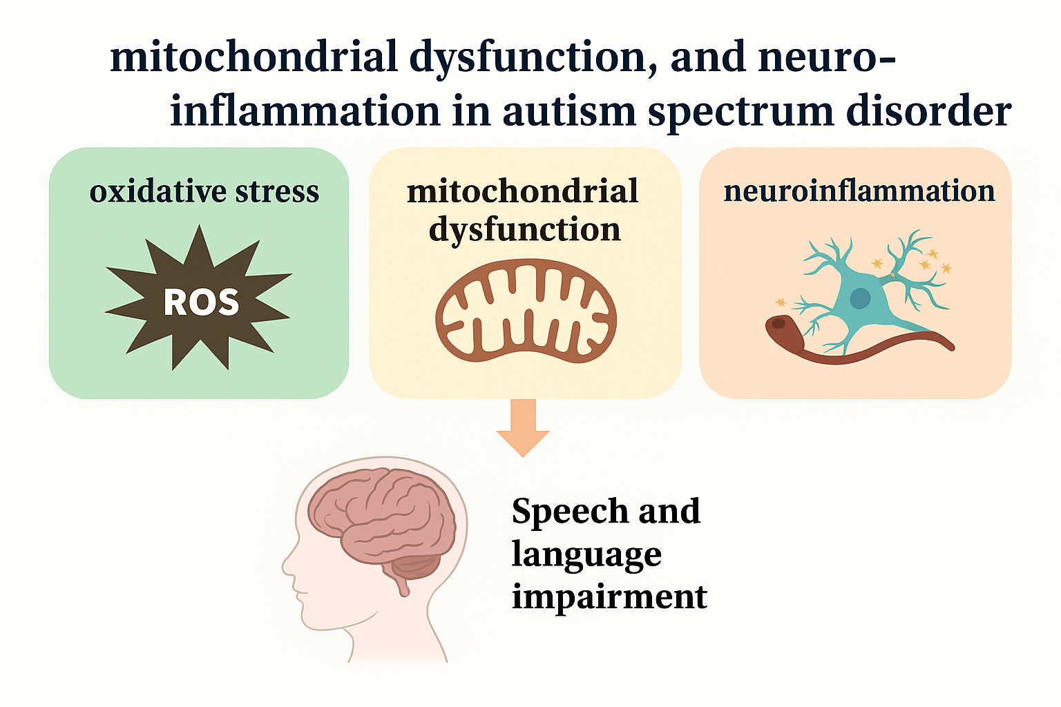
Figure 1. Reactive oxygen species (ROS), mitochondrial dysfunction, and neuroinflammation decrease the functionality of the corticolimbic pathways that play a vital role in communication and language comprehension.
Brainsci 15 00853 g001
Figure 2. Role of oxidative stress markers and ROS in ASD-related biochemical, molecular, and neuronal changes in the brain. ROS-Reactive oxyfen species; 8-OHdG-8-Hydroxy-2-deoxyguanosine; MDA-malondialdehyde.
Brainsci 15 00853 g002
Figure 3. Role of oxidative stress and inflammation in maternal-induced ASD. MIA—Maternal immune activation, IL-interleukin, TNF-Tumor necrosis factor. Light Pink Placenta & Fetus Illustration: Depicts placental structure and developing fetus affected by the pathway. Blue Dots: Symbolize oxidative stress factors crossing the placenta.
Brainsci 15 00853 g003
Table 1. Role of mitochondrial dysfunction in neurological disorders and their association with ASD.
SyndromeMitochondrial Process AffectedEnergy Metabolism ImpactSample SourceGene/Protein InvolvementMitochondrial ImpactNeurological and Developmental Effects
Smith–Lemli–Opitz Syndrome (SLOS)
[47]
Mitochondrial function and cholesterol metabolismDisturbance of cholesterol processing, energy dysregulationFibroblasts from affected individualsDHCR7Accumulation of mitochondria dysfunctional substancesDevelopmental delay, weak muscle tone, behavioral differences
Helsmoortel–Van der Aa Syndrome (HVDAS)
[48]
Cellular respiration efficiencyDeficiency of energy to operate cellsFibroblasts from affected individualsADNPSuppressed mitochondrial oxygen utilization, decreased production of ATPAutism-like characteristics, retarded brain development
DiGeorge Syndrome (DGS)
[49]
Mitochondrial transport and ion balanceFaulty mitochondrial integrity, increased oxidative loadFibroblasts from patients and mouse modelsSLC25A1, SLC25A4Disruptions in mitochondrial carrier proteins, disturbances in calcium homeostasisDeficits in learning, increased risk of psychiatric diseases such as schizophrenia
Angelman Syndrome (AS)
[50]
Gene regulation and electron transportLow levels of ATP production, mitochondrial shortagesFibroblasts and hippocampal cells from UBE3A mutant miceUBE3AETC complex III dysfunction, irregular expression of mitochondrial genesSerious lag of mental development, epilepsy, movement disorders
Fragile X Syndrome (FXS)
[51]
Mitochondrial structure and dynamicsA loss in cellular energy and oxidative stressBrain tissue from FMR1 knockout miceFMR1, MFN1, MFN2, OPA1Loss of fusion protein levels, abridged ATP production, and lesser action in ETC complexes I and IIImpaired cognitive abilities, disrupted synapses in neurons, neurodegeneration
Rett Syndrome (RS)
[52]
Mitochondrial structure and oxidative balanceMore oxidative stress, poor energy consumptionBrain tissue from MECP2 knockout miceMECP2Mitochondria having aberrant morphology, long length of mitochondriaMotor impairment, intellectual disability, the likelihood of a seizure
Cornelia de Lange Syndrome (CdLS)
[53]
Mitochondrial
protein synthesis
Deficient mitochondrial protein synthesisSkin fibroblasts from ASD patientTRMT61A,
MRPS22
Ribosomal mitochondrial defects and compromised complexes of ETC I, III, and IVHeight deficiencies, intellectual disabilities, unusual facial appearance
Phelan–McDermid Syndrome (PMS)
[54]
Electron transport chain performanceReduced supply of ATP, harm by ROSOral samples from PMS patientsSHANK3Nonfunctioning complexes I and IV, and imbalance in the energy schedule of mitochondriaLearning disorders, deficit in neuronal signaling
Disclaimer/Publisher’s Note: The statements, opinions and data contained in all publications are solely those of the individual author(s) and contributor(s) and not of MDPI and/or the editor(s). MDPI and/or the editor(s) disclaim responsibility for any injury to people or property resulting from any ideas, methods, instructions or products referred to in the content.