Article

[18F]FDG PET-CT Imaging of the Low Back in Persistent Spinal Pain Syndrome Type 2: A Pilot Study Towards Improved Diagnosis

by
Lara S. Burmeister
1,2,
Richard L. Witkam
2,
Kris C. P. Vissers
2,*,
Martin Gotthardt
1 and
Dylan J. H. A. Henssen
1,3
1
Department of Medical Imaging, Radboud University Medical Center, 6525GA Nijmegen, The Netherlands
2
Department of Anesthesiology, Pain and Palliative Medicine, Radboud University Medical Center, 6525GA Nijmegen, The Netherlands
3
Department of Nuclear Medicine, University Hospital Leipzig, 04103 Leipzig, Germany
*
Author to whom correspondence should be addressed.
Brain Sci. 2025, 15(7), 724; https://doi.org/10.3390/brainsci15070724
Submission received: 10 June 2025 / Revised: 1 July 2025 / Accepted: 5 July 2025 / Published: 7 July 2025
(This article belongs to the Section Neurotechnology and Neuroimaging)

Abstract

Background/Objectives: Diagnosis of Persistent Spinal Pain Syndrome Type 2 (PSPS-T2) currently lacks objective biomarkers. Therefore, this retrospective study aimed to investigate differences in glucose metabolism in the axial musculoskeletal system in PSPS-T2 patients by means of [18F]FDG PET-CT imaging. Methods: Nine PSPS-T2 patients (five females, four males; mean age of 53 ± 4.82 years) and nine age- and gender-matched healthy controls (five females, four males; mean age of 53 ± 3.91 years) were included. For each participant, 24 regions of interest (ROIs) were manually drawn, including areas of the vertebral endplates, the intervertebral discs, and the psoas muscles. For each ROI, the mean standardized uptake values (SUVs) were assessed. Group differences were evaluated using repeated measures ANOVA with Bonferroni-adjusted post-hoc pairwise comparisons. Additionally, Pearson correlation analyses examined associations between SUVmean values and the Numerical Rating Scale (NRS) pain scores. Results: Results demonstrated significantly higher SUVmean values in healthy controls compared to PSPS-T2 patients, particularly at the superior endplates of L4 and S1, the intervertebral discs at L4-L5 and L5-S1, and the posterior endplates of L4 and L5. Although PSPS-T2 patients exhibited higher SUVmean values than controls in the psoas muscle, these differences were not statistically significant. Additionally, no significant correlations were found between SUVmean values and NRS pain scores, suggesting that metabolic activity alone does not directly reflect pain severity. Conclusions: Despite the limited sample size of this pilot study, the metabolic fingerprint of the axial musculoskeletal system was shown to be distinctly different in PSPS-T2 patients compared to healthy controls. This could lead to an improved understanding of PSPS-T2 pathophysiology and might open new doors for better diagnosis and treatment strategies.

1. Introduction

Persistent spinal pain syndrome type 2 (PSPS-T2) is characterized by persistent or emerging pain of the lower back and/or lower extremities despite anatomically successful surgery of the lower back or spine [1,2]. It is associated with significantly reduced health-related quality of life scores, often exceeding those seen in other chronic pain conditions [3,4]. Despite its high burden, PSPS-T2 remains challenging to diagnose [5].
Although radiological imaging remains essential to exclude serious underlying pathology, such as malignancy or fractions [6], the diagnostic value is limited in the majority of patients with chronic low back pain when no specific cause can be identified [7]. Instead, radiological imaging can even cause more harm than benefit by steering treatment algorithms in the wrong direction [8]. In many cases, symptom severity does not correlate well with structural abnormalities observed in imaging, which highlights the need for validated, phenotype-based approaches that better reflect the underlying pain mechanisms [9,10]. In this light, nuclear imaging techniques might be a promising example.
For instance, one study remarked that [99mTc]HDP SPECT-CT imaging of the low back could help localize a potential pain generator [11]. In line with this, [18F]NaF PET-CT imaging was also found to be able to detect regions of increased osteoblastic activity among patients who had persistent or recurrent back pain following spinal fusion surgery, indicating possible pain-generating areas [12]. This was further investigated in PSPS-T2 patients by Peters et al. in 2015 [13] using [18F]NaF PET-CT imaging. In this study, significantly higher osteoblastic activity was observed in vertebral endplates in patients with the most severe clinical disability [13]. Sophisticated, osteoblastic activity imaging to identify possible bone-related pain-generating areas cannot be used alone to assess the multidimensionality of low back pain in PSPS-T2 patients. Previous studies demonstrated that changes in the (para)spinal muscles, such as atrophy or fatty infiltration, are associated with chronic low back pain [14]. Improved understanding of the biomechanics of low back pain has further highlighted the importance of muscular stabilization of the “neutral zone” range of motion in the low back by use of the (para)spinal muscles [15]. The neutral zone is thereby defined as the region where the spine can move with the greatest ease, with little influence from muscular control that usually comes into play when movement goes beyond this zone. When there is injury to the spine or weakness of the muscle, the zone may increase, which in turn can lead to spinal instability problems in the lower back [16]. In line with these insights, a recent study showed anatomic changes in the psoas muscles in PSPS-T2 patients on lumbar MRI scans, which correlated with various pain-related outcomes [17]. However, to our knowledge, no study has set out to investigate metabolic changes in the axial musculoskeletal structures, specifically in the lower back region, in PSPS-T2 patients.
Therefore, this study aimed to assess the difference in glucose metabolic activity in the vertebrae, disci, and psoas muscle between PSPS-T2 patients and healthy controls using [18F]FDG PET-CT scans.

2. Materials and Methods

2.1. Participants

Subjects were retrospectively included. To be included in this study, adult patients (≥18 years) diagnosed with PSPS-T2 must have undergone an [18F]FDG PET-CT with no signs of other pathologies that could have interfered with the measurements relevant to our outcome parameters (e.g., oncological diseases or infections). Another inclusion criterion was that patients must have received a spinal cord stimulation (SCS) device after the [18F]FDG PET-CT scan. This criterion ensured that all patients had a confirmed clinical indication for SCS implantation based on standard care, and that PET-CT was part of the diagnostic work-up without influencing the treatment decision. Including only patients who went on to receive SCS provided a homogeneous group with comparable disease severity and therapeutic trajectory. Additionally, nine age- and gender-matched controls were included. Controls were only included when [18F]FDG PET-CT showed no signs of possibly interfering with pathology. Indications for the controls to undergo [18F]FDG PET-CT imaging comprised the metabolic evaluation of incidentalomas in the lungs or adrenal glands previously observed on routinely performed anatomical imaging. Patients and controls who refused to share their data anonymously for scientific purposes were excluded from this study.

2.2. Pain Intensity Scores

Pain intensity was assessed using a numerical rating scale (NRS) ranging from 0 to 10 (i.e., 0 = no pain; 10 = worst imaginable pain). The NRS was obtained both prior to the SCS procedure and 12 months after the implantation of SCS.

2.3. [18F]FDG PET-CT Scan Acquisition

Scans were performed at the Department of Nuclear Medicine at the Radboudumc, Nijmegen, The Netherlands. A Siemens Biograph mCT PET UltraHD scanner equipped with a 40-slice adaptive 4D spiral CT was used. Prior to imaging, all participants were prepared with at least 6 h of fasting to ensure low blood glucose and low insulinemia, as this is directly responsible for glucose uptake by non-tumor cells. To ensure these conditions, blood glucose concentration was tested prior to scanning. To ensure optimal imaging conditions, the plasma glucose level needed to be lower than 10 mmol/L). The radionuclide fluorine-18 [18F], which was used in the form of F-18-fluorodeoxyglucose, was intravenously administered as a slow bolus injection. Administered activity was calculated following the recommendations of the European Associations of Nuclear Medicine when using a quadratic relationship between PET acquisition time per bed position and patient weight [18]. To ensure proper tracer distribution and to minimize residual activity, the intravenous line was flushed with 20 mL of 0.9% NaCl. Imaging was initiated 60 min post-injection.

2.4. [18F]FDG PET-CT Data Analysis

Image annotation was carried out using a 3D slicer (https://www.slicer.org/). Regions of interest (ROIs) were drawn at the level of the superior and inferior endplates of the lumbar vertebrae (L1 to L5). Furthermore, ROIs were drawn at the level of the intervertebral discs from level T12/L1 to level L5/S1. Also, three ROIs were drawn at three levels (at level L3, L4, and L5) within the psoas major muscle on each side of the spine at transverse images; therefore, a total of 24 segments were examined for each participant (Figure 1).
In order to correct for the variability of glucose metabolism between subjects, two normalization methods of standardized uptake values (SUVmean and SUVmax) were employed. First, normalization was carried out by use of a volume of interest (VOI) located centrally in the normal-appearing liver parenchyma. Second, blood activity normalization was carried out by using a VOI in the lumen of the left cardiac ventricle.

2.5. Statistical Analysis

Statistical analyses were performed using SPSS (IBM Corporation, Version 29.0.2.0). Differences between groups across spinal levels were evaluated using repeated measures ANOVA to properly handle the repeated measurements within each subject. Following that, post-hoc pairwise comparisons were performed with Bonferroni correction to account for multiple comparisons. Statistical significance was set at p < 0.05, with all reported p-values adjusted using the Bonferroni method to control for the family-wise error rate. Pearson correlation analyses were performed to explore associations between NRS difference scores and SUVmean values.

3. Results

Nine patients (five females, four males; mean age of 53 ± SD 4.82) and nine controls (five females, four males; mean age of 53 years; ±SD 3.91) were included. Patients underwent SCS implantation between 13 and 162 months after the [18F]FDG PET-CT imaging session.
The overall coefficients of variation were 49.5% and 63.1% when the SUVmean values were normalized using the VOI placed in the liver and left ventricle, respectively. In contrast, when the SUVmax values were normalized in the same manner, the overall coefficients of variation were 55.0% and 73.4%, respectively. The SUVmean values, which were normalized using the VOI placed in the liver, resulted in the lowest coefficient of variation. Therefore, these normalized SUVmean values were used for further analyses.
Repeated measures analyses revealed statistically significant differences with higher scores in the control group regarding the SUVmean for the superior endplates (F(1,16) = 6.76, p = 0.019), the discus (F(1,16) = 5.95, p = 0.027), and the posterior endplates (F(1,16) = 6.82, p = 0.019) between groups. Post-hoc Bonferroni-adjusted pairwise comparisons further specified these differences to the superior endplates of L4 (p = 0.048) and S1 (p = 0.037), the discs at L4-L5 (p = 0.043) and L5-S1 (p < 0.001), and the posterior endplates of L4 (p = 0.029) and L5 (p = 0.036), all showing higher SUVmean values in the control group. These p-values, derived from Bonferroni-adjusted post-hoc pairwise comparisons, are detailed in Table 1. Higher SUVmean values in the patient group were only found for the psoas muscle even though the repeated measures analysis for the psoas muscle revealed no statistically significant difference for neither the left psoas muscle (F(1,16) = 0.59, p = 0.453) nor the right psoas muscle (F(1,16) = 0.17, p = 0.686). Pearson correlation analyses revealed no statistically significant correlations between the NRS difference scores and the SUVmean values.

4. Discussion

The present study revealed higher metabolic activity in the region of the lower back in non-painful controls as compared to PSPS-T2 patients. More specifically, SUVmean values were significantly higher in the control group at the superior endplates of L4 and S1, the intervertebral discs at L4-L5 and L5-S1, and the posterior endplates of L4 and L5. We found no significant correlation between the mean SUV values and pain intensity scores. These preliminary results could help provide insights into the biological mechanisms that might lead to PSPS-T2 at the level of the lower back.
Generally, a rise in glucose metabolism in the vertebral structures suggests enhanced metabolic activity, which could be related to bone remodeling processes such as inflammation or degeneration [19,20,21]. However, the lower SUVmean values in the PSPS-T2 patients may suggest an alternative pathological mechanism accompanying this condition. One possible explanation for the observed metabolic differences could be the presence of fibrotic changes. Fibrosis is driven by activated fibroblasts, which play a role in various pathological processes such as scarring, degeneration, and inflammation [22]. Epidural fibrosis, characterized by epidural fat being replaced with scar tissue, is a well-known complication following spinal surgery [23]. Previous research has found a positive association between the number and extent of spinal surgeries and the incidence of fibrosis, as well as a significant relationship between the severity of patients’ symptoms and the extent of fibrosis [24]. Furthermore, intervertebral disc fibrosis is known to play an important role in intervertebral disc degeneration [25,26]. As fibrosis has been described to elicit a homogeneous, mild FDG-uptake [27], the lower SUVmean values in the patient group may be explained by the presence of fibrotic tissue that is less metabolically active compared to the normal physiological uptake that is seen as a consequence of the axial load on bones, ligaments, and muscles. A possible explanation for our findings could thus be that PSPS-T2 patients have a higher degree of spinal and intervertebral disc fibrosis, which may not be adequately captured by [18F]FDG PET-CT imaging.
Fibroblast-activation protein (FAP) is expressed by activated fibroblasts in epidural fibrosis and can be utilized for nuclear imaging [22]. It has been shown that FAP-specific imaging, such as [68GA]FAPI PET-CT, provides a better reflection of fibrosis progression compared to [18F]FDG PET-CT [28]. FAP-specific imaging could hence be a promising alternative to [18F]FDG PET-CT in exploring the role of fibrosis in PSPS-T2.
The iliopsoas muscle is hypothesized to play a crucial role in stabilizing the lumbar spine, and previous studies demonstrated a correlation between its cross-sectional area and decreased pain intensity in individuals experiencing lower back pain [29]. In contrast to previous findings that indicated metabolic changes in the psoas muscle in PSPS-T2 patients [17], we were unable to identify statistically significant differences in glucose metabolism in this muscle group when comparing PSPS-T2 patients to healthy controls. As demonstrated by several studies, low back pain is frequently accompanied by structural changes of the lumbar muscles [30,31]. These alterations consist of muscular atrophy and the infiltration of fat, which are also known to be correlated with inactivity [32,33] Interestingly, there seems to be a reversed relationship in changes between the posterior paraspinal muscles and the psoas muscle, where a higher fat infiltration in the posterior paraspinal muscles, such as the multifidus, is associated with a lower infiltration of fat in the psoas muscle. It is proposed that this could represent a compensating mechanism in which the psoas compensates for the loss in strength in order to stabilize the spine [34,35]. Our findings suggest that even though structural alterations such as atrophy and fatty infiltration may occur, they do not necessarily correspond with increased metabolic activity as shown by [18F]FDG PET-CT. Nonetheless, interpretation of these findings necessitates careful consideration of the movement and activity profiles of both PSPS-T2 patients and healthy controls, as disparities in physical activity may confound the observed metabolic patterns. To enhance the validity of metabolic comparison across cohorts, future studies should include objective assessments of physical activity and further investigate these pathways by integrating additional muscle groups and alternative imaging modalities such as MRI.
As the absence of a significant correlation between SUVmean values and pain intensity scores suggests, metabolic activity alone may not be a direct reflection of the severity of pain in PSPS-T2 patients. This aligns with the broader understanding of chronic pain, which assumes that chronic pain can be seen as a multifactorial condition that is influenced by a variety of mechanisms such as neuroinflammatory processes, central sensitization, and altered processing in the central nervous system [36,37,38]. A possible explanation for the lack of correlation could be the relatively small sample size and the long period over which patients were included in the study. Given that metabolic processes such as inflammation and remodeling evolve over time, it could be possible that some of these changes may have been stabilized by the time of scanning, making them less detectable on [18F]FDG PET-CT imaging. Furthermore, several cognitive behavioral and psychosocial factors, such as anxiety, pain catastrophizing, depression or a person’s coping mechanisms, play an important role in the perception of pain [39,40,41]. In addition to the localized metabolic changes in the spine, all of these mechanisms could have the potential to modulate the experience of pain.

Limitations

When interpreting the current study’s findings, there are several limitations that should be considered. Firstly, the study sample consists of a limited number of participants, which may constrain the extent to which the results can be generalized. Secondly, given the retrospective nature of the study design, it is not possible to assess metabolic changes over time, such as assessing changes before and after SCS implantation or following other major events such as additional surgeries of significant life changes that could influence pain perception or behavior and thereby affect PET imaging outcomes. The conduction of prospective studies that assess the impact of SCS on glucose metabolism may offer further insights into the mechanisms underlying the treatment response. Additionally, even though [18F]FDG PET-CT is indicative of metabolic activity, it is not possible to differentiate between inflammatory, reconstructive, or degenerative processes. Integrating findings from additional biomarkers such as [68GA]FAPI PET-CT or the MRI-based assessment of muscle integrity could further enhance our understanding of the underlying pathophysiology in PSPS-T2.

5. Conclusions

This pilot study explores a potential role for metabolic imaging in identifying pain-related changes with differences in glucose metabolic activity observed in vertebral structures, particularly the endplates and discs, between patients with PSPS-T2 and controls. The absence of significant findings in psoas metabolism and correlations with pain scores may reflect the multifactorial nature of chronic pain in PSPS-T2. Future research should therefore explore multi-modal imaging approaches, combined with psychological assessments, to investigate the interactions between structural, metabolic, and neuropsychological changes in this patient population with dynamic biopsychosocial interactions.

Author Contributions

Conceptualization, L.S.B., R.L.W., K.C.P.V., M.G. and D.J.H.A.H.; methodology, L.S.B., R.L.W., K.C.P.V., M.G. and D.J.H.A.H.; validation, L.S.B. and D.J.H.A.H.; formal analysis, L.S.B., R.L.W. and D.J.H.A.H.; investigation, L.S.B., R.L.W. and D.J.H.A.H.; data curation, L.S.B.; writing—original draft preparation, L.S.B.; writing—review and editing, R.L.W., K.C.P.V., M.G. and D.J.H.A.H.; visualization, L.S.B. and D.J.H.A.H.; supervision, R.L.W., K.C.P.V., M.G. and D.J.H.A.H.; project administration, L.S.B. and D.J.H.A.H. All authors have read and agreed to the published version of the manuscript.

Funding

This research received no external funding.

Institutional Review Board Statement

Ethical review and approval were waived for this study due to its retrospective nature and mere use of completely anonymized data.

Informed Consent Statement

Informed consent was obtained from all subjects involved in the study.

Data Availability Statement

The datasets generated and analyzed during the current study are available from the corresponding author on reasonable request. The data are not publicly available due to their containing information that could compromise the privacy of the participants.

Acknowledgments

Henssen is supported by the Clinician Scientist Programme of Universitätsmedizin Leipzig, which facilitates the integration of clinical practice and scientific research.

Conflicts of Interest

The authors declare no conflicts of interest.

Abbreviations

The following abbreviations are used in this manuscript:
PSPS-T2Persistent Spinal Pain Syndrome Type 2
[18F]FDG PET-CTFluorine-18 Fluorodeoxyglucose Positron Emission Tomography-Computed Tomography
ROIRegion of interest
SUVStandardized uptake value
NRSNumerical Rating Scale
[99mTC]HDP SPECT-CTTechnetium-99m Hydroxymethylene Disphosphonate Single Photon Emission Computed Tomography-Computed Tomography
[18F]NaF PET-CTFluorine-18 Sodium Fluoride Positron Emission Tomography-Computed Tomography
SCSSpinal cord stimulation
NaClSodium Chloride
VOIVolume of interest
FAPFibroblast-activation protein
[68Ga]FAPI PET-CTGallium-68 Fibroblast Activation Inhibitor Positron Emission Tomography-Computed Tomography

References

  1. Clancy, C.; Quinn, A.; Wilson, F. The aetiologies of Failed Back Surgery Syndrome: A systematic review. J. Back Musculoskelet. Rehabil. 2017, 30, 395–402. [Google Scholar] [CrossRef] [PubMed]
  2. Christelis, N.; Simpson, B.; Russo, M.; Stanton-Hicks, M.; Barolat, G.; Thomson, S.; Schug, S.; Baron, R.; Buchser, E.; Carr, D.B.; et al. Persistent Spinal Pain Syndrome: A Proposal for Failed Back Surgery Syndrome and ICD-11. Pain Med. 2021, 22, 807–818. [Google Scholar] [CrossRef] [PubMed]
  3. Manca, A.; Eldabe, S.; Buchser, E.; Kumar, K.; Taylor, R.S. Relationship between health-related quality of life, pain, and functional disability in neuropathic pain patients with failed back surgery syndrome. Value Health 2010, 13, 95–102. [Google Scholar] [CrossRef]
  4. Thomson, S.; Jacques, L. Demographic characteristics of patients with severe neuropathic pain secondary to failed back surgery syndrome. Pain Pract. 2009, 9, 206–215. [Google Scholar] [CrossRef] [PubMed]
  5. Yeo, J. Failed back surgery syndrome-terminology, etiology, prevention, evaluation, and management: A narrative review. J. Yeungnam Med. Sci. 2024, 41, 166–178. [Google Scholar] [CrossRef]
  6. Henschke, N.; Maher, C.G.; Refshauge, K.M.; Herbert, R.D.; Cumming, R.G.; Bleasel, J.; York, J.; Das, A.; McAuley, J.H. Prevalence of and screening for serious spinal pathology in patients presenting to primary care settings with acute low back pain. Arthritis Rheum. 2009, 60, 3072–3080. [Google Scholar] [CrossRef]
  7. Dhagat, P.K.; Jain, M.; Singh, S.N.; Arora, S.; Leelakanth, K. Failed Back Surgery Syndrome: Evaluation with Magnetic Resonance Imaging. J. Clin. Diagn. Res. 2017, 11, Tc06–Tc09. [Google Scholar] [CrossRef]
  8. Hall, A.M.; Aubrey-Bassler, K.; Thorne, B.; Maher, C.G. Do not routinely offer imaging for uncomplicated low back pain. BMJ 2021, 372, n291. [Google Scholar] [CrossRef]
  9. Witkam, R.L.; Buckens, C.F.; van Goethem, J.W.M.; Vissers, K.C.P.; Henssen, D. The current role and future directions of imaging in failed back surgery syndrome patients: An educational review. Insights Imaging 2022, 13, 117. [Google Scholar] [CrossRef]
  10. Babińska, A.; Wawrzynek, W.; Czech, E.; Skupiński, J.; Szczygieł, J.; Łabuz-Roszak, B. No association between MRI changes in the lumbar spine and intensity of pain, quality of life, depressive and anxiety symptoms in patients with low back pain. Neurol. Neurochir. Pol. 2019, 53, 74–82. [Google Scholar] [CrossRef]
  11. Huang, B.-R.; Peng, B.-R.; Pan, L.-K.; Chen, C.-Y. Potential Usefulness of Single Photon Emission Computed Tomography/Computed Tomography in Management of Patients with Failed Back Surgery Syndrome. J. Med. Imaging Health Inform. 2019, 9, 1386–1392. [Google Scholar] [CrossRef]
  12. Pouldar, D.; Bakshian, S.; Matthews, R.; Rao, V.; Manzano, M.; Dardashti, S. Utility of 18F sodium fluoride PET/CT imaging in the evaluation of postoperative pain following surgical spine fusion. Musculoskelet. Surg. 2017, 101, 159–166. [Google Scholar] [CrossRef]
  13. Peters, M.; Willems, P.; Weijers, R.; Wierts, R.; Jutten, L.; Urbach, C.; Arts, C.; van Rhijn, L.; Brans, B. Pseudarthrosis after lumbar spinal fusion: The role of 18F-fluoride PET/CT. Eur. J. Nucl. Med. Mol. Imaging 2015, 42, 1891–1898. [Google Scholar] [CrossRef] [PubMed]
  14. He, K.; Head, J.; Mouchtouris, N.; Hines, K.; Shea, P.; Schmidt, R.; Hoelscher, C.; Stricsek, G.; Harrop, J.; Sharan, A. The Implications of Paraspinal Muscle Atrophy in Low Back Pain, Thoracolumbar Pathology, and Clinical Outcomes After Spine Surgery: A Review of the Literature. Global Spine J. 2020, 10, 657–666. [Google Scholar] [CrossRef] [PubMed]
  15. Freeman, M.D.; Woodham, M.A.; Woodham, A.W. The role of the lumbar multifidus in chronic low back pain: A review. PM&R 2010, 2, 142–146; quiz 141 p following 167. [Google Scholar] [CrossRef]
  16. Panjabi, M.M. The stabilizing system of the spine. Part II. Neutral zone and instability hypothesis. J. Spinal Disord. 1992, 5, 390–396; discussion 397. [Google Scholar] [CrossRef]
  17. Sheldon, B.L.; DiMarzio, M.; Chung, S.H.; Tram, J.; Khazen, O.; Staudt, M.D.; Bondoc, M.; Pilitsis, J.G. Association of Outcomes of Spinal Cord Stimulation for Chronic Low Back Pain and Psoas Measurements Based on Size of Iliopsoas Muscles. Neuromodulation 2022, 25, 121–127. [Google Scholar] [CrossRef]
  18. Boellaard, R.; Delgado-Bolton, R.; Oyen, W.J.; Giammarile, F.; Tatsch, K.; Eschner, W.; Verzijlbergen, F.J.; Barrington, S.F.; Pike, L.C.; Weber, W.A.; et al. FDG PET/CT: EANM procedure guidelines for tumour imaging: Version 2.0. Eur. J. Nucl. Med. Mol. Imaging 2015, 42, 328–354. [Google Scholar] [CrossRef]
  19. Glaudemans, A.W.; de Vries, E.F.; Galli, F.; Dierckx, R.A.; Slart, R.H.; Signore, A. The use of (18)F-FDG-PET/CT for diagnosis and treatment monitoring of inflammatory and infectious diseases. Clin. Dev. Immunol. 2013, 2013, 623036. [Google Scholar] [CrossRef]
  20. Piri, R.; Nøddeskou-Fink, A.H.; Gerke, O.; Larsson, M.; Edenbrandt, L.; Enqvist, O.; Høilund-Carlsen, P.F.; Stochkendahl, M.J. PET/CT imaging of spinal inflammation and microcalcification in patients with low back pain: A pilot study on the quantification by artificial intelligence-based segmentation. Clin. Physiol. Funct. Imaging 2022, 42, 225–232. [Google Scholar] [CrossRef]
  21. Sharma, D.N.; Yerramneni, V.K.; Srivastava, M.K.; Yerragunta, T.; Akurati, S. Role of magnetic resonance imaging and 18-fluorodeoxyglucose positron emission tomography-computed tomography in identifying pain generators in patients with chronic low back pain. J. Craniovertebral Junction Spine 2023, 14, 381–387. [Google Scholar] [CrossRef] [PubMed]
  22. Hotta, M.; Rieger, A.C.; Jafarvand, M.G.; Menon, N.; Farolfi, A.; Benz, M.R.; Calais, J. Non-oncologic incidental uptake on FAPI PET/CT imaging. Br. J. Radiol. 2023, 96, 20220463. [Google Scholar] [CrossRef] [PubMed]
  23. Lubina, Z.I.; Baranovic, S.; Karlak, I.; Novacic, K.; Potocki-Karacic, T.; Lovrić, D. The grading model for the assessment of the total amount of epidural fibrosis in postoperative lumbar spine. Eur. Spine J. 2013, 22, 892–897. [Google Scholar] [CrossRef]
  24. Guner, D.; Asik, I.; Ozgencil, G.E.; Peker, E.; Erden, M.I. The Correlation of Epidural Fibrosis with Epiduroscopic and Radiologic Imaging for Chronic Pain after Back Surgery. Pain Physician 2021, 24, E1219–E1226. [Google Scholar]
  25. Yee, A.; Lam, M.P.; Tam, V.; Chan, W.C.; Chu, I.K.; Cheah, K.S.; Cheung, K.M.; Chan, D. Fibrotic-like changes in degenerate human intervertebral discs revealed by quantitative proteomic analysis. Osteoarthr. Cartil. 2016, 24, 503–513. [Google Scholar] [CrossRef]
  26. Oegema, T.R., Jr.; Johnson, S.L.; Aguiar, D.J.; Ogilvie, J.W. Fibronectin and its fragments increase with degeneration in the human intervertebral disc. Spine (Phila Pa 1976) 2000, 25, 2742–2747. [Google Scholar] [CrossRef] [PubMed]
  27. Tsai, Y.H.; Huang, G.S.; Tang, C.T.; Chang, W.C.; Hsu, Y.C. Case Report: Nerve Root Entrapment Due to Epidural Fibrosis in a Patient with Failed Back Surgery Syndrome: Value of 2-18F-Fluorodeoxyglucose Simultaneous Positron Emission Tomography-Magnetic Resonance Imaging. Front. Med. 2022, 9, 860545. [Google Scholar] [CrossRef]
  28. Ji, H.; Song, X.; Lv, X.; Shao, F.; Long, Y.; Song, Y.; Song, W.; Qiao, P.; Gai, Y.; Jiang, D.; et al. [68Ga]FAPI PET for Imaging and Treatment Monitoring in a Preclinical Model of Pulmonary Fibrosis: Comparison to [18F]FDG PET and CT. Pharmaceuticals 2024, 17, 726. [Google Scholar] [CrossRef]
  29. Mallio, C.A.; Russo, F.; Vadalà, G.; Papalia, R.; Pileri, M.; Mancuso, V.; Bernetti, C.; Volpecina, M.; Di Gennaro, G.; Beomonte Zobel, B.; et al. The importance of psoas muscle on low back pain: A single-center study on lumbar spine MRI. N. Am. Spine Soc. J. 2024, 18, 100326. [Google Scholar] [CrossRef]
  30. Goubert, D.; Oosterwijck, J.V.; Meeus, M.; Danneels, L. Structural Changes of Lumbar Muscles in Non-specific Low Back Pain: A Systematic Review. Pain Physician 2016, 19, E985–E1000. [Google Scholar]
  31. Seyedhoseinpoor, T.; Taghipour, M.; Dadgoo, M.; Sanjari, M.A.; Takamjani, I.E.; Kazemnejad, A.; Khoshamooz, Y.; Hides, J. Alteration of lumbar muscle morphology and composition in relation to low back pain: A systematic review and meta-analysis. Spine J. 2022, 22, 660–676. [Google Scholar] [CrossRef] [PubMed]
  32. Manini, T.M.; Clark, B.C.; Nalls, M.A.; Goodpaster, B.H.; Ploutz-Snyder, L.L.; Harris, T.B. Reduced physical activity increases intermuscular adipose tissue in healthy young adults. Am. J. Clin. Nutr. 2007, 85, 377–384. [Google Scholar] [CrossRef]
  33. Pagano, A.F.; Brioche, T.; Arc-Chagnaud, C.; Demangel, R.; Chopard, A.; Py, G. Short-term disuse promotes fatty acid infiltration into skeletal muscle. J. Cachexia Sarcopenia Muscle 2018, 9, 335–347. [Google Scholar] [CrossRef]
  34. Muellner, M.; Haffer, H.; Chiapparelli, E.; Dodo, Y.; Shue, J.; Tan, E.T.; Zhu, J.; Pumberger, M.; Sama, A.A.; Cammisa, F.P.; et al. Fat infiltration of the posterior paraspinal muscles is inversely associated with the fat infiltration of the psoas muscle: A potential compensatory mechanism in the lumbar spine. BMC Musculoskelet. Disord. 2023, 24, 846. [Google Scholar] [CrossRef]
  35. Özcan-Ekşi, E.E.; Ekşi, M.; Turgut, V.U.; Canbolat, Ç.; Pamir, M.N. Reciprocal relationship between multifidus and psoas at L4-L5 level in women with low back pain. Br. J. Neurosurg. 2021, 35, 220–228. [Google Scholar] [CrossRef] [PubMed]
  36. Schumacher, M.A. Peripheral Neuroinflammation and Pain: How Acute Pain Becomes Chronic. Curr. Neuropharmacol. 2024, 22, 6–14. [Google Scholar] [CrossRef]
  37. Ji, R.R.; Nackley, A.; Huh, Y.; Terrando, N.; Maixner, W. Neuroinflammation and Central Sensitization in Chronic and Widespread Pain. Anesthesiology 2018, 129, 343–366. [Google Scholar] [CrossRef] [PubMed]
  38. Li, W.; Gong, Y.; Liu, J.; Guo, Y.; Tang, H.; Qin, S.; Zhao, Y.; Wang, S.; Xu, Z.; Chen, B. Peripheral and Central Pathological Mechanisms of Chronic Low Back Pain: A Narrative Review. J. Pain Res. 2021, 14, 1483–1494. [Google Scholar] [CrossRef]
  39. Rogers, A.H.; Farris, S.G. A meta-analysis of the associations of elements of the fear-avoidance model of chronic pain with negative affect, depression, anxiety, pain-related disability and pain intensity. Eur. J. Pain 2022, 26, 1611–1635. [Google Scholar] [CrossRef]
  40. Tsuji, T.; Matsudaira, K.; Sato, H.; Vietri, J. The impact of depression among chronic low back pain patients in Japan. BMC Musculoskelet. Disord. 2016, 17, 447. [Google Scholar] [CrossRef]
  41. Meints, S.M.; Edwards, R.R. Evaluating psychosocial contributions to chronic pain outcomes. Prog. Neuropsychopharmacol. Biol. Psychiatry 2018, 87, 168–182. [Google Scholar] [CrossRef] [PubMed]
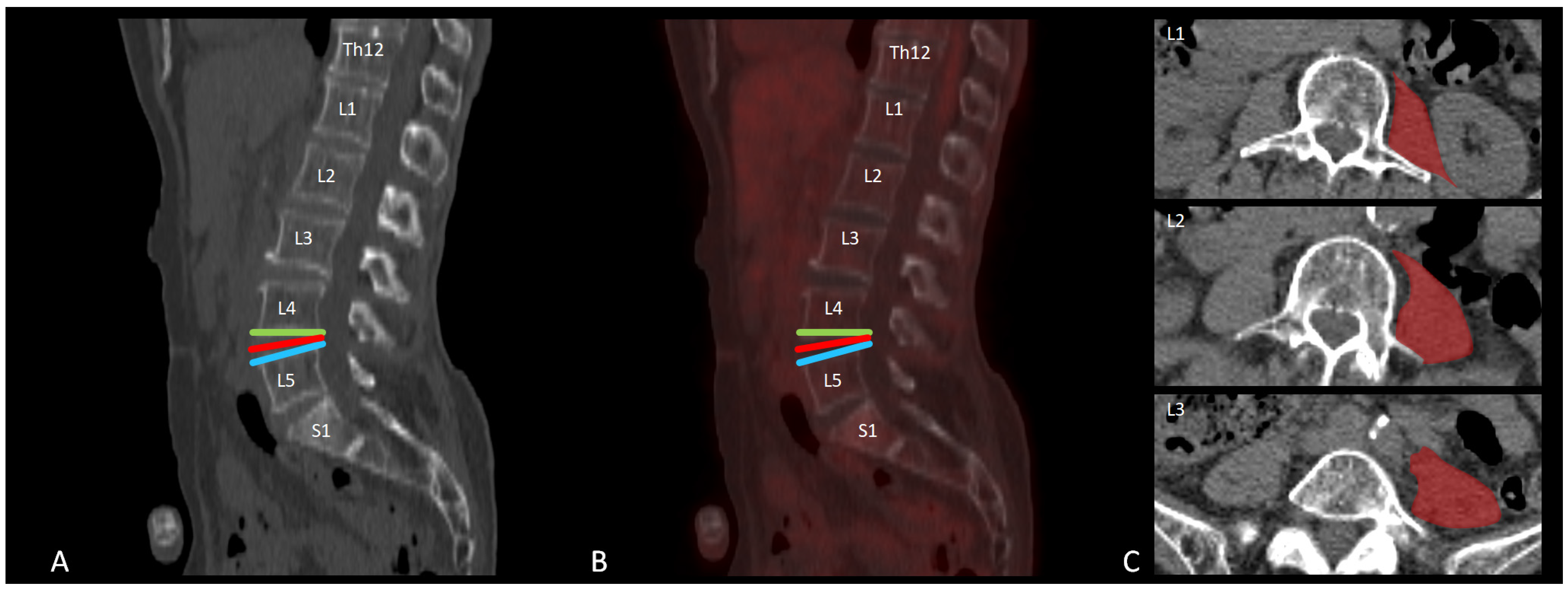
Figure 1. Example of a [18F]FDG PET-CT scan at the level of the lumbar spine of a non-painful individual to illustrate the regions of interest (ROIs) included in this study. (A) Sagittal low-dose CT reconstruction and (B) sagittal fused [18F]FDG-PET-CT image of the lumbar spine with ROIs at the level of L4–L5: superior endplate (green), intervertebral disc (red), and inferior endplate (blue). (C) Transversal low-dose CT images at three lumbar levels (L3–L5) with examples of ROI delineation (red) in the major psoas muscle.
Brainsci 15 00724 g001
Table 1. Overview of the SUVmean values and standard deviations per group and region.
LevelPatientsControlsp-Value *
Superior endplatesL10.55 (0.15)0.71 (0.38)0.266
L20.56 (0.17)0.75 (0.31)0.131
L30.56 (0.20)0.78 (0.31)0.097
L40.55 (0.17)0.80 (0.29)0.048
L50.60 (0.19)0.87 (0.37)0.074
S10.57 (0.21)0.78 (0.20)0.037
DiscusL1–L20.40 (0.10)0.52 (0.21)0.152
L2–L30.39 (0.20)0.47 (0.10)0.293
L3–L40.40 (0.24)0.55 (0.20)0.182
L4–L50.49 (0.26)0.71 (0.15)0.043
L5–S10.40 (0.14)0.75 (0.21)<0.001
T12–L10.44 (0.11)0.61 (0.31)0.134
Psoas leftL30.58 (0.51)0.43 (0.14)0.414
L40.55 (0.48)0.41 (0.15)0.414
L50.49 (0.39)0.41 (0.12)0.573
Psoas rightL30.60 (0.57)0.39 (0.11)0.301
L40.55 (0.55)0.44 (0.13)0.547
L50.46 (0.34)0.57 (0.29)0.446
Posterior endplatesL10.59 (0.13)0.67 (0.29)0.451
L20.57 (0.15)0.66 (0.23)0.331
L30.56 (0.12)0.78 (0.37)0.104
L40.58 (0.16)0.85 (0.30)0.029
L50.53 (0.14)0.79 (0.31)0.036
S10.59 (0.17)0.77 (0.36)0.195
* Adjustment for multiple comparisons: Bonferroni.
Disclaimer/Publisher’s Note: The statements, opinions and data contained in all publications are solely those of the individual author(s) and contributor(s) and not of MDPI and/or the editor(s). MDPI and/or the editor(s) disclaim responsibility for any injury to people or property resulting from any ideas, methods, instructions or products referred to in the content.