Systematic Review

Utilization of Medicinal Plants in Mental Disorders: Neuroplasticity and Neuroprotection in Biomodels

by
Jose Luis Estela-Zape
1,2,*,
Valeria Sanclemente-Cardoza
1,
Lizeth Dayana Noreña-Buitrón
1 and
Leidy Tatiana Ordoñez-Mora
1
1
Faculty of Health, Universidad Santiago de Cali, Cali 760035, Colombia
2
Faculty of Health, Posgrado en Ciencias Biomédicas, Universidad del Valle, Cali 760043, Colombia
*
Author to whom correspondence should be addressed.
Brain Sci. 2025, 15(4), 366; https://doi.org/10.3390/brainsci15040366
Submission received: 25 February 2025 / Revised: 19 March 2025 / Accepted: 20 March 2025 / Published: 31 March 2025
(This article belongs to the Section Neuropsychiatry)

Abstract

:
Background/Objectives: Mental disorders such as anxiety, schizophrenia, and depression are linked to alterations in neuroplasticity and neuroprotection within the central nervous system. While conventional drugs are widely used, medicinal plants are emerging as a promising alternative due to their potential therapeutic effects on neuronal function. This study aimed to explore and analyze the impact of medicinal plants on neuroplasticity and neuroprotection in relation to mental disorders using biomodels. Methods: Data were collected from Scopus, Dimensions, and PubMed by using the search terms “Medicinal plants”, “Neuronal Plasticity”, and “Mental Disorder” in accordance with the guidelines of the PRISMA checklist. Results: A total of twenty-three relevant studies were selected to investigate the association between medicinal plants and mental disorders, focusing on factors such as administered doses and the modulation of neurotransmitters in the context of neuroplasticity and neuroprotection. This review highlights the complexity of study designs, target populations, and methodologies. Of the studies, 86% investigated depression, while 13% focused on anxiety. Regarding neurotransmitter modulation, 47% found that medicinal plants influenced serotonin levels, followed by 27% which found that they affected dopamine; according to the remaining studies, medicinal plants impacted norepinephrine, GABA, and acetylcholine. These findings emphasize the importance of precise dosing and neurotransmitter modulation, suggesting that targeted interactions with neural systems may help clarify the specific effects of these plants on mental health. Conclusions: Research on the effects of medicinal plants on psychiatric disorders in animal models suggests their potential to support neuroplasticity and neuroprotection. Positive impacts on mental health are indicated through the modulation of cytokines, neurotransmitters, and specific signaling pathways.

1. Introduction

The central nervous system (CNS), as the body’s control axis, governs essential functions, including movement, cognition, and emotional regulation [1]. Neuronal dynamics support neuroplasticity and neuroprotection processes, facilitating adaptation and recovery from internal and external damage [2,3]. These processes rely on neurophysiological mechanisms such as synaptogenesis, synaptic plasticity, and neurogenesis, which enable the formation of new connections, the adjustment of existing synaptic strengths, and neuronal genesis [4]. By preserving cellular and neuronal integrity, neuroprotection fosters an environment conducive to neuroplasticity, promoting optimal CNS function [5].
Mental disorders (MDs) in humans, such as anxiety, schizophrenia, and depression, are associated with altered neuroplasticity and neuroprotection mechanisms within the CNS, leading to morphophysiological changes in brain structures and affecting emotional and cognitive functions [6]. Studies indicate that anxiety disrupts synaptic plasticity, impairing adaptive responses to stress. Schizophrenia induces imbalances in synaptic plasticity and neuronal connectivity, impacting perception, cognition, and emotions [7,8]. Depression is linked to reduced neurogenesis in the hippocampus and prefrontal cortex, compromising mood regulation and emotional resilience (Figure 1) [9].
Key brain areas include the amygdala, which mediates responses to fear and anxiety; the hippocampus, whose atrophy is associated with depressive symptoms; and the prefrontal cortex, responsible for emotional regulation and decision making. Genetic and environmental factors influence the regulation of neurotransmitters such as serotonin, dopamine, norepinephrine, GABA, and glutamate. Decreased serotonin levels are linked to depressive symptoms, while reduced dopamine contributes to anhedonia and motivation deficits. The dysregulation of norepinephrine is associated with heightened anxiety, and decreased GABA levels result in increased neuronal excitability. Excess glutamate can lead to synaptic hyperactivity, also associated with anxiety. The activation of the hypothalamic–pituitary–adrenal (HPA) axis in response to chronic stress elevates cortisol levels, resulting in amygdala hyperactivity and reduced hippocampal volume, thereby exacerbating symptoms of anxiety, depression, and stress-related disorders.
Dysfunctions in neuronal dynamics signify brain function deterioration, impacting both mental health and global economic costs. According to the World Health Organization (WHO), the economic burden is estimated at approximately USD one trillion annually, with an alarming 13% increase in MDs [10,11,12].
Standard treatments for mental disorders include antidepressants, mood stabilizers, anxiolytics, and antipsychotics, which modulate neuronal activity and contribute to neuroprotection and neuroplasticity, potentially mitigating cognitive decline in neurodegenerative diseases [13,14]. Research shows that anti-inflammatory agents and antioxidants like vitamins C, E, and A act within the CNS to protect cells from oxidative stress and reduce inflammation, thereby supporting neuronal plasticity. In Parkinson’s disease, certain drugs act as metabolic precursors to dopamine, preventing the degradation of dopaminergic neurons due to mitochondrial dysfunction and apoptotic processes typical of neurodegenerative conditions. Additionally, antidepressants and anxiolytics, particularly neurotransmitter reuptake inhibitors, promote neurogenesis in regions such as the hippocampus, enhancing dendritic growth and neuronal connectivity.
Nevertheless, these therapies face limitations due to complex changes in neuronal dynamics, including receptor imbalances, altered neurotransmitter expression, and reduced neurogenesis. Such factors can lead to decreased efficacy, side effects, and drug resistance, highlighting the need for more effective and tolerable therapeutic alternatives [2].
Medicinal plants have been proposed as a promising alternative in the academic field because of their wide spectrum of therapeutic properties capable of influencing neuronal dynamics, whether in animal or human studies. These properties range from the activation of receptors and enzyme inhibition to the modulation of gene expression and the regulation of neurotransmitters, such as dopamine, serotonin, and gamma-aminobutyric acid (GABA), which contribute to cellular protection and exhibit antioxidant effects [15,16].
Research has shown that Lavandula angustifolia and Valeriana jatamansi can modulate the GABAergic system, addressing anxiety and insomnia [17,18]. In preclinical models, species like Crocus sativus [18], Coffea arabica, and Bacopa monnieri have demonstrated potential in regulating brain-derived neurotrophic factor (BDNF), a key factor in neurogenesis, synaptic plasticity, and neuronal survival, contributing to antidepressant effects during chronic stress [19,20,21].
Studies also suggest that Crocus sativus, Coffea arabica, and Bacopa monnieri exhibit neuroprotective properties, potentially counteracting β-amyloid neurotoxicity, oxidative stress, and glutamate excitotoxicity. Additionally, cannabidiol (CBD) may not only regulate BDNF but also mitigate the adverse effects of tetrahydrocannabinol (THC), supporting neuronal protection and synaptic remodeling [22]. Rhodiola rosea and Ginkgo biloba may reduce inflammation, protect neurons from oxidative damage, enhance synaptic plasticity, and reduce cell death, showing promise for managing depression and schizophrenia [14,23].
The effects of medicinal plants on mental disorders remain insufficiently understood, with inconclusive and heterogeneous findings. While some studies suggest therapeutic potential, significant gaps and inconsistencies persist regarding non-pharmacological treatments. This underscores the need for rigorous research on various plant species documented in scientific literature. Accordingly, a systematic review is proposed to evaluate and analyze the effects of medicinal plants on neuroplasticity and neuroprotection in mental disorders using biomodels. This review aims to consolidate current knowledge, deepening our understanding of the therapeutic properties of medicinal plants and their impact on complex neuronal dynamics associated with mental disorders in animals.

2. Materials and Methods

A systematic review of the literature was carried out according to the guidelines established by the PRISMA checklist [24] to ensure uniformity and quality in the presentation of the reports.

2.1. Search Strategy

2.1.1. Source of Information

A search strategy was developed for SCOPUS, DIMENSIONS, and PUBMED, tailored to each database and incorporating combinations of medical subject headings and free-text terms related to mental disorders, medicinal plants, and study types. The targeted search included indexed terms and free text from sources such as abstracts and ongoing clinical trials. Articles were included in any language and without restriction on the publication date.

2.1.2. Review Question

The research question was formulated and developed by using the PICO strategy: “What are the effects of medicinal plants on neuroplasticity and neuroprotection in mental disorders in biomodels?” The following elements were determined:
-
Population: Biomodels used in the study of mental disorders.
-
Intervention: Administration of medicinal plants.
-
Comparison: Not applicable.
-
Outcomes: Measures of neuroplasticity and neuroprotection.

2.1.3. Search Terms

The search was carried out by using DeCS/MeSH terms, together with the logical operators “OR” and “AND”, with the purpose of constructing a search equation, i.e., (((((((Plants) OR (Medicinal plants)) OR (Phytochemicals)) OR (Traditional Medicine)) OR (Phytotherapy)) AND (Neuronal Plasticity)) AND (Neuroprotective Agents)) AND (Mental Disorder), directly related to the research goal. Appendix A shows the search strategies used in the different databases.

2.1.4. Eligibility and Exclusion Criteria

Studies were included if they evaluated the effects of medicinal plants on mental disorders, specifically anxiety disorder, bipolar disorder, depression, stress, attention deficit hyperactivity disorder, and schizophrenia, considered as primary and secondary outcomes. Only randomized controlled trials (RCTs) and original research articles were included, with no publication date restrictions.
Exclusion criteria were set to omit studies focusing on Alzheimer’s disease, Parkinson’s disease, and Huntington’s disease, as these are classified as neurocognitive disorders in the Diagnostic and Statistical Manual of Mental Disorders (DSM-5) [25]. Additionally, we excluded studies not related to mental disorders, neuroplasticity, or neuroprotection; those involving combinations of plants, synthesized plant-derived compounds, extracts, or commercial medications; and those with incomplete data, reviews, or gray literature.

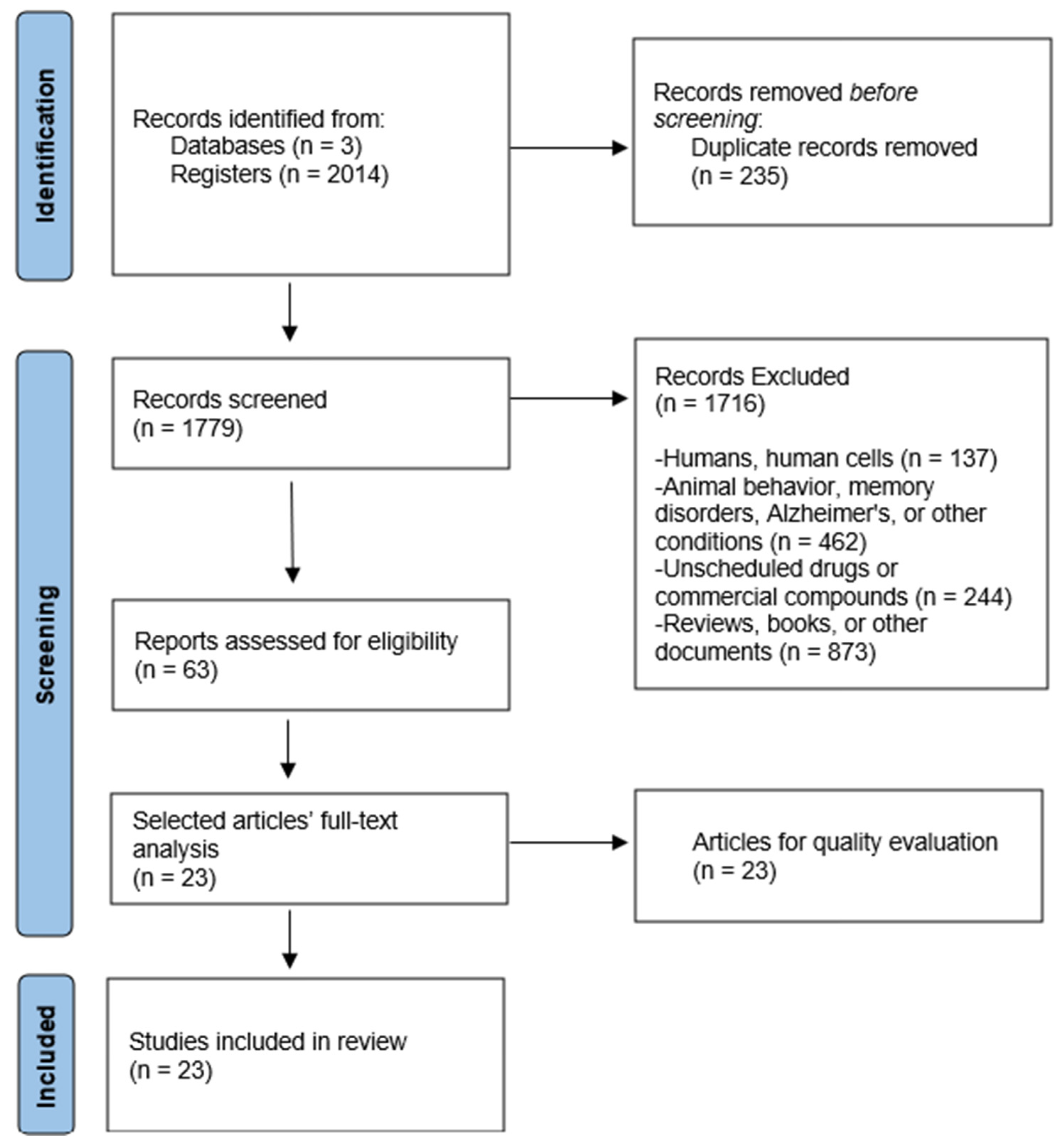
2.2. Study Selection

Studies were selected through a rigorous calibration procedure. Initially, three researchers (L.D.N.B., V.S.C., and L.T.O.M.) independently and blindly carried out the filtering process in various databases.
Each researcher prepared a list of studies that met the inclusion criteria after evaluating the title and abstract of the articles. In situations in which opposition arose among the three reviewers, a fourth reviewer (J.L.E.Z.), an expert on the subject, made the final decision regarding the inclusion of the articles. It is essential to note that the review process was blinded.
Eligibility criteria were applied during the full-text analysis phase in the final selection stage, and any disagreement among the authors regarding the appropriateness, eligibility, quality, or data obtained from the studies was resolved by consensus.

2.3. Quality Evaluation

The SYRCLE Risk of Bias (RoB) tool [26], adapted from the Cochrane RoB tool, was used to conduct a comprehensive methodological assessment of the animal intervention studies included in the analysis. This tool addresses bias factors specific to animal studies and contains ten items: six related to types of bias, one evaluating baseline characteristic similarity between experimental and control groups, one assessing the randomization of the housing conditions, one indicating whether disease induction occurred before or after randomization, and one evaluating blinding. Studies were rated “yes” (green) for low risk of bias, “no” (red) for high risk, and “unclear” (yellow) for uncertain risk. Only studies scoring six or higher were included.

3. Results

A total of 2014 records were initially identified through database analysis. After the removal of duplicates, a detailed review of 1779 articles was conducted, applying inclusion and exclusion criteria to finalize the selection. This process resulted in the identification of 63 relevant studies. Ultimately, 23 articles were included in this review, and their methodological quality was assessed by using the SYRCLE Risk of Bias (RoB) tool [26]. Figure 2 presents a graphical representation of the study selection process.

3.1. Methodological Quality

Out of the 23 studies evaluated, 65.2% (15 studies) obtained scores of 7, 8, and 9 when analyzed by using the RoB tool [26], which reflects a low risk of bias and high methodological quality. In contrast, only 8.69% (two studies) scored 4, suggesting a high risk of bias. The main deficiencies identified in these cases were inadequate randomization procedures, a lack of specification in concealment processes, and difficulties in blinding. A detailed methodological evaluation of each study is presented in Table 1.

3.2. Data Extraction and Synthesis

The study design, target population, and methodology differed among the studies, thus providing a comprehensive approach to analyzing the relationship between medicinal plants and mental disorders, as shown in Table 2. The considerable variability in the study design, populations studied, and methodologies used highlights the complexity inherent in the exploration of the association between medicinal plants and mental disorders.
As shown in Table 3, 86.6% of the studies included focused on depression, while 13.4% addressed anxiety. Regarding neurotransmitter effects, 47% (11 studies) of the medicinal plants studied affected serotonin, 27% (4 studies) influenced dopamine, and the remaining 26% impacted norepinephrine, GABA, and acetylcholine. The attention paid to dosage and neurotransmitter modulation highlights the necessity for precise administration and targeted interaction with neuronal systems to better understand the specific effects of these plants on the nervous system. These findings not only strengthen current knowledge but also provide a robust foundation for future research and clinical applications in psychiatric phytotherapy.

4. Discussion

The analysis conducted in this study suggests the importance of medicinal plants in addressing prevalent psychiatric disorders, including anxiety and depression, highlighting their potential therapeutic potential in the context of neuroplasticity and neuroprotection.

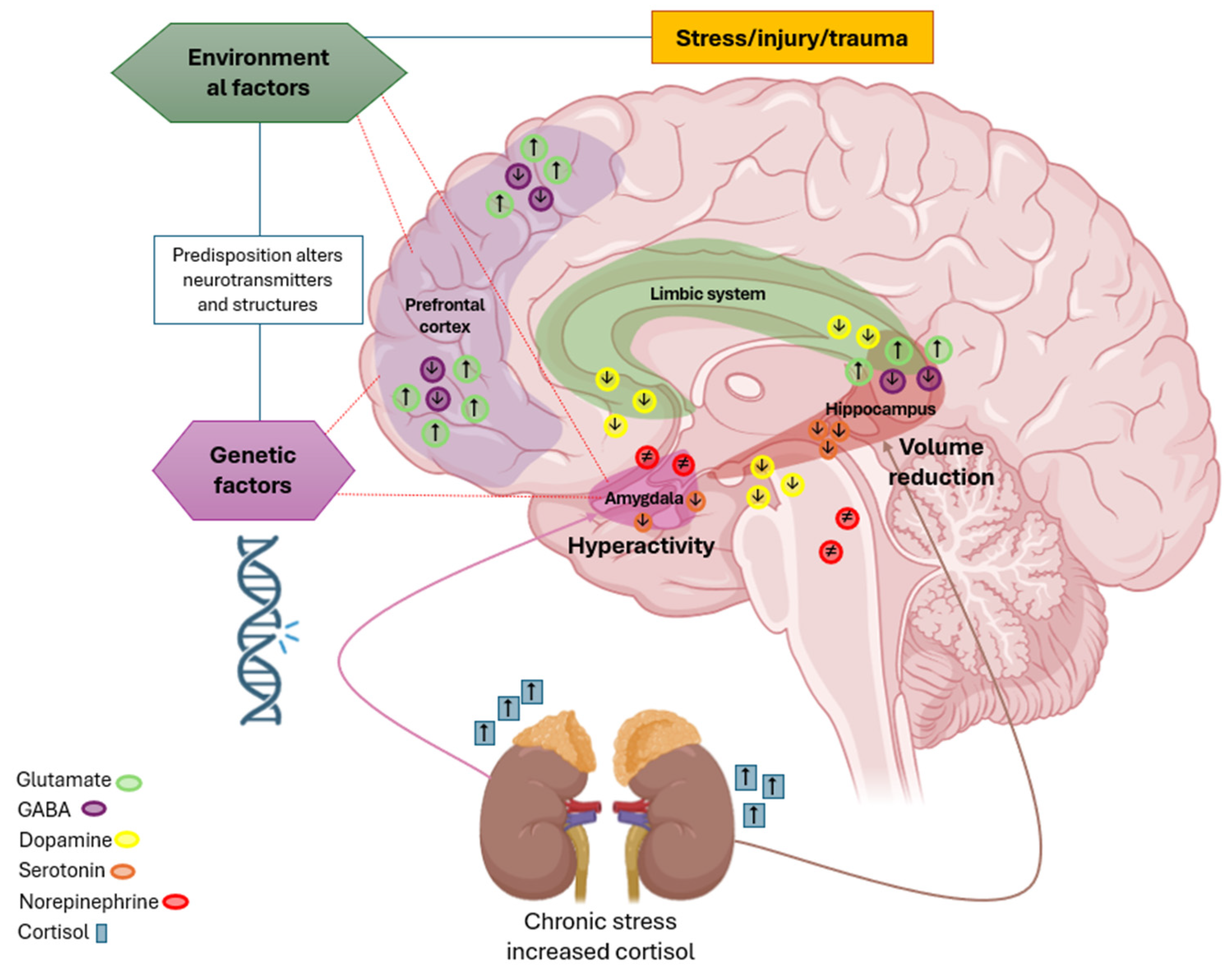
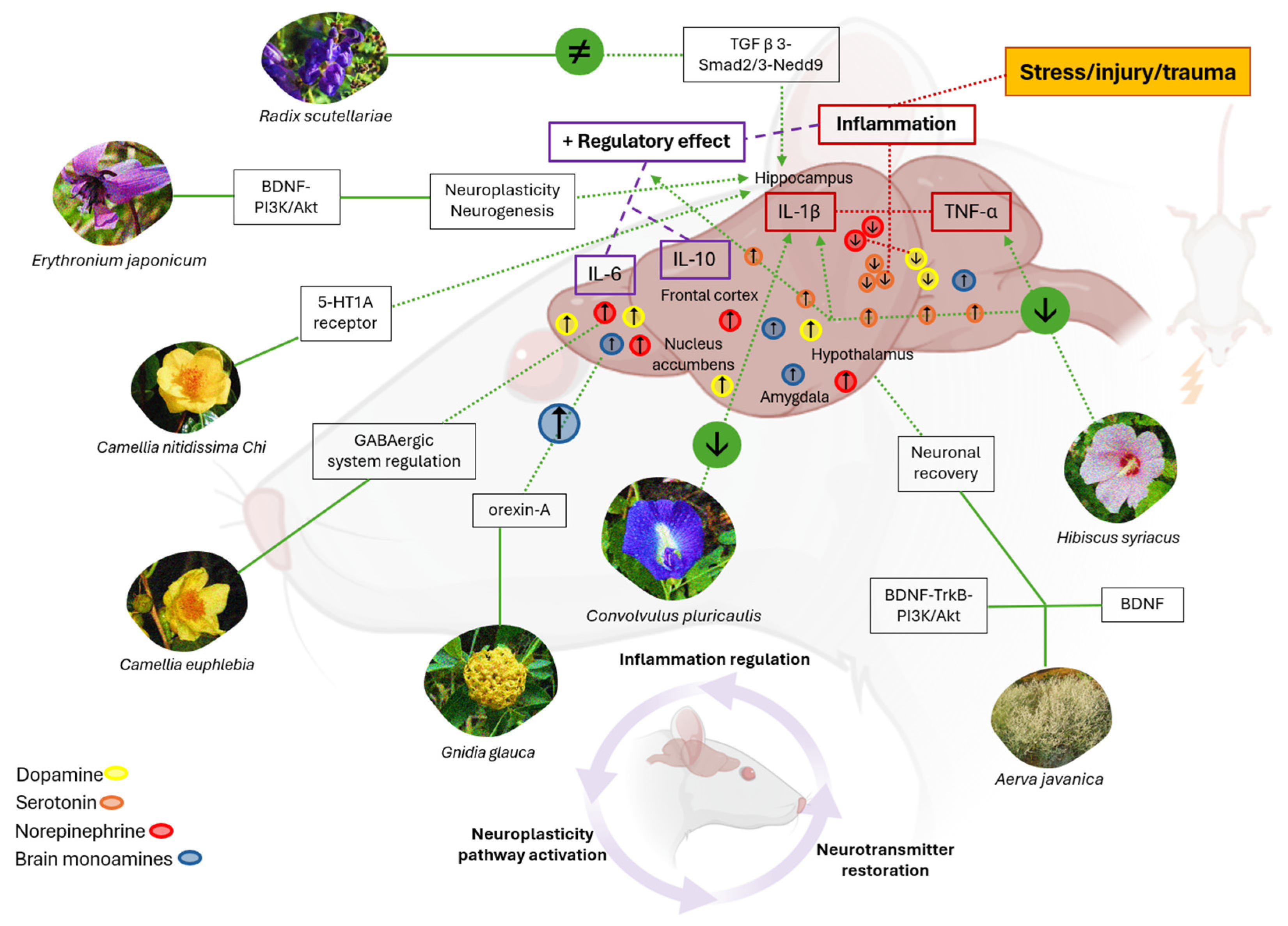
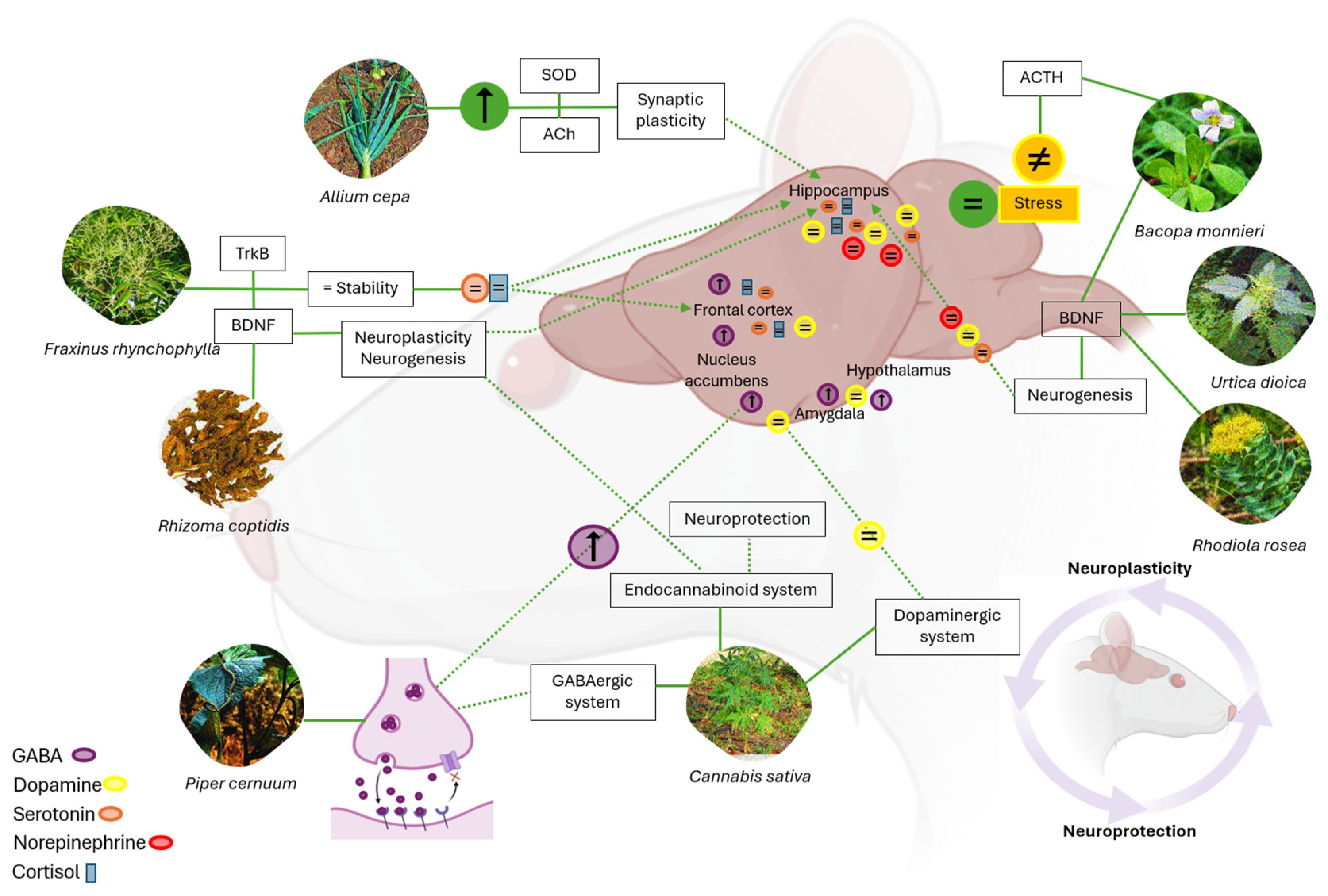
4.1. Phytomedicine Impact on Cytokines, Neurotransmitters, and Neuroplasticity

All gathered studies focused on the effects of phytomedicine in animal models, mostly using adult male mice as a standard population. It should be noted, however, that several studies chose to include female mice because of the greater susceptibility to depression [35,38]. These analyses followed a design that combined in vivo and in vitro approaches, with the specific addition of in silico models [36,40].
Environmental and psychosocial stressors were generated during the trials to induce chronic stress, which allowed us to obtain evidence of destabilization in the balance of cytokines (tumor necrosis factor alpha (TNF-α), interleukin-1 beta (IL-1β), interleukin-6 (IL-6), and interleukin-10 (IL-10)), signaling molecules in the immune response and neuronal communication (Figure 3).
Cytokine imbalance is associated with a reduction in brain amines, including serotonin, dopamine, and norepinephrine, which regulate emotional and cognitive functions. These biochemical changes show a complex interaction between prolonged stress and neurological homeostasis, which may contribute to the development of depression, brain inflammation, and neurotransmitter deregulation. These factors affect the neurochemical mechanisms and neuronal pathways involved in psychiatric disorders [27].
Stress increases inflammatory cytokines (TNF-α, IL-1β, and IL-6), which disrupt neurotransmitter levels (serotonin, dopamine, and norepinephrine), contributing to disorders such as anxiety, depression, and neuroinflammation. Plants like Camellia euphlebia, Camellia nitidissima Chi, Gnidia glauca, Convolvulus pluricaulis, Hibiscus syriacus, and Erythronium japonicum exert anti-inflammatory effects by regulating cytokine levels and restoring neurotransmitter balance, alleviating symptoms of anxiety and depression. Additionally, Aerva javanica and Radix Scutellariae activate neuroplasticity pathways, such as BDNF–PI3K/Akt, which promote neuronal growth and recovery in the hippocampus, mitigating the effects of chronic stress and enhancing overall cognitive function.
Standardized behavioral tests, such as the forced swim test (FST) and the tail suspension test (TST), are essential tools to determine the therapeutic effects of medicinal plants [31]. For example, He et al. (2015) [33] found that the aqueous extract of Camellia euphlebia has anxiolytic effects in the TST, thus alleviating the dysfunction of the GABAergic system and regulating neuronal excitation in mood disorders. Similarly, Tsoi et al. (2022) [37] reported that Camellia nitidissima Chi extract reduces stress and anxiety, thus regulating the hypothalamic–pituitary–adrenocortical (HPA) system and inhibiting chronic corticosterone (CORT) levels. This allows for the modulation of the serotonergic system and 5-HTR1A receptor expression in the hippocampus of mice.
Furthermore, Arika et al. (2019) [38] studied Gnidia glauca using the FST and TST and demonstrated the ability to modulate anxiety through the action of the neuropeptide orexin-A and the association between the increase in brain levels of mood-regulating monoamines. Lim et al. (2020) [41] also found that Erythoronium japonicum leaves activate the BDNF–PI3K/Akt pathway in the hippocampus of mice, involved in neuronal growth, survival, and plasticity. When activated by PI3K/Akt, BDNF appears to influence the regulation of the inflammatory response in the brain, thus exerting anti-inflammatory effects and improving depressive behavior in animal models.
The sucrose preference test (SPT) has been used to evaluate anhedonia, an important marker of depressive disorders in mice [39]. In the study conducted by Kim et al. (2018) [29], the use of Hibiscus syriacus L root reduced pro-inflammatory cytokines (IL-1β, IL-6, IL-8, and TNF-α), which correlated with an increase in the brain levels of serotonin. The findings indicate a potential neuroprotective and inflammation-regulating effect, which could positively impact emotional well-being. Arshad, H.M. et al. (2022) [36], however, demonstrated that the use of Aerva javanica leaves increased BDNF expression through the compound’s quercetin and kaempferol, which generated an antidepressant effect through the BDNF–TrkB–PI3K/Akt pathway.
Other studies [27,39] determined that Convolvulus pluricaulis extract markedly reduced IL-1β in the plasma of rats and decreased elevated levels of ALT liver enzymes. Radix Scutellariae, on the other hand, regulated neuroprotection in the hippocampus of mice, by inhibiting neuronal apoptosis through the TGF β 3–Smad2/3–Nedd9 signaling pathway. These mechanisms may be necessary to preserve neuronal integrity and reduce harmful processes that could contribute to neuropsychiatric diseases.

4.2. Role of Neurotransmitters in Neuroplasticity and Neuroprotection

The impact of medicinal plants on neuroplasticity and neuroprotection in mental disorders has been demonstrated in biomodels studies, which show that their bioactive compounds can modulate neurotransmitters, reduce oxidative stress, and promote neurogenesis [42,49] (Figure 4). Liu et al. (2023) [43] note that the hippocampus regulates neuroplasticity, and in depression, synapses in this region deteriorate, leading to the dysregulation of serotonin (5-HT) levels.
Neurotransmitters such as serotonin and norepinephrine play a key role in regulating mood in depression [48]. Additionally, depression is associated with decreased enzymatic antioxidant defenses and increased levels of reactive oxygen species [44]. Samad et al. (2018) [28] investigated the role of acetylcholine in the development of anxiety and depression. Their findings suggest that Allium cepa has antidepressant and anxiolytic effects by enhancing the activity of superoxide dismutase (SOD), an enzyme that reduces oxidative stress and cellular damage. Additionally, it increases acetylcholinesterase (AChE) activity, which modulates acetylcholine availability in neuronal synapses.
Medicinal plants such as Allium cepa, Fraxinus rhynchophylla, Bacopa monnieri, Urtica dioica, Rhizoma coptidis, Piper cernuum, Rhodiola rosea, and Cannabis sativa have beneficial effects on neuroplasticity and neuroprotection. Allium cepa increases superoxide dismutase (SOD) and acetylcholine, reducing oxidative stress. Fraxinus rhynchophylla normalizes serotonin and cortisol levels, promoting neurogenesis. Bacopa monnieri increases brain-derived neurotrophic factor (BDNF) and regulates adrenocorticotropic hormone (ACTH), supporting new-neuron formation. Urtica dioica modulates the hormonal axis and enhances neuronal regeneration. Rhizoma coptidis increases BDNF, alleviating stress. Piper cernuum enhances synaptic plasticity and modulates neuronal excitability by increasing GABA levels. Rhodiola rosea regulates key neurotransmitters and protects neurons from stress-induced damage. Cannabis sativa, through THC and CBD, influences synaptic plasticity, neurogenesis, and emotional regulation, supporting cognitive health.
Kim, R. et al. (2018) [30] observed that Fraxinus rhynchophylla Hance extract improves anxiety and depressive behavior. This effect can be attributed to the normalization of serotonin and cortisol levels, which affect BDNF and TrkB signaling in the hippocampus of mice. Furthermore, other studies [31,33,38] have determined that extracts from plants, including Bacopa monnieri, Urtica dioica, and Rhizoma coptidis, affect hormonal regulation and neurogenesis in the hippocampus of rats. The extracts appear to affect the production of adrenocorticotropic hormone (ACTH), related to stress, and the increase in BDNF, thus promoting the formation of new neurons. These components could help improve neuronal plasticity and protect brain cells.
Conversely, the study by Maia, M. et al. (2023) [35] examined the anxiolytic potential of Piper cernuum leaves by increasing GABA levels and improving the GABAergic system. The increase in GABA could be associated with the reduction in neuronal excitability and, potentially, could improve synaptic plasticity to mitigate these disorders, considering that GABA is the main inhibitory neurotransmitter in the brain and its alteration is related to anxiety and depression [40,41].
The potential of medicinal plants for neuroplasticity and neuroprotection in psychiatric disorders is an emerging research area. Current evidence, primarily from animal models, indicates that certain plant extracts can affect key neurochemical pathways, such as the BDNF–PI3K/Akt system and the GABAergic system, enhancing mood regulation and reducing brain inflammation. Notable examples include flavonoids from fruits and vegetables, cannabinoids from Cannabis sativa, and adaptogens like Rhodiola rosea, which demonstrate neuroprotective and neuroplastic properties by modulating cortical plasticity and optimizing neuronal synapses.
However, reliance on animal models limits the direct translation of these findings into human clinical practice. Variability in plant extracts and a lack of methodological standardization restrict clinical applicability. The diversity of specific compounds and mechanisms of action further complicates the generalization of results.
To advance this field, rigorous clinical studies are necessary to assess the efficacy and safety of medicinal plants in humans. Additionally, a multidisciplinary and integrative approach is crucial to developing precise and personalized phytotherapeutic treatments, tailored to the specific characteristics of each neuropsychiatric disorder and individual patient needs.
Despite these limitations, this study is notable for its methodological diversity, encompassing molecular analyses and standardized behavioral tests. The integration of vivo, in vitro, and in silico models strengthens the validity and comprehensiveness of the results, providing a solid foundation for future research and therapeutic advancements in phytotherapy with promising prospects for neuropsychiatric disorder treatment.

5. Conclusions

The investigation of molecular mechanisms in medicinal plants using animal models has identified critical pathways that enhance neuroplasticity and provide neuroprotection in psychiatric disorders. Specifically, the studies demonstrate how these plants modulate cytokine expression, regulate neurotransmitter systems, and activate neuroprotective signaling cascades. These findings highlight the therapeutic potential of specific medicinal plants and emphasize the need for focused research to identify the most promising candidates for clinical application. Advanced research could lead to novel treatments for neuropsychiatric disorders, significantly contributing to psychopharmacology and improving mental health outcomes.

Author Contributions

Conceptualization, J.L.E.-Z., V.S.-C. and L.D.N.-B.; methodology, J.L.E.-Z., V.S.-C., L.D.N.-B. and L.T.O.-M.; validation, J.L.E.-Z.; formal analysis J.L.E.-Z., V.S.-C., L.D.N.-B. and L.T.O.-M.; investigation, J.L.E.-Z., V.S.-C. and L.D.N.-B.; resources, J.L.E.-Z.; data curation, J.L.E.-Z., V.S.-C. and L.D.N.-B.; writing—original draft preparation, J.L.E.-Z., V.S.-C. and L.D.N.-B.; writing—review and editing, J.L.E.-Z. and V.S.-C.; visualization, J.L.E.-Z. and L.T.O.-M.; supervision, J.L.E.-Z.; project administration, J.L.E.-Z.; funding acquisition, J.L.E.-Z. and L.T.O.-M. All authors have read and agreed to the published version of the manuscript.

Funding

This research has been funded by the General Directorate of Investigations of Universidad Santiago de Cali under call No. DGI 01-2025.

Conflicts of Interest

The authors declare no conflicts of interest.

Abbreviations

The following abbreviations are used in this manuscript:
CNScentral nervous system
MDsmental disorders
HPAhypothalamic–pituitary–adrenal
WHOWorld Health Organization
GABAgamma-aminobutyric acid
CBDcannabidiol
BDNFbrain-derived neurotrophic factor
THCtetrahydrocannabinol
TNF-αtumor necrosis factor alpha
IL-1βinterleukin-1 beta
IL-6interleukin-6
IL-10interleukin-10
AChEacetylcholinesterase

Appendix A

Table A1. Search strategies for different databases.
DatabaseSearch DateSearch EquationArticles Found
Scopus1 October 2025(((((((Plants) OR (Medicinal plants)) OR (Phytochemicals)) OR (Traditional Medicine)) OR (Phytotherapy)) AND (Neuronal Plasticity)) AND (Neuroprotective Agents)) AND (Mental Disorder) 1950
PubMed1 November 2025(((((((Plants) OR (Medicinal plants)) OR (Phytochemicals)) OR (Traditional Medicine)) OR (Phytotherapy)) AND (Neuronal Plasticity)) AND (Neuroprotective Agents)) AND (Mental Disorder)46
Dimensions1 May 2025(((((((Plants) OR (Medicinal plants)) OR (Phytochemicals)) OR (Traditional Medicine)) OR (Phytotherapy)) AND (Neuronal Plasticity)) AND (Neuroprotective Agents)) AND (Mental Disorder)18

References

  1. Colucci-D’Amato, L.; Speranza, L.; Volpicelli, F. Neurotrophic Factor BDNF, Physiological Functions and Therapeutic Potential in Depression, Neurodegeneration and Brain Cancer. Int. J. Mol. Sci. 2020, 21, 7777. [Google Scholar] [CrossRef] [PubMed]
  2. Wu, A.; Zhang, J. Neuroinflammation, memory, and depression: New approaches to hippocampal neurogenesis. J. Neuroinflammation 2023, 20, 283. [Google Scholar] [CrossRef] [PubMed]
  3. Magee, J.C.; Grienberger, C. Synaptic Plasticity Forms and Functions. Annu. Rev. Neurosci. 2020, 43, 95–117. [Google Scholar] [CrossRef] [PubMed]
  4. Price, R.B.; Duman, R. Neuroplasticity in cognitive and psychological mechanisms of depression: An integrative model. Mol. Psychiatry 2020, 25, 530–543. [Google Scholar] [CrossRef]
  5. Budzynska, B.; Faggio, C.; Kruk-Slomka, M.; Samec, D.; Nabavi, S.F.; Sureda, A.; Devi, K.P.; Nabavi, S.M. Rutin as Neuroprotective Agent: From Bench to Bedside. Curr. Med. Chem. 2019, 26, 5152–5164. [Google Scholar] [CrossRef]
  6. Troubat, R.; Barone, P.; Leman, S.; Desmidt, T.; Cressant, A.; Atanasova, B.; Brizard, B.; El Hage, W.; Surget, A.; Belzung, C.; et al. Neuroinflammation and depression: A review. Eur. J. Neurosci. 2021, 53, 151–171. [Google Scholar] [CrossRef]
  7. Petković, A.; Chaudhury, D. Encore: Behavioural animal models of stress, depression and mood disorders. Front. Behav. Neurosci. 2022, 16, 931964. [Google Scholar] [CrossRef]
  8. Lashgari, N.A.; Roudsari, N.M.; Shamsnia, H.S.; Shayan, M.; Momtaz, S.; Abdolghaffari, A.H. TLR/mTOR inflammatory signaling pathway: Novel insight for the treatment of schizophrenia. Can. J. Physiol. Pharmacol. 2023, 102, 150–160. [Google Scholar] [CrossRef]
  9. Fathinezhad, Z.; Sewell, R.D.E.; Lorigooini, Z.; Rafieian-Kopaei, M. Depression and Treatment with Effective Herbs. Curr. Pharm. Des. 2019, 25, 738–745. [Google Scholar] [CrossRef]
  10. World Health Organization. The WHO Special Initiative for Mental Health (‎2019–2023)‎: Universal Health Coverage for Mental Health. World Health Organization. 2019. Licencia: CC BY-NC-SA 3.0 IGO. Available online: https://iris.who.int/handle/10665/310981 (accessed on 7 December 2023).
  11. Arias, D.; Saxena, S.; Verguet, S. Quantifying the global burden of mental disorders and their economic value. eClinicalMedicine 2022, 54, 101675. [Google Scholar] [CrossRef]
  12. Moitra, M.; Owens, S.; Hailemariam, M.; Wilson, K.S.; Mensa-Kwao, A.; Gonese, G.; Kamamia, C.K.; White, B.; Young, D.M.; Collins, P.Y. Global Mental Health: Where We Are and Where We Are Going. Curr. Psychiatry Rep. 2023, 25, 301–311. [Google Scholar] [CrossRef]
  13. Asher, G.N.; Gerkin, J.; Gaynes, B.N. Complementary Therapies for Mental Health Disorders. Med. Clin. N. Am. 2017, 101, 847–864. [Google Scholar] [CrossRef] [PubMed]
  14. Rosson, S.; de Filippis, R.; Croatto, G.; Collantoni, E.; Pallottino, S.; Guinart, D.; Brunoni, A.R.; Dell’osso, B.; Pigato, G.; Hyde, J.; et al. Brain stimulation and other biological non-pharmacological interventions in mental disorders: An umbrella review. Neurosci. Biobehav. Rev. 2022, 139, 104743. [Google Scholar] [CrossRef]
  15. Goodwin, G.M.; Stein, D.J. Generalised Anxiety Disorder and Depression: Contemporary Treatment Approaches. Adv. Ther. 2021, 38 (Suppl. 2), 45–51. [Google Scholar] [CrossRef] [PubMed]
  16. Sarris, J. Herbal medicines in the treatment of psychiatric disorders: 10-year updated review. Phytother. Res. 2018, 32, 1147–1162. [Google Scholar] [CrossRef]
  17. López, V.; Nielsen, B.; Solas, M.; Ramírez, M.J.; Jäger, A.K. Exploring Pharmacological Mechanisms of Lavender (Lavandula angustifolia) Essential Oil on Central Nervous System Targets. Front. Pharmacol. 2017, 8, 280. [Google Scholar] [CrossRef]
  18. Ota, M.; Ni, H.; Maki, Y.; Kato, D.; Moriguchi, S.; Nakayama, S.; Oiwa, Y.; Ishiuchi, K.; Makino, T. Binding activity of Valeriana fauriei root extract on GABAA receptor flunitrazepam sites and distribution of its active ingredients in the brain of mice—A comparison with that of V. officinalis root. J. Ethnopharmacol. 2021, 278, 114262. [Google Scholar] [CrossRef]
  19. Lopresti, A.L.; Drummond, P.D. Saffron (Crocus sativus) for depression: A systematic review of clinical studies and examination of underlying antidepressant mechanisms of action. Hum. Psychopharmacol. 2014, 29, 517–527. [Google Scholar] [CrossRef]
  20. El Midaoui, A.; Ghzaiel, I.; Vervandier-Fasseur, D.; Ksila, M.; Zarrouk, A.; Nury, T.; Khallouki, F.; El Hessni, A.; Ibrahimi, S.O.; Latruffe, N.; et al. Saffron (Crocus sativus L.): A Source of Nutrients for Health and for the Treatment of Neuropsychiatric and Age-Related Diseases. Nutrients 2022, 14, 597. [Google Scholar] [CrossRef]
  21. Hirshler, Y.; Doron, R. Neuroplasticity-related mechanisms underlying the antidepressant-like effects of traditional herbal medicines. Eur. Neuropsychopharmacol. 2017, 27, 945–958. [Google Scholar] [CrossRef]
  22. Sangiovanni, E.; Brivio, P.; Dell’Agli, M.; Calabrese, F. Botanicals as Modulators of Neuroplasticity: Focus on BDNF. Neural Plast. 2017, 2017, 5965371. [Google Scholar] [CrossRef] [PubMed]
  23. Campos, A.C.; Fogaça, M.V.; Scarante, F.F.; Joca, S.R.L.; Sales, A.J.; Gomes, F.V.; Sonego, A.B.; Rodrigues, N.S.; Galve-Roperh, I.; Guimarães, F.S. Plastic and Neuroprotective Mechanisms Involved in the Therapeutic Effects of Cannabidiol in Psychiatric Disorders. Front. Pharmacol. 2017, 8, 269. [Google Scholar] [CrossRef] [PubMed]
  24. Page, M.J.; McKenzie, J.E.; Bossuyt, P.M.; Boutron, I.; Hoffmann, T.C.; Mulrow, C.D.; Shamseer, L.; Tetzlaff, J.M.; Akl, E.A.; Brennan, S.E.; et al. The PRISMA 2020 statement: An updated guideline for reporting systematic reviews. BMJ 2021, 372, n71. [Google Scholar] [CrossRef] [PubMed]
  25. American Psychiatric Association. Diagnostic and Statistical Manual of Mental Disorders, 5th ed.; American Psychiatric Association: Arlington, VA, USA, 2013. [Google Scholar] [CrossRef]
  26. Hooijmans, C.R.; Rovers, M.M.; de Vries, R.B.; Leenaars, M.; Ritskes-Hoitinga, M.; Langendam, M.W. SYRCLE’s risk of bias tool for animal studies. BMC Med. Res. Methodol. 2014, 14, 43. [Google Scholar] [CrossRef]
  27. Gupta, G.L.; Fernandes, J. Protective effect of Convolvulus pluricaulis against neuroinflammation associated depressive behavior induced by chronic unpredictable mild stress in rat. Biomed. Pharmacother. 2019, 109, 1698–1708. [Google Scholar] [CrossRef]
  28. Samad, N.; Saleem, A. Administration of Allium cepa L. bulb attenuates stress-produced anxiety and depression and improves memory in male mice. Metab. Brain Dis. 2018, 33, 271–281. [Google Scholar] [CrossRef]
  29. Kim, Y.H.; Im, A.R.; Park, B.K.; Paek, S.H.; Choi, G.; Kim, Y.R.; Whang, W.K.; Lee, K.H.; Oh, S.-E.; Lee, M.Y. Antidepressant-Like and Neuroprotective Effects of Ethanol Extract from the Root Bark of Hibiscus syriacus L. BioMed Res. Int. 2018, 2018, 7383869. [Google Scholar] [CrossRef]
  30. Kim, Y.R.; Park, B.K.; Kim, Y.H.; Shim, I.; Kang, I.C.; Lee, M.Y. Antidepressant Effect of Fraxinus rhynchophylla Hance Extract in a Mouse Model of Chronic Stress-Induced Depression. BioMed Res. Int. 2018, 2018, 8249563. [Google Scholar] [CrossRef]
  31. Kumar, S.; Mondal, A.C. Neuroprotective, Neurotrophic and Anti-oxidative Role of Bacopa monnieri on CUS Induced Model of Depression in Rat. Neurochem. Res. 2016, 41, 3083–3094. [Google Scholar] [CrossRef]
  32. Chen, W.C.; Lai, Y.S.; Lin, S.H.; Lu, K.H.; Lin, Y.E.; Panyod, S.; Ho, C.-T.; Sheen, L.-Y. Anti-depressant effects of Gastrodia elata Blume and its compounds gastrodin and 4-hydroxybenzyl alcohol, via the monoaminergic system and neuronal cytoskeletal remodeling. J. Ethnopharmacol. 2016, 182, 190–199. [Google Scholar] [CrossRef]
  33. He, D.; Wang, X.; Zhang, P.; Luo, X.; Li, X.; Wang, L.; Li, S.; Xu, Y. Evaluation of the Anxiolytic and Antidepressant Activities of the Aqueous Extract from Camellia euphlebia Merr. ex Sealy in Mice. Evid.-Based Complement. Altern. Med. 2015, 2015, 618409. [Google Scholar] [CrossRef] [PubMed]
  34. Patel, S.S.; Udayabanu, M. Urtica dioica extract attenuates depressive like behavior and associative memory dysfunction in dexamethasone induced diabetic mice. Metab. Brain Dis. 2014, 29, 121–130. [Google Scholar] [CrossRef] [PubMed]
  35. Maia, M.A.; Jurcevic, J.D.; Malheiros, A.; Cazarin, C.A.; Dalmagro, A.P.; do Espírito Santo, C.; da Silva, L.M.; de Souza, M.M. Neuropharmacology Potential of the Hydroalcoholic Extract from the Leaves of Piper cernuum: Anxiolytic, Hypnotic, and Antidepressant-Like Effects. Evid.-Based Complement. Altern. Med. 2023, 2023, 1183809. [Google Scholar] [CrossRef]
  36. Arshad, H.M.; Ahmad, F.U.; Lodhi, A.H. Methanolic Extract of Aerva javanica Leaves Prevents LPS-Induced Depressive Like Behavior in Experimental Mice. Drug Des. Devel. Ther. 2022, 16, 4179–4204. [Google Scholar] [CrossRef] [PubMed]
  37. Tsoi, B.; Gao, C.; Yan, S.; Du, Q.; Yu, H.; Li, P. Camellia nitidissima Chi extract promotes adult hippocampal neurogenesis and attenuates chronic corticosterone-induced depressive behaviours through regulating Akt/GSK3β/CREB signaling pathway. J. Funct. Foods 2022, 95, 105199. [Google Scholar] [CrossRef]
  38. Arika, W.M.; Kibiti, C.M.; Njagi, J.M.; Ngugi, M.P. Effects of DCM Leaf Extract of Gnidia glauca (Fresen) on Locomotor Activity, Anxiety, and Exploration-Like Behaviors in High-Fat Diet-Induced Obese Rats. Behav. Neurol. 2019, 2019, 7359235. [Google Scholar] [CrossRef]
  39. Zhao, F.; Zhang, C.; Xiao, D.; Zhang, W.; Zhou, L.; Gu, S.; Qu, R. Radix Scutellariae Ameliorates Stress-Induced Depressive-Like Behaviors via Protecting Neurons through the TGFβ3-Smad2/3-Nedd9 Signaling Pathway. Neural Plast. 2020, 2020, 8886715. [Google Scholar] [CrossRef]
  40. Qi, Y.; Ni, S.; Heng, X.; Qu, S.; Ge, P.; Zhao, X.; Yao, Z.; Guo, R.; Yang, N.; Zhang, Q.; et al. Uncovering the Potential Mechanisms of Coptis chinensis Franch. for Serious Mental Illness by Network Pharmacology and Pharmacology-Based Analysis. Drug Des. Devel. Ther. 2022, 16, 325–342. [Google Scholar] [CrossRef]
  41. Lim, D.W.; Park, J.; Han, D.; Lee, J.; Kim, Y.T.; Lee, C. Anti-Inflammatory Effects of Asian Fawn Lily (Erythronium japonicum) Extract on Lipopolysaccharide-Induced Depressive-Like Behavior in Mice. Nutrients 2020, 12, 3809. [Google Scholar] [CrossRef]
  42. Yu, H.; Shao, S.; Xu, J.; Guo, H.; Zhong, Z.; Xu, J. Persimmon leaf extract alleviates chronic social defeat stress-induced depressive-like behaviors by preventing dendritic spine loss via inhibition of serotonin reuptake in mice. Chin. Med. 2022, 17, 65. [Google Scholar] [CrossRef]
  43. Liu, E.Y.; Yang, C.L.; Tsai, J.C.; Cheng, H.Y.; Peng, W.H. Antidepressive mechanisms of rhynchophylline in mice with chronic unpredictable stress-induced depression. J. Ethnopharmacol. 2023, 309, 116302. [Google Scholar] [CrossRef] [PubMed]
  44. Ghazizadeh, J.; Hamedeyazdan, S.; Torbati, M.; Farajdokht, F.; Fakhari, A.; Mahmoudi, J.; Araj-Khodaei, M.; Sadigh-Eteghad, S. Melissa officinalis L. hydro-alcoholic extract inhibits anxiety and depression through prevention of central oxidative stress and apoptosis. Exp. Physiol. 2020, 105, 707–720. [Google Scholar] [CrossRef] [PubMed]
  45. Guo, M.; Pei, W.J.; Liu, L.; Chen, K.; Cheng, Y.; Piao, X.L. Neuroprotective effects of gypenosides on LPS-induced anxiety and depression-like behaviors. Int. Immunopharmacol. 2024, 143 Pt 1, 113367. [Google Scholar] [CrossRef] [PubMed]
  46. Fernandes E Mendonça, L.M.; Joshi, A.B.; Bhandarkar, A.; Shaikh, S.; Fernandes, S.; Joshi, H.; Joshi, S. Potential anxiolytic therapeutics from Hybanthus enneaspermus (L.) F. Muell.—Mitigate anxiety by plausibly modulating the GABAA-Cl channel. Neurochem. Int. 2024, 178, 105804. [Google Scholar] [CrossRef]
  47. Tao, X.; Zhou, Y.; Wang, Z.; Wang, L.; Xia, T.; Yan, M.; Chang, Q. Cajaninstilbene acid ameliorates depression-like behaviors in mice by suppressing TLR4/NF-κB mediated neuroinflammation and promoting autophagy. Behav. Brain Res. 2024, 471, 115142. [Google Scholar] [CrossRef]
  48. Li, J.; Zhang, M.; Pei, Y.; Yang, Q.; Zheng, L.; Wang, G.; Sun, Y.; Yang, W.; Liu, L. The total alkaloids of Sophora alopecuroides L. improve depression-like behavior in mice via BDNF-mediated AKT/mTOR signaling pathway. J. Ethnopharmacol. 2023, 316, 116723. [Google Scholar] [CrossRef]
  49. Estela-Zape, J.L.; Libreros-Chica, D.C.; Noreña-Buitrón, L.D.; Sierra-Olea, J.M. Efectos terapéuticos de las plantas medicinales en los trastornos de ansiedad: Revisión exploratoria. Psiquiatr. Biológica 2024, 31, 100495. [Google Scholar] [CrossRef]
Figure 1. Pathophysiology of anxiety, depression, and stress. Note: Upward arrows imply increase, downward arrows decrease, sign (≠) inhibition of the neurotransmitter.
Brainsci 15 00366 g001
Figure 2. Flowchart of study selection process.
Brainsci 15 00366 g002
Figure 3. Regulation of cytokines, neurotransmitters, and neuroplasticity. Note: Ascending arrows imply increase, descending arrows decrease, sign (≠) inhibition of the neurotransmitter.
Brainsci 15 00366 g003
Figure 4. Neuroplasticity and neuroprotection effects of medicinal plants. Note: Ascending arrows imply increase, descending arrows decrease, sign (≠) inhibition, sign = neurotransmitter regulation.
Brainsci 15 00366 g004
Table 1. Risk of bias (RoB).
12345678910
Gupta, G. et al. (2019) [27]Brainsci 15 00366 i001Brainsci 15 00366 i001Brainsci 15 00366 i001Brainsci 15 00366 i001Brainsci 15 00366 i002Brainsci 15 00366 i001Brainsci 15 00366 i002Brainsci 15 00366 i001Brainsci 15 00366 i001Brainsci 15 00366 i0018
Samad, N. et al. (2017) [28]Brainsci 15 00366 i001Brainsci 15 00366 i001Brainsci 15 00366 i001Brainsci 15 00366 i001Brainsci 15 00366 i002Brainsci 15 00366 i001Brainsci 15 00366 i002Brainsci 15 00366 i001Brainsci 15 00366 i001Brainsci 15 00366 i0018
Kim, Y. et al. (2018) [29]Brainsci 15 00366 i001Brainsci 15 00366 i001Brainsci 15 00366 i001Brainsci 15 00366 i001Brainsci 15 00366 i002Brainsci 15 00366 i001Brainsci 15 00366 i002Brainsci 15 00366 i001Brainsci 15 00366 i001Brainsci 15 00366 i0018
Kim, Y. et al. (2018) [30]Brainsci 15 00366 i001Brainsci 15 00366 i001Brainsci 15 00366 i001Brainsci 15 00366 i001Brainsci 15 00366 i002Brainsci 15 00366 i001Brainsci 15 00366 i002Brainsci 15 00366 i001Brainsci 15 00366 i001Brainsci 15 00366 i0018
Kumar, S. et al. (2016) [31]Brainsci 15 00366 i001Brainsci 15 00366 i001Brainsci 15 00366 i001Brainsci 15 00366 i001Brainsci 15 00366 i002Brainsci 15 00366 i001Brainsci 15 00366 i002Brainsci 15 00366 i001Brainsci 15 00366 i001Brainsci 15 00366 i0018
Chen, W. et al. (2016) [32]Brainsci 15 00366 i001Brainsci 15 00366 i001Brainsci 15 00366 i001Brainsci 15 00366 i001Brainsci 15 00366 i002Brainsci 15 00366 i001Brainsci 15 00366 i002Brainsci 15 00366 i001Brainsci 15 00366 i001Brainsci 15 00366 i0018
He, D. et al. (2015) [33]Brainsci 15 00366 i001Brainsci 15 00366 i001Brainsci 15 00366 i001Brainsci 15 00366 i001Brainsci 15 00366 i002Brainsci 15 00366 i001Brainsci 15 00366 i002Brainsci 15 00366 i001Brainsci 15 00366 i001Brainsci 15 00366 i0018
Patel, S. et al. (2014) [34]Brainsci 15 00366 i001Brainsci 15 00366 i001Brainsci 15 00366 i001Brainsci 15 00366 i001Brainsci 15 00366 i002Brainsci 15 00366 i001Brainsci 15 00366 i002Brainsci 15 00366 i001Brainsci 15 00366 i001Brainsci 15 00366 i0018
Maia, M. et al. (2023) [35]Brainsci 15 00366 i003Brainsci 15 00366 i001Brainsci 15 00366 i001Brainsci 15 00366 i003Brainsci 15 00366 i002Brainsci 15 00366 i001Brainsci 15 00366 i001Brainsci 15 00366 i001Brainsci 15 00366 i001Brainsci 15 00366 i0017
Arshad, H. et al. (2022) [36]Brainsci 15 00366 i003Brainsci 15 00366 i001Brainsci 15 00366 i001Brainsci 15 00366 i003Brainsci 15 00366 i002Brainsci 15 00366 i001Brainsci 15 00366 i002Brainsci 15 00366 i001Brainsci 15 00366 i001Brainsci 15 00366 i0016
Tsoi, B. (2022) [37]Brainsci 15 00366 i001Brainsci 15 00366 i001Brainsci 15 00366 i001Brainsci 15 00366 i001Brainsci 15 00366 i002Brainsci 15 00366 i001Brainsci 15 00366 i001Brainsci 15 00366 i001Brainsci 15 00366 i001Brainsci 15 00366 i0019
Arika, W. et al. (2019) [38]Brainsci 15 00366 i001Brainsci 15 00366 i001Brainsci 15 00366 i001Brainsci 15 00366 i001Brainsci 15 00366 i002Brainsci 15 00366 i001Brainsci 15 00366 i002Brainsci 15 00366 i001Brainsci 15 00366 i001Brainsci 15 00366 i0018
Zhao, F. et al. (2020) [39]Brainsci 15 00366 i002Brainsci 15 00366 i001Brainsci 15 00366 i002Brainsci 15 00366 i002Brainsci 15 00366 i002Brainsci 15 00366 i001Brainsci 15 00366 i001Brainsci 15 00366 i001Brainsci 15 00366 i001Brainsci 15 00366 i0016
Qi, Y. et al. (2022) [40]Brainsci 15 00366 i001Brainsci 15 00366 i001Brainsci 15 00366 i001Brainsci 15 00366 i001Brainsci 15 00366 i002Brainsci 15 00366 i001Brainsci 15 00366 i002Brainsci 15 00366 i001Brainsci 15 00366 i001Brainsci 15 00366 i0018
Lim, D. et al. (2020) [41]Brainsci 15 00366 i003Brainsci 15 00366 i001Brainsci 15 00366 i001Brainsci 15 00366 i003Brainsci 15 00366 i002Brainsci 15 00366 i003Brainsci 15 00366 i001Brainsci 15 00366 i001Brainsci 15 00366 i001Brainsci 15 00366 i0016
Yu, H. et al. (2022) [42]Brainsci 15 00366 i001Brainsci 15 00366 i001Brainsci 15 00366 i002Brainsci 15 00366 i001Brainsci 15 00366 i001Brainsci 15 00366 i002Brainsci 15 00366 i001Brainsci 15 00366 i001Brainsci 15 00366 i001Brainsci 15 00366 i0018
Liu, E.Y. et al. (2023) [43]Brainsci 15 00366 i001Brainsci 15 00366 i001Brainsci 15 00366 i003Brainsci 15 00366 i001Brainsci 15 00366 i003Brainsci 15 00366 i002Brainsci 15 00366 i003Brainsci 15 00366 i001Brainsci 15 00366 i001Brainsci 15 00366 i0016
Ghazizadeh, J. et al. (2020) [44]Brainsci 15 00366 i001Brainsci 15 00366 i001Brainsci 15 00366 i002Brainsci 15 00366 i001Brainsci 15 00366 i002Brainsci 15 00366 i001Brainsci 15 00366 i001Brainsci 15 00366 i001Brainsci 15 00366 i001Brainsci 15 00366 i0018
Zhao, F. et al. (2020) [39]Brainsci 15 00366 i002Brainsci 15 00366 i001Brainsci 15 00366 i003Brainsci 15 00366 i003Brainsci 15 00366 i003Brainsci 15 00366 i003Brainsci 15 00366 i003Brainsci 15 00366 i001Brainsci 15 00366 i001Brainsci 15 00366 i0014
Guo, M. et al. (2024) [45]Brainsci 15 00366 i002Brainsci 15 00366 i001Brainsci 15 00366 i003Brainsci 15 00366 i003Brainsci 15 00366 i001Brainsci 15 00366 i003Brainsci 15 00366 i001Brainsci 15 00366 i001Brainsci 15 00366 i001Brainsci 15 00366 i0016
Fernandes, L.M. et al. (2024) [46]Brainsci 15 00366 i002Brainsci 15 00366 i001Brainsci 15 00366 i003Brainsci 15 00366 i002Brainsci 15 00366 i003Brainsci 15 00366 i003Brainsci 15 00366 i003Brainsci 15 00366 i001Brainsci 15 00366 i001Brainsci 15 00366 i0014
Tao, X. et al. (2024) [47]Brainsci 15 00366 i002Brainsci 15 00366 i001Brainsci 15 00366 i003Brainsci 15 00366 i001Brainsci 15 00366 i002Brainsci 15 00366 i002Brainsci 15 00366 i002Brainsci 15 00366 i001Brainsci 15 00366 i001Brainsci 15 00366 i0016
Li, J. et al. (2023) [48]Brainsci 15 00366 i001Brainsci 15 00366 i001Brainsci 15 00366 i002Brainsci 15 00366 i001Brainsci 15 00366 i001Brainsci 15 00366 i001Brainsci 15 00366 i001Brainsci 15 00366 i001Brainsci 15 00366 i001Brainsci 15 00366 i0019
Green: Low risk, Yellow: Some concerns, Red: High risk.
Table 2. Design of studies found.
Author and YearPlantsFamilyObjectiveSample SizeStudy DesignControl GroupInstrumentsResults
Gupta, G. et al., 2019 [27]Convolvulus pluricaulisConvolvulaceaeEffects on neuroinflammation and monoamines in depression36 male Wistar ratsIn vivo, in vitro6 groups: no stress + control, CUMS + control, 3 CPE groups, and CUMS + fluoxetineChromatography, acute toxicity, and blood and brain extractionCPE reduced cytokines and enhanced neurotransmitter levels
Samad, N. et al., 2017 [28]Allium cepaAmarilidáceasImpact on biochemical and behavioral changes24 male albino Wistar ratsIn vivo, in vitroOnion extract vs. controlBrain tissue extractionImproved anxiety, depression, and memory
Kim, Y. et al., 2018 [29]Hibiscus syriacusMalvaceaeEffects on depressive behaviors and neurotrophic factors36 male C57/BL6 miceIn vivo, in vitro6 groups: saline and stress + treatmentsCell culture and neuroblastoma cellsReduced corticosterone levels
Kim, Y. et al., 2018 [30]Fraxinus rhynchophyllaOleaceaePrevention of depressive behavior post-stressMale C57BL/6 miceIn vivo, in vitroPBS controls and FX treatmentsBrain tissue samplesFX reduced depressive behaviors via serotonin modulation
Kumar, S. et al., 2016 [31]Bacopa monnieriPlantaginaceaeNeuroprotective effects on stress-induced depression32 male Sprague-Dawley ratsIn vivo and in vitro4 groups: stress, BME, and IMI treatmentsBiochemical assays and brain sectioningBME reversed depressive effects by enhancing antioxidant levels
Chen, W. et al., 2016 [32]Gastrodia elataOrchidaceaeAntidepressant compounds and neurogenesis40 male Sprague-Dawley ratsIn vivo and in vitro4 groups: WGE, GAS, and HBA treatmentsHPLC-UVWGE modulated monoamine metabolism
He, D. et al., 2015 [33]Camellia euphlebiaTheaceaeAnxiolytic and antidepressant activities30 male Kunming miceIn vivo and in vitro5 groups: NaCl, diazepam, fluoxetine, and CEEBrain homogenizationIncreased neurotransmitters and dopamine release
Patel, S. et al., 2014 [34]Urtica dioicaUrticaceaeEffects on diabetes-induced cognitive impairmentAdult Swiss albino miceIn vivo and in vitro5 groups: dexamethasone and UD treatmentsHPLC-UVReversed depressive behaviors by reducing oxidative stress
Maia, M. et al., 2023 [35]Piper cernuumPiperaceaeNeuropharmacological effectsFemale Swiss miceIn vivo and in vitroGABA estimation via spectrophotometryGABA levelsExhibited antidepressant and anxiolytic properties
Arshad, H. et al., 2022 [36]Aer Aerva javanicaAmaranthaceaePharmacological activities in LPS-induced depression60 male Swiss albino miceIn vivo, in vitro, and in silico6 groups: saline, imipramine, and Aj Cr treatmentsMolecular dockingAj Cr showed antidepressant effects
Tsoi, B., 2022 [37]Camellia nitidissimaTheaceaeHippocampal neurogenesis and corticosterone-induced depression72 male C57BL/6 N miceIn vivo and in vitro6 groups: control, CORT, and CNC treatmentsPlasma analysis and hippocampal neuron cultureCNC improved behavior through Akt/GSK3β/CREB signaling
Arika, W. et al., 2019 [38]Gnidia glaucaThymelaeaceaeEffects on locomotor and anxiety-like behaviors30 female ratsIn vivo6 groups: diet + control and treatmentsGC-MSIncreased locomotor and exploratory behavior
Zhao, F. et al., 2020 [39]Radix ScutellariaeLamiaceaeAntidepressant effects in CUMS model50 male adult ICR miceIn vivo and in vitroCUMS, fluoxetine, and RS treatmentsHippocampus extractionImproved behaviors via TGF β pathway
Qi, Y. et al., 2022 [40]Coptis chinensisRanunculaceaeTherapeutic mechanism in severe mental disorders60 male SPF C57BL/6 miceIn vivo, in vitro, and in silico6 groups: DZP and RC treatmentsBlood and brain tissue extraction and molecular dockingDemonstrated anxiolytic effects
Lim, D. et al., 2020 [41]Erythronium japonicumLiliáceasAnti-inflammatory effects in LPS-induced depression50 male ICR miceIn vivo and in vitro5 groups: sham, control, and treatmentsHippocampus extraction dose of EJEReduced neuroinflammation and depressive behaviors
Yu H; et al., 2022 [42]Diospyros kaki ThunbEbenaceaeActivity on neurotransmitters in depressionCD-1 male miceIn vivo and in vitro4 groups: low or high doses of PLE or fluoxetineGolgi staining and immunofluorescenceIt relieved depressive behaviors by inhibiting serotonin reuptake
Liu E; et al., 2023 [43]Uncaria rhynchophyllaRubiáceasAntidepressant effects of RHC57BL/6 male miceIn vivo and in vitro6 groups with different doses of RH or fluoxetine Western blot testIncreased 5-HT levels in the cortex and hippocampus
Ghazizadeh J et al., 2020 [44]Melissa officinalisLamiaceaeAntidepressant effects of MO60 male albino BALB/c miceIn vivo and in vitro5 randomized groups with different stress techniquesHomogenization and TBAR assayIt attenuated stress-induced anxious and depressive behaviors
Zhao F. et al., 2020 [39]Radix ScutellariaeLamiaceaeAntidepressant effects and action on the TGF β signaling pathwayAdult male ICR miceIn vivo2 groups Immunohistochemistry and Nissl stainingReversed the decrease in TGF β 3 protein
Guo, M; et al., 2024 [45]Gynostemma pentaphyllumCucurbitaceae. G. pentaphyllumNeuroprotective effects of Gyp on anxiety and depressionMiceIn vivoGyp and fluoxetine hydrochlorideUltrasonic sonication and resin chromatographyImproved anxiety and depression
Fernandes, LM; et al., 2024 [46] Hybanthus enneaspermusViolaceaeAnxiolytic activity of ethanolic extract of Hybanthus enneaspermusMiceIn vivo and in silicoHybanthus enneaspermus and diazepam hydrochlorideExtraction, fractionation, and biofractionSignificantly mitigated anxiety
Tao, X; et al., 2024 [47]Ácido cajaninstilbenoFabaceaeEffects of CSA on depressive behaviorMale C57BL/6 J and BALB/ miceIn vivo and in vitro2 groups: CSAMolecular analysisIt exerted antidepressant effects
Li, J; et al., 2023 [48]Sophora alopecuroides L.FabáceasAmeliorative effect of Sophora alopecuroides L. on depressive behaviorMiceIn vivo and in vitroALK from Sophora alopecuroides L.Molecular biology and incubationIt showed antidepressant effects
CPE: Convolvulus pluricaulis extract; BDNF: brain-derived neurotrophic factor; ACTH: adrenocorticotropic hormone; CREB: cAMP response element-binding protein; GAS: Gastrodia elata stem extract; HBA: herbaceous extract from Gastrodia elata; 5-HT: serotonin; DA: dopamine; GABA: gamma-aminobutyric acid; FGVβ3: transforming growth factor beta 3; Nedd9: neural precursor cell expressed developmentally down-regulated 9; PI3K: phosphoinositide 3-kinase; Akt: protein kinase B; TGF β 3: transforming growth factor beta 3; SMAD2/3: SMAD family member 2/3; PFC: prefrontal cortex; IL-1β: interleukin-1 beta; NLRP3: NOD-like receptor family pyrin domain containing 3; ASC; apoptosis-associated speck-like protein containing a CARD; TLR4: Toll-like receptor 4; NF-κB: nuclear factor kappa-light-chain-enhancer of activated B cells; CSA: Cajanus cajan extract; ALKs: alkaloids.
Table 3. Influence on mental illnesses, doses, neurotransmitters, and nervous system.
Author
and Year
Plants/SegmentMental
Disorders
Administration Duration and DosageNeurotransmittersEffects on the
Nervous System
Gupta, G. et al., 2019 [27]Convolvulus pluricaulis (dried leaves)Depression50–100 mg/kg CPE or 10 mg/kg fluoxetine, once daily for 7 daysSerotonin and norepinephrineRestored serotonin and norepinephrine levels in the hippocampus and prefrontal cortex
Samad, N. et al., 2017 [28]Allium cepa (stem)Anxiety and depression200 mg/kg/day for 14 daysAcetylcholineIncreased brain acetylcholine, enhancing memory processes through neuroplasticity
Kim, Y. et al., 2018 [29]Hibiscus syriacus
(root)
Depression and stress200 mg/kg for 22 daysSerotoninReduced depressive behavior via CREB/BDNF signaling, enhancing cognitive function
Kim, Y. et al., 2018 [30]Fraxinus rhynchophyl
(stem)
Depression100–400 mg/kg for 2 weeksSerotoninIncreased serotonin, decreased cortisol, and elevated BDNF in the hippocampus
Kumar, S. et al., 2016 [31]Bacopa monnieri (leaves)Depression80 mg/kgBDNFImproved behavioral anomalies and increased ACTH, corticosterone, BDNF, and hippocampal neurogenesis
Chen, W. et al., 2016 [32]Gastrodia elata
(stem)
Depression500 mg/kg WGE, 100 mg/kg GAS, and HBA for 2 weeksSerotonin and monoaminesDecreased monoamine turnover and influenced the dopaminergic system
El, D. et al., 2015 [33]Camellia euphlebia
(leaves)
Anxiety and depression100–400 mg/kg/day for 7 daysGABA, norepinephrine, and dopamineIncreased 5-HT and DA levels, providing anxiolytic and antidepressant effects
Patel, S. et al., 2014 [34]Urtica dioica
(leaves)
Depression50–100 mg/kg/dayAcetylcholineModulated acetylcholine release, improving memory and depressive symptoms
Maia, M. et al., 2023 [35]Piper cernuum (leaves)Depression and anxiety50–150 mg/kg for 15 daysGABA and serotoninIncreased GABA levels, optimizing neurotransmission
Arshad, H. et al., 2022 [36]Aerva javanic
(leaves)
Depression100–500 mg/kg for 14 daysNorepinephrine, dopamine, catecholamines, and BDNFNormalized BDNF levels, reduced oxidative stress, and mitigated depressive behavior
Tsoi, B; 2022 [37]Camellia nitidissima
(dried leaves)
Depression and anxiety10–50 mg/kg for 40 daysSerotoninIncreased serotonin levels and promoted neurogenesis
Arika, W. et al., 2019 [38]Gnidia glauca
(fresh leaves)
Anxiety200–300 mg/kg for 12 weeksGABA and dopamineAnxiolytic effects through dopamine release and GABAergic activation
Zhao, F. et al., 2020 [39]Radix Scutellariae
(dried leaves)
Depression1.5 g/kg for 4 weeksFGVβ3 and Nedd9Modulated neuroprotection, anxiolytic effects, and TGF β 3–Smad2/3–Nedd9 pathway
Qi, Y. et al., 2022 [40]Coptis chinensis (fresh leaves)Anxiety146–584 mg/kg/day for 6 daysDopamine and serotoninProvided neuroprotection by regulating inflammatory factors
Lim, D. et al., 2020 [41]Erythronium japonicum
(leaves)
Depression100–300 mg/kg for 7 daysBDNFReduced inflammatory cytokines and improved depressive behavior by activating BDNF–PI3K/Akt pathway
Yu, H. et al., 2022 [42]Diospyros kaki Thunb
(leaves)
Depression 30–60 mg/kg or fluoxetine 10.0 mg/kg for 10 daysSerotoninInhibits 5HT reuptake and regulates the BDNF signaling pathway in the cortex
Liu, E. et al., 2023 [43]Uncaria rhynchophylla
(leaves)
Depression RH at 25 mg/kg or fluoxetine 10 mg/kg for 28 daysSerotonin Significantly increased 5-HT levels in the cortex
Ghazizadeh, J; et al., 2020 [44]Melissa officinalis
(leaves)
Depression and anxietyMO at 50, 75, and 150 mg kg, for 14 daysSerotoninAnti-inflammatory, antimicrobial, antioxidant, sedative, and neuroprotective effects
Zhao, F. et al., 2020 [39]Radix Scutellariae
(root)
DepressionRS at 0.75 g/kg and fluoxetine at 1.5 g/kg for 4 weeksSerotonin and GABAMediated the TGF β 3–Smad2/3–Nedd9 signaling pathway, potential mechanism of the neuroprotective effect
Guo, M. et al., 2024 [45]Gynostemma pentaphyllum (leaves)Anxiety and depressionGyp at 50, 100, or 200 mg/kg with fluoxetine hydrochlorideNLRP3/Caspase-1/ASC in PFCOptimization in cytokine expression in the hippocampus and PFC, with IL-1β showing the most pronounced regulation
Fernandes, LM. et al., 2024 [46] Hybanthus enneaspermus (leaves)Anxiety400 mg/kg Hybanthus enneaspermusGABA, 5-HT, NA, and DAImproved GABA levels, attenuated glutamate, and enhanced levels of NA, 5-HT, DA, and antioxidant enzymes
Tao, X. et al., 2024 [47]Ácid cajaninstilbeno: Cajanus cajan
(legume)
DepressionGroup 1: CSA (7.5, 15, and 30 mg/kg)
Group 2: CSA (7.5–30 mg/kg)
TLR4/NF-κBIt counteracted the activation of the TLR4/NF-κB pathway and the reduction in autophagy levels
Li, J. et al., 2023 [48]Sophora alopecuroides L. (leaves)DepressionALK from Sophora alopecuroides L.BDNF–AKT–mTORAntidepressant effect of ALKs from Sophora alopecuroides L. based on the BDNF–AKT–mTOR signaling pathway of the prefrontal cortex
CPE: Convolvulus pluricaulis extract; 5-HT: serotonin; DA: dopamine; GABA: gamma-aminobutyric acid; FGVβ3: transforming growth factor beta 3; Nedd9: neural precursor cell expressed developmentally down-regulated 9; BDNF: brain-derived neurotrophic factor; ACTH: adrenocorticotropic hormone; CREB: cAMP response element-binding protein; GAS: gastrodin; HBA: hesperidin; GABA: gamma-aminobutyric acid; NA: norepinephrine; CPE: Convolvulus pluricaulis extract; UD: Urtica dioica extract; HEPC: Piper cernuum hydroalcoholic extract; Aj Cr: Aerva javanica crude leaf extract; CNC: Camellia nitidissima extract; DCM: Gnidia glauca dichloromethane extract; RS: Radix Scutellariae extract; DZP: diazepam; RC: reference control; EJE: Erythronium japonicum extract; LPS: lipopolysaccharide; HPLC: high-performance liquid chromatography; PBS: phosphate-buffered saline; SK-N-SH: human neuroblastoma cells; CORT: corticosterone; RU486: mifepristone; PLE: Caqui leaves; RH: Uncaria rhynchophylla; MO: Melissa officinalis; TBAR: Thiobarbituric Acid Reactive Substances Assay; TLR4/NF-κB: Toll-like receptor 4/nuclear factor kappa-light-chain-enhancer of activated B cells; Gyp: gypenosides; CSA: cajaninstilbene acid; ALKs: alkaloids.
Disclaimer/Publisher’s Note: The statements, opinions and data contained in all publications are solely those of the individual author(s) and contributor(s) and not of MDPI and/or the editor(s). MDPI and/or the editor(s) disclaim responsibility for any injury to people or property resulting from any ideas, methods, instructions or products referred to in the content.

