Review

Unveiling the Important Role of Gut Microbiota and Diet in Multiple Sclerosis

1
Department of Neurology, Cantonal Hospital Zenica, Crkvice 67, 72000 Zenica, Bosnia and Herzegovina
2
Department of Physiology, School of Medicine, University of Zenica, Travnička 1, 72000 Zenica, Bosnia and Herzegovina
3
Department of Neurosurgery, Cantonal Hospital Zenica, Crkvice 67, 72000 Zenica, Bosnia and Herzegovina
4
Department of Doctoral Studies, School of Medicine, University of Tuzla, 75000 Tuzla, Bosnia and Herzegovina
5
Internal Medicine Clinic, University Clinical Center of Tuzla, Ulica prof. dr. Ibre Pašića, 75000 Tuzla, Bosnia and Herzegovina
6
Department of Physiology, School of Medicine, University of Tuzla, Univerzitetska 1, 75000 Tuzla, Bosnia and Herzegovina
7
Department of Maxillofacial Surgery, Cantonal Hospital Zenica, Crkvice 67, 72000 Zenica, Bosnia and Herzegovina
8
Department of Neurology, School of Medicine, University of Zenica, Travnička 1, 72000 Zenica, Bosnia and Herzegovina
9
Department of Anatomy, School of Medicine, University of Zenica, Travnička 1, 72000 Zenica, Bosnia and Herzegovina
10
Department of Neurosurgery, University Hospital Marburg, Baldingerstr., 35033 Marburg, Germany
*
Author to whom correspondence should be addressed.
Brain Sci. 2025, 15(3), 253; https://doi.org/10.3390/brainsci15030253
Submission received: 27 December 2024 / Revised: 23 February 2025 / Accepted: 25 February 2025 / Published: 27 February 2025

Abstract

:
Multiple sclerosis (MS) is a chronic inflammatory disease of the central nervous system (CNS), characterized by neurodegeneration, axonal damage, demyelination, and inflammation. Recently, gut dysbiosis has been linked to MS and other autoimmune conditions. Namely, gut microbiota has a vital role in regulating immune function by influencing immune cell development, cytokine production, and intestinal barrier integrity. While balanced microbiota fosters immune tolerance, dysbiosis disrupts immune regulation, damages intestinal permeability, and heightens the risk of autoimmune diseases. The critical factor in shaping the gut microbiota and modulating immune response is diet. Research shows that high-fat diets rich in saturated fats are associated with disease progression. Conversely, diets rich in fruits, yogurt, and legumes may lower the risk of MS onset and progression. Specific dietary interventions, such as the Mediterranean diet (MD) and ketogenic diet, have shown potential to reduce inflammation, support neuroprotection, and promote CNS repair. Probiotics, by restoring microbial balance, may also help mitigate immune dysfunction noted in MS. Personalized dietary strategies targeting the gut microbiota hold promise for managing MS by modulating immune responses and slowing disease progression. Optimizing nutrient intake and adopting anti-inflammatory diets could improve disease control and quality of life. Understanding gut-immune interactions is essential for developing tailored nutritional therapies for MS patients.

1. Introduction

Gut microbiota is an assortment of microorganisms inhabiting the mammalian intestines. They have a crucial role in different aspects of the host’s health, including digestion, nutrient absorption, the functioning of the immune system, and metabolism [1,2]. Microbial communities inhabit every part of the human body, as shown in Figure 1, with particularly dense colonization occurring in the intestines, forming what is known as the microbiota or commensal microflora [3,4,5]. The host and its gut microbiota together form a complex entity known as the ‘superorganism’, combining human and bacterial genes [6,7,8]. Over 1000 distinct bacterial species and approximately 1014 bacterial cells are thought to be present in the adult human gut [8]. Furthermore, recent studies have noted that gut microbiota can influence both distant and nearby organ systems, thus making it an important factor in the onset or progression of a variety of disorders [2,9]. Maintaining the host’s homeostasis depends on the general balance of the gut microbial community’s composition, as well as the existence or lack of significant species that influence the host’s health [1,10].
Aerobes and facultative anaerobes are two to three orders of magnitude inferior to stringent anaerobes, which make up the bulk of the gut microbiota, according to [1]. The number of bacterial species found in the human gut has been estimated to be between 500 and 1000, although estimates vary greatly throughout studies [1,6]. There are also more than 50 bacterial phyla that have been described to date, including Proteobacteria, Verrucomicrobia, Actinobacteria, Fusobacteria, and Cyanobacteria, that are present in trace amounts. Hence, the human gut microbiota is dominated by two of them: the Bacteroidetes and the Firmicutes [1,2]. Collectively, all microbes and their genetic components are marked as the ’microbiome’. Estimates suggest that the microbiome contains 100 times more genes than the human genome [2]. They serve as a crucial barrier against colonization by harmful microbes, a concept known as colonization resistance [2,3]. Recent studies also underscore the gut microbiota’s important role in immune function and host energy metabolism [2,11]. These include maintaining the integrity of the mucosal immune system and intestinal epithelial balance [4,6].

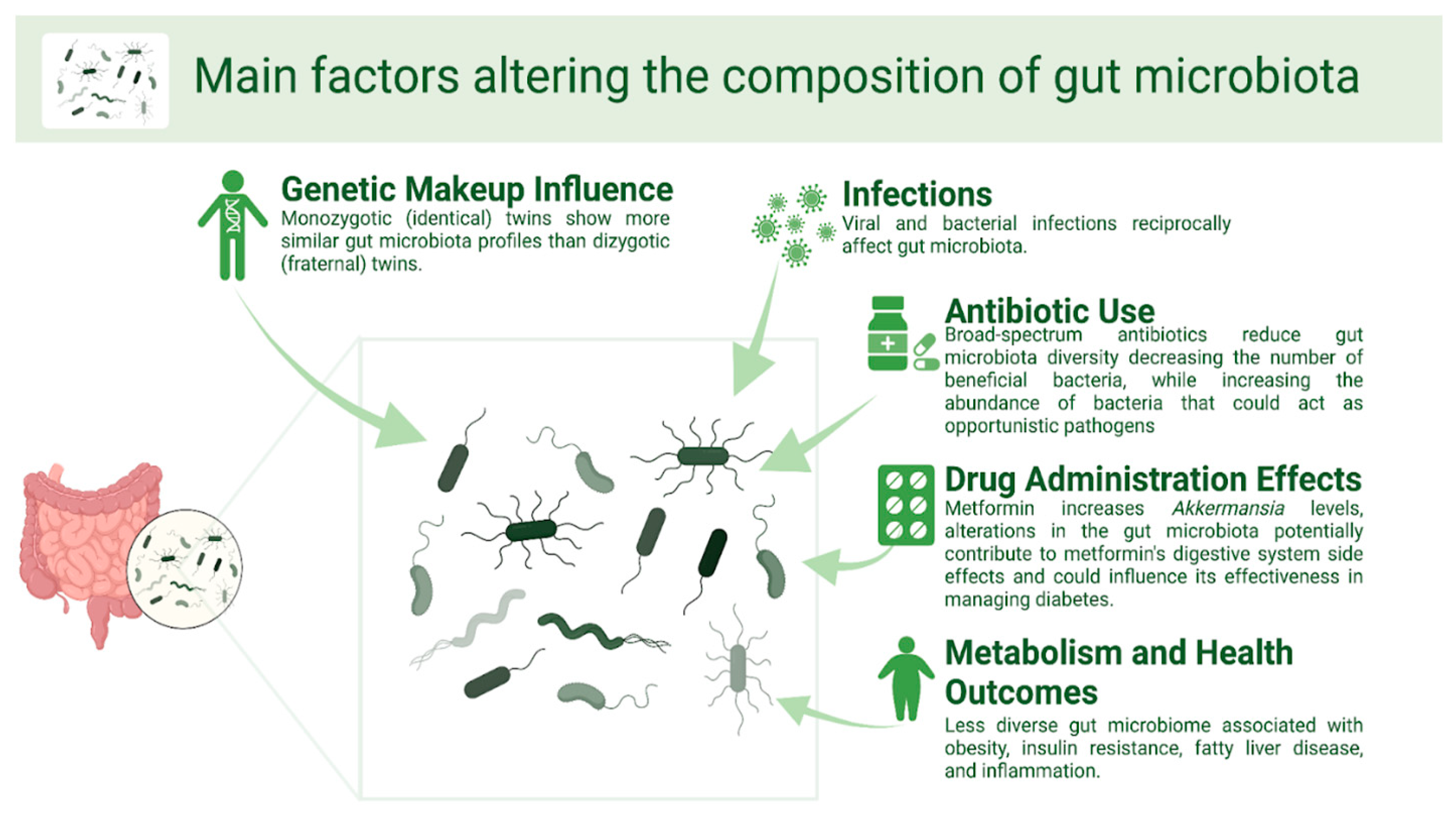
Main Factors Altering the Composition of Gut Microbiota

A wide palette of internal and external variables affects the composition of the gut microbiota, as shown in Figure 2. Among them is the host’s genetic composition [12]. Family members exhibit greater similarity in microbiota communities compared to unrelated individuals, with monozygotic twins showing more similar gut microbiota profiles than dizygotic twins [12,13,14]. Infections, both viral and bacterial, also have a reciprocal relationship with the gut microbiota [15,16,17]. Chou et al. [15] registered that the clearance of hepatitis B virus infection in a mouse model depends on the renewal of the healthy gut microbiota. The alteration of gut microbiota was also noted in studies investigating the effects of an enteropathogenic infection with Citrobacter rodentium on the microbiota composition in mice [17], as well as in a study investigating the Clostridium difficile infection that noted reduced microbial diversity in infected patients when compared to the healthy subjects [18]. Also, C. difficile infection is a typical result of severe dysbiosis in the gut microbiota [19,20,21,22].
The gut microbiota is also shaped by different types of drugs, especially antibiotics [23,24]. Broad-spectrum antibiotics decrease bacterial diversity and the number of beneficial bacteria, while increasing the abundance of potentially opportunistic bacteria [25]. For example, the administration of clindamycin has been linked with the longest-lasting effects on the gut microbiota [26,27]. Gut dysbiosis caused by early antibiotic exposure in neonates is a potential risk factor for inflammatory bowel disease later in life [28,29,30]. Also, antibiotic usage in children could contribute to obesity later in life by altering the gut flora, according to studies conducted in both people and animals [31,32,33,34]. Other studies demonstrated that antibiotics could reduce body weight and increase insulin sensitivity by modifying gut bacteria [35,36]. For example, this significantly contributes metabolically to the host organism by fermenting complex indigestible dietary carbohydrates and proteins, creating short-chain fatty acids as fermentation by-products. Additionally, they are pivotal in synthesizing vitamins, facilitating ion absorption, and converting dietary polyphenolic compounds into their bioactive forms [10,11]. Therefore, while the role of antibiotics in the rising rates of obesity, particularly in childhood, remains unclear, their profound impact on gut microbiota composition is undeniable.
This is also confirmed by various products that have shown an antidiabetic property by changing the gut flora, such as berberine [37,38]. Also, metformin is commonly prescribed to treat Type 2 diabetes, and it has recently been noted that metformin administration can also modify the composition of the gut [39,40,41]. For instance, in a study inducing obesity in mice through a high-fat diet, metformin increased levels of Akkermansia, a bacterium known for degrading mucin, and more prominently in obese mice [42]. Similarly, recent human studies have confirmed metformin’s impact on the gut microbiota [41]. This indicates that alterations in the gut bacteria potentially contribute to metformin’s digestive system side effects and could influence its effectiveness in managing diabetes.
In addition, drugs that are often used for the treatment of MS relapse, marked as disease-modifying treatments (DMTs), aim to decrease relapse rates and MS progression. DMTs, including interferon-β (IFN-β), fingolimod, and ocrelizumab, increase the gut microbiota diversity and reduce pro-inflammatory bacteria. This indicates their influence on the gut microbiota composition and their potential use as a mechanism that improves their therapeutic effects. Also, as a recent systematic review noted, DMTs do not always impact the diversity of the microbiota as a whole, but they do lead to variations at the taxonomic level, thus, having consequences on the course of the disease. The study argued their potential role in modulating disease activity in MS patients through microbiome alterations, which should be confirmed in further studies and larger clinical trials [43].
The role of food-ingested bacteria in shaping the gut microbiome was previously underestimated, largely due to methodological limitations that have since been addressed [43]. Extensive research in both mice and humans has established that high-calorie diets contribute significantly to conditions like obesity and Type 2 diabetes [44,45]. However, emerging evidence indicates that the critical connection between diet and these conditions lies within the gut microbiota [46,47]. Human gut microbiota has been described by 16S ribosomal RNA sequencing studies as various enterotypes based on the types of bacteria present. These enterotypes have been significantly linked to long-term diets, especially diets high in protein and animal fat [48,49]. In their study, Wu et al. [50] demonstrated that diets rich in protein and animal fat correlated with higher levels of Bacteroides, whereas carbohydrate-rich diets were linked with Prevotella. Additionally, they conducted controlled feeding experiments with 10 subjects, observing that the gut microbiome changed after 24 h of initiating either a high-fat, low-fiber diet or a low-fat, high-fiber diet [50]. These findings strongly suggest that diet has a key role in shaping enterotype distribution within the gut microbiota.
Therefore, recognizing diet as a pivotal factor in gut microbiome creates an opportunity for intervention. Interventional studies have demonstrated that dietary modifications cause rapid and substantial alterations in gut microbiome [48]. The Western diet has been implicated as a significant contributor to this phenomenon. Also, yogurt consumption is perceived as beneficial to health, potentially due to its ability to modify the host gut microbiota alongside other nutritional factors [51]. Despite limited studies for decades, recent advancements in high-throughput sequencing technology and bioinformatics have led to a surge in research, including clinical trials over the past eight years, investigating how yogurt consumption influences changes in the gut microbiota [52,53]. Recent studies suggest that consuming probiotic bacteria in yogurt and other fermented dairy products positively impacts the host’s gut microbiome [12]. Fermented dairy products introduce numerous lactic acid bacteria into the gastrointestinal tract, potentially altering the intestinal environment by reducing lipopolysaccharide production and enhancing gut epithelial cell integrity through increased tight junction formation [12]. Another potential avenue of gut microbiota altering is the transplantation of the gut microbiome from healthy donors with the aim of promoting the abundance of healthy bacteria [19,20]. However, it still remains to be determined whether sustained dietary modifications can establish enduring changes in bacterial enterotypes and even be used as a potential novel therapeutic approach to the treatment of gut dysbiosis. Developing specialized dietary therapies for MS patients requires an understanding of the gut–brain axis. Namely, through immune response modulation, personalized dietary interventions targeting the gut microbiota could be beneficial for patients with MS. Adopting anti-inflammatory diets and optimizing nutritional habits has the potential to enhance quality of life (QoL) for MS patients, as well as to help with disease management. Further studies and larger clinical trials are required to provide a more thorough understanding of complex interactions between gut microbiota and MS onset and progression. The clinical implications of these findings hold the promise of effective dietary treatments and microbiota-targeted drugs as supplemental techniques in the management of MS.

2. The Complex Relationship Between Gut Microbiota and Multiple Sclerosis

Recently, MS has been associated with gut microbiota dysbiosis [54,55]. It is a complex neurological disease defined by chronic inflammation and immune-mediated damage to the central nervous system (CNS). Increasing incidence of MS cases in the last 10 years is most likely influenced by environmental factors, including changes in dietary habits that impact the gut microbiome [56,57,58,59,60]. Research into the association between gut microbiota, diet, and the immune system in MS has recently garnered significant interest. This complex relationship underscores the challenge of investigating specific dietary components that influence the gut–brain axis.

2.1. Gut Microbiota Composition in MS Patients

Initial proof of gut dysbiosis in MS was observed in a Japanese population, coinciding with a recent rise in MS cases in Japan [61]. It is proposed that the main cause of gut dysbiosis is dietary change [62]. Namely, Westernized lifestyles and diets, known for promoting inflammation in the gut [57], have been implicated as a key factor in increasing MS risk. Conversely, adopting a healthy diet has shown potential to promote intestinal microbiota towards an anti-inflammatory status, supporting dietary interventions in MS management [57,63]. Additionally, studies have repeatedly noted that patients with MS have a distinct composition of gut microbiota compared to healthy patients [60,61]. These findings indicate that particular microbial communities can trigger harmful immune responses in individuals with MS. Modifying the gut microbiota to restore balance and improve immune control shows potential as a treatment for MS, such as through probiotics, prebiotics, and dietary treatments [58,59,60]. However, further larger clinical trials should confirm these findings.

2.2. Underlying Mechanisms of Gut Microbiota Interactions with the Immune System in MS

One of the most significant mechanisms underlying the interplay between gut microbiota and the immune system is via modulating the intestinal barrier function [64,65,66]. The gut barrier is composed of tightly organized epithelial cells, responsible for preventing pathogens and toxins from entering the bloodstream [67,68]. Gut dysbiosis can disrupt this tight barrier, leading to a condition marked as “leaky gut”. This enables microbial antigens and other pro-inflammatory molecules to enter the circulation system, triggering immune responses that can contribute to systemic inflammation and the onset of autoimmunity [69]. Berer et al. [70] noted that the gut microbiota derived from patients with MS also triggers the development of spontaneous autoimmune encephalomyelitis in mice [70], indicating that gut dysbiosis promotes inflammatory immune response, as shown in Figure 3. There are fewer favorable bacteria, such as Bacteroides, as well as more potentially harmful bacteria, like Akkermansia and Methanobrevibacter, consequently affecting the function of immune cells, like regulatory T cells (Tregs) and Th17 cells, in fulfilling their role of keeping the immune system in check and stopping autoimmunity onset [62]. For example, Cekanaviciute et al. [71] discovered that the gut bacteria of MS patients can influence the functions of T cells and exacerbate the MS symptoms in animal models [71]. Furthermore, the gut bacteria also regulates the formation of short-chain fatty acids (SCFAs) by degrading dietary fibers to create SCFAs, such as propionate, butyrate, and acetate, which have immunomodulatory properties [72].
In addition, the reciprocal relationship between the intestines and the CNS, marked as the gut–brain axis, highlights the influence of gut bacteria on MS. This axis includes neurological, hormonal, and immunological processes that enable the intestines to affect CNS function and vice versa [73]. For example, Thirion et al. [74] discovered notable changes in gut microbiota composition in MS patients. However, the main question that remains to be further researched is focused on investigating whether the gut dysbiosis is a cause or a consequence of MS. Hence, dysbiosis is linked to MS through various alterations of the human immune system, with alterations of gut microbiota having a key role in promoting inflammation. Consequently, this could trigger the onset or exacerbation of MS. On the other hand, MS itself can promote gut dysbiosis through changes in the gut–brain axis, as well as due to the use of various immunomodulatory drugs in the MS treatment. This could indicate that dysbiosis is a secondary effect of MS. Nevertheless, the current opinion is that the link between gut dysbiosis and MS is bidirectional [74,75,76]. Therefore, their relationship is complex, and a better understanding could lead to the engineering of novel treatments for MS, mostly focused on inflammation reduction.

3. Therapeutic Potential of Gut Microbiota Modulation in MS

Gaining a comprehensive understanding of gut microbiota and its relationship with immunity is essential for the development of precise and focused therapies for both adult and pediatric patients with MS [76,77,78,79,80,81,82]. An important mechanism includes the synthesis of SCFAs that have demonstrated anti-inflammatory properties via interacting with G protein-coupled receptors (GPCRs), like GPR41 and GPR43 on immune cells [72]. Research indicates that SCFAs can enhance the development of regulatory T cells (Tregs), responsible for preserving immunological tolerance and inhibiting autoimmune reactions [62,72]. Also, aryl hydrocarbon receptor (AHR), a ligand-activated transcription factor expressed on different immune cells, including T cells and dendritic cells, interacts with gut microbiota, thus having the potential to modulate immune responses and inflammation within the CNS [83,84,85]. Moreover, activation of the AHR can affect the functioning of microglia in the CNS, which in turn has an impact on neuroinflammation [86].
Multiple studies have examined the capacity of probiotics to improve symptoms and slow down the progression of MS, such as the use of particular probiotic strains, including Lactobacillus spp. and Bifidobacterium spp. [87,88,89]. Prebiotics can also improve intestinal barrier integrity, reduce systemic inflammation, and modify immunological responses by encouraging the proliferation of commensal bacteria that produce SCFAs [90]. Also, a study that researched the effect of aerobic exercise with probiotic intake on the myelination of nerve fibers in a cuprizone-induced mouse model of MS noted that lifestyle interventions have the potential to alleviate inflammatory processes in the brains of MS patients [91]. Therefore, dietary interventions aimed at modifying gut microbiota composition represent a non-invasive and potentially cost-effective additional therapeutic approach to managing MS.

3.1. The Mediterranean Diet

The MD, initially investigated by Keys in the 1960s, mirrors the dietary habits of Mediterranean populations, emphasizing seasonal, communal eating, rest, physical activity, and local foods [92,93]. It limits consumption of dairy and meat and increases consumption of fruits, vegetables, nuts, legumes, whole grains, and fish. It also recommends the use of olive oil and moderate amounts of alcohol, particularly red wine [94]. In the general population, the MD demonstrates favorable health effects, such as a lower incidence of a number of chronic illnesses, including several neurodegenerative conditions like depression and cognitive decline [95]. This could be attributed to the diverse phytochemical compounds in its foods, known for their nutraceutical properties.
Because of their anti-inflammatory and antioxidant properties, they lower serum levels of inflammatory factors, including C-reactive protein and interleukin-(IL-) 6 [96]. Also, the lignans, tocopherols, phenolic acids, flavonoids, stilbenes, carotenoids, and unsaturated fatty acids, present in various foods within the MD, render them “functional foods”. Extra virgin olive oil (EVOO), a staple of the MD, is renowned for its rich content of phytochemical compounds, particularly polyphenols, which confer various health benefits. Its main lipid constituents are triglycerides, consisting predominantly of oleic acid (73.6%), followed by palmitic acid (13.7%) and linoleic acid (7.85%) [97]. EVOO contains over 230 bioactive molecules, including phenols, sterols, chlorophylls, carotenoids, alcohols, and esters, exhibiting neuroprotective properties by inhibiting pathways associated with inflammation and oxidative stress. Daily consumption of EVOO can provide significant health benefits, including protection against oxidative stress-related damage [98].
The function of MD in MS has been investigated in recent research [99,100,101,102,103]. The relationship between MS severity and MD adherence was examined in a single-center study involving 106 patients. According to the study, individuals who adhered to the MD had less severe symptoms of MS than patients who did not. Importantly, no individual component of the MD showed an independent association with MS severity, suggesting that the combined dietary pattern plays a synergistic role over individual food intake [92].
Researchers evaluated the effects of the Western diet (WD) and a high-vegetable/low-protein (HV/LP) diet, which is similar to the MD, in people with RRMS in a pilot trial. The gut composition of the HV/LP group had more butyrate-producing Lachnospiraceae bacteria, fewer pro-inflammatory PD-1+ and IL-17+ T cells, and more anti-inflammatory PD-L1+ monocytes. Clinically, during follow-up, the EDSS score and relapse rate significantly decreased in the HV/LP group. However, the number of participants was small, and larger studies are required to confirm these findings [57]. Additionally, a recent study on individuals with MS found a positive correlation between circulating Th17 cell levels and meat consumption. The meat consumption was negatively correlated with the relative abundance of Bacteroides thetaiotaomicron, which has a high capacity for polysaccharide digestion. B. thetaiotaomicron was inversely associated with circulating Th17 cells, while Th17 cells were positively linked to meat intake [104], as noted in Table 1.

3.2. Ketogenic Diet

The ketogenic diet (KD) is a dietary intervention based on high-fat, low-carbohydrate intake, inducing ketosis and modulating various metabolic pathways [118]. Widely used in clinical practice for drug-resistant epilepsy and inflammatory conditions like Febrile infection-related epilepsy syndrome [119]. Beta-hydroxybutyrate (BHB) and acetoacetate (ACA) represent the main ketone products generated by KD. These compounds have potential anti-inflammatory and neuroprotective effects, as they help reduce oxidative stress, support mitochondrial function, regulate epigenetic changes, and influence the gut microbiome composition [120]. KD’s mechanisms of action involve targeting immune activation pathways and inflammatory mediators, including adenosine, ketone bodies, PPAR-γ, NLRP3 inflammasome, and gut microbiota [121,122]. BHB, a key anti-inflammatory agent generated during the KD inhibits the activation of IL-1β [123] mediated by the NLRP3 inflammasome, potentially contributing to the diet’s anti-inflammatory properties [124]. Dysregulation of the inflammasome is implicated in autoimmune disorders, including MS and EAE, where NLRP3 mediates immune cell migration to the CNS, contributing to neuro-inflammation [125]. KD has been shown to improve inflammatory markers, disability, cognition, and disease progression in rats with EAE. Ketone bodies from KD provide an alternative source of energy for the brain and may reduce neuroinflammation by inhibiting NLRP3. They also enhance mitochondrial biogenesis and redox balance [125,126].
Effective dietary interventions for MS can modulate inflammation, protect against 452 neurodegenerations, or promote nervous system repair. The impact may result from direct metabolite actions, gut microbiota metabolites, or diet-mediated alterations in gut bacteria composition [58,59,60]. The nutrition plan includes two phases: adaptation and maintenance. The adaptation phase lasts four weeks with 20 g/day carbohydrate intake to establish ketosis. In the one-month maintenance phase, carbohydrate consumption increases by 5 g each week up to 40 g/day, maintaining low glycemic index and load for sustained ketosis and stable blood sugar levels. A similar nutritional approach was tested in a clinical trial, showing good compliance in MS patients, and the study noted that the KD is a cost-effective complementary therapeutic option for MS. The protocol follows international ketogenic guidelines, with a caloric deficit of 300–500 kcal varying with BMI. Daily water intake is set at 0.4 mL per kg of body weight [96].
Recent studies have noted several important effects of KD on MS pathology. Notably, the KD has been shown to reduce hippocampal demyelination, inhibit the activation of microglia and astrocytes, and modulate the SIRT1/PPAR-γ and SIRT1/P-Akt/mTOR pathways [126]. Furthermore, KD affects the expression of several enzymes implicated in MS’s inflammatory response. It suppresses the systemic production of arachidonate 5-lipoxygenase (ALOX5), cyclooxygenase (COX) 1, and COX2, which are essential for the synthesis of pro-inflammatory eicosanoids and linked to inflammation and demyelination in multiple sclerosis [106]. Furthermore, it was also reported that the KD group exhibited significantly reduced levels of serum neurofilament light chain (sNfL) compared to the common diet group [127]. These findings suggest that the KD could be a potential therapeutic strategy for managing MS through multiple pathways.
The gut microbiota of patients with MS may also be impacted by the KD. When compared to MS patients who did not follow the KD, a six-month KD intervention reduced six bacterial groups, such as Bacteroides and Faecalibacterium prausnitzii. The only bacteria that did not quickly decline after the KD intervention was Akkermansia. Bacterial concentrations started to recover after 12 weeks and reached levels comparable to healthy controls after 23–24 weeks, except for Akkermansia, which declined post-KD, as shown in Table 1 [107]. Despite these initial results, the long-term use of KD in MS patients requires careful evaluation due to potential risks, such as an increase in apoB-containing lipoproteins, which could elevate cardiovascular disease risk [128]. A recent meta-analysis of 11 studies including 608 subjects found that modified MD improved both fatigue and QoL, while low-fat diets only improved fatigue. Fasting, calorie-restricted, and anti-inflammatory diets had no significant impact, and ketogenic diets showed mixed results. However, all these studies require larger clinical trials to reach a general consensus about appropriate diet for MS, as well as their safety and efficacy [129].

3.3. Calorie Restriction

The term “dietary restriction” (DR) refers to controlled dietary regimens used in experimental models, involving reduced energy intake without causing malnutrition [130]. These regimens have shown promising protective effects against EAE, a widely studied model for MS [130]. Although the precise mechanisms by which DR affects the gut microbiota in MS remain largely unknown, recent studies on intermittent fasting (IF), a special type of DR, have shown improvements in EAE pathology and clinical outcomes [108,127]. The principal DR consists of a daily calorie restriction (CR) and IF involves periodic elimination or a significant reduction in food intake for specific intervals. Intermittent fasting (IF) comes in several forms, each offering a unique approach to calorie restriction [131,132,133]. IF has been associated with alterations in microbiota composition, enriching it with beneficial bacteria, such as Lactobacillaceae, Bacteroidaceae, and Prevotellaceae. Remarkably, fecal microbiome transplantation from IF-treated mice to mice on a normal dietary regimen has shown promising therapeutic potential in ameliorating EAE symptoms. These findings suggest a complex interplay between dietary restriction, gut microbiota, and MS pathogenesis, warranting further investigation into their potential therapeutic implications [111]. IF also reduced the accumulation of monocytes in the spinal cord of EAE mice. Monocytes from mice that fasted had downregulated expression of pro-inflammatory genes such as TNF-a, IL-1b, CXCL2, and CXCL10 compared to those from non-fasted mice [115].
In a study by Fitzgerald et al. [134], the safety and practicality of various CR diets were researched in 36 patients with MS. It was noted that traditional CR led to greater weight loss compared to IF, although adherence was lower in the IF group. Both CR and IF significantly improved emotional well-being in patients with MS, notably reducing depression scores. This is offering additional emotional health benefits, which are crucial for the holistic management of MS [134]. Also, Wingo et al. [135] explored the effects of a time-restricted eating (TRE) protocols for adults with RRMS. Over 8 weeks, 12 participants ate within an 8 h window and fasted for the remaining 16 h. The findings suggest that TRE may help manage MS symptoms, mostly cognitive and motor functions. Positive participant feedback highlights that further trials are needed to confirm its effectiveness [135].
When it comes to QoL and diets, CR and fasting did not significantly impact fatigue or QoL, low-fat diets only improved fatigue, and modified MD significantly improved both fatigue and QoL [129,136]. In a systematic review by Lin et al. it was shown that IF is a promising dietary intervention for managing MS symptoms, as it can help with weight loss and enhance the QoL through mechanisms such as calorie restriction, metabolic changes, improved insulin sensitivity, better gut health, and inflammation reduction. However, variations in fasting protocols complicate the generalization of results, and the effects of IF on the immune system is not yet fully understood [137]. Therefore, extensive clinical trials are essential to explore the therapeutic potential and underlying mechanisms of IF and other diets in the context of MS.

3.4. Low-Salt Diet

It has recently been investigated that sodium consumption may be a dietary risk factor for the development and course of MS, which is supported by studies on mouse models. They show that a high-sodium diet increases EAE severity, enhances BBB permeability, and promotes increased numbers of peripherally generated and CNS-infiltrating Th17 cells [138,139,140,141]. The upregulated production of Th17 cells results in increased levels of interleukin-17 (IL-17), whereas reduced salt intake leads to the induction of the anti-inflammatory cytokine IL-10 [142,143]. Sodium chloride modulates the renin-angiotensin and studies indicated that a rise in systolic blood pressure induced by high salt intake is linked to disruptions in white matter integrity in young normotensive individuals [144,145].
Studies have not yet identified an association between dietary sodium intake and the risk of MS, including pediatric-onset MS [146,147,148,149,150,151]. A study observing patients with relapsing-remitting MS over a 2-year period demonstrated a positive association between MS exacerbation rates and sodium intake [149]. In addition, the evaluation of the association between urinary sodium concentration and MS progression and activity, encompassing a 5-year follow-up period, found that salt intake did not significantly impact the transformation from clinically isolated syndrome (CIS) to MS, nor did it influence clinical or magnetic resonance imaging (MRI) outcomes [150]. However, this should be confirmed and further investigated in larger studies.

4. Role of Probiotics, Prebiotics, and Postbiotics in MS

4.1. Probiotics

Based on the current definition, “probiotics are live microorganisms that, when administered in adequate amounts, confer a health effect on the host” [152]. The microbiota, crucial for host survival by defending against pathogens and providing nutritional benefits, outnumbers human cells tenfold, supporting the concept of humans as “symbiotic” organisms living in harmony with their microbiota [153,154]. The ability to modulate the host’s immune responses by displacing pathogens through competitive exclusion, secreting protective mediators, and providing essential nutrients are potential benefits of probiotics, as shown in Figure 4 [153,155]. For example, before being immunized, marmoset twins were split into two groups and fed either a water-based (WBD) or yogurt-based (YBD) diet. Reduced demyelination and a weakened pro-inflammatory response from T cells, B cells, and cytokines were observed in those following the YBD diet. A few marmosets on the YBD diet showed no signs of EAE. Only after immunization did these marmosets’ gut microbiome composition change, most likely as a result of the interplay between their immune system responses and food [156].
Also, administering a combination of probiotics to mice in the chronic phase of Theiler’s encephalomyelitis virus (TMEV) infection, which is a murine model for primary progressive MS, decreased disease severity and the onset of motor disability. This treatment also influenced gut microbiota, resulting in higher levels of Bacteroidetes, Actinobacteria, Tenericutes, and TM7 taxa. Additionally, the study revealed reductions in gliosis, leukocyte infiltration, and the expression of IL-1β and IL-6 in the CNS, alongside elevated butyrate and acetate in plasma [157]. Also, a systematic review demonstrated that probiotics influenced the immune system by promoting the production of anti-inflammatory cytokines, such as IL-4, IL-10, and TGF-β, and regulatory T cells (Tregs), while lowering the levels of Th1 and Th17 cells and the levels of pro-inflammatory cytokines, such as IL-17, IFN-γ, GM-CSF, and TNF-α [158].
Clinical trials have also shown that administering probiotics, like capsules containing Lactobacillus and Bifidobacterium strains, resulted in improvements in disability, depression, and overall health, along with reduced expression of pro-inflammatory cytokines [89,159,160]. In their study, Tankou et al. [161] examined the effects of probiotics on gut microbiota. MS patients and healthy volunteers observed changes in microbiota composition after a two-month probiotic treatment (Lactobacillus, Bifidobacterium, and Streptococcus), including decreased α-diversity and increased relative abundance of the indicated species. However, stopping probiotics caused microbial and immunological alterations to reverse. The findings’ importance is limited by constraints including the small sample size, short duration, and confounding factors like dietary practices and disease-modifying medicine, which calls for more investigation [161]. Lactobacilli also confirmed their anti-inflammatory role by reducing Th1 and Th17 cytokines through IL-10 induction [162], as the administration of L. casei T2 strain led to the demyelination effects of cuprizone (CPZ) in mice [163].

4.2. Prebiotics

Prebiotics are products that intestinal bacteria in hosts use to support the survival and activity of particular bacterial genera and species [164,165]. They are mostly galactans and inulin [165]. Bacterial strains have the ability to break down prebiotics, which are indigestible to the host, into products that either support specific bacteria or participate in other bacterial metabolic processes, including the production of lactate, succinate, folates, indoles, secondary bile acids, and SCFA. By stimulating the growth of Bifidobacteria and other SCFA producers, prebiotic administration can alter the composition of the gut microbiome. Furthermore, prebiotics have the ability to activate cytokine expression and interact with immune cell receptors [166].
Therefore, prebiotics’ immunomodulatory effects rely on changes in the microbiota population through the synthesis of fermentation products like SCFAs. It is also achieved through a mechanism unrelated to the gut microbiota, as was noted by modifying B-cell responses by long-chain b2-1 fructans in germ-free mice [167,168]. Anthropometric markers, impairment levels, and measures of systemic inflammation were all correlated with dietary fiber consumption in MS patients, according to a clinical trial [169]. In addition, by altering the gut microbiota, recent research on pomegranate peel extract as a prebiotic has demonstrated that it can reduce the clinical symptoms of EAE, prevent DC activation and Th17 cell differentiation, and trigger the production of immunoregulatory cytokines, thereby establishing prebiotics as promising therapeutic candidates for further research on EAE and MS [170].

4.3. Postbiotics

Postbiotics are bioactive substances that are produced when fibers (prebiotics) are broken down and digested by the beneficial bacteria in the gastrointestinal tract (probiotic bacteria). Postbiotics show several advantages over probiotics in terms of safety and stability. They are safer for vulnerable populations, including immunocompromised individuals and infants, as there is risk associated with live probiotics strains. Unlike probiotics, postbiotics contain no living organisms, which enhances their consistency and reliability [171,172,173]. The primary metabolites of bacterial anaerobic fermentation of indigestible polysaccharides, such as resistant starch and dietary fiber in the human colon, are postbiotics, specifically SCFAs acetate and butyrate [174].
Recently, various studies have noted a reduction in serum butyrate levels among MS patients, which correlates with a decrease in SCFA-producing bacteria [175,176]. Additionally, MS patients show elevated acetate levels, the most abundant SCFA produced by gut bacteria [177]. SCFAs may have a similar role in MS as they do in experimental autoimmune encephalomyelitis, according to animal models [178]. It is also highlighted that SCFAs play a bidirectional role in regulating autoimmune inflammation in MS [179,180,181]. SCFAs, particularly acetate, propionate, and butyrate, have the potential to influence CNS autoimmunity, possibly via BBB transporters, highlighting their relevance in MS pathogenesis [177,182,183]. Serum levels of propionate (PA) and circulating follicular Tregs are favorably correlated with butyrate and IL-10 [184], and acetate and L-17+ [185]. In contrast, acetate levels were shown to be marginally lower in patients with RRMS or CIS, and the ratios of acetate/butyrate and acetate/(propionate + butyrate) were considerably lower in MS patients when compared to healthy controls [186].
However, research on SCFA intake for EAE and MS is still in the early stages. In experimental models, butyrate has shown to prevent CNS autoimmunity by reducing demyelination and inflammation [187]. Methyl butyrate, administered post-EAE induction, alleviated clinical symptoms and improved CNS histopathology [188]. Also, PA has been studied in both mice and humans. In EAE mice, PA intake led to a higher levels of Treg cells (CD4+ CD25+ Foxp3+), improving their clinical course compared to controls, as well as decreasing demyelination [189]. Also, in obese patients with MS, PA supplementation significantly reduced Th17 cell frequencies [190]. However, further studies and larger clinical trials should confirm the effectiveness and safety of probiotics, prebiotics and postbiotics.

5. Gut Dysbiosis in Pediatric-Onset MS

Analyzing the microbial composition and functional profiles of patients with pediatric-onset MS in comparison to healthy controls has been the main focus of recent studies, especially due to the technology using sophisticated sequencing techniques, such as meta-genomic analysis [76,78]. For example, the upregulation of the lipopolysaccharide biosynthesis pathway has been observed in pediatric-onset MS [76,78]. This was also noted in a study investigating the gut microbiota characteristics associated with MRI lesion burden in pediatric-onset MS [79].
In addition, breastfeeding has an important role in the gut composition in children, as it could potentially be a protective factor in pediatric-onset MS. Several studies have shown that breastfeeding promotes the growth of beneficial bacteria, such as Bifidobacteria, in the infant gut [80]. Breastfeeding has been shown to have an impact on the gut microbial community in late infancy, even after the inclusion of solid foods in children’s diet [80]. Breastfeeding provides infants with bioactive components that influence their microbiota, exposing them to microbial communities from breast milk and the breast surface [81]. Emerging research indicates a potential entero-mammary route for microbial transfer, suggesting that maternal probiotic supplementation could potentially modulate the gut microbiota of infants [80]. However, further studies need to investigate the role of breastfeeding in pediatric-onset MS.

The Role of Nutrition for Patients with Pediatric-Onset MS

As previously mentioned, early life environmental factors, including diet, can influence the risk of developing MA in pediatric patients, where early lifestyle modifications can have long-lasting impacts. Research using the experimental autoimmune encephalomyelitis (EAE) model has investigated the effects of diet on MS and its progression, which is influenced by gut microbiota, enzyme activity, and vascular risk factors. There are no specific dietary guidelines for MS, although some studies indicate that a healthy diet and lifestyle improve clinical parameters and QoL [54,93]. For example, a recent study on children with early-onset pediatric MS noted that high-fat diets, especially those rich in saturated fats, were linked to a higher risk of disease progression, while sugar consumption did not significantly affect relapse risk. On the contrary, a healthy childhood diet including fruits, yogurt, and legumes was associated with a decreased risk of developing MS in adulthood [191]. Another case–control study with 95 participants (44 pediatric-onset MS cases, 51 controls) from the Canadian Pediatric Demyelinating Disease Network study examined the relationship between diet, intestinal microbiota, and MS. Participants completed a food-frequency questionnaire by age 21, and 59 provided stool samples. The findings revealed that a higher MD score and increased consumption of fiber and iron were associated with a decreased likelihood of pediatric-onset MS [77].
Diet, rather than MS, explained individual variations in gut microbiota [102]. The effects of eating vegetables and saturated fat on MS were examined in a multicenter trial involving pediatric patients with RRMS or clinically isolated syndrome (CIS). According to the study, the risk of relapsing tripled for every 10% increase in saturated fat consumption. On the other hand, the risk of relapse decreased by 50% for every extra cup equivalent of veggies. Therefore, vegetable intake provided protection, whereas fat intake was linked to an increased risk of relapse in pediatric MS, with saturated fat driving this association [103].
In the pediatric study, McDonald et al. [148] investigated the effect of different sodium intakes, but detected no significant differences in higher sodium intakes or those exceeding nutrient reference values between cases and controls. While adult and pediatric MS share similarities in presentation and pathophysiology, pediatric MS exhibits distinct clinical features and disease progression patterns. In the systematic review by Zostawa et al. (2017), it has been noted that excess sodium chloride intake may potentially contribute to inflammation in autoimmune and neurodegenerative diseases, as evidenced in both experimental and clinical settings, albeit with varying outcomes. However, current evidence suggests that adopting a low-salt diet (5 g/day) could help in preventing and treating autoimmune diseases, such as MS [151]. Nevertheless, larger clinical trials should investigate dietary interventions for pediatric patients with MS.

6. Conclusions

An imbalance in the gut microbiota, known as gut dysbiosis, has recently been linked to the onset and progression of autoimmune diseases, such as multiple sclerosis (MS). Diet plays a crucial role in forming the gut microbiota of a host. For example, a Western-style diet, which is characterized by high intakes of saturated fats and processed foods, has been related to the progression of MS [91]. On the other hand, the Mediterranean diet with lots of fruits, vegetables, whole grains, and healthy fats, has been linked to better clinical results and a lower risk of MS. Therefore, customized dietary approaches that target the gut microbiota by modifying immune response and gut inflammation, as well as the gut–brain axis, have the potential for more efficient management of MS. Apart from probiotics and prebiotics, diets that alter the composition of the gut microbiota offer a non-invasive and possibly economical method of MS. Despite the promising therapeutic potential of microbiota-based therapies in MS, several challenges and unanswered questions remain. One major challenge is the heterogeneity of gut microbiota composition among individuals, which complicates the identification of universal microbial targets for therapeutic intervention [33]. Furthermore, the dynamic nature of the gut microbiota and its interactions with host genetics, diet, lifestyle factors, and medications necessitate personalized approaches to microbiota modulation in clinical settings [34].
This review provides a comprehensive analysis of the interactions among gut microbiota, diet (including probiotics, prebiotics, and postbiotics), and their impact on the pathogenesis and progression of MS. Previous research has established a link between gut dysbiosis and MS, indicating that an imbalanced gut microbiota can disrupt immune responses and exacerbate inflammation. Building upon this foundation, the review explores the mechanisms through which gut microbiota modulates immune functions in MS patients. Additionally, it discusses how specific dietary patterns such as Mediterranean and ketogenic diets can influence disease progression and immune regulation in MS. The potential therapeutic role of probiotics in restoring microbial equilibrium is also emphasized based on current literature. The review consolidates evidence supporting the development of personalized dietary strategies aimed at enhancing gut microbiota as a promising non-invasive approach to managing MS. Furthermore, it investigates the reciprocal relationship between the gut and brain in MS, underscoring the need for rigorous clinical trials to validate these findings.

Author Contributions

Conceptualization, A.D.K., E.B. (Emir Begagić) and M.P.; methodology, A.D.K. and M.P.; software, S.H.; validation, A.B., E.B. (Emir Begagić) and H.B.; investigation, A.D.K., E.B. (Emir Begagić), S.H., A.B., E.B. (Emir Bećirović), H.H., L.T.L., S.K.V., H.B., T.K. and M.P.; resources, M.P.; data curation, E.B. (Emir Begagić); writing—original draft preparation, A.D.K., E.B. (Emir Bećirović), H.H. and M.P.; writing—review and editing, E.B. (Emir Begagić), L.T.L., S.K.V. and H.B.; visualization, S.H. and H.H.; supervision, M.P. All authors have read and agreed to the published version of the manuscript.

Funding

This research received no external funding.

Data Availability Statement

Data are contained within the article.

Conflicts of Interest

The authors declare no conflicts of interest.

References

  1. Sekirov, I.; Russell, S.L.; Antunes, L.C.; Finlay, B.B. Gut microbiota in health and disease. Physiol. Rev. 2010, 90, 859–904. [Google Scholar] [CrossRef] [PubMed]
  2. Bien, J.; Palagani, V.; Bozko, P. The intestinal microbiota dysbiosis and Clostridium difficile infection: Is there a relationship with inflammatory bowel disease? Ther. Adv. Gastroenterol. 2013, 6, 53–68. [Google Scholar] [CrossRef]
  3. Falony, G.; Vlachou, A.; Verbrugghe, K.; De Vuyst, L. Cross-feeding between Bifidobacterium longum BB536 and acetate-converting, butyrate-producing colon bacteria during growth on oligofructose. Appl. Environ. Microbiol. 2006, 72, 7835–7841. [Google Scholar] [CrossRef]
  4. Cash, H.L.; Whitham, C.V.; Behrendt, C.L.; Hooper, L.V. Symbiotic bacteria direct expression of an intestinal bactericidal lectin. Science 2006, 313, 1126–1130. [Google Scholar] [CrossRef] [PubMed]
  5. Oktaviono, Y.H.; Dyah Lamara, A.; Saputra, P.B.T.; Arnindita, J.N.; Pasahari, D.; Saputra, M.E.; Suasti, N.M.A. The roles of trimethylamine-N-oxide in atherosclerosis and its potential therapeutic aspect: A literature review. Biomol. Biomed. 2023, 23, 936–948. [Google Scholar] [CrossRef] [PubMed]
  6. Zhang, X.; Chen, B.; Zhao, L.; Li, H. The gut microbiota: Emerging evidence in autoimmune diseases. Trends Mol. Med. 2020, 26, 862–873. [Google Scholar] [CrossRef]
  7. Peterson, J.; Garges, S.; Giovanni, M.; McInnes, P.; Wang, L.; Schloss, J.A.; Bonazzi, V.; McEwen, J.E.; Wetterstrand, K.A.; Deal, C.; et al. The NIH Human Microbiome Project. Genome Res. 2009, 19, 2317–2323. [Google Scholar] [CrossRef]
  8. Džidić-Krivić, A.; Kusturica, J.; Sher, E.K.; Selak, N.; Osmančević, N.; Karahmet Farhat, E.; Sher, F. Effects of intestinal flora on pharmacokinetics and pharmacodynamics of drugs. Drug Metab. Rev. 2023, 55, 126–139. [Google Scholar] [CrossRef]
  9. Marchesi, J.; Shanahan, F. The normal intestinal microbiota. Curr. Opin. Infect. Dis. 2007, 20, 508–513. [Google Scholar] [CrossRef]
  10. Farhat, E.K.; Sher, E.K.; Džidić-Krivić, A.; Banjari, I.; Sher, F. Functional biotransformation of phytoestrogens by gut microbiota with impact on cancer treatment. J. Nutr. Biochem. 2023, 118, 109368. [Google Scholar] [CrossRef]
  11. Wen, L.; Duffy, A. Factors Influencing the Gut Microbiota, Inflammation, and Type 2 Diabetes. J. Nutr. 2017, 147, 1468s–1475s. [Google Scholar] [CrossRef]
  12. Goodrich, J.K.; Waters, J.L.; Poole, A.C.; Sutter, J.L.; Koren, O.; Blekhman, R.; Beaumont, M.; Van Treuren, W.; Knight, R.; Bell, J.T.; et al. Human genetics shape the gut microbiome. Cell 2014, 159, 789–799. [Google Scholar] [CrossRef]
  13. Blekhman, R.; Goodrich, J.K.; Huang, K.; Sun, Q.; Bukowski, R.; Bell, J.T.; Spector, T.D.; Keinan, A.; Ley, R.E.; Gevers, D.; et al. Host genetic variation impacts microbiome composition across human body sites. Genome Biol. 2015, 16, 191. [Google Scholar] [CrossRef] [PubMed]
  14. Thompson-Chagoyán, O.C.; Maldonado, J.; Gil, A. Aetiology of inflammatory bowel disease (IBD): Role of intestinal microbiota and gut-associated lymphoid tissue immune response. Clin. Nutr. 2005, 24, 339–352. [Google Scholar] [CrossRef]
  15. Chou, H.H.; Chien, W.H.; Wu, L.L.; Cheng, C.H.; Chung, C.H.; Horng, J.H.; Ni, Y.H.; Tseng, H.T.; Wu, D.; Lu, X.; et al. Age-related immune clearance of hepatitis B virus infection requires the establishment of gut microbiota. Proc. Natl. Acad. Sci. USA 2015, 112, 2175–2180. [Google Scholar] [CrossRef] [PubMed]
  16. Zilberman-Schapira, G.; Zmora, N.; Itav, S.; Bashiardes, S.; Elinav, H.; Elinav, E. The gut microbiome in human immunodeficiency virus infection. BMC Med. 2016, 14, 83. [Google Scholar] [CrossRef] [PubMed]
  17. Hoffmann, C.; Hill, D.A.; Minkah, N.; Kirn, T.; Troy, A.; Artis, D.; Bushman, F. Community-wide response of the gut microbiota to enteropathogenic Citrobacter rodentium infection revealed by deep sequencing. Infect. Immun. 2009, 77, 4668–4678. [Google Scholar] [CrossRef]
  18. Zhang, L.; Dong, D.; Jiang, C.; Li, Z.; Wang, X.; Peng, Y. Insight into alteration of gut microbiota in Clostridium difficile infection and asymptomatic C. difficile colonization. Anaerobe 2015, 34, 1–7. [Google Scholar] [CrossRef]
  19. Youngster, I.; Russell, G.H.; Pindar, C.; Ziv-Baran, T.; Sauk, J.; Hohmann, E.L. Oral, capsulized, frozen fecal microbiota transplantation for relapsing Clostridium difficile infection. JAMA 2014, 312, 1772–1778. [Google Scholar] [CrossRef]
  20. Bashan, A.; Gibson, T.E.; Friedman, J.; Carey, V.J.; Weiss, S.T.; Hohmann, E.L.; Liu, Y.Y. Universality of human microbial dynamics. Nature 2016, 534, 259–262. [Google Scholar] [CrossRef]
  21. Seekatz, A.M.; Young, V.B. Clostridium difficile and the microbiota. J. Clin. Investig. 2014, 124, 4182–4189. [Google Scholar] [CrossRef]
  22. Blanchi, J.; Goret, J.; Mégraud, F. Clostridium difficile Infection: A Model for Disruption of the Gut Microbiota Equilibrium. Dig. Dis. 2016, 34, 217–220. [Google Scholar] [CrossRef] [PubMed]
  23. Kang, M.J.; Kim, H.G.; Kim, J.S.; Oh, D.G.; Um, Y.J.; Seo, C.S.; Han, J.W.; Cho, H.J.; Kim, G.H.; Jeong, T.C.; et al. The effect of gut microbiota on drug metabolism. Expert. Opin. Drug Metab. Toxicol. 2013, 9, 1295–1308. [Google Scholar] [CrossRef] [PubMed]
  24. Yoo, D.H.; Kim, I.S.; Van Le, T.K.; Jung, I.H.; Yoo, H.H.; Kim, D.H. Gut microbiota-mediated drug interactions between lovastatin and antibiotics. Drug Metab. Dispos. 2014, 42, 1508–1513. [Google Scholar] [CrossRef] [PubMed]
  25. Modi, S.R.; Collins, J.J.; Relman, D.A. Antibiotics and the gut microbiota. J. Clin. Investig. 2014, 124, 4212–4218. [Google Scholar] [CrossRef]
  26. Cho, I.; Yamanishi, S.; Cox, L.; Methé, B.A.; Zavadil, J.; Li, K.; Gao, Z.; Mahana, D.; Raju, K.; Teitler, I.; et al. Antibiotics in early life alter the murine colonic microbiome and adiposity. Nature 2012, 488, 621–626. [Google Scholar] [CrossRef]
  27. Kozyrskyj, A.L.; Ernst, P.; Becker, A.B. Increased risk of childhood asthma from antibiotic use in early life. Chest 2007, 131, 1753–1759. [Google Scholar] [CrossRef]
  28. Shaw, S.Y.; Blanchard, J.F.; Bernstein, C.N. Association between the use of antibiotics in the first year of life and pediatric inflammatory bowel disease. Am. J. Gastroenterol. 2010, 105, 2687–2692. [Google Scholar] [CrossRef]
  29. Alatawi, H.; Mosli, M.; Saadah, O.I.; Annese, V.; Al-Hindi, R.; Alatawy, M.; Al-Amrah, H.; Alshehri, D.; Bahieldin, A.; Edris, S. Attributes of intestinal microbiota composition and their correlation with clinical primary non-response to anti-TNF-α agents in inflammatory bowel disease patients. Bosn. J. Basic. Med. Sci. 2022, 22, 412–426. [Google Scholar] [CrossRef]
  30. Chen, Z.; Gu, Q.; Chen, R. Promotive role of IRF7 in ferroptosis of colonic epithelial cells in ulcerative colitis by the miR-375-3p/SLC11A2 axis. Biomol. Biomed. 2023, 23, 437–449. [Google Scholar] [CrossRef]
  31. Trasande, L.; Blustein, J.; Liu, M.; Corwin, E.; Cox, L.M.; Blaser, M.J. Infant antibiotic exposures and early-life body mass. Int. J. Obes. 2013, 37, 16–23. [Google Scholar] [CrossRef] [PubMed]
  32. Mahana, D.; Trent, C.M.; Kurtz, Z.D.; Bokulich, N.A.; Battaglia, T.; Chung, J.; Müller, C.L.; Li, H.; Bonneau, R.A.; Blaser, M.J. Antibiotic perturbation of the murine gut microbiome enhances the adiposity, insulin resistance, and liver disease associated with high-fat diet. Genome Med. 2016, 8, 48. [Google Scholar] [CrossRef] [PubMed]
  33. Fujisaka, S.; Ussar, S.; Clish, C.; Devkota, S.; Dreyfuss, J.M.; Sakaguchi, M.; Soto, M.; Konishi, M.; Softic, S.; Altindis, E.; et al. Antibiotic effects on gut microbiota and metabolism are host dependent. J. Clin. Investig. 2016, 126, 4430–4443. [Google Scholar] [CrossRef] [PubMed]
  34. Reijnders, D.; Goossens, G.H.; Hermes, G.D.; Neis, E.P.; van der Beek, C.M.; Most, J.; Holst, J.J.; Lenaerts, K.; Kootte, R.S.; Nieuwdorp, M.; et al. Effects of Gut Microbiota Manipulation by Antibiotics on Host Metabolism in Obese Humans: A Randomized Double-Blind Placebo-Controlled Trial. Cell Metab. 2016, 24, 63–74. [Google Scholar] [CrossRef]
  35. Membrez, M.; Blancher, F.; Jaquet, M.; Bibiloni, R.; Cani, P.D.; Burcelin, R.G.; Corthesy, I.; Macé, K.; Chou, C.J. Gut microbiota modulation with norfloxacin and ampicillin enhances glucose tolerance in mice. Faseb J. 2008, 22, 2416–2426. [Google Scholar] [CrossRef]
  36. Chou, C.J.; Membrez, M.; Blancher, F. Gut decontamination with norfloxacin and ampicillin enhances insulin sensitivity in mice. In Nestle Nutrition Workshop Series Pediatric Program; Nestec: Basel, Switzerland, 2008; Volume 62, pp. 127–137; discussion 137–140. [Google Scholar] [CrossRef]
  37. Han, J.; Lin, H.; Huang, W. Modulating gut microbiota as an anti-diabetic mechanism of berberine. Med. Sci. Monit. 2011, 17, Ra164–Ra167. [Google Scholar] [CrossRef]
  38. Chang, W.; Chen, L.; Hatch, G.M. Berberine as a therapy for type 2 diabetes and its complications: From mechanism of action to clinical studies. Biochem. Cell Biol. 2015, 93, 479–486. [Google Scholar] [CrossRef]
  39. Hou, K.; Zhang, S.; Wu, Z.; Zhu, D.; Chen, F.; Lei, Z.N.; Liu, W.; Xiao, C.; Chen, Z.S. Reconstruction of intestinal microecology of type 2 diabetes by fecal microbiota transplantation: Why and how. Bosn. J. Basic. Med. Sci. 2022, 22, 315–325. [Google Scholar] [CrossRef]
  40. Lee, H.; Ko, G. Effect of metformin on metabolic improvement and gut microbiota. Appl. Environ. Microbiol. 2014, 80, 5935–5943. [Google Scholar] [CrossRef]
  41. Forslund, K.; Hildebrand, F.; Nielsen, T.; Falony, G.; Le Chatelier, E.; Sunagawa, S.; Prifti, E.; Vieira-Silva, S.; Gudmundsdottir, V.; Pedersen, H.K.; et al. Disentangling type 2 diabetes and metformin treatment signatures in the human gut microbiota. Nature 2015, 528, 262–266. [Google Scholar] [CrossRef]
  42. Shin, N.R.; Lee, J.C.; Lee, H.Y.; Kim, M.S.; Whon, T.W.; Lee, M.S.; Bae, J.W. An increase in the Akkermansia spp. population induced by metformin treatment improves glucose homeostasis in diet-induced obese mice. Gut 2014, 63, 727–735. [Google Scholar] [CrossRef] [PubMed]
  43. Tsai, C.C.; Jette, S.; Tremlett, H. Disease-modifying therapies used to treat multiple sclerosis and the gut microbiome: A systematic review. J. Neurol. 2024, 271, 1108–1123. [Google Scholar] [CrossRef]
  44. Field, A.E.; Willett, W.C.; Lissner, L.; Colditz, G.A. Dietary fat and weight gain among women in the Nurses’ Health Study. Obesity 2007, 15, 967–976. [Google Scholar] [CrossRef] [PubMed]
  45. Winzell, M.S.; Ahrén, B. The high-fat diet-fed mouse: A model for studying mechanisms and treatment of impaired glucose tolerance and type 2 diabetes. Diabetes 2004, 53 (Suppl. S3), S215–S219. [Google Scholar] [CrossRef]
  46. Musso, G.; Gambino, R.; Cassader, M. Obesity, diabetes, and gut microbiota: The hygiene hypothesis expanded? Diabetes Care 2010, 33, 2277–2284. [Google Scholar] [CrossRef]
  47. Sonnenburg, E.D.; Smits, S.A.; Tikhonov, M.; Higginbottom, S.K.; Wingreen, N.S.; Sonnenburg, J.L. Diet-induced extinctions in the gut microbiota compound over generations. Nature 2016, 529, 212–215. [Google Scholar] [CrossRef]
  48. David, L.A.; Maurice, C.F.; Carmody, R.N.; Gootenberg, D.B.; Button, J.E.; Wolfe, B.E.; Ling, A.V.; Devlin, A.S.; Varma, Y.; Fischbach, M.A.; et al. Diet rapidly and reproducibly alters the human gut microbiome. Nature 2014, 505, 559–563. [Google Scholar] [CrossRef]
  49. Arumugam, M.; Raes, J.; Pelletier, E.; Le Paslier, D.; Yamada, T.; Mende, D.R.; Fernandes, G.R.; Tap, J.; Bruls, T.; Batto, J.M.; et al. Enterotypes of the human gut microbiome. Nature 2011, 473, 174–180. [Google Scholar] [CrossRef] [PubMed]
  50. Wu, G.D.; Chen, J.; Hoffmann, C.; Bittinger, K.; Chen, Y.Y.; Keilbaugh, S.A.; Bewtra, M.; Knights, D.; Walters, W.A.; Knight, R.; et al. Linking long-term dietary patterns with gut microbial enterotypes. Science 2011, 334, 105–108. [Google Scholar] [CrossRef]
  51. Mackowiak, P.A. Recycling metchnikoff: Probiotics, the intestinal microbiome and the quest for long life. Front. Public Health 2013, 1, 52. [Google Scholar] [CrossRef]
  52. Merenstein, D.J.; Tan, T.P.; Molokin, A.; Smith, K.H.; Roberts, R.F.; Shara, N.M.; Mete, M.; Sanders, M.E.; Solano-Aguilar, G. Safety of Bifidobacterium animalis subsp. lactis (B. lactis) strain BB-12-supplemented yogurt in healthy adults on antibiotics: A phase I safety study. Gut Microbes 2015, 6, 66–77. [Google Scholar] [CrossRef]
  53. Uyeno, Y.; Sekiguchi, Y.; Kamagata, Y. Impact of consumption of probiotic lactobacilli-containing yogurt on microbial composition in human feces. Int. J. Food Microbiol. 2008, 122, 16–22. [Google Scholar] [CrossRef]
  54. Bronzini, M.; Maglione, A.; Rosso, R.; Matta, M.; Masuzzo, F.; Rolla, S.; Clerico, M. Feeding the gut microbiome: Impact on multiple sclerosis. Front. Immunol. 2023, 14, 1176016. [Google Scholar] [CrossRef] [PubMed]
  55. Li, J.; Huang, Y.; Zhang, Y.; Liu, P.; Liu, M.; Zhang, M.; Wu, R. S1P/S1PR signaling pathway advancements in autoimmune diseases. Biomol. Biomed. 2023, 23, 922–935. [Google Scholar] [CrossRef] [PubMed]
  56. Ordoñez-Rodriguez, A.; Roman, P.; Rueda-Ruzafa, L.; Campos-Rios, A.; Cardona, D. Changes in Gut Microbiota and Multiple Sclerosis: A Systematic Review. Int. J. Environ. Res. Public Health 2023, 20, 4624. [Google Scholar] [CrossRef] [PubMed]
  57. Saresella, M.; Mendozzi, L.; Rossi, V.; Mazzali, F.; Piancone, F.; LaRosa, F.; Marventano, I.; Caputo, D.; Felis, G.E.; Clerici, M. Immunological and Clinical Effect of Diet Modulation of the Gut Microbiome in Multiple Sclerosis Patients: A Pilot Study. Front. Immunol. 2017, 8, 1391. [Google Scholar] [CrossRef]
  58. Asghari, K.M.; Dolatkhah, N.; Ayromlou, H.; Mirnasiri, F.; Dadfar, T.; Hashemian, M. The Effect of Probiotic Supplementation on the Clinical and Para-Clinical Findings of Multiple Sclerosis: A Randomized Clinical Trial. Sci. Rep. 2023, 13, 18577. [Google Scholar] [CrossRef]
  59. Kouchaki, E.; Tamtaji, O.R.; Salami, M.; Bahmani, F.; Daneshvar Kakhaki, R.; Akbari, E.; Tajabadi-Ebrahimi, M.; Jafari, P.; Asemi, Z. Clinical and Metabolic Response to Probiotic Supplementation in Patients with Multiple Sclerosis: A Randomized, Double-Blind, Placebo-Controlled Trial. Clin. Nutr. 2017, 36, 1245–1249. [Google Scholar] [CrossRef]
  60. Straus Farber, R.; Walker, E.L.; Diallo, F.; Onomichi, K.; Riley, C.; Zhang, L.; Zhu, W.; De Jager, P.L.; Xia, Z. A Randomized Cross-Over Trial of Prebiotics and Probiotics in Multiple Sclerosis: Trial Feasibility, Supplement Tolerability, and Symptom Abatement. Mult. Scler. Relat. Disord. 2024, 89, 105762. [Google Scholar] [CrossRef]
  61. Miyake, S.; Kim, S.; Suda, W.; Oshima, K.; Nakamura, M.; Matsuoka, T.; Chihara, N.; Tomita, A.; Sato, W.; Kim, S.W.; et al. Dysbiosis in the Gut Microbiota of Patients with Multiple Sclerosis, with a Striking Depletion of Species Belonging to Clostridia XIVa and IV Clusters. PLoS ONE 2015, 10, e0137429. [Google Scholar] [CrossRef]
  62. Jangi, S.; Gandhi, R.; Cox, L.M.; Li, N.; von Glehn, F.; Yan, R.; Patel, B.; Mazzola, M.A.; Liu, S.; Glanz, B.L.; et al. Alterations of the human gut microbiome in multiple sclerosis. Nat. Commun. 2016, 7, 12015. [Google Scholar] [CrossRef] [PubMed]
  63. Jayasinghe, M.; Prathiraja, O.; Kayani, A.M.A.; Jena, R.; Caldera, D.; Silva, M.S.; Singhal, M.; Pierre, J., Jr. The Role of Diet and Gut Microbiome in Multiple Sclerosis. Cureus 2022, 14, e28975. [Google Scholar] [CrossRef]
  64. Afzaal, M.; Saeed, F.; Shah, Y.A.; Hussain, M.; Rabail, R.; Socol, C.T.; Hassoun, A.; Pateiro, M.; Lorenzo, J.M.; Rusu, A.V.; et al. Human gut microbiota in health and disease: Unveiling the relationship. Front. Microbiol. 2022, 13, 999001. [Google Scholar] [CrossRef]
  65. Papiri, G.; D’Andreamatteo, G.; Cacchiò, G.; Alia, S.; Silvestrini, M.; Paci, C.; Luzzi, S.; Vignini, A. Multiple Sclerosis: Inflammatory and Neuroglial Aspects. Curr. Issues Mol. Biol. 2023, 45, 1443–1470. [Google Scholar] [CrossRef] [PubMed]
  66. Yoo, J.Y.; Groer, M.; Dutra, S.V.O.; Sarkar, A.; McSkimming, D.I. Gut Microbiota and Immune System Interactions. Microorganisms 2020, 8, 1587. [Google Scholar] [CrossRef] [PubMed]
  67. Wang, X.; Yuan, W.; Yang, C.; Wang, Z.; Zhang, J.; Xu, D.; Sun, X.; Sun, W. Emerging role of gut microbiota in autoimmune diseases. Front. Immunol. 2024, 15, 1365554. [Google Scholar] [CrossRef]
  68. Takiishi, T.; Fenero, C.I.M.; Câmara, N.O.S. Intestinal barrier and gut microbiota: Shaping our immune responses throughout life. Tissue Barriers 2017, 5, e1373208. [Google Scholar] [CrossRef]
  69. Kinashi, Y.; Hase, K. Partners in Leaky Gut Syndrome: Intestinal Dysbiosis and Autoimmunity. Front. Immunol. 2021, 12, 673708. [Google Scholar] [CrossRef]
  70. Berer, K.; Gerdes, L.A.; Cekanaviciute, E.; Jia, X.; Xiao, L.; Xia, Z.; Liu, C.; Klotz, L.; Stauffer, U.; Baranzini, S.E.; et al. Gut microbiota from multiple sclerosis patients enables spontaneous autoimmune encephalomyelitis in mice. Proc. Natl. Acad. Sci. USA 2017, 114, 10719–10724. [Google Scholar] [CrossRef]
  71. Cekanaviciute, E.; Yoo, B.B.; Runia, T.F.; Debelius, J.W.; Singh, S.; Nelson, C.A.; Kanner, R.; Bencosme, Y.; Lee, Y.K.; Hauser, S.L.; et al. Gut bacteria from multiple sclerosis patients modulate human T cells and exacerbate symptoms in mouse models. Proc. Natl. Acad. Sci. USA 2017, 114, 10713–10718. [Google Scholar] [CrossRef]
  72. Arpaia, N.; Campbell, C.; Fan, X.; Dikiy, S.; van der Veeken, J.; deRoos, P.; Liu, H.; Cross, J.R.; Pfeffer, K.; Coffer, P.J.; et al. Metabolites Produced by Commensal Bacteria Promote Peripheral Regulatory T-Cell Generation. Nature 2013, 504, 451–455. [Google Scholar] [CrossRef] [PubMed]
  73. Appleton, J. The Gut-Brain Axis: Influence of Microbiota on Mood and Mental Health. Integr. Med. 2018, 17, 28–32. [Google Scholar]
  74. Thirion, F.; Sellebjerg, F.; Fan, Y.; Lyu, L.; Hansen, T.H.; Pons, N.; Levenez, F.; Quinquis, B.; Stankevic, E.; Søndergaard, H.B.; et al. The Gut Microbiota in Multiple Sclerosis Varies with Disease Activity. Genome Med. 2023, 15, 1. [Google Scholar] [CrossRef]
  75. Chen, J.; Chia, N.; Kalari, K.R.; Yao, J.Z.; Novotna, M.; Paz Soldan, M.M.; Luckey, D.H.; Marietta, E.V.; Jeraldo, P.R.; Chen, X.; et al. Multiple Sclerosis Patients Have a Distinct Gut Microbiota Compared to Healthy Controls. Sci. Rep. 2016, 6, 28484. [Google Scholar] [CrossRef]
  76. Schoeps, V.A.; Zhou, X.; Horton, M.K.; Zhu, F.; McCauley, K.E.; Nasr, Z.; Virupakshaiah, A.; Gorman, M.P.; Benson, L.A.; Weinstock-Guttman, B.; et al. Short-Chain Fatty Acid Producers in the Gut Are Associated with Pediatric Multiple Sclerosis Onset. Ann. Clin. Transl. Neurol. 2024, 11, 169–184. [Google Scholar] [CrossRef] [PubMed]
  77. Tremlett, H.; Zhu, F.; Arnold, D.; Bar-Or, A.; Bernstein, C.N.; Bonner, C.; Forbes, J.D.; Graham, M.; Hart, J.; Knox, N.C.; et al. The gut microbiota in pediatric multiple sclerosis and demyelinating syndromes. Ann. Clin. Transl. Neurol. 2021, 8, 2252–2269. [Google Scholar] [CrossRef] [PubMed]
  78. Mirza, A.I.; Zhu, F.; Knox, N.; Forbes, J.D.; Van Domselaar, G.; Bernstein, C.N.; Graham, M.; Marrie, R.A.; Hart, J.; Yeh, E.A.; et al. Metagenomic Analysis of the Pediatric-Onset Multiple Sclerosis Gut Microbiome. Neurology 2022, 98, e1050–e1063. [Google Scholar] [CrossRef] [PubMed]
  79. Zhu, F.; Zhao, Y.; Arnold, D.L.; Bar-Or, A.; Bernstein, C.N.; Bonner, C.; Graham, M.; Hart, J.; Knox, N.; Marrie, R.A.; et al. A Cross-Sectional Study of MRI Features and the Gut Microbiome in Pediatric-Onset Multiple Sclerosis. Ann. Clin. Transl. Neurol. 2024, 11, 486–496. [Google Scholar] [CrossRef]
  80. Matsuyama, M.; Gomez-Arango, L.F.; Fukuma, N.M.; Morrison, M.; Davies, P.S.W.; Hill, R.J. Breastfeeding: A key modulator of gut microbiota characteristics in late infancy. J. Dev. Orig. Health Dis. 2019, 10, 206–213. [Google Scholar] [CrossRef]
  81. van den Elsen, L.W.J.; Garssen, J.; Burcelin, R.; Verhasselt, V. Shaping the Gut Microbiota by Breastfeeding: The Gateway to Allergy Prevention? Front. Pediatr. 2019, 7, 47. [Google Scholar] [CrossRef]
  82. Kirby, T.O.; Ochoa-Repáraz, J. The Gut Microbiome in Multiple Sclerosis: A Potential Therapeutic Avenue. Med. Sci. 2018, 6, 69. [Google Scholar] [CrossRef] [PubMed]
  83. Rothhammer, V.; Mascanfroni, I.D.; Bunse, L.; Takenaka, M.C.; Kenison, J.E.; Mayo, L.; Chao, C.C.; Patel, B.; Yan, R.; Blain, M.; et al. Type I Interferons and Microbial Metabolites of Tryptophan Modulate Astrocyte Activity and Central Nervous System Inflammation via the Aryl Hydrocarbon Receptor. Nat. Med. 2016, 22, 586–597. [Google Scholar] [CrossRef]
  84. Rothhammer, V.; Kenison, J.; Li, Z.; Tjon, E.; Takenaka, M.; Chao, C.; Alves de Lima, K.; Borucki, D.; Kaye, J.; Quintana, F. Aryl Hydrocarbon Re-Ceptor Activation in Astrocytes by Laquinimod Ameliorates Autoimmune Inflammation in the CNS. Neurol. Neuroimmunol. Neuroinflamm 2021, 8, e946. [Google Scholar] [CrossRef] [PubMed]
  85. Abdullah, A.; Maged, M.; Hairul-Islam, M.I.; Osama, I.A.; Maha, H.; Manal, A.; Hamza, H. Activation of aryl hydrocarbon receptor signaling by a novel agonist ameliorates autoimmune encephalomyelitis. PLoS ONE 2019, 14, e0215981, Erratum in PLoS ONE 2019, 14, e0223429. [Google Scholar] [CrossRef]
  86. Sheu, M.L.; Pan, L.Y.; Yang, C.N.; Sheehan, J.; Pan, L.Y.; You, W.C.; Wang, C.C.; Pan, H.C. Thrombin-Induced Microglia Activation Modulated through Aryl Hydrocarbon Receptors. Int. J. Mol. Sci. 2023, 24, 1416. [Google Scholar] [CrossRef] [PubMed]
  87. Rahimlou, M.; Hosseini, S.A.; Majdinasab, N.; Haghighizadeh, M.H.; Husain, D. Effects of long-term administration of Multi-Strain Probiotic on circulating levels of BDNF, NGF, IL-6 and mental health in patients with multiple sclerosis: A randomized, double-blind, placebo-controlled trial. Nutr. Neurosci. 2022, 25, 411–422. [Google Scholar] [CrossRef]
  88. Hasaniani, N.; Ghasemi-Kasman, M.; Halaji, M.; Rostami-Mansoor, S. Bifidobacterium breve Probiotic Compared to Lactobacillus casei Causes a Better Reduction in Demyelination and Oxidative Stress in Cuprizone-Induced Demyelination Model of Rat. Mol. Neurobiol. 2024, 61, 498–509. [Google Scholar] [CrossRef]
  89. Mirashrafi, S.; Hejazi Taghanaki, S.Z.; Sarlak, F.; Moravejolahkami, A.R.; Hojjati Kermani, M.A.; Haratian, M. Effect of probiotics supplementation on disease progression, depression, general health, and anthropometric measurements in relapsing-remitting multiple sclerosis patients: A systematic review and meta-analysis of clinical trials. Int. J. Clin. Pract. 2021, 75, e14724. [Google Scholar] [CrossRef]
  90. Megur, A.; Daliri, E.B.; Baltriukienė, D.; Burokas, A. Prebiotics as a Tool for the Prevention and Treatment of Obesity and Diabetes: Classification and Ability to Modulate the Gut Microbiota. Int. J. Mol. Sci. 2022, 23, 6097. [Google Scholar] [CrossRef]
  91. Sajedi, D.; Shabani, R.; Elmieh, A. The Effect of Aerobic Training With the Consumption of Probiotics on the Myelination of Nerve Fibers in Cuprizone-induced Demyelination Mouse Model of Multiple Sclerosis. Basic. Clin. Neurosci. 2023, 14, 73–86. [Google Scholar] [CrossRef]
  92. Guglielmetti, M.; Al-Qahtani, W.H.; Ferraris, C.; Grosso, G.; Fiorini, S.; Tavazzi, E.; Greco, G.; La Malfa, A.; Bergamaschi, R.; Tagliabue, A. Adherence to Mediterranean Diet Is Associated with Multiple Sclerosis Severity. Nutrients 2023, 15, 4009. [Google Scholar] [CrossRef] [PubMed]
  93. Stoiloudis, P.; Kesidou, E.; Bakirtzis, C.; Sintila, S.A.; Konstantinidou, N.; Boziki, M.; Grigoriadis, N. The Role of Diet and Interventions on Multiple Sclerosis: A Review. Nutrients 2022, 14, 1150. [Google Scholar] [CrossRef] [PubMed]
  94. Finicelli, M.; Di Salle, A.; Galderisi, U.; Peluso, G. The Mediterranean Diet: An Update of the Clinical Trials. Nutrients 2022, 14, 2956. [Google Scholar] [CrossRef] [PubMed]
  95. Dinu, M.; Pagliai, G.; Casini, A.; Sofi, F. Mediterranean diet and multiple health outcomes: An umbrella review of meta-analyses of observational studies and randomised trials. Eur. J. Clin. Nutr. 2018, 72, 30–43. [Google Scholar] [CrossRef]
  96. Di Majo, D.; Cacciabaudo, F.; Accardi, G.; Gambino, G.; Giglia, G.; Ferraro, G.; Candore, G.; Sardo, P. Ketogenic and Modified Mediterranean Diet as a Tool to Counteract Neuroinflammation in Multiple Sclerosis: Nutritional Suggestions. Nutrients 2022, 14, 2384. [Google Scholar] [CrossRef]
  97. Carnovale, E.; Nutrizione, I.n.d.; Marletta, L. Tabelle di Composizione Degli Alimenti; Edra: Tuscany, Italy, 1997. [Google Scholar]
  98. Accardi, G.; Aiello, A.; Gargano, V.; Gambino, C.M.; Caracappa, S.; Marineo, S.; Vesco, G.; Carru, C.; Zinellu, A.; Zarcone, M.; et al. Nutraceutical effects of table green olives: A pilot study with Nocellara del Belice olives. Immun. Ageing 2016, 13, 11. [Google Scholar] [CrossRef]
  99. Esposito, S.; Sparaco, M.; Maniscalco, G.T.; Signoriello, E.; Lanzillo, R.; Russo, C.; Carmisciano, L.; Cepparulo, S.; Lavorgna, L.; Gallo, A.; et al. Lifestyle and Mediterranean diet adherence in a cohort of Southern Italian patients with Multiple Sclerosis. Mult. Scler. Relat. Disord. 2021, 47, 102636. [Google Scholar] [CrossRef]
  100. Bohlouli, J.; Namjoo, I.; Borzoo-Isfahani, M.; Poorbaferani, F.; Moravejolahkami, A.R.; Clark, C.C.T.; Hojjati Kermani, M.A. Modified Mediterranean diet v. traditional Iranian diet: Efficacy of dietary interventions on dietary inflammatory index score, fatigue severity and disability in multiple sclerosis patients. Br. J. Nutr. 2022, 128, 1274–1284. [Google Scholar] [CrossRef]
  101. Katz Sand, I.; Benn, E.K.T.; Fabian, M.; Fitzgerald, K.C.; Digga, E.; Deshpande, R.; Miller, A.; Gallo, S.; Arab, L. Randomized-controlled trial of a modified Mediterranean dietary program for multiple sclerosis: A pilot study. Mult. Scler. Relat. Disord. 2019, 36, 101403. [Google Scholar] [CrossRef]
  102. Mirza, A.; Zhu, F.; Knox, N.; Black, L.; Daly, A.; Bonner, C.; Domselaar, G.; Bernstein, C.; Marrie, R.; Hart, J.; et al. Mediterranean Diet and Associations with the Gut Microbiota and Pediatric-Onset Multiple Sclerosis: A Trivariate Analysis. 2023. Available online: https://pubmed.ncbi.nlm.nih.gov/39030379/ (accessed on 26 December 2024).
  103. Azary, S.; Schreiner, T.; Graves, J.; Waldman, A.; Belman, A.; Guttman, B.W.; Aaen, G.; Tillema, J.M.; Mar, S.; Hart, J.; et al. Contribution of dietary intake to relapse rate in early paediatric multiple sclerosis. J. Neurol. Neurosurg. Psychiatry 2018, 89, 28–33. [Google Scholar] [CrossRef]
  104. Cantoni, C.; Lin, Q.; Dorsett, Y.; Ghezzi, L.; Liu, Z.; Pan, Y.; Chen, K.; Han, Y.; Li, Z.; Xiao, H.; et al. Alterations of host-gut microbiome interactions in multiple sclerosis. EBioMedicine 2022, 76, 103798. [Google Scholar] [CrossRef] [PubMed]
  105. Barone, M.; Mendozzi, L.; D’Amico, F.; Saresella, M.; Rampelli, S.; Piancone, F.; La Rosa, F.; Marventano, I.; Clerici, M.; d’Arma, A.; et al. Influence of a High-Impact Multidimensional Rehabilitation Program on the Gut Microbiota of Patients with Multiple Sclerosis. Int. J. Mol. Sci. 2021, 22, 7173. [Google Scholar] [CrossRef] [PubMed]
  106. Bock, M.; Karber, M.; Kuhn, H. Ketogenic diets attenuate cyclooxygenase and lipoxygenase gene expression in multiple sclerosis. EBioMedicine 2018, 36, 293–303. [Google Scholar] [CrossRef] [PubMed]
  107. Swidsinski, A.; Dörffel, Y.; Loening-Baucke, V.; Gille, C.; Göktas, Ö.; Reißhauer, A.; Neuhaus, J.; Weylandt, K.H.; Guschin, A.; Bock, M. Reduced Mass and Diversity of the Colonic Microbiome in Patients with Multiple Sclerosis and Their Improvement with Ketogenic Diet. Front. Microbiol. 2017, 8, 1141. [Google Scholar] [CrossRef]
  108. Esquifino, A.I.; Cano, P.; Jimenez-Ortega, V.; Fernández-Mateos, M.P.; Cardinali, D.P. Immune response after experimental allergic encephalomyelitis in rats subjected to calorie restriction. J. Neuroinflamm. 2007, 4, 6. [Google Scholar] [CrossRef]
  109. Piccio, L.; Stark, J.L.; Cross, A.H. Chronic calorie restriction attenuates experimental autoimmune encephalomyelitis. J. Leukoc. Biol. 2008, 84, 940–948. [Google Scholar] [CrossRef]
  110. Choi, I.Y.; Piccio, L.; Childress, P.; Bollman, B.; Ghosh, A.; Brandhorst, S.; Suarez, J.; Michalsen, A.; Cross, A.H.; Morgan, T.E.; et al. A Diet Mimicking Fasting Promotes Regeneration and Reduces Autoimmunity and Multiple Sclerosis Symptoms. Cell Rep. 2016, 15, 2136–2146. [Google Scholar] [CrossRef]
  111. Cignarella, F.; Cantoni, C.; Ghezzi, L.; Salter, A.; Dorsett, Y.; Chen, L.; Phillips, D.; Weinstock, G.M.; Fontana, L.; Cross, A.H.; et al. Intermittent Fasting Confers Protection in CNS Autoimmunity by Altering the Gut Microbiota. Cell Metab. 2018, 27, 1222–1235.e1226. [Google Scholar] [CrossRef]
  112. Bai, M.; Wang, Y.; Han, R.; Xu, L.; Huang, M.; Zhao, J.; Lin, Y.; Song, S.; Chen, Y. Intermittent caloric restriction with a modified fasting-mimicking diet ameliorates autoimmunity and promotes recovery in a mouse model of multiple sclerosis. J. Nutr. Biochem. 2021, 87, 108493. [Google Scholar] [CrossRef]
  113. Razeghi Jahromi, S.; Ghaemi, A.; Alizadeh, A.; Sabetghadam, F.; Moradi Tabriz, H.; Togha, M. Effects of Intermittent Fasting on Experimental Autoimune Encephalomyelitis in C57BL/6 Mice. Iran. J. Allergy Asthma Immunol. 2016, 15, 212–219. [Google Scholar] [PubMed]
  114. Kafami, L.; Raza, M.; Razavi, A.; Mirshafiey, A.; Movahedian, M.; Khorramizadeh, M.R. Intermittent feeding attenuates clinical course of experimental autoimmune encephalomyelitis in C57BL/6 mice. Avicenna J. Med. Biotechnol. 2010, 2, 47–52. [Google Scholar] [PubMed]
  115. Jordan, S.; Tung, N.; Casanova-Acebes, M.; Chang, C.; Cantoni, C.; Zhang, D.; Wirtz, T.H.; Naik, S.; Rose, S.A.; Brocker, C.N.; et al. Dietary Intake Regulates the Circulating Inflammatory Monocyte Pool. Cell 2019, 178, 1102–1114.e1117. [Google Scholar] [CrossRef]
  116. Fitzgerald, K.C.; Bhargava, P.; Smith, M.D.; Vizthum, D.; Henry-Barron, B.; Kornberg, M.D.; Cassard, S.D.; Kapogiannis, D.; Sullivan, P.; Baer, D.J.; et al. Intermittent calorie restriction alters T cell subsets and metabolic markers in people with multiple sclerosis. EBioMedicine 2022, 82, 104124. [Google Scholar] [CrossRef]
  117. Yi, B.; Titze, J.; Rykova, M.; Feuerecker, M.; Vassilieva, G.; Nichiporuk, I.; Schelling, G.; Morukov, B.; Choukèr, A. Effects of dietary salt levels on monocytic cells and immune responses in healthy human subjects: A longitudinal study. Transl. Res. 2015, 166, 103–110. [Google Scholar] [CrossRef] [PubMed]
  118. Prabhakar, A.; Quach, A.; Zhang, H.; Terrera, M.; Jackemeyer, D.; Xian, X.; Tsow, F.; Tao, N.; Forzani, E.S. Acetone as biomarker for ketosis buildup capability--a study in healthy individuals under combined high fat and starvation diets. Nutr. J. 2015, 14, 41. [Google Scholar] [CrossRef]
  119. Sourbron, J.; Klinkenberg, S.; van Kuijk, S.M.J.; Lagae, L.; Lambrechts, D.; Braakman, H.M.H.; Majoie, M. Ketogenic diet for the treatment of pediatric epilepsy: Review and meta-analysis. Childs Nerv. Syst. 2020, 36, 1099–1109. [Google Scholar] [CrossRef] [PubMed]
  120. Gough, S.M.; Casella, A.; Ortega, K.J.; Hackam, A.S. Neuroprotection by the Ketogenic Diet: Evidence and Controversies. Front. Nutr. 2021, 8, 782657. [Google Scholar] [CrossRef]
  121. Koh, S.; Dupuis, N.; Auvin, S. Ketogenic diet and Neuroinflammation. Epilepsy Res. 2020, 167, 106454. [Google Scholar] [CrossRef]
  122. Yao, A.; Li, Z.; Lyu, J.; Yu, L.; Wei, S.; Xue, L.; Wang, H.; Chen, G.Q. On the nutritional and therapeutic effects of ketone body D-β-hydroxybutyrate. Appl. Microbiol. Biotechnol. 2021, 105, 6229–6243. [Google Scholar] [CrossRef]
  123. Youm, Y.H.; Nguyen, K.Y.; Grant, R.W.; Goldberg, E.L.; Bodogai, M.; Kim, D.; D’Agostino, D.; Planavsky, N.; Lupfer, C.; Kanneganti, T.D.; et al. The ketone metabolite β-hydroxybutyrate blocks NLRP3 inflammasome-mediated inflammatory disease. Nat. Med. 2015, 21, 263–269. [Google Scholar] [CrossRef]
  124. Yamanashi, T.; Iwata, M.; Kamiya, N.; Tsunetomi, K.; Kajitani, N.; Wada, N.; Iitsuka, T.; Yamauchi, T.; Miura, A.; Pu, S.; et al. Beta-hydroxybutyrate, an endogenic NLRP3 inflammasome inhibitor, attenuates stress-induced behavioral and inflammatory responses. Sci. Rep. 2017, 7, 7677. [Google Scholar] [CrossRef] [PubMed]
  125. Broz, P.; Dixit, V.M. Inflammasomes: Mechanism of assembly, regulation and signalling. Nat. Rev. Immunol. 2016, 16, 407–420. [Google Scholar] [CrossRef]
  126. Liu, C.; Zhang, N.; Zhang, R.; Jin, L.; Petridis, A.K.; Loers, G.; Zheng, X.; Wang, Z.; Siebert, H.C. Cuprizone-Induced Demyelination in Mouse Hippocampus Is Alleviated by Ketogenic Diet. J. Agric. Food Chem. 2020, 68, 11215–11228. [Google Scholar] [CrossRef] [PubMed]
  127. Bock, M.; Steffen, F.; Zipp, F.; Bittner, S. Impact of Dietary Intervention on Serum Neurofilament Light Chain in Multiple Sclerosis. Neurol. Neuroimmunol. Neuroinflamm 2022, 9, 1102. [Google Scholar] [CrossRef]
  128. O’Neill, B.; Raggi, P. The ketogenic diet: Pros and cons. Atherosclerosis 2020, 292, 119–126. [Google Scholar] [CrossRef] [PubMed]
  129. Snetselaar, L.G.; Cheek, J.J.; Fox, S.S.; Healy, H.S.; Schweizer, M.L.; Bao, W.; Kamholz, J.; Titcomb, T.J. Efficacy of Diet on Fatigue and Quality of Life in Multiple Sclerosis: A Systematic Review and Network Meta-analysis of Randomized Trials. Neurology 2023, 100, e357–e366. [Google Scholar] [CrossRef]
  130. Cantoni, C.; Dorsett, Y.; Fontana, L.; Zhou, Y.; Piccio, L. Effects of dietary restriction on gut microbiota and CNS autoimmunity. Clin. Immunol. 2022, 235, 108575. [Google Scholar] [CrossRef]
  131. Templeman, I.; Gonzalez, J.T.; Thompson, D.; Betts, J.A. The role of intermittent fasting and meal timing in weight management and metabolic health. Proc. Nutr. Soc. 2020, 79, 76–87. [Google Scholar] [CrossRef]
  132. Wei, M.; Brandhorst, S.; Shelehchi, M.; Mirzaei, H.; Cheng, C.W.; Budniak, J.; Groshen, S.; Mack, W.J.; Guen, E.; Di Biase, S.; et al. Fasting-mimicking diet and markers/risk factors for aging, diabetes, cancer, and cardiovascular disease. Sci. Transl. Med. 2017, 9, eaai8700. [Google Scholar] [CrossRef]
  133. Brandhorst, S.; Choi, I.Y.; Wei, M.; Cheng, C.W.; Sedrakyan, S.; Navarrete, G.; Dubeau, L.; Yap, L.P.; Park, R.; Vinciguerra, M.; et al. A Periodic Diet that Mimics Fasting Promotes Multi-System Regeneration, Enhanced Cognitive Performance, and Healthspan. Cell Metab. 2015, 22, 86–99. [Google Scholar] [CrossRef]
  134. Fitzgerald, K.C.; Vizthum, D.; Henry-Barron, B.; Schweitzer, A.; Cassard, S.D.; Kossoff, E.; Hartman, A.L.; Kapogiannis, D.; Sullivan, P.; Baer, D.J.; et al. Effect of intermittent vs. daily calorie restriction on changes in weight and patient-reported outcomes in people with multiple sclerosis. Mult. Scler. Relat. Disord. 2018, 23, 33–39. [Google Scholar] [CrossRef] [PubMed]
  135. Wingo, B.C.; Rinker, J.R., 2nd; Green, K.; Peterson, C.M. Feasibility and acceptability of time-restricted eating in a group of adults with multiple sclerosis. Front. Neurol. 2022, 13, 1087126. [Google Scholar] [CrossRef]
  136. Spain, R.I.; Piccio, L.; Langer-Gould, A.M. The Role of Diet in Multiple Sclerosis: Food for Thought. Neurology 2023, 100, 167–168. [Google Scholar] [CrossRef] [PubMed]
  137. Lin, X.; Wang, S.; Gao, Y. The effects of intermittent fasting for patients with multiple sclerosis (MS): A systematic review. Front. Nutr. 2023, 10, 1328426. [Google Scholar] [CrossRef] [PubMed]
  138. Hucke, S.; Wiendl, H.; Klotz, L. Implications of dietary salt intake for multiple sclerosis pathogenesis. Mult. Scler. 2016, 22, 133–139. [Google Scholar] [CrossRef]
  139. Probst, Y.; Mowbray, E.; Svensen, E.; Thompson, K. A Systematic Review of the Impact of Dietary Sodium on Autoimmunity and Inflammation Related to Multiple Sclerosis. Adv. Nutr. 2019, 10, 902–910. [Google Scholar] [CrossRef]
  140. Kleinewietfeld, M.; Manzel, A.; Titze, J.; Kvakan, H.; Yosef, N.; Linker, R.A.; Muller, D.N.; Hafler, D.A. Sodium chloride drives autoimmune disease by the induction of pathogenic TH17 cells. Nature 2013, 496, 518–522. [Google Scholar] [CrossRef]
  141. Krementsov, D.N.; Case, L.K.; Hickey, W.F.; Teuscher, C. Exacerbation of autoimmune neuroinflammation by dietary sodium is genetically controlled and sex specific. Faseb J. 2015, 29, 3446–3457. [Google Scholar] [CrossRef]
  142. Zhou, X.; Zhang, L.; Ji, W.J.; Yuan, F.; Guo, Z.Z.; Pang, B.; Luo, T.; Liu, X.; Zhang, W.C.; Jiang, T.M.; et al. Variation in dietary salt intake induces coordinated dynamics of monocyte subsets and monocyte-platelet aggregates in humans: Implications in end organ inflammation. PLoS ONE 2013, 8, e60332. [Google Scholar] [CrossRef]
  143. Luo, T.; Ji, W.J.; Yuan, F.; Guo, Z.Z.; Li, Y.X.; Dong, Y.; Ma, Y.Q.; Zhou, X.; Li, Y.M. Th17/Treg Imbalance Induced by Dietary Salt Variation Indicates Inflammation of Target Organs in Humans. Sci. Rep. 2016, 6, 26767. [Google Scholar] [CrossRef]
  144. Stegbauer, J.; Lee, D.H.; Seubert, S.; Ellrichmann, G.; Manzel, A.; Kvakan, H.; Muller, D.N.; Gaupp, S.; Rump, L.C.; Gold, R.; et al. Role of the renin-angiotensin system in autoimmune inflammation of the central nervous system. Proc. Natl. Acad. Sci. USA 2009, 106, 14942–14947. [Google Scholar] [CrossRef] [PubMed]
  145. Maillard, P.; Seshadri, S.; Beiser, A.; Himali, J.J.; Au, R.; Fletcher, E.; Carmichael, O.; Wolf, P.A.; DeCarli, C. Effects of systolic blood pressure on white-matter integrity in young adults in the Framingham Heart Study: A cross-sectional study. Lancet Neurol. 2012, 11, 1039–1047. [Google Scholar] [CrossRef] [PubMed]
  146. Nourbakhsh, B.; Graves, J.; Casper, T.C.; Lulu, S.; Waldman, A.; Belman, A.; Greenberg, B.; Weinstock-Guttman, B.; Aaen, G.; Tillema, J.M.; et al. Dietary salt intake and time to relapse in paediatric multiple sclerosis. J. Neurol. Neurosurg. Psychiatry 2016, 87, 1350–1353. [Google Scholar] [CrossRef]
  147. Cortese, M.; Yuan, C.; Chitnis, T.; Ascherio, A.; Munger, K.L. No association between dietary sodium intake and the risk of multiple sclerosis. Neurology 2017, 89, 1322–1329. [Google Scholar] [CrossRef]
  148. McDonald, J.; Graves, J.; Waldman, A.; Lotze, T.; Schreiner, T.; Belman, A.; Greenberg, B.; Weinstock-Guttman, B.; Aaen, G.; Tillema, J.M.; et al. A case-control study of dietary salt intake in pediatric-onset multiple sclerosis. Mult. Scler. Relat. Disord. 2016, 6, 87–92. [Google Scholar] [CrossRef]
  149. Farez, M.F.; Fiol, M.P.; Gaitán, M.I.; Quintana, F.J.; Correale, J. Sodium intake is associated with increased disease activity in multiple sclerosis. J. Neurol. Neurosurg. Psychiatry 2015, 86, 26–31. [Google Scholar] [CrossRef] [PubMed]
  150. Fitzgerald, K.C.; Munger, K.L.; Hartung, H.P.; Freedman, M.S.; Montalbán, X.; Edan, G.; Wicklein, E.M.; Radue, E.W.; Kappos, L.; Pohl, C.; et al. Sodium intake and multiple sclerosis activity and progression in BENEFIT. Ann. Neurol. 2017, 82, 20–29. [Google Scholar] [CrossRef]
  151. Zostawa, J.; Adamczyk, J.; Sowa, P.; Adamczyk-Sowa, M. The influence of sodium on pathophysiology of multiple sclerosis. Neurol. Sci. 2017, 38, 389–398. [Google Scholar] [CrossRef]
  152. Hill, C.; Guarner, F.; Reid, G.; Gibson, G.R.; Merenstein, D.J.; Pot, B.; Morelli, L.; Canani, R.B.; Flint, H.J.; Salminen, S.; et al. Expert consensus document. The International Scientific Association for Probiotics and Prebiotics consensus statement on the scope and appropriate use of the term probiotic. Nat. Rev. Gastroenterol. Hepatol. 2014, 11, 506–514. [Google Scholar] [CrossRef]
  153. Thursby, E.; Juge, N. Introduction to the human gut microbiota. Biochem. J. 2017, 474, 1823–1836. [Google Scholar] [CrossRef]
  154. Bull, M.J.; Plummer, N.T. Part 1: The Human Gut Microbiome in Health and Disease. Integr. Med. 2014, 13, 17–22. [Google Scholar]
  155. Valizadeh, S.; Majdi Seghinsara, A.; Maleki Chollou, K.; Bahadori, A.; Abbaszadeh, S.; Taghdir, M.; Behniafar, H.; Riahi, S.M. The efficacy of probiotics in experimental autoimmune encephalomyelitis (an animal model for MS): A systematic review and meta-analysis. Lett. Appl. Microbiol. 2021, 73, 408–417. [Google Scholar] [CrossRef] [PubMed]
  156. Kap, Y.S.; Bus-Spoor, C.; van Driel, N.; Dubbelaar, M.L.; Grit, C.; Kooistra, S.M.; Fagrouch, Z.C.; Verschoor, E.J.; Bauer, J.; Eggen, B.J.L.; et al. Targeted Diet Modification Reduces Multiple Sclerosis-like Disease in Adult Marmoset Monkeys from an Outbred Colony. J. Immunol. 2018, 201, 3229–3243. [Google Scholar] [CrossRef] [PubMed]
  157. Mestre, L.; Carrillo-Salinas, F.J.; Feliú, A.; Mecha, M.; Alonso, G.; Espejo, C.; Calvo-Barreiro, L.; Luque-García, J.L.; Estevez, H.; Villar, L.M.; et al. How oral probiotics affect the severity of an experimental model of progressive multiple sclerosis? Bringing commensal bacteria into the neurodegenerative process. Gut Microbes 2020, 12, 1813532. [Google Scholar] [CrossRef]
  158. Morshedi, M.; Hashemi, R.; Moazzen, S.; Sahebkar, A.; Hosseinifard, E.-S. Immunomodulatory and anti-inflammatory effects of probiotics in multiple sclerosis: A systematic review. J. Neuroinflamm. 2019, 16, 231. [Google Scholar] [CrossRef]
  159. Jiang, J.; Chu, C.; Wu, C.; Wang, C.; Zhang, C.; Li, T.; Zhai, Q.; Yu, L.; Tian, F.; Chen, W. Efficacy of probiotics in multiple sclerosis: A systematic review of preclinical trials and meta-analysis of randomized controlled trials. Food Funct. 2021, 12, 2354–2377. [Google Scholar] [CrossRef]
  160. Patterson, E.; Tan, H.T.T.; Groeger, D.; Andrews, M.; Buckley, M.; Murphy, E.F.; Groeger, J.A. Bifidobacterium longum 1714 improves sleep quality and aspects of well-being in healthy adults: A randomized, double-blind, placebo-controlled clinical trial. Sci. Rep. 2024, 14, 3725. [Google Scholar] [CrossRef]
  161. Tankou, S.K.; Regev, K.; Healy, B.C.; Tjon, E.; Laghi, L.; Cox, L.M.; Kivisäkk, P.; Pierre, I.V.; Hrishikesh, L.; Gandhi, R.; et al. A probiotic modulates the microbiome and immunity in multiple sclerosis. Ann. Neurol. 2018, 83, 1147–1161. [Google Scholar] [CrossRef]
  162. Lavasani, S.; Dzhambazov, B.; Nouri, M.; Fåk, F.; Buske, S.; Molin, G.; Thorlacius, H.; Alenfall, J.; Jeppsson, B.; Weström, B. A novel probiotic mixture exerts a therapeutic effect on experimental autoimmune encephalomyelitis mediated by IL-10 producing regulatory T cells. PLoS ONE 2010, 5, e9009. [Google Scholar] [CrossRef]
  163. Gharehkhani Digehsara, S.; Name, N.; Esfandiari, B.; Karim, E.; Taheri, S.; Tajabadi-Ebrahimi, M.; Arasteh, J. Effects of Lactobacillus casei Strain T2 (IBRC-M10783) on the Modulation of Th17/Treg and Evaluation of miR-155, miR-25, and IDO-1 Expression in a Cuprizone-Induced C57BL/6 Mouse Model of Demyelination. Inflammation 2021, 44, 334–343. [Google Scholar] [CrossRef]
  164. Gibson, G.R.; Hutkins, R.; Sanders, M.E.; Prescott, S.L.; Reimer, R.A.; Salminen, S.J.; Scott, K.; Stanton, C.; Swanson, K.S.; Cani, P.D.; et al. Expert consensus document: The International Scientific Association for Probiotics and Prebiotics (ISAPP) consensus statement on the definition and scope of prebiotics. Nat. Rev. Gastroenterol. Hepatol. 2017, 14, 491–502. [Google Scholar] [CrossRef] [PubMed]
  165. Sanders, M.E.; Merenstein, D.J.; Reid, G.; Gibson, G.R.; Rastall, R.A. Probiotics and prebiotics in intestinal health and disease: From biology to the clinic. Nat. Rev. Gastroenterol. Hepatol. 2019, 16, 605–616. [Google Scholar] [CrossRef] [PubMed]
  166. Enam, F.; Mansell, T.J. Prebiotics: Tools to manipulate the gut microbiome and metabolome. J. Ind. Microbiol. Biotechnol. 2019, 46, 1445–1459. [Google Scholar] [CrossRef] [PubMed]
  167. Fransen, F.; Sahasrabudhe, N.; Elderman, M.; Bosveld, M.; El Aidy, S.; Hugenholtz, F.; Borghuis, T.; Kousemaker, B.; Winkel, S.; Jongh, C.; et al. β2→1-Fructans Modulate the Immune System In Vivo in a Microbiota-Dependent and -Independent Fashion. Front. Immunol. 2017, 8, 154. [Google Scholar] [CrossRef]
  168. Yahfoufi, N.; Mallet, J.F.; Graham, E.; Matar, C. Role of probiotics and prebiotics in immunomodulation. Curr. Opin. Food Sci. 2018, 20, 82–91. [Google Scholar] [CrossRef]
  169. Moravejolahkami, A.R.; Paknahad, Z.; Chitsaz, A. Dietary intake of energy and fiber in MS patients; an approach to prebiotics role. Nutr. Food Sci. 2019, 49, 1039–1050. [Google Scholar] [CrossRef]
  170. Lu, X.Y.; Han, B.; Deng, X.; Deng, S.Y.; Zhang, Y.Y.; Shen, P.X.; Hui, T.; Chen, R.H.; Li, X.; Zhang, Y. Pomegranate peel extract ameliorates the severity of experimental autoimmune encephalomyelitis via modulation of gut microbiota. Gut Microbes 2020, 12, 1857515. [Google Scholar] [CrossRef]
  171. Piqué, N.; Berlanga, M.; Miñana-Galbis, D. Health Benefits of Heat-Killed (Tyndallized) Probiotics: An Overview. Int. J. Mol. Sci. 2019, 20, 2534. [Google Scholar] [CrossRef]
  172. Li, G.; Xie, F.; Yan, S.; Hu, X.; Jin, B.; Wang, J.; Wu, J.; Yin, D.; Xie, Q. Subhealth: Definition, criteria for diagnosis and potential prevalence in the central region of China. BMC Public. Health 2013, 13, 446. [Google Scholar] [CrossRef]
  173. Ma, L.; Tu, H.; Chen, T. Postbiotics in Human Health: A Narrative Review. Nutrients 2023, 15, 291. [Google Scholar] [CrossRef]
  174. Dalile, B.; Van Oudenhove, L.; Vervliet, B.; Verbeke, K. The role of short-chain fatty acids in microbiota-gut-brain communication. Nat. Rev. Gastroenterol. Hepatol. 2019, 16, 461–478. [Google Scholar] [CrossRef] [PubMed]
  175. Saresella, M.; Marventano, I.; Barone, M.; La Rosa, F.; Piancone, F.; Mendozzi, L.; d’Arma, A.; Rossi, V.; Pugnetti, L.; Roda, G.; et al. Alterations in Circulating Fatty Acid Are Associated With Gut Microbiota Dysbiosis and Inflammation in Multiple Sclerosis. Front. Immunol. 2020, 11, 1390. [Google Scholar] [CrossRef] [PubMed]
  176. Levi, I.; Gurevich, M.; Perlman, G.; Magalashvili, D.; Menascu, S.; Bar, N.; Godneva, A.; Zahavi, L.; Chermon, D.; Kosower, N.; et al. Potential role of indolelactate and butyrate in multiple sclerosis revealed by integrated microbiome-metabolome analysis. Cell Rep. Med. 2021, 2, 100246. [Google Scholar] [CrossRef] [PubMed]
  177. Silva, Y.P.; Bernardi, A.; Frozza, R.L. The Role of Short-Chain Fatty Acids From Gut Microbiota in Gut-Brain Communication. Front. Endocrinol. 2020, 11, 25. [Google Scholar] [CrossRef]
  178. Melbye, P.; Olsson, A.; Hansen, T.H.; Søndergaard, H.B.; Bang Oturai, A. Short-chain fatty acids and gut microbiota in multiple sclerosis. Acta Neurol. Scand. 2019, 139, 208–219. [Google Scholar] [CrossRef]
  179. Furusawa, Y.; Obata, Y.; Fukuda, S.; Endo, T.A.; Nakato, G.; Takahashi, D.; Nakanishi, Y.; Uetake, C.; Kato, K.; Kato, T.; et al. Commensal microbe-derived butyrate induces the differentiation of colonic regulatory T cells. Nature 2013, 504, 446–450. [Google Scholar] [CrossRef]
  180. Kespohl, M.; Vachharajani, N.; Luu, M.; Harb, H.; Pautz, S.; Wolff, S.; Sillner, N.; Walker, A.; Schmitt-Kopplin, P.; Boettger, T.; et al. The Microbial Metabolite Butyrate Induces Expression of Th1-Associated Factors in CD4(+) T Cells. Front. Immunol. 2017, 8, 1036. [Google Scholar] [CrossRef]
  181. Mizuno, M.; Noto, D.; Kaga, N.; Chiba, A.; Miyake, S. The dual role of short fatty acid chains in the pathogenesis of autoimmune disease models. PLoS ONE 2017, 12, e0173032. [Google Scholar] [CrossRef]
  182. Vijay, N.; Morris, M.E. Role of monocarboxylate transporters in drug delivery to the brain. Curr. Pharm. Des. 2014, 20, 1487–1498. [Google Scholar] [CrossRef]
  183. Ríos-Covián, D.; Ruas-Madiedo, P.; Margolles, A.; Gueimonde, M.; de Los Reyes-Gavilán, C.G.; Salazar, N. Intestinal Short Chain Fatty Acids and their Link with Diet and Human Health. Front. Microbiol. 2016, 7, 185. [Google Scholar] [CrossRef]
  184. Trend, S.; Leffler, J.; Jones, A.P.; Cha, L.; Gorman, S.; Brown, D.A.; Breit, S.N.; Kermode, A.G.; French, M.A.; Ward, N.C.; et al. Associations of serum short-chain fatty acids with circulating immune cells and serum biomarkers in patients with multiple sclerosis. Sci. Rep. 2021, 11, 5244. [Google Scholar] [CrossRef] [PubMed]
  185. Pérez-Pérez, S.; Domínguez-Mozo, M.I.; Alonso-Gómez, A.; Medina, S.; Villarrubia, N.; Fernández-Velasco, J.I.; García-Martínez, M.; García-Calvo, E.; Estévez, H.; Costa-Frossard, L.; et al. Acetate correlates with disability and immune response in multiple sclerosis. PeerJ 2020, 8, e10220. [Google Scholar] [CrossRef] [PubMed]
  186. Olsson, A.; Gustavsen, S.; Nguyen, T.D.; Nyman, M.; Langkilde, A.R.; Hansen, T.H.; Sellebjerg, F.; Oturai, A.B.; Bach Søndergaard, H. Serum Short-Chain Fatty Acids and Associations with Inflammation in Newly Diagnosed Patients with Multiple Sclerosis and Healthy Controls. Front. Immunol. 2021, 12, 661493. [Google Scholar] [CrossRef]
  187. Calvo-Barreiro, L.; Eixarch, H.; Cornejo, T.; Costa, C.; Castillo, M.; Mestre, L.; Guaza, C.; Martínez-Cuesta, M.D.C.; Tanoue, T.; Honda, K.; et al. Selected Clostridia Strains from The Human Microbiota and their Metabolite, Butyrate, Improve Experimental Autoimmune Encephalomyelitis. Neurotherapeutics 2021, 18, 920–937. [Google Scholar] [CrossRef]
  188. Wang, C.; Yang, J.; Xie, L.; Saimaier, K.; Zhuang, W.; Han, M.; Liu, G.; Lv, J.; Shi, G.; Li, N.; et al. Methyl Butyrate Alleviates Experimental Autoimmune Encephalomyelitis and Regulates the Balance of Effector T Cells and Regulatory T Cells. Inflammation 2022, 45, 977–991. [Google Scholar] [CrossRef] [PubMed]
  189. Haghikia, A.; Jörg, S.; Duscha, A.; Berg, J.; Manzel, A.; Waschbisch, A.; Hammer, A.; Lee, D.H.; May, C.; Wilck, N.; et al. Dietary Fatty Acids Directly Impact Central Nervous System Autoimmunity via the Small Intestine. Immunity 2015, 43, 817–829. [Google Scholar] [CrossRef]
  190. Haase, S.; Mäurer, J.; Duscha, A.; Lee, D.H.; Balogh, A.; Gold, R.; Müller, D.N.; Haghikia, A.; Linker, R.A. Propionic Acid Rescues High-Fat Diet Enhanced Immunopathology in Autoimmunity via Effects on Th17 Responses. Front. Immunol. 2021, 12, 701626. [Google Scholar] [CrossRef]
  191. Langer-Gould, A.; Brara, S.M.; Beaber, B.E.; Koebnick, C. Childhood obesity and risk of pediatric multiple sclerosis and clinically isolated syndrome. Neurology 2013, 80, 548–552. [Google Scholar] [CrossRef]
Figure 1. The essential roles and anatomical distribution of human microbiota, with their involvement in digestion, nutrient absorption, immune function, and metabolism. The distribution of microbiota across body regions shows the highest concentration in the gastrointestinal (GI) tract (30%), followed by the mouth (26%), skin (21%), airways (14%), and urogenital apparatus (9%). The human gut microbiota is dominated by Firmicutes and Bacteroidetes, with smaller proportions of other bacterial phyla such as Proteobacteria, Verrucomicrobia, Actinobacteria, Fusobacteria, and Cyanobacteria. GI, gastrointestinal tract. Adopted from [7]. Created in BioRender. Hadzic, S. (2025) https://BioRender.com/a99u000 (accessed on 26 December 2024).
Brainsci 15 00253 g001
Figure 2. Gut microbiota can be influenced by numerous intrinsic and extrinsic factors. Created in BioRender. Hadzic, S. (2025) https://BioRender.com/f02l310 (accessed on 26 December 2024).
Brainsci 15 00253 g002
Figure 3. Gut microbiota is a dynamic ecosystem that responds to various intrinsic and extrinsic factors, changing the composition of bacteria inside the gut, which is shown in the left part of Figure 3. Present bacteria then influence intestinal barrier function or produce molecules that act on the cells of the immune system modulating its activity, as represented in the right part of the figure. SCFAs, short-chain fatty acids; CNS, central nervous system; AHR, Aryl hydrocarbon receptor; LPS, Lipopolysaccharides; T cells, T lymphocytes; LAB, lactic acid bacteria. Adopted from [6]. Created in BioRender. Hadzic, S. (2025) https://BioRender.com/.
Brainsci 15 00253 g003
Figure 4. Overview of the progression from prebiotics to probiotics and finally to postbiotics, and their roles in gut health and immune modulation. Prebiotics, such as FOS and GOS, support the growth of beneficial bacteria and enhance the immune response by promoting Tregs and balancing Th1/Th17 ratios. Probiotics (e.g., Lactobacillus and Bifidobacterium species) further regulate immune responses, influencing Treg and Th2 pathways. Postbiotics, including SCFAs like acetate, PA, and butyrate, help maintain immune tolerance by promoting Treg activity and reducing inflammatory responses such as Th17 activation. Created with https://www.canva.com/. TNF-α, Tumor Necrosis Factor-alpha; Tregs, regulatory T cells; Th1/Th17, T helper cell subtypes; FOS, Fructooligosaccharides; GOS, Galactooligosaccharides; SCFAs, short-chain fatty acids; PA, Propionate; CD4+ CD25+ Foxp3+, markers for T regulatory cells; GATA-3, transcription factor for Th2 cells; ROR-γt, transcription factor for Th17 cells; IL-10, Interleukin 10 (anti-inflammatory cytokine).
Brainsci 15 00253 g004
Table 1. Effects of different dietary interventions on the immune system and gut microbiota composition.
DietEffectImmune SystemGut MicrobiotaReferences
Mediterranean
diet
PD-L1+ monocytes
Gut Treg suppression
Th2 cells
-
Lachnospiraceae 
-
Bacteroidaceae 
-
Barnesiella 
-
Sutterella 
-
Oscillospira 
-
Bacteroides thetaiotamicron 
[57,104,105]
IL-17+, PD-1+ T cells
Th17 cells
-
Coriobacteriaceae (Collinsella) 
-
Peptostreptococcaceae 
-
Ruminococcus 
Ketogenic
diet
Inhibition of microglia activation (EAE)
Peripheral lymphocytes count (EAE)
Enzymes COX1, COX2 and ALOX5
-
Bacteroides 
-
Faecalibacterium Prausnitzii 
-
Akkermansia 
[106,107]
Caloric restrictionCorticosterone, adiponectin (EAE)
Tregs number
Naïve T cells
BDNF
-
Lactinobacillaceae, Bacteroiddaceae and Prevotellaceae (EAE) 
-
Lactobacillus johnsonii, Lactobacillus reuteri, Lactobacillus murinus, and Lactobacillus sp. ASF360 
-
Faecalibacterium 
-
Lachnospiracea incertae sedis 
-
Blautia 
[108,109,110,111,112,113,114,115,116]
IL-6, leptin (EAE)
T cells, B cells and INF–γ (EAE)
Total CD4+ T cells
Pro-inflammatory cytokines (EAE)
Th1 and Th17 cells (EAE)
TNFα, IL-1β, CXCL2 and CXCL10 (EAE)
Memory T cell effector memory reductions in Th1
-
Akkermansia 
Low-salt dietIL-10
Treg cells
n/d[117]
Th17 cells
- IL-6, IL-23
n/d, not defined; ↑, stimulation; ↓, inhibition.
Disclaimer/Publisher’s Note: The statements, opinions and data contained in all publications are solely those of the individual author(s) and contributor(s) and not of MDPI and/or the editor(s). MDPI and/or the editor(s) disclaim responsibility for any injury to people or property resulting from any ideas, methods, instructions or products referred to in the content.

