Abstract
Background: Posterior cervical fusion (PCF) is widely used for cervical spinal cord decompression with/without fusion. In our hybrid operating room, intraoperative computed tomography (iCT) is routinely used to verify screw placement. This study analyzed clinical and radiological outcomes after PCF and evaluated iCT benefits for detecting screw misplacement. Methods: Nineteen patients underwent PCF between March 2012 and April 2016 for degenerative (n = 6), neoplastic (n = 7), and traumatic (n = 6) conditions. Seven patients had primary PCF, while twelve underwent PCF following anterior fusion due to segmental instability with cervical malalignment (n = 11) or tumor progression (n = 1). Results: The mean patient age was 59 ± 11 years, with 63% male patients. The median follow-up was 21 months. PCF averaged 4.74 segments (range: 1–9). At follow-up, 79% reported pain improvement and normal sensorimotor function. Of six patients with preoperative paresis, five showed improved muscle strength. No persistent gait disturbances occurred. Complications requiring revision occurred in four patients (21%): three surgical site infections and one cerebrospinal fluid leak. One perioperative death occurred (5%). iCT detected incorrect screw placement in seven patients (36%), allowing the immediate repositioning of eight screws, preventing later revision surgeries. The overall fusion rate was 92%. Conclusions: PCF with iCT is safe and effective for various cervical spine pathologies, yielding good long-term clinical outcomes. iCT effectively detects and enables immediate correction of screw malposition, reducing revision surgery needs. This imaging modality demonstrates high sensitivity and specificity for identifying clinically relevant screw malpositions.
Keywords:
cervical spine; fusion; posterior; surgical technique; outcome; hybrid OR; intraoperative CT 1. Introduction
A wide range of cervical spine disorders can be managed with various surgical treatments. These disorders include degenerative and neoplastic pathologies affecting the three columns of the spine, which can lead to micro or macro instability, resulting in the compression of the nerve roots or spinal cord. In cases of spinal cord injury accompanied by progressive neurological deficits, early decompression surgery performed within 24 h of symptom onset has been associated with improved neurological outcomes [1]. However, the optimal timing of surgical intervention for cervical spondylotic myelopathy remains more controversial. While some studies [2] have reported better short-term relief of neck pain following surgery compared to conservative management, long-term benefits have not been consistently demonstrated [3]. As a result, surgical intervention is typically recommended only for patients presenting with moderate to severe signs of myelopathy or progressive neurological deficits [4].
Several surgical techniques have been described for the decompression of the cervical spine, including both anterior and posterior approaches, with or without fusion. Among the well-established anterior techniques are anterior cervical discectomy and fusion (ACDF) and anterior cervical corpectomy and fusion (ACCF) [5,6,7,8]. The use of an anterior plate can provide additional stability, aiming to enhance fusion rates and prevent pseudoarthrosis [9]. Alternative posterior approaches include laminoplasty, laminotomy, and laminectomy, which may be combined with fusion of the affected segments. However, surgical procedures involving screw placement carry an inherent risk of misplacement, potentially leading to neurological or vascular injuries and necessitating revision surgery.
Since 2010, our institution has used a hybrid operating room equipped with intraoperative computed tomography (iCT) to confirm the correct placement of screws and minimize the need for revision surgery. In a previous study, we reported the high sensitivity and specificity of iCT in identifying incorrect pedicle screw placement during lumbar spinal instrumentation [10].
While ACCF and ACDF have remained the standard techniques for decompression and fusion of the cervical spine at our institution, the posterior approach has recently gained more frequent use in the management of degenerative and neoplastic pathologies, as well as cervical spine injuries. The current study aims to evaluate the accuracy of iCT in posterior cervical spine fusion and to report the associated clinical and radiological outcomes.
2. Materials and Methods
This study included patients who underwent posterior cervical fusion in our hybrid operating room between March 2012 and April 2016. Lateral mass screws were used for levels C3 to C6, while transpedicular screws were preferred for C7 and the thoracic spine. One patient underwent an isolated C1–C2 fusion using the Harms/Goel technique, and another patient received a fusion from the occiput to C5 combined with an atlantoaxial fusion as described by Magerl.
The following patient characteristics were recorded: age at time of surgery, patient history, neurological status, previous cervical spine surgeries including the approach, adverse events, and revision surgeries during the follow-up period. Postoperative immobilization of the cervical spine was achieved using either a stiff cervical collar or a halo vest.
Physicians from our department conducted neurological examinations at the initial presentation, discharge, and last follow-up. The clinical assessment included level of neck pain and radicular pain using the visual analog scale (VAS [11]), as well as assessing sensorimotor deficits, gait disturbance, and deep tendon reflexes. For patients who were still alive, an additional long-term follow-up assessment was performed in the outpatient clinic. In these patients, we additionally collected the Neck Disability Index (NDI) [12], the Nurick Scale [13], the modified Macnab Criteria [14], and Odom’s Criteria [15].
Routine imaging, including anterior–posterior, lateral, and flexion–extension X-rays, computed tomography (CT), and magnetic resonance imaging (MRI), was performed before and after surgery. The first authors and an independent radiologist analyzed these images. The radiological endpoints included the fusion rate using the Lenke and Bridwell fusion classification [16], the number of misplaced screws, and any loosening or fractures of the screws. Additionally, changes in alignment and signs of myelopathy were assessed.
Before 2010, both transpedicular and lateral mass screw placement was guided by conventional two-directional X-rays (lateral and anterior–posterior). Although postoperative CT scans were routinely performed during this period, the rate of misplaced screws was not systematically documented.
In 2010, the Cantonal Hospital Aarau installed a hybrid operating room equipped with a Philips AlluraXper FD20 (Philips Healthcare, Best, The Netherlands) angiography suite featuring cone beam CT with a rotating C-arm. This system was chosen for its versatility in both diagnostic imaging and interventional endovascular procedures. For spinal screw placement, the C-arm rotates around the target area to generate a three-dimensional scan, enabling immediate verification of screw positioning. All patients in this study underwent intraoperative CT scanning. When significant screw misplacement was detected, immediate repositioning was performed until correct placement was confirmed by final intraoperative CT. As a standard practice at our institution, patients underwent a postoperative CT scan with thin-slice reconstructions of 0.5 mm in the axial plane and 3 mm in the sagittal/coronal planes to rule out any relevant screw misplacement. This is performed due to safety considerations and quality control, as the postoperative CT scan provides better imaging quality compared to the iCT. The postoperative CT scans were then used to validate the iCT findings and to calculate specificity, sensitivity, and accuracy.
The intraoperative CT scan software does not allow us to measure distances in millimeters with precise accuracy. As a result, we defined four categories to classify screw placement. A screw was considered to have correct positioning if it was entirely surrounded by the substance of the pedicle or lateral mass. If a screw extended beyond the cortical bone of the pedicle or lateral mass but did not protrude more than one-third of its diameter, it was recorded as a minor violation. Screws that bulged from the bony borders by more than one-third of their diameter but did not exceed the full diameter were classified as moderate violations. Any screw protrusion exceeding the full diameter was labeled as a severe violation.
Neuromonitoring with somatosensory evoked potentials (SSEPs) and motor evoked potentials (MEPs) is not routinely performed during spinal fusion procedures at our institution. However, these monitoring techniques are regularly employed during the treatment of intradural pathologies.
Statistical Analysis
We present the results using percentages, means with standard deviations, and medians for various parameters, including patient characteristics, extent of spinal fusion, symptoms at discharge, follow-up and long-term outcomes, radiological findings, complications, and adverse events. Additionally, we evaluated the sensitivity (percentage of detected condition), specificity (percentage of detected absence of condition), and accuracy (percentage of overall correct assignment) of the iCT scan in assessing screw placement, using the postoperative CT scan as the reference.
3. Results
3.1. Patient Characteristics
Between March 2012 and April 2016, a total of 19 patients, consisting of 12 males (63%) and 7 females (37%), underwent posterior cervical fusion at our institution. The mean age at the time of surgery was 59 (±11) years.
Among these patients, seven (37%) had a neoplastic mass in the vertebral column, while six patients (32%) had degenerative changes as the indication for surgery. The remaining six patients (32%) had suffered a traumatic cervical spine injury with fracture. Despite the wide variation in the indications for cervical posterior fusion within our series, the different entities demonstrated similar results. In most cases, neck pain was the primary indication for surgery (Table 1).

Table 1.
Patient characteristics.
3.2. Extent of Surgical Spinal Fusion
Prior to the posterior cervical fusion, 12 patients (63%) had already undergone an anterior approach for cervical fusion. For seven patients (37%), posterior spinal fusion was the primary treatment option. The cervical fusion procedures involved a range of one to nine segments, with a mean of 4.74 segments. In six cases (32%), the fusion was confined to the cervical spine. However, in five additional posterior fusions (26%), it was necessary to include the occipito-cervical junction. Furthermore, in 11 cases (58%), the fusion extended to the upper thoracic spine. One patient (5%) developed kyphosis following a chordoma resection via an anterior approach (ACCF) and required a fusion spanning nine segments from the occipito-cervical junction to Th2 (Figure 1 and Table 2).
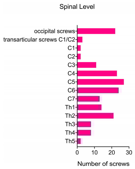
Figure 1.
Screw placement: quantity and anatomical distribution.
Table 2.
Overview of total number and level of screw placement.
Following the surgery, patients were immobilized with a stiff cervical collar until the first follow-up, which occurred 4 to 6 weeks postoperatively. In three cases involving fusion to the upper thoracic spine, a corset was prescribed. One patient (5%) who underwent fusion from the occipito-cervical junction to C6 required a halo vest for immobilization.
3.3. Symptoms at Discharge
At the time of initial presentation, 16 patients (84%) reported neck pain, while 4 patients (21%) experienced radicular pain. Upon clinical examination, sensory deficits were found in seven patients (37%), and paresis was present in six cases (32%). Gait disturbance and hyperreflexia were each observed in two patients (11%). At discharge, 15 patients (94%) in the neck pain group demonstrated improvement in their pain levels, with 1 patient becoming completely pain-free. One patient (6%) showed no change compared to their preoperative pain level. No new postoperative neck pain was reported at discharge. All patients with radicular pain experienced improvement, with one patient in this group becoming pain-free after surgery. Motor deficits improved in three out of six patients (50%), with one patient having no residual paresis at discharge. Muscle strength remained stable in the other three cases (50%). Sensory disturbances improved in four out of seven patients (57%), with two patients (29%) recovering normal sensibility and three patients (43%) remaining unchanged. Regarding myelopathy symptoms, both patients (100%) in the gait disturbance group had no residual symptoms at discharge. As anticipated, hyperreflexia did not normalize during the brief period until discharge.
3.4. Follow-Up and Clinical Long-Term Outcome
Among the 19 patients in this study, 15 had a clinical follow-up of at least 12 months, with a median follow-up duration of 21 months after surgery. One patient died 22 days after the surgery, and no follow-up data could be recorded after discharge. At the last follow-up, 11 out of 15 patients (73%) who had complained of pain before surgery reported an improvement in their overall pain level, including both radicular and neck pain. Three patients (20%) reported no changes, and one patient (7%) experienced an increase in neck pain compared to their preoperative state. Neurological examination revealed that 15 patients (83%) had no sensorimotor deficits. An increase in muscular strength was recorded in four out of five patients (80%), with two patients (33%) achieving normal strength. One patient (20%) remained stable. Among the six patients in the sensory deficit group, only one patient (17%) had a residual sensory deficit with unchanged symptoms. Neither of the two patients with preoperative gait impairment showed residual gait disturbance at the last follow-up (Figure 2A,B and Figure 3).
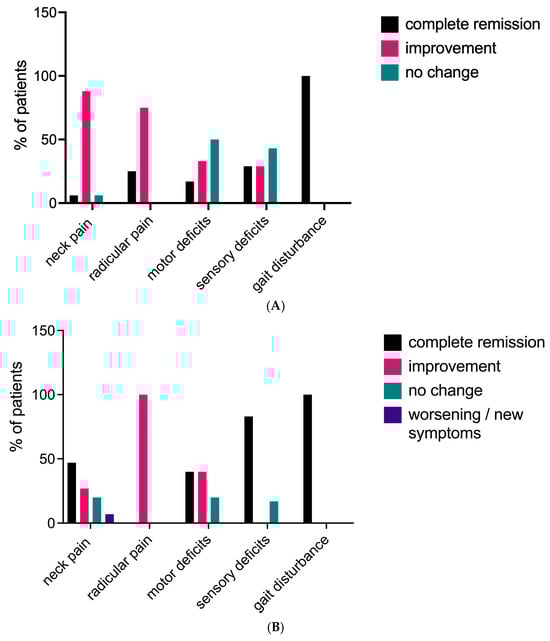
Figure 2.
(A) Clinical course of symptoms at discharge. (B) Evolution of symptoms at final follow-up.
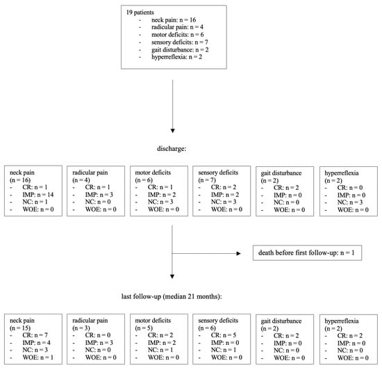
Figure 3.
Flowchart demonstrating the clinical course. CR: complete remission, IMP: improvement, NC: no change, WOE: worsening or new symptoms.
Seven patients (37%) were prospectively followed up in our outpatient clinic. Their Neck Disability Index Score (NDI) ranged from 0 to 24 points. However, six patients (86%) showed only mild or no disability (NDI < 15). The corresponding median for NDI was 4. The Nurick Scale was assessed in two patients with myelopathy symptoms and gait disturbance. The median Nurick Scale score was 0. According to the modified Macnab criteria, a good or excellent outcome was achieved in five patients (71%). In terms of Odom’s criteria, five patients (71%) reported a good or excellent outcome. There was no poor outcome in our current series.
3.5. Radiological Findings
Long-term radiological follow-up was not routinely conducted, with a median radiological follow-up of 12 months. Thirteen out of nineteen patients (68%) underwent a CT scan of the fused segments at least 12 months after the surgery. In this group, the fusion rate was evaluated using the Lenke and Bridwell classification system. A solid fusion across the entire fused area was observed in nine patients (69%). Among these, five cases (38%) achieved trabeculated bilateral fusion (Lenke and Bridwell Grade A), while four cases (31%) had unilateral fusion (Grade B). Small, non-solid fusion masses (Grade C) were observed in three patients (23%). One patient (8%) had a non-union in one or more of the fused segments (Grade D) (Table 3).
Table 3.
Clinical and radiological scores.
During the radiological follow-up, screw loosening was observed in five patients (38%), involving a total of 17 screws. However, due to the absence of symptoms, no revision surgery for screw replacement was necessary.
3.6. Complications and Adverse Events
Four patients (21%) required revision surgery due to surgical site infection (n = 3) or cerebrospinal fluid leak (n = 1). One patient underwent additional surgery using an anterior approach after experiencing a screw fracture and impaction of a previously placed intervertebral cage. In two patients with persistent neck pain, the hardware was removed.
Since the PCF surgery, four patients (21%) died. The indication for surgery in these cases was neoplastic lesions in three patients and degenerative disease in one patient. There was one case (5%) of perioperative mortality within 30 days of surgery. The patient had prostate cancer with osseous metastases and initially underwent an anterior vertebrectomy of C6 and C7 with anterior plate placement, followed by a posterior fusion from C5 to Th4 10 days later. Although the initial postoperative course was uneventful, with the patient being discharged to rehabilitation 7 days after surgery, the patient died in the rehabilitation facility 15 days later. The exact cause of death remains unknown.
3.7. Sensitivity, Specificity, and Accuracy of iCT
In our series, iCT was used in all cases. For upper posterior cervical spine approaches, including C6, lateral mass screws were used, while pedicle screws were the preferred technique for fusion below C6. A total of 151 lateral mass and pedicle screws were evaluated using iCT. Based on the iCT findings, eight screws (5.3%) were repositioned intraoperatively. Correct screw placement was confirmed with a final iCT scan at the end of the surgery. Additionally, all patients underwent a postoperative CT scan, which confirmed the correct placement of 110 screws (72.8%). A minor violation was found in 24 screws (15.9%), a moderate violation in 13 screws (8.6%), and a severe violation in 4 screws (2.6%). No additional surgeries were necessary to reposition the screws.
The sensitivity, specificity, and accuracy of the iCT scans were calculated using the postoperative CT scan as a reference. For correct screw placement, the sensitivity, specificity, and accuracy were 92.7%, 82.9%, and 90.0%, respectively. For minor violations, the values were 70.8%, 91.3%, and 88.1%, respectively. For moderate violations, the sensitivity, specificity, and accuracy were 69.2%, 98.6%, and 96.0%, respectively. For severe screw violations, the sensitivity was 75%, specificity was 100%, and accuracy was 99.3%. Refer to Table 4 and Table 5.
Table 4.
Assessment of screw placement—comparison of intraoperative versus postoperative CT.
Table 5.
Sensitivity, specificity, and accuracy of intraoperative CT.
Radiation exposure was documented in 100% of the cases. The mean cumulative air kerma for the intraoperative CT was 43.51 (±42.93) mGy, compared to 66.6 mGy for the postoperative CT scans of the cervical spine. The mean corresponding cumulative dose area product (DAP) was 15.51 (±15.72) Gy cm2 in the iCT and 13.15 Gy cm2 in the regular postoperative scan.
4. Discussion
4.1. Clinical Outcome
Our study demonstrated good short- and long-term outcomes, regardless of previous surgical interventions on the cervical spine or the underlying condition. At the 2-year follow-up, 79% of patients reported overall pain improvement, and functional recovery was achieved in up to 88% of cases. These findings are consistent with a systematic review and meta-analysis by Youssef et al. [17], which included data from 1238 patients who underwent posterior spinal fusion. They reported similar favorable outcomes, with significant improvements in pain levels and overall function, as assessed by the Japanese Orthopedic Association (JOA) and modified Japanese Orthopedic Association (mJOA) scores. Similarly, Anderson et al. [18] conducted a systematic review of 11 studies and described comparable results, with functional improvement in 70% to 95% of patients and a significant improvement in JOA scores.
In our series, 15 out of 19 patients had a follow-up of at least 12 months. Among all patients, statistically significant improvements were observed in all evaluated categories, including pain, sensorimotor deficits, and gait disturbance. The 15 patients with longer follow-up showed a trend towards progressively better results over time. Although there was no statistical significance between the two groups due to the small sample size, we would expect the four patients who were not followed for at least 12 months to continue improving rather than deteriorating.
The use of iCT in this study allowed for the immediate identification and direct replacement of eight misplaced screws, resulting in improved screw placement accuracy and potentially avoiding the need for subsequent revision surgeries. Patients who underwent intraoperative screw repositioning demonstrated comparable outcomes to those who did not require screw adjustment, with no significant differences in pain reduction, neurological function, or quality-of-life measures in both short- and long-term follow-up. Based on these findings, it is reasonable to hypothesize that the implementation of iCT technology could lead to better clinical outcomes for patients undergoing spinal instrumentation procedures. However, to thoroughly evaluate the impact of iCT on patient outcomes and to establish its superiority over alternative techniques, such as navigated screw placement, further controlled and prospective studies are necessary.
4.2. Surgical Approach
Various surgical approaches to the cervical spine, with or without fusion of the segment, have been described in the literature. Anterior cervical approaches include anterior cervical discectomy with cage only or with an additional plate, as well as corpectomy with fusion. For posterior approaches, laminotomy, laminoplasty, and laminectomy with or without fusion are the most common techniques. In cases of symptomatic cervical disk herniations, anterior cervical discectomy and fusion (ACDF) is a well-established and routine procedure. However, this approach is associated with typical complications such as injury to the recurrent laryngeal nerve and dysphagia [19]. In cervical spondylotic myelopathy (CSM), spondylotic changes and deformities lead to the compression of the spinal cord. The most favorable approach for treating CSM remains controversial [20].
A retrospective study conducted at our institution found that posterior decompression without fusion was an effective treatment option for CSM, significantly relieving symptoms in patients without signs of cervical spinal instability prior to surgery [21]. In contrast, other authors have recommended avoiding stand-alone laminectomy due to the inherent risk of delayed postoperative kyphosis [22]. For patients with pre-existing neck pain, kyphosis, and signs of instability, posterior fusion should generally be the preferred approach [23].
Asher et al. [24] published a multicenter analysis comparing anterior and posterior approaches. They found that patients undergoing posterior fusion were more likely to receive a fusion involving more than three levels compared to the anterior fusion group. While the length of hospital stay was significantly longer in the posterior group, long-term reported outcomes and complication rates were similar between the two approaches [24].
A prospective study [25] involving a total of 264 patients compared anterior discectomy and fusion with posterior decompression (laminoplasty or laminectomy with fusion). In this series, the anterior approach showed favorable results. However, patients receiving posterior decompression were more likely to suffer from multi-level pathologies and were generally older. After adjusting for these confounding factors, similar results were reported for both anterior and posterior approaches [25].
In trauma patients, the choice of surgical approach depends on additional factors such as the presence and volume of herniated disks, the presence of bone fragments, concomitant spondylotic changes narrowing the spinal canal, the presence of uni- or bilateral facet dislocation, and the patient’s neurological status. Finally, it is important to note that posterior fusion of the cervical spine is a more demanding technique, and the surgeon’s training level and experience play a crucial role.
In our current series, 63% of patients were initially treated using an anterior approach. Posterior fusion was the first choice for patients with preoperative instability or malalignment of the cervical spine caused by fractures or tumor growth. Posterior fusion following anterior fusion was performed if symptoms persisted, mainly in cases of malalignment and instability.
4.3. Adverse Events and Revision Rate
Other studies have reported complication rates of 11% and 11.6% [23,26]. We attribute the higher perioperative complication rate in our series to the small number of patients included. All patients who underwent revision surgery showed a good outcome with complication-free wound healing. More significant surgical complications, especially those resulting in new persistent neurological deficits or long-term disability, did not occur. Notably, due to the use of iCT, no additional surgery was required at a later stage to correct the alignment of misplaced screws.
4.4. Radiological Outcomes and Fusion Rates
At the final radiological follow-up, 92% of patients showed signs of partial or complete fusion (Lenke and Bridwell Grades A to C). This finding is consistent with the fusion rates reported in previous studies, which typically ranged from 89% to 100% [27,28]. Only one patient showed a non-union at their last follow-up, which was 7.33 years after posterior fusion. Clinical outcomes in patients with incomplete fusion (Lenke and Bridwell Grades C and D) were comparable to those with solid fusion.
4.5. Intraoperative CT Scan
In comparison to the postoperative CT scan, iCT showed a good sensitivity and specificity. All four screws with severe violation were registered on the iCT scan. Of the 13 screws showing a moderate violation, only one misplaced screw was missed on iCT. A previous study by Nevzati E. et al. [10], which was conducted at our institution, evaluated the accuracy of intraoperative CT scans for pedicle screws in the lumbar and caudal thoracic spine. They reported sensitivity and specificity rates for moderate and severe violations exceeding 86%, which slightly surpasses the sensitivity of our results. We attribute this difference to the smaller diameter of cervical lateral mass and pedicle screws, where differences in resolution between iCT and regular CT scans become more important for detecting screw misplacement.
Some authors argue that lateral mass screw placement does not require intraoperative radiographic control, as they are less likely to be misplaced when using anatomical landmarks [29]. This is consistent with the results in our series, where only one out of the eight intraoperatively corrected screws was a lateral mass screw at the C4 level, while the other seven were transpedicular screws. We routinely performed an iCT scan, as it offered the possibility to further reduce the risk of screw misplacement with a reasonably moderate increase in radiation exposure and only a minor extension of the operation time.
Given the high sensitivity (92.7%), specificity (82.9%), and accuracy (90.0%) reported in this study, it can be argued that a routine postoperative CT scan may be unnecessary, as it offers no significant additional benefits compared to iCT.
It is important to note that the use of an iCT scan is just one method to decrease the number of screw misplacements. Several studies have demonstrated the advantages of using intraoperative navigation [30,31,32,33]. Navigated screw placement is used in many centers and has recently also been established at our institution. Each technique offers distinct advantages and limitations. The angiography suite provides versatility beyond spinal surgery, supporting various interventional procedures. While navigation technology reduces radiation exposure for both patients and surgical staff, it may require larger incisions for reference frame placement, potentially increasing soft tissue dissection compared to intraoperative CT.
At our institution, intraoperative CT remained the standard of care for spinal fusion procedures. However, the recent implementation of navigation technology may reduce the need for routine intraoperative CT scanning in the future, as it independently enables accurate screw placement. The measured mean values for cumulative air kerma and dose area product are slightly lower than the values measured in the postoperative CT scans. However, it is important to note that these measurements are difficult to compare. The iCT scans are performed in a flat panel angiography suite with a rotating C-arm, while the postoperative CT scans use a multidetector computed tomography system. As a result, the actual radiation doses affecting the patient are not accurately represented by the measured values. A study by Jones and Odisio [34] used a phantom to compare the radiation doses when using the two different systems. They found that the mean central axis dose in the multidetector CT system was 41–69% lower compared to the flat panel CT scan. However, the measured noise was much higher in the multidetector CT systems. When noise magnitudes were matched, similar radiation doses were expected between the two systems.
At our institution, cervical fusion procedures are primarily performed via anterior approach without intraoperative CT guidance. The AlluraXper FD20 system maintains cost-effectiveness through its diverse applications beyond spine surgery. The system supports multiple procedures including lumbar spinal fusion, surgical management of intracranial hemorrhages, and vascular neurosurgery. Additionally, the angiography suite serves both neurosurgical and vascular surgery departments. In centers with higher case volumes, the system may achieve cost-effectiveness through spinal fusion procedures alone.
4.6. Study Limitations
This retrospective study had several limitations. First, the cohort size was relatively small, which may limit the generalizability of the findings. Our institutional preference for the anterior approach to cervical fusion limits posterior cervical fusion procedures to approximately six cases annually.
Second, due to the retrospective nature of the study, clinical scores such as VAS, NDI, Nurick Scale, and modified Macnab Criteria were not available for all patients, which could have provided a more comprehensive assessment of patient outcomes. Third, among the 13 patients with long-term radiological follow-up, 3 exhibited fusion masses with apparent cracks, suggesting that these may not represent solid fusions. Finally, CT scans were typically performed on symptomatic patients suspected of non-union, which could have led to a biased detection of lower fusion rates.
5. Conclusions
The findings of this study suggest that posterior cervical fusion performed in a hybrid OR setting is a safe and effective treatment option for various cervical spinal pathologies. Most patients (88%) achieved good or excellent long-term clinical outcomes, with 84% reporting pain improvement and a fusion rate of 94%. The high sensitivity and specificity of iCT for detecting relevant screw malposition prevented the need for any revision surgery for screw replacement in this complex and surgically demanding area of the spine. These results highlight the potential benefits of using advanced intraoperative imaging technologies in the surgical management of cervical spine disorders. Future prospective studies with larger cohorts are needed to fully evaluate the efficacy of intraoperative CT for spinal fusion procedures, particularly in comparison with alternative techniques such as spinal navigation.
Author Contributions
Conceptualization, J.C.K. and J.F.; methodology, J.C.K. and A.D.; data collection, A.D. and J.C.K.; formal analysis, A.D. and J.C.K.; writing—original draft preparation, A.D. and J.C.K.; writing—review and editing, A.K., A.D., J.C.K. and J.F.; supervision, J.C.K. All authors have read and agreed to the published version of the manuscript.
Funding
This research received no external funding.
Institutional Review Board Statement
All procedures performed in studies involving human participants were in accordance with the ethical standards of the institutional and national research committee and with the 1964 Helsinki Declaration and its later amendments or comparable ethical standards. The study was approved by the local ethics committee of northwestern and central Switzerland (EKNZ Nr.2018-00521, approval date 27 June 2018). All patients or their families provided written voluntary informed consent prior to study enrollment.
Informed Consent Statement
Informed consent was obtained from all subjects involved in the study. All patients or their families provided written voluntary informed consent prior to study enrollment.
Data Availability Statement
The authors will share the data upon reasonable request.
Conflicts of Interest
The authors declare no conflicts of interest.
Abbreviations
Posterior cervical fusion (PCF); computed tomography (CT); intraoperative computed tomography (iCT); magnetic reesonance imaging (MRI); cervical spondylotic myelopathy (CSM); anterior cervical discectomy and fusion (ACDF); anterior cervical corpectomy and fusion (ACCF); visual analog scale (VAS); Neck Disability Index (NDI); Japanese Orthopedic Association Score (JOA); modified Japanese Orthopedic Association Score (mJOA); dose area product (DAP).
References
- Fehlings, M.G.; Vaccaro, A.; Wilson, J.R.; Singh, A.; Cadotte, D.W.; Harrop, J.S.; Aarabi, B.; Shaffrey, C.; Dvorak, M.; Fisher, C.; et al. Early versus delayed decompression for traumatic cervical spinal cord injury: Results of the Surgical Timing in Acute Spinal Cord Injury Study (STASCIS). PLoS ONE 2012, 7, e32037. [Google Scholar] [CrossRef]
- Persson, L.C.; Carlsson, C.A.; Carlsson, J.Y. Long-lasting cervical radicular pain managed with surgery, physiotherapy, or a cervical collar. A prospective, randomized study. Spine 1997, 22, 751–758. [Google Scholar] [CrossRef]
- Nikolaidis, I.; Fouyas, I.P.; Sandercock, P.A.; Statham, P.F. Surgery for cervical radiculopathy or myelopathy. Cochrane Database Syst Rev. 2010, 2010, CD001466. [Google Scholar] [CrossRef] [PubMed]
- Fehlings, M.G.; Tetreault, L.A.; Riew, K.D.; Middleton, J.W.; Aarabi, B.; Arnold, P.M.; Brodke, D.S.; Burns, A.S.; Carette, S.; Chen, R.; et al. A Clinical Practice Guideline for the Management of Patients With Degenerative Cervical Myelopathy: Recommendations for Patients With Mild, Moderate, and Severe Disease and Nonmyelopathic Patients With Evidence of Cord Compression. Glob. Spine J. 2017, 7 (Suppl. S3), 70S–83S. [Google Scholar] [CrossRef] [PubMed]
- Mummaneni, P.V.; Kaiser, M.G.; Matz, P.G.; Anderson, P.A.; Groff, M.W.; Heary, R.F.; Holly, L.T.; Ryken, T.C.; Choudhri, T.F.; Vresilovic, E.J.; et al. Cervical surgical techniques for the treatment of cervical spondylotic myelopathy. J. Neurosurg. Spine 2009, 11, 130–141. [Google Scholar] [CrossRef] [PubMed]
- Emery, S.E.; Bohlman, H.H.; Bolesta, M.J.; Jones, P.K. Anterior cervical decompression and arthrodesis for the treatment of cervical spondylotic myelopathy. Two to seventeen-year follow-up. J. Bone Jt. Surg. Am. 1998, 80, 941–951. [Google Scholar] [CrossRef] [PubMed]
- Hilibrand, A.S.; Fye, M.A.; Emery, S.E.; Palumbo, M.A.; Bohlman, H.H. Impact of smoking on the outcome of anterior cervical arthrodesis with interbody or strut-grafting. J. Bone Joint Surg. Am. 2001, 83-A, 668–673. [Google Scholar] [CrossRef]
- Koller, H.; Reynolds, J.; Zenner, J.; Forstner, R.; Hempfing, A.; Maislinger, I.; Kolb, K.; Tauber, M.; Resch, H.; Mayer, M.; et al. Mid- to long-term outcome of instrumented anterior cervical fusion for subaxial injuries. Eur. Spine J. 2009, 18, 630–653. [Google Scholar] [CrossRef]
- Kawakami, M.; Tamaki, T.; Iwasaki, H.; Yoshida, M.; Ando, M.; Yamada, H. A comparative study of surgical approaches for cervical compressive myelopathy. Clin. Orthop. Relat. Res. 2000, 381, 129–136. [Google Scholar] [CrossRef] [PubMed]
- Nevzati, E.; Fandino, J.; Schatlo, B.; Heimberg, M.; Marbacher, S.; Remonda, L.; Fathi, A.-R. Validation and accuracy of intraoperative CT scan using the Philips AlluraXper FD20 angiography suite for assessment of spinal instrumentation. Br. J. Neurosurg. 2017, 31, 741–746. [Google Scholar] [CrossRef] [PubMed]
- McCormack, H.M.; Horne, D.J.; Sheather, S. Clinical applications of visual analogue scales: A critical review. Psychol. Med. 1988, 18, 1007–1019. [Google Scholar] [CrossRef] [PubMed]
- Vernon, H.; Mior, S. The Neck Disability Index: A study of reliability and validity. J. Manipulative Physiol. Ther. 1991, 14, 409–415. [Google Scholar]
- Nurick, S. The pathogenesis of the spinal cord disorder associated with cervical spondylosis. Brain 1972, 95, 87–100. [Google Scholar] [CrossRef]
- Macnab, I. Negative disc exploration. An analysis of the causes of nerve-root involvement in sixty-eight patients. J. Bone Joint Surg. Am. 1971, 53, 891–903. [Google Scholar] [CrossRef] [PubMed]
- Odom, G.L.; Finney, W.; Woodhall, B. Cervical disk lesions. J. Am. Med. Assoc. 1958, 166, 23–28. [Google Scholar] [CrossRef]
- Lenke, L.G.; Betz, R.R.; Harms, J.; Bridwell, K.H.; Clements, D.H.; Lowe, T.G.; Blanke, K.R.N. Adolescent idiopathic scoliosis: A new classification to determine extent of spinal arthrodesis. J. Bone Jt. Surg. Am. 2001, 83-A, 1169–1181. [Google Scholar] [CrossRef]
- Youssef, J.A.; Heiner, A.D.; Montgomery, J.R.; Tender, G.C.; Lorio, M.P.; Morreale, J.M.; Phillips, F.M. Outcomes of posterior cervical fusion and decompression: A systematic review and meta-analysis. Spine J. 2019, 19, 1714–1729. [Google Scholar] [CrossRef] [PubMed]
- Anderson, P.A.; Matz, P.G.; Groff, M.W.; Heary, R.F.; Holly, L.T.; Kaiser, M.G.; Mummaneni, P.V.; Ryken, T.C.; Choudhri, T.F.; Vresilovic, E.J.; et al. Laminectomy and fusion for the treatment of cervical degenerative myelopathy. J. Neurosurg. Spine 2009, 11, 150–156. [Google Scholar] [CrossRef] [PubMed]
- Lee, M.J.; Bazaz, R.; Furey, C.G.; Yoo, J. Risk factors for dysphagia after anterior cervical spine surgery: A two-year prospective cohort study. Spine J. 2007, 7, 141–147. [Google Scholar] [CrossRef]
- Witwer, B.P.; Trost, G.R. Cervical spondylosis: Ventral or dorsal surgery. Neurosurgery 2007, 60 (Supp1. S1), S130–S136. [Google Scholar] [CrossRef]
- Woernle, K.; Marbacher, S.; Khamis, A.; Landolt, H.; Fandino, J. Clinical Outcome after Laminectomy without Fusion for Cervical Spondylotic Myelopathy. Open J. Mod. Neurosurg. 2015, 5, 41–48. [Google Scholar] [CrossRef][Green Version]
- Albert, T.J.; Vacarro, A. Postlaminectomy kyphosis. Spine 1998, 23, 2738–2745. [Google Scholar] [CrossRef] [PubMed]
- Lau, D.; Winkler, E.A.; Than, K.D.; Chou, D.; Mummaneni, P.V. Laminoplasty versus laminectomy with posterior spinal fusion for multilevel cervical spondylotic myelopathy: Influence of cervical alignment on outcomes. J. Neurosurg. Spine 2017, 27, 508–517. [Google Scholar] [CrossRef] [PubMed]
- Asher, A.L.; Devin, C.J.; Kerezoudis, P.; Chotai, S.; Nian, H.; Harrell, F.E., Jr.; Sivaganesan, A.; McGirt, M.J.; Archer, K.R.; Foley, K.T.; et al. Comparison of Outcomes Following Anterior vs. Posterior Fusion Surgery for Patients With Degenerative Cervical Myelopathy: An Analysis From Quality Outcomes Database. Neurosurgery 2019, 84, 919–926. [Google Scholar] [CrossRef] [PubMed]
- Fehlings, M.G.; Barry, S.; Kopjar, B.; Yoon, S.T.; Arnold, P.; Massicotte, E.M.; Vaccaro, A.; Brodke, D.S.; Shaffrey, C.; Smith, J.S.; et al. Anterior versus posterior surgical approaches to treat cervical spondylotic myelopathy: Outcomes of the prospective multicenter AOSpine North America CSM study in 264 patients. Spine 2013, 38, 2247–2252. [Google Scholar] [CrossRef] [PubMed]
- Audat, Z.A.; Fawareh, M.D.; Radydeh, A.M.; Obeidat, M.M.; Odat, M.A.; Bashaireh, K.M.; Barbarawi, M.M.; Nusairat, M.T.; Ibraheem, A.B.; Audat, M.Z. Anterior versus posterior approach to treat cervical spondylotic myelopathy, clinical and radiological results with long period of follow-up. SAGE Open Med. 2018, 6, 2050312118766199. [Google Scholar] [CrossRef]
- Muffoletto, A.J.; Hadjipavlou, A.G.; Jensen, R.E.; Nauta, H.J.; Necessary, J.T.; Norcross-Nechay, K. Techniques and pitfalls of cervical lateral mass plate fixation. Am. J. Orthop. 2000, 29, 897–903. [Google Scholar]
- Kumar, V.G.; Rea, G.L.; Mervis, L.J.; McGregor, J.M. Cervical spondylotic myelopathy: Functional and radiographic long-term outcome after laminectomy and posterior fusion. Neurosurgery 1999, 44, 771–777, Discussion 777–778. [Google Scholar] [CrossRef]
- Katonis, P.; Papadakis, S.A.; Galanakos, S.; Paskou, D.; Bano, A.; Sapkas, G.; Hadjipavlou, A.G. Lateral mass screw complications: Analysis of 1662 screws. J. Spinal. Disord. Tech. 2011, 24, 415–420. [Google Scholar] [CrossRef]
- Scarone, P.; Vincenzo, G.; Distefano, D.; Del Grande, F.; Cianfoni, A.; Presilla, S.; Reinert, M. Use of the Airo mobile intraoperative CT system versus the O-arm for transpedicular screw fixation in the thoracic and lumbar spine: A retrospective cohort study of 263 patients. J. Neurosurg. Spine 2018, 29, 397–406. [Google Scholar] [CrossRef] [PubMed]
- Farah, K.; Coudert, P.; Graillon, T.; Blondel, B.; Dufour, H.; Gille, O.; Fuentes, S. Prospective Comparative Study in Spine Surgery Between O-Arm and Airo Systems: Efficacy and Radiation Exposure. World Neurosurg. 2018, 118, e175–e184. [Google Scholar] [CrossRef] [PubMed]
- Mandelka, E.; Gierse, J.; Zimmermann, F.; Gruetzner, P.A.; Franke, J.; Vetter, S.Y. Implications of navigation in thoracolumbar pedicle screw placement on screw accuracy and screw diameter/pedicle width ratio. Brain Spine 2023, 3, 101780. [Google Scholar] [CrossRef]
- Ille, S.; Baumgart, L.; Obermueller, T.; Meyer, B.; Krieg, S.M. Clinical efficiency of operating room-based sliding gantry CT as compared to mobile cone-beam CT-based navigated pedicle screw placement in 853 patients and 6733 screws. Eur. Spine J. 2021, 30, 3720–3730. [Google Scholar] [CrossRef] [PubMed]
- Jones, A.K.; Odisio, B.C. Comparison of radiation dose and image quality between flat panel computed tomography and multidetector computed tomography in a hybrid CT-angiography suite. J. Appl. Clin. Med. Phys. 2020, 21, 121–127. [Google Scholar] [CrossRef]
Disclaimer/Publisher’s Note: The statements, opinions and data contained in all publications are solely those of the individual author(s) and contributor(s) and not of MDPI and/or the editor(s). MDPI and/or the editor(s) disclaim responsibility for any injury to people or property resulting from any ideas, methods, instructions or products referred to in the content. |
© 2025 by the authors. Licensee MDPI, Basel, Switzerland. This article is an open access article distributed under the terms and conditions of the Creative Commons Attribution (CC BY) license (https://creativecommons.org/licenses/by/4.0/).