Article

Amyloid-PET and White Matter Hyperintensities Have Independent Effects on Baseline Cognitive Function and Synergistic Effects on Longitudinal Executive Function

1
Sanders-Brown Center on Aging, College of Medicine, University of Kentucky, Lexington, KY 40506, USA
2
Department of Behavioral Science, College of Medicine, University of Kentucky, Lexington, KY 40506, USA
3
Department of Epidemiology, College of Public Health, University of Kentucky, Lexington, KY 40536, USA
4
Department of Neurology, College of Medicine, University of Kentucky, Lexington, KY 40506, USA
5
Department of Radiology, College of Medicine, University of Kentucky, Lexington, KY 40536, USA
6
Department of Neuroscience, College of Medicine, University of Kentucky, Lexington, KY 40536, USA
7
Department of Physiology, College of Medicine, University of Kentucky, Lexington, KY 40506, USA
*
Author to whom correspondence should be addressed.
Brain Sci. 2023, 13(2), 218; https://doi.org/10.3390/brainsci13020218
Submission received: 3 January 2023 / Revised: 21 January 2023 / Accepted: 23 January 2023 / Published: 28 January 2023
(This article belongs to the Section Neurodegenerative Diseases)

Abstract

:
Co-occurrence of beta amyloid (Aβ) and white matter hyperintensities (WMHs) increase the risk of dementia and both are considered biomarkers of preclinical dementia. Moderation and mediation modeling were used to define the interplay between global and regional Aβ and WMHs measures in relation to executive function (EF) and memory composite scores outcomes at baseline and after approximately 2 years across a sample of 714 clinically normal participants from the Alzheimer’s Disease Neuroimaging Initiative (ADNI 2). The moderation regression analysis showed additive effects of Aβ and WMHs over baseline memory and EF scores (p = 0.401 and 0.061, respectively) and synergistic effects over follow-up EF (p < 0.05). Through mediation analysis, the data presented demonstrate that WMHs effects, mediated by global and regional amyloid burden, are responsible for baseline cognitive performance deficits in memory and EF. These findings suggest that Aβ and WMHs contribute to baseline cognition independently while WMHs volumes exert effects on baseline cognitive performance directly and through influences on Aβ accumulation.

1. Introduction

Alzheimer’s disease (AD) and subcortical vascular dementia are considered the most common pathologic contributors to dementia in the aging population. Both frequently coexist in over 80% of community dwelling adults with dementia [1]. Cerebral small vessel disease (CSVD) has also been linked to the pathogenesis of AD and is largely responsible for the development of subcortical vascular dementia [2]. AD and CSVD share multiple risk factors [3], occurring concomitantly in over 50% of individuals with dementia [4], and there may be substantial overlap between these two conditions in terms of clinical, pathological and radiological findings. Co-occurrence of beta amyloid (Aβ) (a hallmark pathologic feature of AD) and white matter hyperintensities (WMHs) (reflecting CSVD burden) increase the risk of dementia [5].
WMHs and Aβ are also key drivers of cognitive decline in healthy older adults, and both are considered biomarkers of preclinical dementia [6,7]. Neuroimaging studies examining the combined impact of Aβ burden and WMHs on cognition have largely examined these variables independently rather than examining the potential interplay between these key pathologic hallmarks of dementia. Several studies have addressed this issue of interplay between Aβ and WMHs with often contradictory results [6,7,8,9,10,11,12,13,14]. Notably, many of these studies focused on global measures of WMHs and Aβ without exploration of regional effects in relation to stage of disease. Studying the effect of Aβ and WMHs on cognition without emphasizing the importance of lesion location can miss the functional consequences of the two pathologies. Previous studies investigating the impact of regional distribution of WMHs on executive function (EF) found that WMHs in all cortical regions (frontal areas, occipital, parietal and temporal) are associated with deficits in EF scores [15,16,17], but regional quantification of Aβ and its interaction with WMH volume has not been fully investigated in these prior studies.
Aβ deposition is hypothesized as being the initial step in the neuropathological development of AD and dementia [18], but findings from previous studies have also shown that WMHs often occur prior to the presence of amyloid-β plaques in preclinical AD [19,20,21] supporting a retrograde degeneration hypothesis [22,23]. Additionally, evidence from recent studies has suggested that the relationship between WMHs and Aβ is strongly determined by the spatial distribution of the two pathologies [24]. The majority of published studies have emphasized the spatial heterogeneity of WMHs [25,26], but have left the potential heterogeneous influences of regional Aβ deposition relatively unexplored.
Since mixed dementia is widely recognized as the norm rather than the exception, in the present study we sought to explore the relation between regional and global Aβ and WMHs with cognitive function (EF and memory) scores in cognitively normal (CN) older adults at baseline, and further examine the relation between WMHs and regional Aβ deposition in relation to cognitive performance changes over time.

2. Materials and Methods

Data used in the preparation of this article were obtained from the Alzheimer’s Disease Neuroimaging Initiative (ADNI) database (adni.loni.usc.edu). The ADNI was launched in 2003 as a public–private partnership, led by Principal Investigator Michael W. Weiner, MD. The primary goal of ADNI has been to test whether serial magnetic resonance imaging (MRI), positron emission tomography (PET), other biological markers and clinical and neuropsychological assessment can be combined to measure the progression of mild cognitive impairment (MCI) and early Alzheimer’s disease (AD).

2.1. Participants

Summary data from the Alzheimer’s Disease Neuroimaging Initiative (ADNI 2) at http://adni.loni.usc.edu (accessed on 24 July 2021) [27] were used in the present analyses. Only participants with a mini-mental state examination (MMSE) score greater than or equal to 26 at baseline meeting these inclusion criteria were included in the analysis: (1) complete demographic information (i.e., age, sex, education) and ApoE genotype available; (2) neurocognitive composite metrics for EF and memory at baseline and after approximately 2 years, (3) PET Florbetapir for focal and global Aβ quantification; and (4) T2 FLAIR scan acquisition with WMHs volume quantification. Details of ADNI inclusion criteria, clinical procedures and methodology are available elsewhere [28,29]. These criteria were developed to specifically investigate predisease and preclinical disease states that may lead to further cognitive impairment and dementia.
Participants with missing data required for the analyses were excluded. A total of 53 participants were excluded from the longitudinal analysis on the basis of missing data required for the analysis. Excluded participants did not differ significantly from these included in the analysis in regards to age, sex, education, ApoE or baseline MMSE scores (data not shown).

2.2. Imaging Analysis

2.2.1. White Matter Hyperintensity Quantification Method

WMH volumes were quantified using the 4-tissue segmentation method described previously [30]. Briefly, first step was co-registration of the FLAIR to the 3D T1 image inhomogeneity-corrected and non-linearly aligned to a minimal deformation template (MDT) using the T1 transformation and the FMRIB Software Library (FSL) toolbox [31,32]. Modified Bayesian probability and structure prior probability maps were used to estimate WMH in the MDT. Binary WMHs masks were then created using 3.5 SD threshold above the mean. The segmented WMHs masks were then back-transformed into native space for tissue WMH volumes calculation. An Expectation–Maximization (EM) algorithm was used for segmentation to isolate gray, white, and CSF measurements in template-space. Transforming these masks back to each image’s native space produced rough estimate 3-tissue segmentations. Finally, WMHs were ultimately subtracted from segmented white matter volume and reported in cubic millimeters.

2.2.2. Calculation of Florbetapir Cortical Summary Values

Preprocessing of the AV-45 PET scans and computation of the global AV-45 PET values were done centrally by the ADNI core as described previously [33]. Briefly, each subject’s florbetapir image was coregistered using SPM8 to that subject’s MRI image that was closest in time to the florbetapir scan. Freesurfer processing was carried out to skull-strip, segment and delineate cortical and subcortical regions in all MRI scans [34,35]. Volume-weighted florbetapir means from a cortical summary region were extracted. A single binary cortical summary region composed of all the subregions was created to calculate the mean uptake across each region. We used the summary data of global and regional results (frontal, parietal and cingulate regions that have been most frequently associated with EF performance [36,37,38,39]). SUVr determination was based on the whole cerebellum reference region. Details regarding regions of interest forming the subregions frontal, parietal and cingulate have been described previously [33].

2.3. Composite Measures of Executive Function and Memory: ADNI_EF and ADNI_Memory

The specific tests included in ADNI executive function (ADNI-EF) composite scores are Category Fluency (animal and vegetable naming), Trail Making Tests A and B, Digit Span backwards, Wechsler adult intelligence scale-revised (WAIS-R) Digit Symbol Substitution and 5 Clock Drawing items (circle, symbol, numbers, hands, time) [40]. The memory composite (ADNI_Memory) included the Rey auditory verbal learning test (RAVLT), the cognitive component of the Alzheimer’s disease assessment scale (ADAS-Cog) and Wechsler logical memory scale scores [40]. We used the ADNI-EF and ADNI-Memory scores corresponding to the baseline scan and EF and memory scores within one to three years from the baseline as follow-up scores.

2.4. Statistical Analyses

All statistical analyses were conducted using SPSS, version 26.0 (SPSS Inc., Chicago, IL, USA). Significance was set at p < 0.05, with correction based on the number of the analysis. We used multiple regression models including the interaction term and the main effects of Aβ and WMHs to investigate the independent and combined associations of these pathologies with cognition (ADNI-EF and ADNI-Memory). WMH volumes were logarithmically transformed due to the positive skewed distribution for the statistical analysis. Moderation analyses assessing the relationship between Aβ and WMHs were fit to the data using the following equation:
Ŷ = b0 + b1WMHs + b2 Aβ + b3WMH*Aβ +bX
where Ŷ represents the mean scores of composite measures of cognitive (executive or memory) function and bX represents beta coefficients and adjustment covariates. Age (continuous values), sex (indicator variable), education (continuous values), ApoE (dummy indicator variable) and total intracranial volumes (continuous values) were entered as covariates in all analyses. The models were built using Hayes’ PROCESS macro for SPSS (model 1) [41]. Longitudinal analyses examined the association of the interaction between baseline Aβ burden and WMHs with mean follow-up cognitive (ADNI-EF or ADNI-Memory) scores after controlling for baseline ADNI cognitive scores and time following the baseline scan acquisitions and cognitive testing. To consider the regional Aβ burden effects in addition to global Aβ burden and WMHs on baseline and change in ADNI-EF and ADNI-Memory, moderated regressions were run for amyloid deposition (SUVr) in each of the a priori selected cortical regions (Frontal, cingulate and parietal), adjusting for age, sex, education, ApoE and intracranial volumes.
Assuming that WMH is causally related to Aβ deposition, which is in turn causally related to cognitive (ADNI-EF or ADNI-Memory) scores, mediation models were built to estimate the relationship between WMH (independent variable) and cognitive function (dependent variable) and Aβ (mediator variable). This model was built using Hayes’ PROCESS macro for SPSS (model 4) [41]. Significance was tested using 5000 bootstrap samples to calculate bias-corrected 95% confidence intervals. Indirect effects with bootstrapped 95% confidence intervals not crossing zero were considered significant. Age, sex, education, ApoE and intracranial volumes were entered as covariates in the mediation regression models. Baseline ADNI composite scores and times between the two cognitive scores tests were added as covariates in the mediation regression models examining longitudinal cognitive score changes.

3. Results

The demographic, clinical and imaging characteristics of the included participants are provided in Table 1. Briefly, the sample included 326 women and 388 men with a mean age of 73.13 ± 7.4 years; participants were highly educated on average (16.3 ± 2.6 years).

3.1. Testing the Interaction Term and the Main Effect of Global and Regional Aβ Burden and WMHs with Cognition

The moderation regression analysis detected a marginally statistically significant interaction effect between Aβ burden and WMHs in relation to baseline EF (p-value = 0.061), and a significant interaction term between the biomarkers in relation to longitudinal executive functions with p-value = 0.036. Overall, when amyloid burden was higher, the association between log WMHs and ADNI-EF was stronger. (+1 SD above sample mean) (Figure 1A). Regarding memory composite scores, the models did not detect any statistically significant interaction effect between global Aβ burden and WMHs in relation to baseline ADNI-memory scores or follow-up ADNI-memory scores (p-Value 0.401 and 0.937 respectively).
The regional models examining the interaction between Aβ burden in discrete cortical regions and WMH volumes detected marginally significant interaction between frontal, parietal and cingulate Aβ burden and WMHs in relation to baseline EF performance with p-value 0.061, 0.059 and 0.071, respectively. In contrast, the models failed to detect even marginally significant interaction effects between regional Aβ burden and WMHs with baseline memory (p-values for interaction term were in frontal Aβ 0.360, parietal Aβ 0.353 and cingulate Aβ 0.411).
The moderation analysis detected a significant interaction effect of frontal and cingulate Aβ burden and WMHs on longitudinal EF performance with p value 0.033 for frontal and 0.024 for cingulate (Figure 1B,C). The (regression) slopes illustrating the negative association of WMH volume with longitudinal executive function performance for three levels of regional Aβ burden and WMHs (−1, 0 and 1 SD using sample’s mean) are shown in Figure 1. In contrast to the results for EF, the moderation regression analysis failed to detect a statistically significant interaction effect between regional Aβ burden and WMH in relation to follow-up ADNI-memory scores. Results regarding the moderation models between global and regional Aβ burden and WMHs with baseline and follow-up cognitive (ADNI-EF and ADNI-Mem) scores are displayed in Supplementary tables.
The (regression) slopes representing WMH’s negative associations with executive functions for high levels of regional Aβ burden (+1 SD using sample’s mean). Figure 1A showed the interaction term of global Aβ burden and WMHs with Follow-up ADNI-EF scores. Overall, when amyloid burden was higher, the association between log WMH and ADNI-EF was stronger. (+1SD above sample mean). Figure 1B showed the interaction term of frontal Aβ burden and WMHs with Follow-up ADNI-EF scores. Figure 1C showed the interaction term of baseline cingulate Aβ burden and WMHs with Follow-up ADNI-EF scores. Figure 1D showed the interaction term of baseline parietal Aβ burden and WMHs with Follow-up ADNI-EF scores.

3.2. Testing the Mediation Effects of Global and Regional Aβ Burden on WMH-Related Cognitive Performance

Mediation analyses were conducted to assess whether global and regional Aβ burdens act as potential mediators of the relationship between WMHs and cognitive performance. Path models of the mediation effect are presented in Table 2. The path analysis revealed that global Aβ deposition mediates the relationship between WMHs and baseline cognitive (EF and memory) scores (see Figure 2A,B). The bootstrap confidence interval (CI) for the indirect effect demonstrated that the indirect effect of WMH on baseline EF performance (a × b = −0.045; CI: −0.09, −0.01) and baseline memory scores (CI: −0.01, −0.006) through global Aβ burden were significant. Both frontal and parietal Aβ SUVr mediated the effect of WMH on baseline EF and memory performance. In contrast, there was no significant mediation effect by cingulate Aβ SUVr for baseline cognitive performance (see Table 2 and Supplementary Materials figures). The other model explored whether global and regional Aβ SUVr mediated the relationship between WMHs and follow-up cognitive (EF and memory) scores after adding baseline cognitive scores and times since the initial cognitive test as the covariates. Results demonstrated that neither global Aβ SUVr (see Figure 2C,D) nor regional Aβ SUVr mediates these associations as the CI for the indirect effect was crossing the zero and did not reach statistical significance. While our path analyses were conducted to assess whether global and regional Aβ burden act as potential mediators of the relationship between WMHs and ADNI-memory scores, we tested its opposite direction, and no significant mediating effects were detected for baseline or follow-up ADNI-memory scores. (See Supplementary Materials figures)
As such, path (a) expresses one unit increase in WMH associated with increase of Aβ SUVr, whereas path (b) expresses the change in cognitive function (EF and memory) associated with an increment of Aβ SUVr. Path (c’) shows the direct effect of WMHs on cognitive function scores and path (c) shows the total effect. The bootstrap confidence interval (CI) for the indirect effect demonstrated that the indirect effect of WMH on baseline EF performance (a × b = −0.045; CI: −0.09, −0.01) (Figure 2A) and baseline memory scores (CI: −0.01, −0.006) (Figure 2B) through global Aβ burden were significant. In contrast, the CI for the indirect mediation effects of WMHs on follow-up EF scores ((a × b = −0.015; CI: −0.045, 0.009) (Figure 2C)) and follow-up ADNI-memory scores ((CI: −0.031, 0.002) (Figure 2D)) through global Aβ SUVr were crossing zero. (WMH, white matter hyperintensities; SUVr, standard uptake value ratio; Aβ, beta amyloid; CI, confidence intervals).

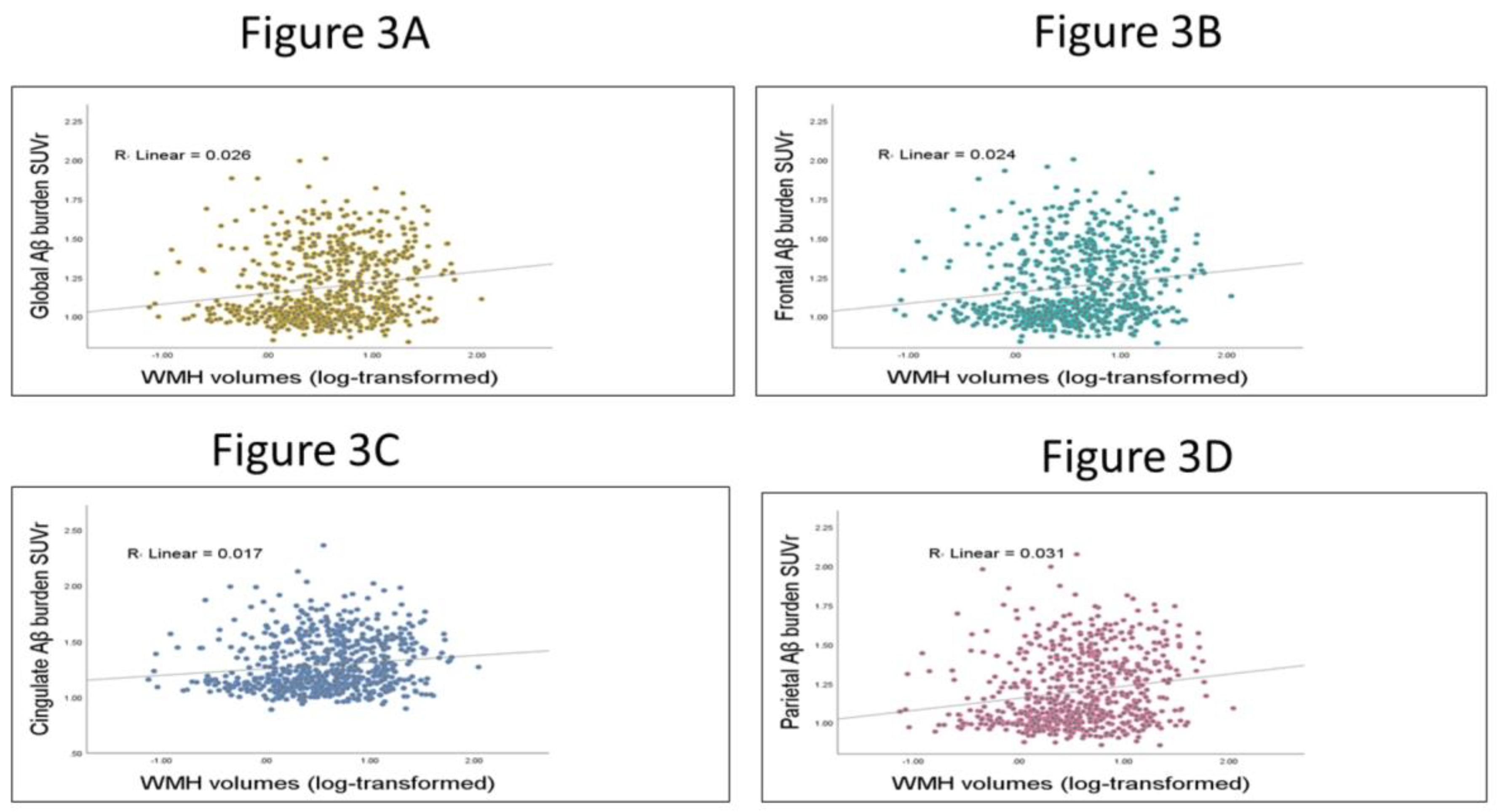
3.3. Testing the Relation between WMHs and Global and Regional Aβ SUVr

Bivariate plots were used to show the association between regional and global Aβ burden, and WMH volumes. (See Figure 3)
Figure 3: The correlation between regional and global Aβ burden, and WMH volumes.
The scatterplots show the relation between Aβ burden and WMH volumes. In Figure 3A, the Pearson correlation between global Aβ and WMH volumes was 0.162. Figure 3B, the Pearson correlation between frontal Aβ and WMH volumes was 0.155. In Figure 3C, the Pearson correlation between cingulate Aβ and WMH volumes was 0.130. Finally, in Figure 3D the Pearson correlation between parietal Aβ and WMH volumes was 0.177.

4. Discussion

The present results indicate that there is an independent effect of both Aβ and WMHs on cognitive performance at baseline and a synergistic interaction between these baseline biomarkers of pathology on future EF performance. This synergistic effect was not seen in future memory performance. The other main finding, demonstrated through mediation analysis, is that the extent of WMH-dependent changes in baseline EF was mediated by both global and regional Aβ burdens. Thus, WMH accumulation appears to increase Aβ deposition which in turn influences cognition longitudinally.
The present moderation analysis provides evidence of an additive effect of WMHs and Aβ in relation to baseline cognitive performance, which is consistent with the findings from other cross-section biomarker studies [6,9,10,42]. The present study extends the results from previous studies by analyzing the effect of baseline biomarkers of Aβ and WMHs on future cognitive performance. The data demonstrate that the combined effect of Aβ burden and WMHs on follow-up EF scores is greater than the sum of the two individual biomarker effects supporting a hypothesis of synergistic interactions between both Aβ and WMHs. The synergistic effect between the baseline biomarkers of pathology on follow-up EF scores was not seen in follow-up memory scores. This finding may be related to the population studied as ADNI participants are largely well-educated and ADNI excludes participants with severe CSVD. One previous study showed that CN participants with low WMH volumes underwent low to no changes in memory performance, independent of amyloid burden [11]. Additionally, findings from another study have demonstrated that the association between WMH volumes and both EF and memory function were significant in demented participants, but that the relationship between memory and WMHs was not detected in CN participants, such as those included in the present study [43]. It is also possible that the synergistic effect seen in follow-up EF performance but not memory performance may involve alterations in brain function and structure networks [44], WMH burden and/or Aβ deposition that were not included in the present analysis.
The present data further demonstrate that regional Aβ burden in frontal and cingulate regions are most related to WMHs effect on future EF change. These data suggest that this relation might be explained by WMHs’ impacts on connectivity within the executive control, and default mode networks [44]. Indeed, the present findings support the hypothesis that executive function performance is dependent on both intact white matter pathway connectivity, as well as pathologic integrity of the cortical regions themselves [12,45,46] and that there is a synergistic interaction between baseline biomarkers of pathology on longitudinal cognitive functions that is dependent on frontal–subcortical circuitry in cognitive aging and pAD [13].
Studying the independent associations of WMHs and Aβ burden with baseline and follow-up cognitive performance, the present data demonstrate that baseline EF performance is explained by both global amyloid burden and WMHs, but that Aβ burden was the only predictor of follow-up EF performance and baseline memory performance (See Supplementary Tables S1 and S2). Consistent with our findings, one recent study from the University of California, Davis Alzheimer’s Disease Research Center (UCD ADRC) diversity cohort found that WMHs were related to baseline EF, but not longitudinal change in EF [11]. Conversely, the same group has argued that baseline WMHs likely represent only a small percentage of the final WMHs [47], suggesting that longitudinal EF performance may be most related to progression of WMHs, rather than solely associated with baseline WMHs. It is also possible that these findings are related to the population studied, as ADNI excludes participants with severe vascular risk factors that may limit analysis of the full impact of WMH and other cerebrovascular injury in this cohort [9,48]. It should also be pointed out that baseline WMH was one of the predictors for follow-up memory performance with Aβ burden. These findings emphasize the important role of WMHs as a risk factor for future memory decline even if they may not affect baseline memory performance at the stage of pAD [11,49].
The present data clearly demonstrate that WMHs are associated with global and regional Aβ burden pathology, exerting an effect on baseline cognitive performance through multiple pathways. Consistent with our findings, a recently published study based on the C6 project in the Medical Imaging Trial Network of Canada (MITNEC-C6), which differs from ADNI in being a WMH-enriched cohort [50] demonstrated that the relation between global WMH and cognition was mediated by global Aβ burden and cortical atrophy. The authors interpreted their results to suggest that Aβ burden appears to be aggravated in participants with WMHs supporting a synergistic pathophysiological process. They further hypothesized that the relationship between WMHs and Aβ burden may be related to impaired general cerebral perfusion [5,51] or to vascular pathology affecting amyloid clearance pathways [8]. Another possible explanation presented by the authors focused on myelin damage [52], which has been shown to promote Aβ oligomerization, eventually contributing to amyloid deposition [53]. These potential mechanisms may help explain the topographic patterns of regional Aβ deposition in relation to WMH effects on critical brain networks seen in the present study. The hypothesis of a regional association between Aβ and WMHs is supported by the present data.
One major limitation of the present study includes the possibility of selection bias as ADNI excludes participants with significant evidence for cerebrovascular disease, which may limit discoveries into the interactions between Aβ and WMHs that may exist in more generalizable samples. Further studies examining the interaction between Aβ and WMHs in participants with higher levels of CSVD including lacunes, small subcortical infarctions, microinfarcts and microbleeds are needed to more fully investigate the associations of Aβ with CSVD. Additionally, the sample of participants evaluated is relatively small for moderation analyses but was restricted by the availability of extant data. Longitudinal outcome analyses may also be limited by the relatively short follow-up period in the ADNI 2 participants studied. It is possible that a more extended follow-up period would help to clarify further the relationships between Aβ and WMHs across progressive stages of cognitive decline, including MCI and early dementia stages. Strengths of the present study include the use of ADNI data allowing analysis in a large sample of relatively homogeneous CN participants that have been classified extensively for the presence of pAD and CSVD. An additional strength is the use of both mediation and moderation analyses in studying the relation between cognitive function and the effects of the two primary drivers of dementia in the population (Aβ and WMHs) within discrete brain regions.

5. Conclusions

While to what extent WMHs affect Aβ burden and cognition remain unclear, the present data demonstrate that baseline Aβ burden and WMH volumes have both independent associations with EF and memory scores at baseline, as well as synergistic associations with future EF performance. The extent of WMH-dependent changes in baseline cognitive performance was related to both the direct effect of WMHs and an indirect effect moderated through global and regional Aβ burdens. The biological relationships between regional Aβ and WMHs responsible for this indirect effect need further investigation across a broader profile of the disease course of dementia.

Supplementary Materials

The following supporting information can be downloaded at https://www.mdpi.com/article/10.3390/brainsci13020218/s1, Figure S1: Path models of the mediation effect of WMHs between global and regional Aβ burden and baseline ADNI-memory scores; Figure S2: Path models of the mediation effect of baseline WMH volumes on follow-up ADNI-memory performance; Table S1: The interaction between global and regional Aβ burden and WMHs on cross-section and longitudinal performance in EF; Table S2: The interaction term and the main effect of global and regional Aβ burden and WMHs with memory score.

Author Contributions

Conceptualization, D.G.A. and G.A.J.; methodology, D.G.A.; software, D.G.A.; formal analysis, D.G.A.; investigation, D.G.A. and G.A.J.; resources, G.A.J.; writing—original draft preparation, D.G.A. and G.A.J.; writing—review and editing, E.L.A., A.A.B., R.E.K., B.T.G., Y.J., D.M.W. and G.A.J.; supervision, G.A.J.; funding acquisition, G.A.J. All authors have read and agreed to the published version of the manuscript.

Funding

This work was supported by NIH/NIA P30 AG072946 and the generous support of the Robert T. and Nyles Y. McCowan endowment to the Sanders-Brown Center on Aging and the B. Wayne Hughes Memorial Fund.

Institutional Review Board Statement

Ethical approval was obtained by the ADNI investigators (http://www.adni-info.org/pdfs/adni_protocol_9_19_08.pdf). Data accessed on 24 July 2021. This study was approved by the institutional review boards of all the participating institutions within ADNI. It is important to highlight that we did not implement the data acquisition ourselves; our study was carried out on the publicly and freely available data from the ADNI repository. Where the process is usually started by submitting a research protocol to ADNI. Once the proposal is protocol; ADNI would grant the research team the data access, for download, through their website (http://adni.loni.usc.edu/data-samples/access-data/). “Data availability statement: The dataset, employed in this study, is owned by a third-party organization; the Alzheimer’s Disease Neuroimaging Initiative (ADNI). Data are publicly and freely available from the http://adni.loni.usc.edu/data-samples/access-data/ Institutional Data Access / Ethics Committee (contact via http://adni.loni.usc.edu/data-samples/access-data/) upon sending a request that includes the proposed algorithm uses the ADNI data repository. All ADNI studies are conducted according to the Good Clinical Practice guidelines, the Declaration of Helsinki, and U.S. 21 CFR Part 50 (Protection of Human Subjects), and Part 56 (Institutional Review Boards). Written informed consent was obtained from all participants before protocol-specific procedures were performed. The ADNI protocol was approved by the Institutional Review Boards of all of the participating institutions. Ethics committees/institutional review boards that approved the study are: Albany Medical Center Committee on Research Involving Human Subjects Institutional Review Board, Boston University Medical Campus and Boston Medical Center Institutional Review Board, Butler Hospital Institutional Review Board, Cleveland Clinic Institutional Review Board, Columbia University Medical Center Institutional Review Board, Duke University Health System Institutional Review Board, Emory Institutional Review Board, Georgetown University Institutional Review Board, Health Sciences Institutional Review Board, Houston Methodist Institutional Review Board, Howard University Office of Regulatory Research Compliance, Icahn School of Medicine at Mount Sinai Program for the Protection of Human Subjects, Indiana University Institutional Review Board, Institutional Review Board of Baylor College of Medicine, Jewish General Hospital Research Ethics Board, Johns Hopkins Medicine Institutional Review Board, Lifespan - Rhode Island Hospital Institutional Review Board, Mayo Clinic Institutional Review Board, Mount Sinai Medical Center Institutional Review Board, Nathan Kline Institute for Psychiatric Research & Rockland Psychiatric Center Institutional Review Board, New York University Langone Medical Center School of Medicine Institutional Review Board, Northwestern University Institutional Review Board, Oregon Health and Science University Institutional Review Board, Partners Human Research Committee Research Ethics, Board Sunnybrook Health Sciences Centre, Roper St. Francis Healthcare Institutional Review Board, Rush University Medical Center Institutional Review Board, St. Joseph’s Phoenix Institutional Review Board, Stanford Institutional Review Board, The Ohio State University Institutional Review Board, University Hospitals Cleveland Medical Center Institutional Review Board, University of Alabama Office of the IRB, University of British Columbia Research Ethics Board, University of California Davis Institutional Review Board Administration, University of California Los Angeles Office of the Human Research Protection Program, University of California San Diego Human Research Protections Program, University of California San Francisco Human Research Protection Program, University of Iowa Institutional Review Board, University of Kansas Medical Center Human Subjects Committee, University of Kentucky Medical Institutional Review Board, University of Michigan Medical School Institutional Review Board, University of Pennsylvania Institutional Review Board, University of Pittsburgh Institutional Review Board, University of Rochester Research Subjects Review Board, University of South Florida Institutional Review Board, University of Southern, California Institutional Review Board, UT Southwestern Institution Review Board, VA Long Beach Healthcare System Institutional Review Board, Vanderbilt University Medical Center Institutional Review Board, Wake Forest School of Medicine Institutional Review Board, Washington University School of Medicine Institutional Review Board, Western Institutional Review Board, Western University Health Sciences Research Ethics Board, and Yale University Institutional Review Board.”

Informed Consent Statement

Informed consent was obtained from all subjects involved in the study.

Data Availability Statement

Summary data from the Alzheimer’s Disease Neuroimaging Initiative (ADNI 2) could be found at http://adni.loni.usc.edu. Data accessed on 24 July 2021.

Acknowledgments

We thank Alzheimer’s Disease Neuroimaging Initiative for access to their data and the many participants who made this study possible. Data collection and sharing for this project was funded by the Alzheimer’s Disease Neuroimaging Initiative (ADNI) (National Institutes of Health Grant U01 AG024904) and DOD ADNI (Department of Defense award number W81XWH-12-2-0012). ADNI is funded by the National Institute on Aging, the National Institute of Biomedical Imaging and Bioengineering and through generous contributions from the following: AbbVie, Alzheimer’s Association; Alzheimer’s Drug Discovery Foundation; Araclon Biotech; BioClinica, Inc.; Biogen; Bristol-Myers Squibb Company; CereSpir, Inc.; Cogstate; Eisai Inc.; Elan Pharmaceuticals, Inc.; Eli Lilly and Company; EuroImmun; F. Hoffmann-La Roche Ltd. and its affiliated company Genentech, Inc.; Fujirebio; GE Healthcare; IXICO Ltd.; Janssen Alzheimer Immunotherapy Research & Development, LLC.; Johnson & Johnson Pharmaceutical Research & Development LLC.; Lumosity; Lundbeck; Merck & Co., Inc.; Meso Scale Diagnostics, LLC.; NeuroRx Research; Neurotrack Technologies; Novartis Pharmaceuticals Corporation; Pfizer Inc.; Piramal Imaging; Servier; Takeda Pharmaceutical Company; and Transition Therapeutics. The Canadian Institutes of Health Research is providing funds to support ADNI clinical sites in Canada. Private sector contributions are facilitated by the Foundation for the National Institutes of Health (www.fnih.org) on 24 July 2021 . The grantee organization is the Northern California Institute for Research and Education, and the study is coordinated by the Alzheimer’s Therapeutic Research Institute at the University of Southern California. ADNI data are disseminated by the Laboratory for Neuro Imaging at the University of Southern California.

Conflicts of Interest

The authors do not have any conflict of interest in relation to the submitted manuscript to disclose.

References

  1. Chui, H.C.; Ramirez-Gomez, L. Clinical and imaging features of mixed Alzheimer and vascular pathologies. Alzheimer’s Res. Ther. 2015, 7, 21. [Google Scholar] [CrossRef] [Green Version]
  2. Kim, H.W.; Hong, J.; Jeon, J. Cerebral small vessel disease and Alzheimer’s disease: A review. Front. Neurol. 2020, 11, 927. [Google Scholar] [CrossRef]
  3. Sato, N.; Morishita, R. The roles of lipid and glucose metabolism in modulation of β-amyloid, tau, and neurodegeneration in the pathogenesis of Alzheimer disease. Front. Aging Neurosci. 2015, 7, 199. [Google Scholar] [CrossRef] [PubMed] [Green Version]
  4. Schneider, J.A.; Arvanitakis, Z.; Bang, W.; Bennett, D.A. Mixed brain pathologies account for most dementia cases in community-dwelling older persons. Neurology 2007, 69, 2197–2204. [Google Scholar] [CrossRef] [PubMed]
  5. Iadecola, C. The overlap between neurodegenerative and vascular factors in the pathogenesis of dementia. Acta Neuropathol. 2010, 120, 287–296. [Google Scholar] [CrossRef] [PubMed] [Green Version]
  6. Vemuri, P.; Lesnick, T.G.; Przybelski, S.A.; Knopman, D.S.; Preboske, G.M.; Kantarci, K.; Raman, M.R.; Machulda, M.M.; Mielke, M.M.; Lowe, V.J.; et al. Vascular and amyloid pathologies are independent predictors of cognitive decline in normal elderly. Brain 2015, 138, 761–771. [Google Scholar] [CrossRef] [PubMed] [Green Version]
  7. Hedden, T.; Mormino, E.C.; Amariglio, R.E.; Younger, A.P.; Schultz, A.; Becker, J.A.; Buckner, R.L.; Johnson, K.A.; Sperling, R.A.; Rentz, D.M. Cognitive profile of amyloid burden and white matter hyperintensities in cognitively normal older adults. J. Neurosci. 2012, 32, 16233–16242. [Google Scholar] [CrossRef] [PubMed] [Green Version]
  8. Roseborough, A.; Ramirez, J.; Black, S.E.; Edwards, J.D. Associations between amyloid β and white matter hyperintensities: A systematic review. Alzheimer’s Dement. 2017, 13, 1154–1167. [Google Scholar] [CrossRef]
  9. Marchant, N.L.; Reed, B.R.; DeCarli, C.S.; Madison, C.M.; Weiner, M.W.; Chui, H.C.; Jagust, W.J. Cerebrovascular disease, beta-amyloid, and cognition in aging. Neurobiol. Aging 2012, 33, 1006.e25–1006.e36. [Google Scholar] [CrossRef] [Green Version]
  10. Park, J.-H.; Seo, S.W.; Kim, C.; Kim, S.H.; Kim, G.H.; Kim, S.T.; Jeon, S.; Lee, J.M.; Oh, S.J.; Kim, J.S.; et al. Effects of cerebrovascular disease and amyloid beta burden on cognition in subjects with subcortical vascular cognitive impairment. Neurobiol. Aging 2014, 35, 254–260. [Google Scholar] [CrossRef]
  11. Han, J.W.; Maillard, P.; Harvey, D.; Fletcher, E.; Martinez, O.; Johnson, D.K.; Olichney, J.M.; Farias, S.T.; Villeneuve, S.; Jagust, W.; et al. Association of vascular brain injury, neurodegeneration, amyloid, and cognitive trajectory. Neurology 2020, 95, e2622–e2634. [Google Scholar] [CrossRef] [PubMed]
  12. Kim, H.J.; Im, K.; Kwon, H.; Lee, J.-M.; Kim, C.; Kim, Y.J.; Jung, N.-Y.; Cho, H.; Ye, B.S.; Noh, Y.; et al. Clinical effect of white matter network disruption related to amyloid and small vessel disease. Neurology 2015, 85, 63–70. [Google Scholar] [CrossRef] [PubMed] [Green Version]
  13. Dupont, P.S.; Bocti, C.; Joannette, M.; Lavallée, M.M.; Nikelski, J.; Vallet, G.T.; Chertkow, H.; Joubert, S. Amyloid burden and white matter hyperintensities mediate age-related cognitive differences. Neurobiol. Aging 2020, 86, 16–26. [Google Scholar]
  14. Lee, M.J.; Seo, S.W.; Na, D.L.; Kim, C.; Park, J.H.; Kim, G.H.; Kim, C.H.; Noh, Y.; Cho, H.; Kim, H.J.; et al. Synergistic effects of ischemia and β-amyloid burden on cognitive decline in patients with subcortical vascular mild cognitive impairment. JAMA Psychiatry 2014, 71, 412–422. [Google Scholar] [CrossRef]
  15. Brugulat-Serrat, A.; Salvadó, G.; Sudre, C.H.; Grau-Rivera, O.; Suárez-Calvet, M.; Falcon, C.; Sánchez-Benavides, G.; Gramunt, N.; Fauria, K.; Cardoso, M.J.; et al. Patterns of white matter hyperintensities associated with cognition in middle-aged cognitively healthy individuals. Brain Imaging Behav. 2020, 14, 2012–2023. [Google Scholar] [CrossRef] [Green Version]
  16. Garnier-Crussard, A.; Bougacha, S.; Wirth, M.; André, C.; Delarue, M.; Landeau, B.; Mézenge, F.; Kuhn, E.; Gonneaud, J.; Chocat, A.; et al. White matter hyperintensities across the adult lifespan: Relation to age, Aβ load, and cognition. Alzheimers Res. Ther. 2020, 12, 127. [Google Scholar] [CrossRef]
  17. Tullberg, M.; Fletcher, E.; DeCarli, C.; Mungas, D.; Reed, B.R.; Harvey, D.J.; Weiner, M.W.; Chui, H.C.; Jagust, W.J. White matter lesions impair frontal lobe function regardless of their location. Neurology 2004, 63, 246–253. [Google Scholar] [CrossRef]
  18. Jack, C.R., Jr.; Knopman, D.S.; Jagust, W.J.; Petersen, R.C.; Weiner, M.W.; Aisen, P.S.; Shaw, L.M.; Vemuri, P.; Wiste, H.J.; Weigand, S.D.; et al. Tracking pathophysiological processes in Alzheimer’s disease: An updated hypothetical model of dynamic biomarkers. Lancet Neurol. 2013, 12, 207–216. [Google Scholar] [CrossRef] [Green Version]
  19. Lee, S.; Viqar, F.; Zimmerman, M.E.; Narkhede, A.; Tosto, G.; Benzinger, T.L.; Marcus, D.S.; Fagan, A.M.; Goate, A.; Fox, N.C.; et al. White matter hyperintensities are a core feature of Alzheimer’s disease: Evidence from the dominantly inherited Alzheimer network. Ann. Neurol. 2016, 79, 929–939. [Google Scholar] [CrossRef] [PubMed] [Green Version]
  20. Iturria-Medina, Y.I.; Sotero, R.C.; Toussaint, P.J.; Mateos-Pérez, J.M.; Evans, A.C. Early role of vascular dysregulation on late-onset Alzheimer’s disease based on multifactorial data-driven analysis. Nat. Commun. 2016, 7, 11934. [Google Scholar] [CrossRef] [Green Version]
  21. Moscoso, A.; Rey-Bretal, D.; Silva-Rodríguez, J.; Aldrey, J.M.; Cortes, J.; Pias-Peleteiro, J.; Ruibal, A.; Aguiar, P.; Alzheimer’s Disease Neuroimaging Initiative. White matter hyperintensities are associated with subthreshold amyloid accumulation. Neuroimage 2020, 218, 116944. [Google Scholar] [CrossRef]
  22. Kanaan, N.M.; Pigino, G.F.; Brady, S.T.; Lazarov, O.; Binder, L.I.; Morfini, G.A. Axonal degeneration in Alzheimer’s disease: When signaling abnormalities meet the axonal transport system. Exp. Neurol. 2013, 246, 44–53. [Google Scholar] [CrossRef] [Green Version]
  23. Salvadores, N.; Gerónimo-Olvera, C.; Court, F.A. Axonal Degeneration in AD: The Contribution of Aβ and Tau. Front. Aging Neurosci. 2020, 12, 581767. [Google Scholar] [CrossRef] [PubMed]
  24. Lorenzini, L.; Ansems, L.T.; Alves, I.L.; Ingala, S.; García, D.V.; Tomassen, J.; Sudre, C.; Salvadó, G.; Shekari, M.; Operto, G.; et al. Regional associations of white matter hyperintensities and early cortical amyloid pathology. Brain Commun. 2022, 4, fcac150. [Google Scholar] [CrossRef] [PubMed]
  25. Pålhaugen, L.; Sudre, C.H.; Tecelao, S.; Nakling, A.; Almdahl, I.S.; Kalheim, L.F.; Cardoso, M.J.; Johnsen, S.H.; Rongve, A.; Aarsland, D.; et al. Brain amyloid and vascular risk are related to distinct white matter hyperintensity patterns. J. Cereb. Blood Flow Metab. 2021, 41, 1162–1174. [Google Scholar] [CrossRef] [PubMed]
  26. Weaver, N.A.; Doeven, T.; Barkhof, F.; Biesbroek, J.M.; Groeneveld, O.N.; Kuijf, H.J.; Prins, N.D.; Scheltens, P.; Teunissen, C.E.; van der Flier, W.M.; et al. Cerebral amyloid burden is associated with white matter hyperintensity location in specific posterior white matter regions. Neurobiol. Aging 2019, 84, 225–234. [Google Scholar] [CrossRef]
  27. Mueller, S.G.; Weiner, M.W.; Thal, L.J.; Petersen, R.C.; Jack, C.R.; Jagust, W.; Trojanowski, J.Q.; Toga, A.W.; Beckett, L. Ways toward an early diagnosis in Alzheimer’s disease: The Alzheimer’s Disease Neuroimaging Initiative (ADNI). Alzheimer’s Dement. 2005, 1, 55–66. [Google Scholar] [CrossRef] [Green Version]
  28. Jack, C.R., Jr.; Bernstein, M.A.; Fox, N.C.; Thompson, P.; Alexander, G.; Harvey, D.; Borowski, B.; Britson, P.J.; LWhitwell, J.; Ward, C.; et al. The Alzheimer’s disease neuroimaging initiative (ADNI): MRI methods. J. Magn. Reson. Imaging 2008, 27, 685–691. [Google Scholar] [CrossRef] [Green Version]
  29. Petersen, R.C.; Aisen, P.S.; Beckett, L.A.; Donohue, M.C.; Gamst, A.C.; Harvey, D.J.; Jack, C.R.; Jagust, W.J.; Shaw, L.M.; Toga, A.W.; et al. Alzheimer’s disease neuroimaging initiative (ADNI): Clinical characterization. Neurology 2010, 74, 201–209. [Google Scholar] [CrossRef] [Green Version]
  30. DeCarli, C.; Maillard, P.; Fletcher, E. Four Tissue Segmentation in ADNI II. Alzheimer’s Disease Neuroimaging Initiative. Department of Neurology and Neurology and Center for Neuroscience, University of California at Davis. 2013. Available online: https://www.alz.washington.edu/WEB/adni_proto.pdf (accessed on 18 December 2013).
  31. Jenkinson, M.; Beckmann, C.F.; Behrens, T.E.; Woolrich, M.W.; Smith, S.M. Fsl. Neuroimage 2012, 62, 782–790. [Google Scholar] [CrossRef] [PubMed] [Green Version]
  32. Smith, S.M.; Jenkinson, M.; Woolrich, M.W.; Beckmann, C.F.; Behrens, T.E.; Johansen-Berg, H.; Bannister, P.R.; De Luca, M.; Drobnjak, I.; Flitney, D.E.; et al. Advances in functional and structural MR image analysis and implementation as FSL. Neuroimage 2004, 23, S208–S219. [Google Scholar] [CrossRef] [PubMed]
  33. Jagust, W.J.; Landau, S.M.; Koeppe, R.A.; Reiman, E.M.; Chen, K.; Mathis, C.A.; Price, J.C.; Foster, N.L.; Wang, A.Y. The Alzheimer’s disease neuroimaging initiative 2 PET core: 2015. Alzheimer’s Dement. 2015, 11, 757–771. [Google Scholar] [CrossRef] [PubMed] [Green Version]
  34. Fischl, B.; Sereno, M.; Dale, A. Cortical surface-based analysis: II: Inflation, flattening, and a surface-based coordinate system. Neuroimage 1999, 9, 195–207. [Google Scholar] [CrossRef] [PubMed] [Green Version]
  35. Fischl, B.; Salat, D.H.; Busa, E.; Albert, M.; Dieterich, M.; Haselgrove, C.; van der Kouwe, A.; Killiany, R.; Kennedy, D.; Klaveness, S.; et al. Whole brain segmentation: Automated labeling of neuroanatomical structures in the human brain. Neuron 2002, 33, 341–355. [Google Scholar] [CrossRef] [PubMed] [Green Version]
  36. Nowrangi, M.A.; Lyketsos, C.; Rao, V.; Munro, C.A. Systematic review of neuroimaging correlates of executive functioning: Converging evidence from different clinical populations. J. Neuropsychiatry Clin. Neurosci. 2014, 26, 114–125. [Google Scholar] [CrossRef] [Green Version]
  37. Pérez-Cordón, A.; Monté-Rubio, G.; Sanabria, A.; Rodriguez-Gomez, O.; Valero, S.; Abdelnour, C.; Marquié, M.; Espinosa, A.; Ortega, G.; Hernandez, I.; et al. Subtle executive deficits are associated with higher brain amyloid burden and lower cortical volume in subjective cognitive decline: The FACEHBI cohort. Sci. Rep. 2020, 10, 17721. [Google Scholar] [CrossRef]
  38. Ranasinghe, K.G.; Hinkley, L.B.; Beagle, A.J.; Mizuiri, D.; Dowling, A.F.; Honma, S.M.; Finucane, M.M.; Scherling, C.; Miller, B.L.; Nagarajan, S.S.; et al. Regional functional connectivity predicts distinct cognitive impairments in Alzheimer’s disease spectrum. Neuroimage Clin. 2014, 5, 385–395. [Google Scholar] [CrossRef] [Green Version]
  39. Ali, D.G.; Bahrani, A.A.; Barber, J.M.; El Khouli, R.H.; Gold, B.T.; Harp, J.P.; Jiang, Y.; Wilcock, D.M.; Jicha, G.A. Amyloid-PET Levels in the Precuneus and Posterior Cingulate Cortices Are Associated with Executive Function Scores in Preclinical Alzheimer’s Disease Prior to Overt Global Amyloid Positivity. J. Alzheimer’s Dis. 2022, 88, 1127–1135. [Google Scholar] [CrossRef]
  40. Gibbons, L.E.; Carle, A.C.; Mackin, R.S.; Mukherjee, S.; Insel, P.; Curtis, S.M. Composite measures of executive function and memory: ADNI_EF and ADNI_Mem. Alzheimer’s Dis. Neuroimaging Initiat. 2012, 6, 517–527. [Google Scholar]
  41. Hayes, A.F. Introduction to Mediation, Moderation, and Conditional Process Analysis: A Regression-Based Approach; Guilford Publications: New York, NY, USA, 2017. [Google Scholar]
  42. Marchant, N.L.; Reed, B.R.; Sanossian, N.; Madison, C.M.; Kriger, S.; Dhada, R.; Mack, W.J.; DeCarli, C.; Weiner, M.W.; Mungas, D.M.; et al. The aging brain and cognition: Contribution of vascular injury and aβ to mild cognitive dysfunction. JAMA Neurol. 2013, 70, 488–495. [Google Scholar] [CrossRef] [Green Version]
  43. Carmichael, O.; Mungas, D.; Beckett, L.; Harvey, D.; Farias, S.T.; Reed, B.; Olichney, J.; Miller, J.; DeCarli, C. MRI predictors of cognitive change in a diverse and carefully characterized elderly population. Neurobiol. Aging 2012, 33, 83–95.e2. [Google Scholar] [CrossRef] [PubMed] [Green Version]
  44. Lin, C.; Ly, M.; Karim, H.T.; Wei, W.; Snitz, B.E.; Klunk, W.E.; Aizenstein, H.J. The effect of amyloid deposition on longitudinal resting-state functional connectivity in cognitively normal older adults. Alzheimers Res. Ther. 2020, 12, 7. [Google Scholar] [CrossRef] [PubMed]
  45. Wang, Z.; Williams, V.J.; Stephens, K.A.; Kim, C.; Bai, L.; Zhang, M.; Salat, D.H. The effect of white matter signal abnormalities on default mode network connectivity in mild cognitive impairment. Hum. Brain Mapp. 2020, 41, 1237–1248. [Google Scholar] [CrossRef] [Green Version]
  46. Kumar, D.; Vipin, A.; Wong, B.; Ng, K.P.; Kandiah, N. Differential Effects of Confluent and Nonconfluent White Matter Hyperintensities on Functional Connectivity in Mild Cognitive Impairment. Brain Connect. 2020, 10, 547–554. [Google Scholar] [CrossRef] [PubMed]
  47. Maillard, P.; Carmichael, O.; Fletcher, E.; Reed, B.; Mungas, D.; DeCarli, C. Coevolution of white matter hyperintensities and cognition in the elderly. Neurology 2012, 79, 442–448. [Google Scholar] [CrossRef] [Green Version]
  48. Carey, C.L.; Kramer, J.H.; Josephson, S.A.; Mungas, D.; Reed, B.R.; Schuff, N.; Weiner, M.W.; Chui, H.C. Subcortical lacunes are associated with executive dysfunction in cognitively normal elderly. Stroke 2008, 39, 397–402. [Google Scholar] [CrossRef] [Green Version]
  49. Kumar, D.; Yatawara, C.; Wang, B.; Wong, B.; Tan, Y.J.; Zailan, F.Z.; Ng, K.P.; Kandiah, N. APOE4 and Confluent White Matter Hyperintensities Have a Synergistic Effect on Episodic Memory Impairment in Prodromal Dementia. J. Alzheimer’s Dis. 2022, 87, 1103–1114. [Google Scholar] [CrossRef]
  50. Ottoy, J.; Ozzoude, M.; Zukotynski, K.; Adamo, S.; Scott, C.; Gaudet, V.; Ramirez, J.; Swardfager, W.; Cogo-Moreira, H.; Lam, B.; et al. Vascular burden and cognition: Mediating roles of neurodegeneration and amyloid PET. Alzheimers Dement. 2022. [Google Scholar] [CrossRef]
  51. Zlokovic, B.V. Neurovascular pathways to neurodegeneration in Alzheimer’s disease and other disorders. Nat. Rev. Neurosci. 2011, 12, 723–738. [Google Scholar] [CrossRef]
  52. Park, M.; Moon, Y.; Han, S.-H.; Kim, H.K.; Moon, W.-J. Myelin loss in white matter hyperintensities and normal-appearing white matter of cognitively impaired patients: A quantitative synthetic magnetic resonance imaging study. Eur. Radiol. 2019, 29, 4914–4921. [Google Scholar] [CrossRef]
  53. Bartzokis, G. Alzheimer’s disease as homeostatic responses to age-related myelin breakdown. Neurobiol. Aging 2011, 32, 1341–1371. [Google Scholar] [CrossRef] [PubMed]
Figure 1. Longitudinal EF performance at low (−1 SD), moderate mean (0 SD) and high (1 SD) levels of baseline global and regional Aβ burden and WMHs.
Brainsci 13 00218 g001
Figure 2. Path models of the mediation effect of global and Aβ burden between WMHs and cognitive performance.
Brainsci 13 00218 g002
Figure 3. Scatterplot between global and regional Aβ burden and WMH volumes.
Brainsci 13 00218 g003
Table 1. Characteristics of participants.
CharacteristicRangeMean (SD)Sample Size
Age (years)55–9673.13 (7.41)714
Education (years)10–2016.31 (2.6)714
Global Aβ burden (SUVr)0.84–2.011.19 (0.22)714
Frontal Aβ burden (SUVr)0.83–2.011.19 (0.23)714
Cingulate Aβ burden (SUVr)0.89–2.361.29 (0.22)714
Parietal Aβ burden (SUVr)0.87–2.081.20 (0.22)714
WMH volumes in cubic centimeters0.07–61.026.94 (8.54)714
WMH volumes (log-transformed)−1.13 to 1.790.57 (0.52)714
Total intracranial volumes1084.29 to 1861.82411.88 (135.97)714
ADNI-EF (baseline)−3.01 to 2.990.54 (0.94)714
ADNI-EF (follow-up) −3.01 to 2.990.50 (1.06)661
ADNI-memory (baseline)−2.8 to 3.140.56 (0.81)714
ADNI-memory (follow-up)−2.62 to 3.060.52 (0.94)661
Time between test (years)1 to 31.86 (0.49)661
APOE e4 allele N(%) copy 12 to 458 (8.1)714
APOE e4 allele N(%) copy 23 to 4300 (42.1)714
Sex male N (%) 388 (54.3)714
Key: Aβ, Beta Amyloid; WMH, White Matter Hyperintensity; ADNI-EF, Executive Function; SD, Standard Deviation; SUVr, Standardized Uptake Value Ratio.
Table 2. The mediation effects of global and regional Aβ burden on WMH-related cognitive performance.
Path (a)
Coefficient β
(p-Value)
Path (b)
Coefficient β (p-Value)
Direct Effect of WMH Path (c’)
Coefficient β (p-Value)
Total Effect Path (c)
Coefficient β (p-Value)
CI for Indirect Effect
Cognitive FunctionAβ Burden
Baseline EFGlobal Aβ SUVr0.039 (0.018)−1.152 (<0.001)−0.195 (0.004)−0.239 (0.001)0.09 and0.01
Frontal Aβ SUVr0.037 (0.032)−1.093 (<0.001)−0.199 (0.003)−0.239 (0.001)0.008 and0.005
Cingulate Aβ SUVr0.031 (0.085)−0.979 (<0.001)−0.216 (0.002)−0.239 (0.001)−0.057 and 0.02
Parietal Aβ SUVr0.052 (0.007)−1.114 (<0.001)−0.182 (0.007)−0.239 (0.001)0.11 and0.02
Baseline memoryGlobal Aβ SUVr0.039 (0.018)−1.253 (<0.001)−0.073 (0.209)−0.119 (0.047)0.01 and0.006
Frontal Aβ SUVr0.037 (0.033)−1.148 (<0.001)−0.077 (0.178)−0.119 (0.047)0.11 and0.01
Cingulate Aβ SUVr0.03 (0.085)−1.013 (0.001)−0.089 (0.121)−0.119 (0.047)−0.07 and 0.004
Parietal Aβ SUVr0.052 (0.002)−1.174 (<0.001)−0.059 (0.302)−0.119 (0.047)0.11 and0.02
Follow-up EF scores Global Aβ SUVr0.02 (0.24)−0.744 (<0.001)−0.099 (0.072)−0.113 (0.044)−0.045 and 0.009
Frontal Aβ SUVr0.018 (0.314)−0.689 (<0.001)−0.102 (0.061)−0.113 (0.044)−0.045 and 0.009
Cingulate Aβ SUVr0.016 (0.385)−0.685 (<0.001)−0.101 (0.065)−0.113 (0.044)−0.04 and 0.014
Parietal Aβ SUVr0.029 (0.088)−0.717 (<0.001)−0.092 (0.093)−0.113 (0.044)−0.05 and 0.01
Follow-up
memory scores
Global Aβ SUVr0.028 (0.084)−0.441 (<0.001)−0.113 (0.003)−0.125 (0.001)−0.031 and 0.002
Frontal Aβ SUVr0.026 (0.127)−0.418 (<0.001)−0.114 (0.004)−0.125 (0.001)−0.028 and 0.04
Cingulate Aβ SUVr0.021 (0.238)−0.373 (<0.001)−0.117 (0.002)−0.125 (0.001)−0.024 and 0.05
Parietal Aβ SUVr0.039 (0.019)−0.408 (<0.001)−0.109 (0.004)−0.125 (0.001)−0.035 and 0.002
Path (a) expresses the change in Aβ SUVr with one unit increase in WMH, whereas path (b) expressed decrement of cognitive function associated with an increment of Aβ SUVr. Path (c’) shows the direct effect of WMH on cognitive function and path (c) shows the total effect. Indirect effects with bootstrapped 95% confidence intervals not crossing zero were considered significant (bold in the table).
Disclaimer/Publisher’s Note: The statements, opinions and data contained in all publications are solely those of the individual author(s) and contributor(s) and not of MDPI and/or the editor(s). MDPI and/or the editor(s) disclaim responsibility for any injury to people or property resulting from any ideas, methods, instructions or products referred to in the content.

