
The Role of the Inner Nuclear Layer for Perception of Persisting Tiling Inside a Monocular Scotoma

Abstract
1. Introduction
2. Materials and Methods
2.1. Retinal Imaging
2.2. Perimetry
2.3. Pupillometry
2.4. Search Strategy and Selection Criteria
3. Results
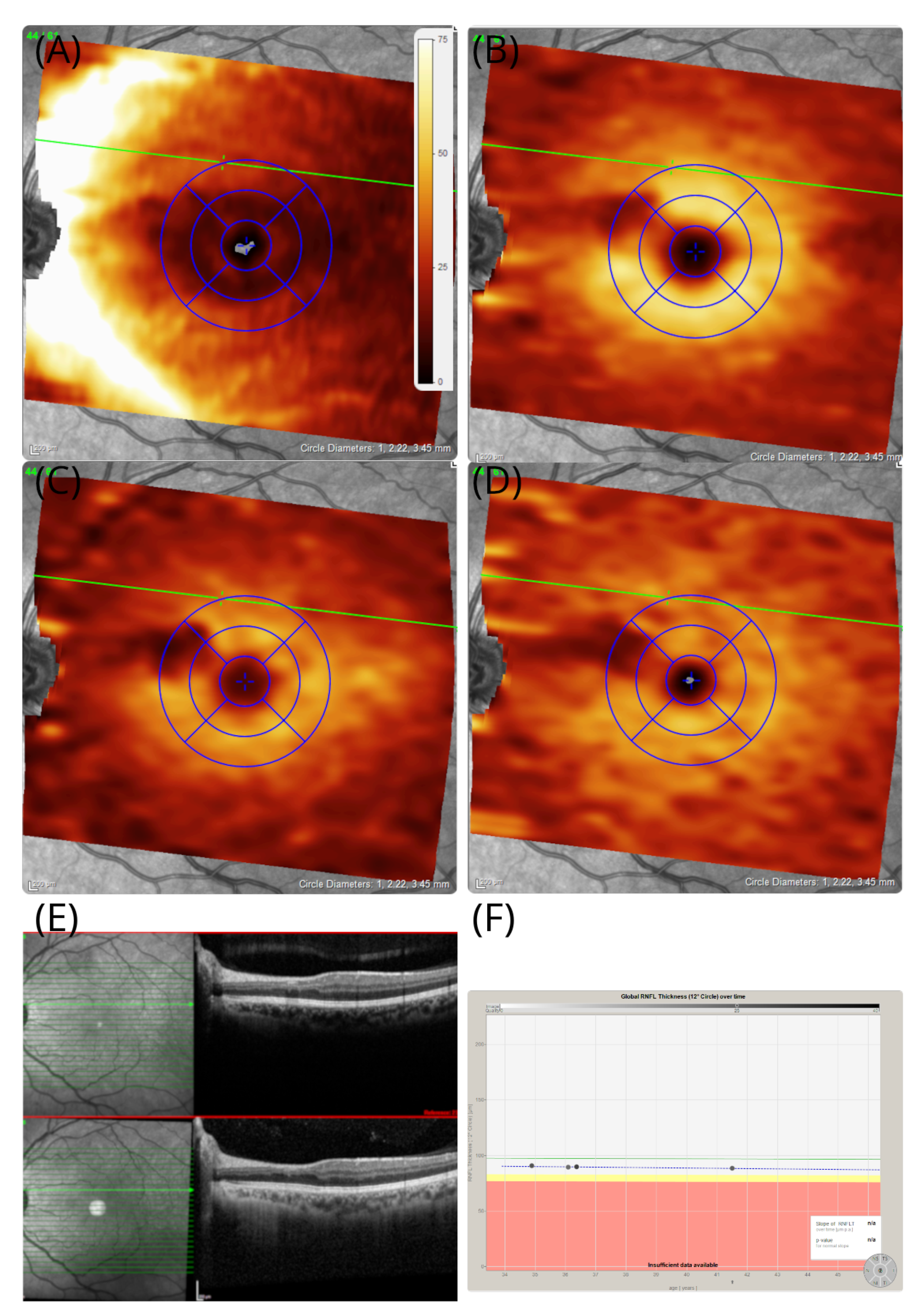
3.1. Case #1
3.2. Case #2
4. Discussion
Author Contributions
Funding
Institutional Review Board Statement
Informed Consent Statement
Data Availability Statement
Acknowledgments
Conflicts of Interest
Abbreviations
| AZOOR | Acute zonal occult outer retinopathy |
| CBS | Charles Bonnet Syndrome |
| CB1&2 | Cannabinoid receptor |
| GCL | Ganglion cell layer |
| INL | Inner nuclear layer |
| IPL | Inner plexiform layer |
| OCT | Optical coherence tomography |
| OCTA | Optical coherence tomography angiography |
| RNFL | Retinal nerve fibre layer |
References
- Plant, G.T. The fortification spectra of migraine. Br. Med. J. (Clin. Res. Ed.) 1986, 293, 1613–1617. [Google Scholar] [CrossRef] [PubMed]
- Ferrari, M.D.; Klever, R.R.; Terwindt, G.M.; Ayata, C.; van den Maagdenberg, A.M.J.M. Migraine pathophysiology: Lessons from mouse models and human genetics. Lancet Neurol. 2015, 14, 65–80. [Google Scholar] [CrossRef]
- Leao, A. Spreading depression of activity in the cerebral cortex. J. Neurophysiol. 1944, 7, 359–390. [Google Scholar] [CrossRef]
- Grüsser, O.J.; Hagner, M. On the history of deformation phosphenes and the idea of internal light generated in the eye for the purpose of vision. Doc. Ophthalmol. 1990, 74, 57–85. [Google Scholar] [CrossRef]
- Balk, L.J.; Petzold, A. Influence of the eye-tracking-based follow-up function in retinal nerve fiber layer thickness using fourier-domain optical coherence tomography. Investig. Ophthalmol. Vis. Sci. 2013, 54, 3045. [Google Scholar] [CrossRef][Green Version]
- Tewarie, P.; Balk, L.; Costello, F.; Green, A.; Martin, R.; Schippling, S.; Petzold, A. The OSCAR-IB Consensus Criteria for Retinal OCT Quality Assessment. PLoS ONE 2012, 7, e34823. [Google Scholar] [CrossRef]
- Aytulun, A.; Cruz-Herranz, A.; Aktas, O.; Balcer, L.J.; Balk, L.; Barboni, P.; Blanco, A.A.; Calabresi, P.A.; Costello, F.; Sanchez-Dalmau, B.; et al. The APOSTEL 2.0 Recommendations for Reporting Quantitative Optical Coherence Tomography Studies. Neurology 2021, 97, 68–79. [Google Scholar] [CrossRef]
- DeLange, J.M.; Cutrer, F.M. Our Evolving Understanding of Migraine with Aura. Curr. Pain Headache Rep. 2014, 18, 453. [Google Scholar] [CrossRef]
- Rowe, F.; VIS Group UK. Visual Perceptual Consequences of Stroke. Strabismus 2009, 17, 24–28. [Google Scholar] [CrossRef]
- Santhouse, A.M. Visual hallucinatory syndromes and the anatomy of the visual brain. Brain 2000, 123, 2055–2064. [Google Scholar] [CrossRef]
- Hahamy, A.; Wilf, M.; Rosin, B.; Behrmann, M.; Malach, R. How do the blind ‘see’? The role of spontaneous brain activity in self-generated perception. Brain 2020, 144, 340–353. [Google Scholar] [CrossRef] [PubMed]
- Toosy, A.T.; Roberton, B.J.; Jayaram, H.; Plant, G.T. Monocular complex visual hallucinations and their suppression by eye closure. Eye 2005, 20, 732–733. [Google Scholar] [CrossRef] [PubMed][Green Version]
- Cogan, D.G. Visual hallucinations as release phenomena. Albrecht Graefes Arch. Klin. Exp. Ophthalmol. 1973, 188, 139–150. [Google Scholar] [CrossRef]
- Blom, J.D. Alice in Wonderland syndrome. Neurol. Clin. Pract. 2016, 6, 259–270. [Google Scholar] [CrossRef] [PubMed]
- Halpern, J. Hallucinogen persisting perception disorder: What do we know after 50 years? Drug Alcohol Depend. 2003, 69, 109–119. [Google Scholar] [CrossRef]
- Bouskila, J.; Harrar, V.; Javadi, P.; Beierschmitt, A.; Palmour, R.; Casanova, C.; Bouchard, J.F.; Ptito, M. Cannabinoid Receptors CB1 and CB2 Modulate the Electroretinographic Waves in Vervet Monkeys. Neural Plast. 2016, 2016, 1253245. [Google Scholar] [CrossRef]
- Brown, P.; Kapoor, R. Saccades. J. Clin. Neuroophthalmol. 1993, 13, 77. [Google Scholar]
- Grosberg, B.M.; Solomon, S. Retinal migraine: Two cases of prolonged but reversible monocular visual defects. Cephalalgia 2006, 26, 754–757. [Google Scholar] [CrossRef]
- Hamel, C. Retinitis pigmentosa. Orphanet J. Rare Dis. 2006, 1, 40. [Google Scholar] [CrossRef]
- Canamary, A.M.; Takahashi, W.Y.; Sallum, J.M.F. Autoimmune retinopathy: A Review. Int. J. Retin. Vitr. 2018, 4. [Google Scholar] [CrossRef]
- Monson, D.M.; Smith, J.R. Acute Zonal Occult Outer Retinopathy. Surv. Ophthalmol. 2011, 56, 23–35. [Google Scholar] [CrossRef] [PubMed]
- Kraus, N.; Niedeggen, M.; Hesselmann, G. Negative affect impedes perceptual filling-in in the uniformity illusion. Conscious. Cogn. 2022, 98, 103258. [Google Scholar] [CrossRef] [PubMed]
- Reinhard, J.I.; Damm, I.; Ivanov, I.V.; Trauzettel-Klosinski, S. Eye Movements During Saccadic and Fixation Tasks in Patients with Homonymous Hemianopia. J. Neuro-Ophthalmol. 2014, 34, 354–361. [Google Scholar] [CrossRef] [PubMed]


Publisher’s Note: MDPI stays neutral with regard to jurisdictional claims in published maps and institutional affiliations. |
© 2022 by the authors. Licensee MDPI, Basel, Switzerland. This article is an open access article distributed under the terms and conditions of the Creative Commons Attribution (CC BY) license (https://creativecommons.org/licenses/by/4.0/).
Share and Cite
Gandhewar, R.; Jurkute, N.; Petzold, A. The Role of the Inner Nuclear Layer for Perception of Persisting Tiling Inside a Monocular Scotoma. Brain Sci. 2022, 12, 1542. https://doi.org/10.3390/brainsci12111542
Gandhewar R, Jurkute N, Petzold A. The Role of the Inner Nuclear Layer for Perception of Persisting Tiling Inside a Monocular Scotoma. Brain Sciences. 2022; 12(11):1542. https://doi.org/10.3390/brainsci12111542
Chicago/Turabian StyleGandhewar, Rishikesh, Neringa Jurkute, and Axel Petzold. 2022. "The Role of the Inner Nuclear Layer for Perception of Persisting Tiling Inside a Monocular Scotoma" Brain Sciences 12, no. 11: 1542. https://doi.org/10.3390/brainsci12111542
APA StyleGandhewar, R., Jurkute, N., & Petzold, A. (2022). The Role of the Inner Nuclear Layer for Perception of Persisting Tiling Inside a Monocular Scotoma. Brain Sciences, 12(11), 1542. https://doi.org/10.3390/brainsci12111542