
Transcranial Magnetic Stimulation as a Tool to Investigate Motor Cortex Excitability in Sport


 ,
,  ,
,  ,
,  and
and 
Abstract
1. Introduction
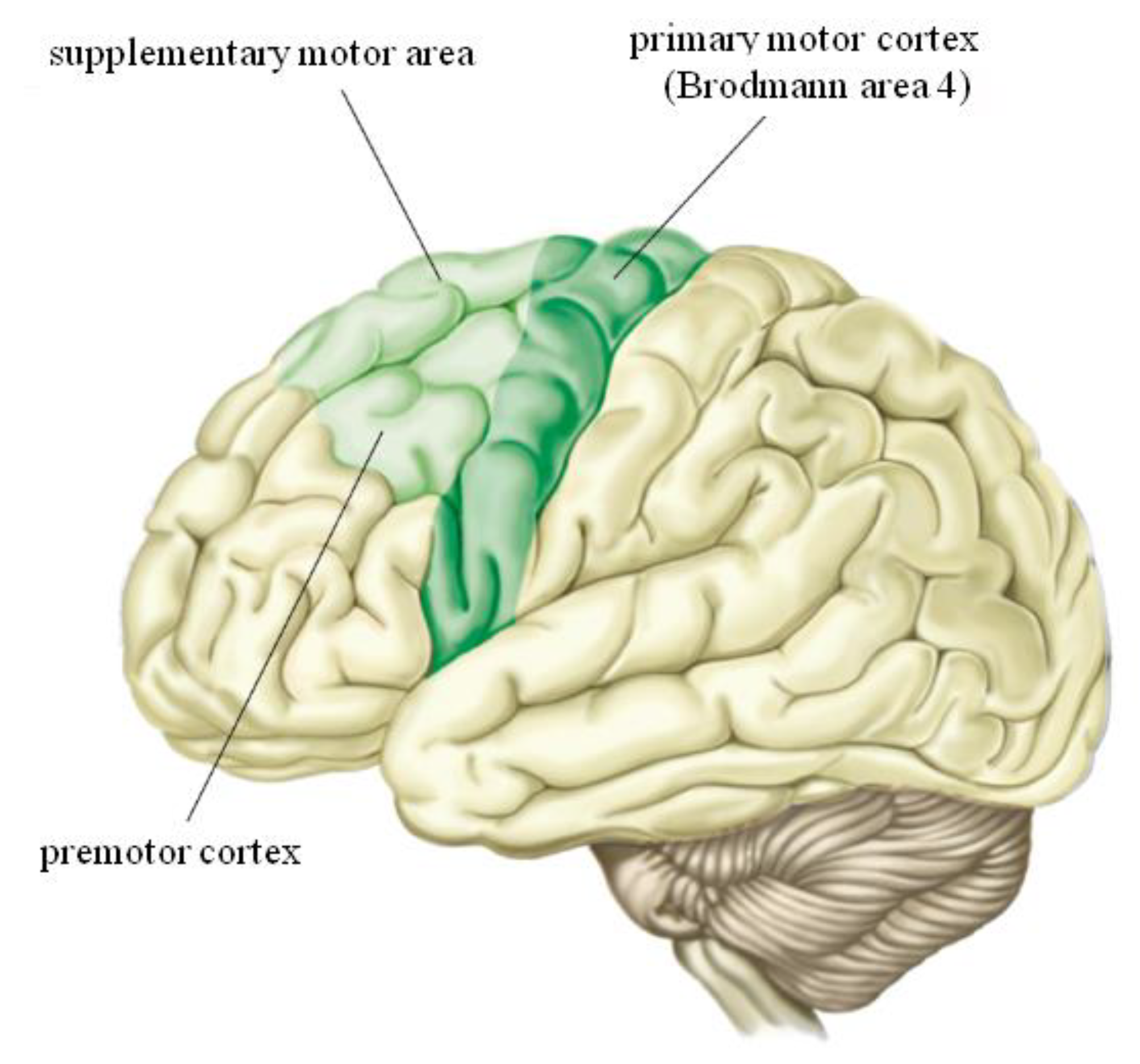
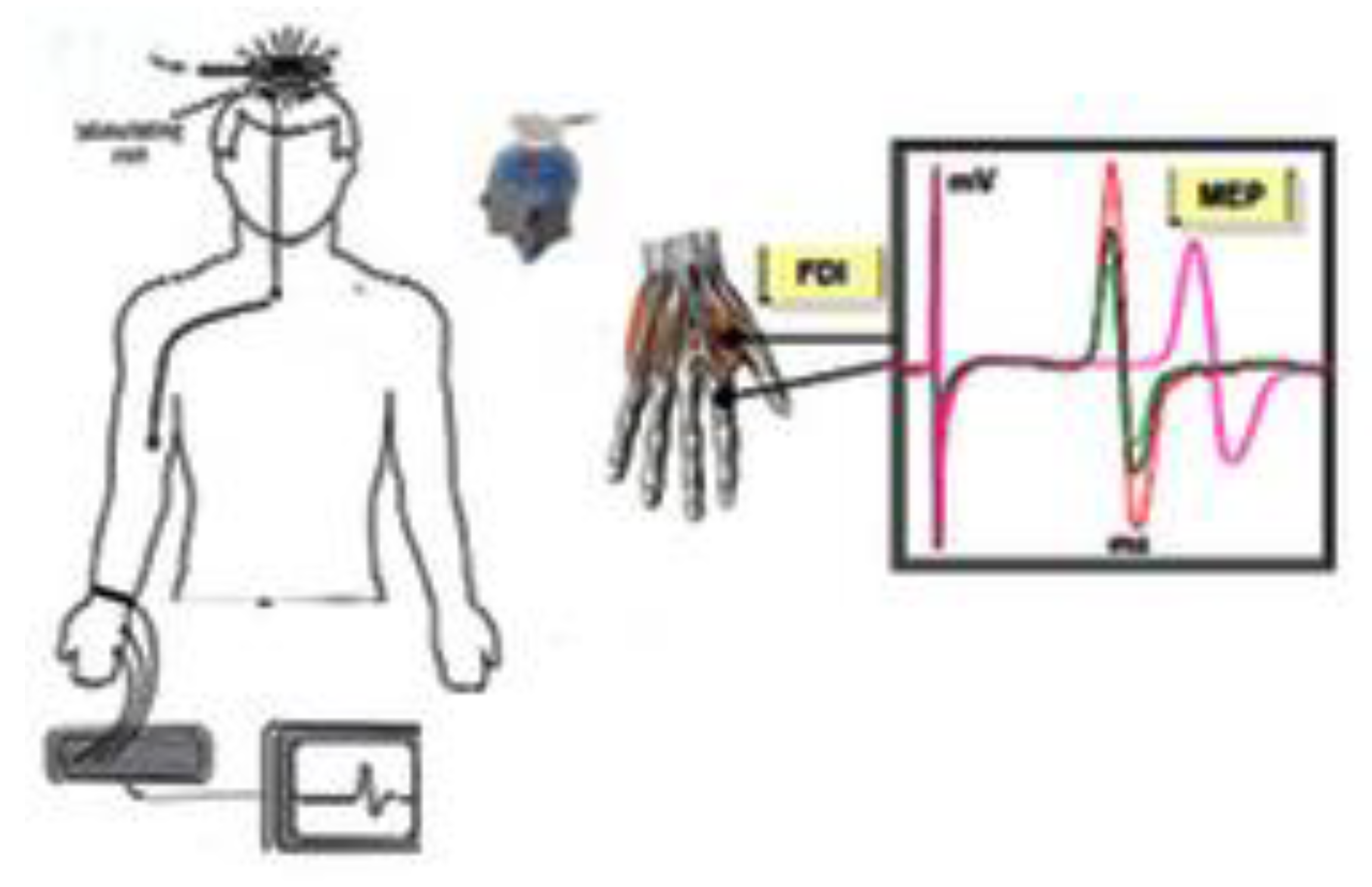
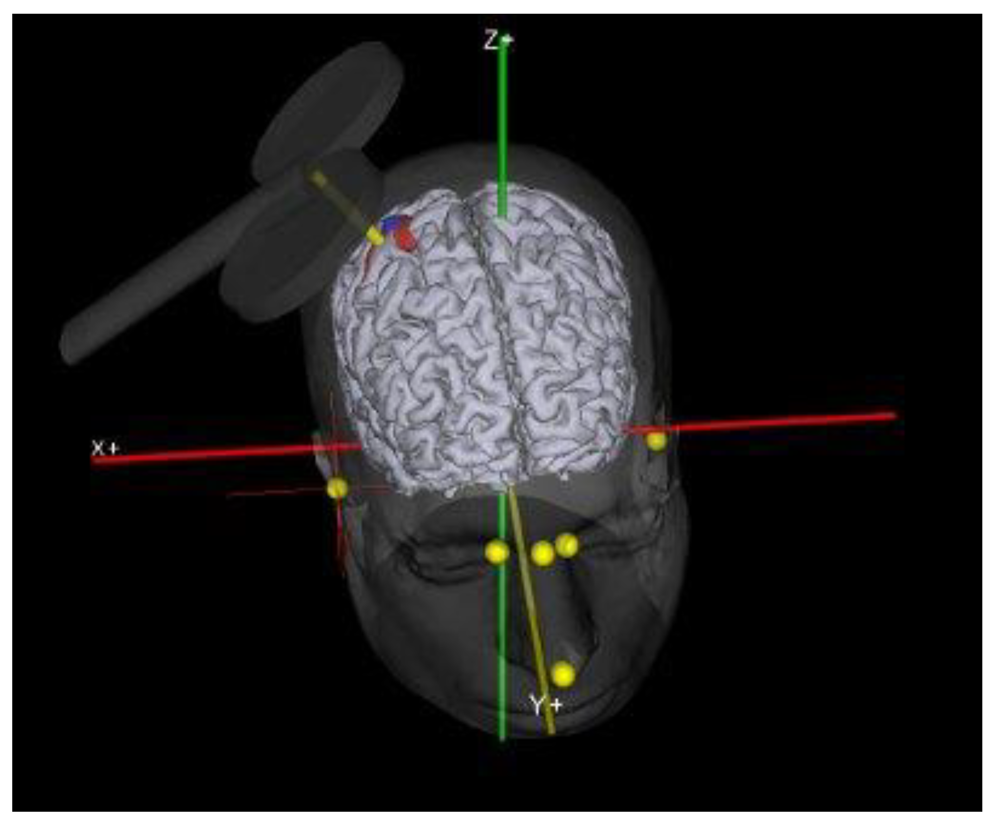
2. Transcranial Magnetic Stimulation (TMS)
3. Cortical Excitability and Physical Exercise
3.1. Skill Training
3.2. Fatigue
3.3. Aerobic and Anaerobic Exercise
4. The Use of TMS in Sport Science
5. Conclusions
Author Contributions
Funding
Conflicts of Interest
References
- Missitzi, J.; Gentner, R.; Geladas, N.; Politis, P.; Karandreas, N.; Classen, J.; Klissouras, V. Plasticity in human motor cortex is in part genetically determined. J. Physiol. 2011, 589, 297–306. [Google Scholar] [CrossRef] [PubMed]
- Buccino, G.; Binkofski, F.; Fink, G.R.; Fadiga, L.; Fogassi, L.; Gallese, V.; Seitz, R.J.; Zilles, K.; Rizzolatti, G.; Freund, H.J. Action observation activates premotor and parietal areas in a somatotopic manner: An fMRI study. Eur. J. Neurosci. 2001, 13, 400–404. [Google Scholar] [CrossRef] [PubMed]
- Bütefisch, C.M.; Khurana, V.; Kopylev, L.; Cohen, L.G. Enhancing encoding of a motor memory in the primary motor cortex by cortical stimulation. J. Neurophysiol. 2004, 91, 2110–2116. [Google Scholar] [CrossRef] [PubMed]
- Stefan, K.; Classen, J.; Celnik, P.; Cohen, L.G. Concurrent action observation modulates practice-induced motor memory formation. Eur. J. Neurosci. 2008, 27, 730–738. [Google Scholar] [CrossRef] [PubMed]
- Messina, G.; Zannella, C.; Monda, V.; Dato, A.; Liccardo, D.; De Blasio, S.; Valenzano, A.; Moscatelli, F.; Messina, A.; Cibelli, G.; et al. The Beneficial Effects of Coffee in Human Nutrition. Biol. Med. 2015, 7, 1. [Google Scholar]
- Classen, J.; Wolters, A.; Stefan, K.; Wycislo, M.; Sandbrink, F.; Schmidt, A.; Kunesch, E. Chapter 59 Paired associative stimulation. Suppl. Clin. Neurophysiol. 2004, 57, 563–569. [Google Scholar]
- Celnik, P.; Stefan, K.; Hummel, F.; Duque, J.; Classen, J.; Cohen, L.G. Encoding a motor memory in the older adult by action observation. Neuroimage 2006, 29, 677–684. [Google Scholar] [CrossRef]
- Rossini, P.M.; Barker, A.T.; Berardelli, A.; Caramia, M.D.; Caruso, G.; Cracco, R.Q.; Dimitrijević, M.R.; Hallett, M.; Katayama, Y.; Lücking, C.H. Non-invasive electrical and magnetic stimulation of the brain, spinal cord and roots: Basic principles and procedures for routine clinical application. Report of an IFCN committee. Electroencephalogr. Clin. Neurophysiol. 1994, 91, 79–92. [Google Scholar] [CrossRef]
- Chen, R. Intracortical Circuits and Their Interactions. In Cortical Connectivity; Springer: Heidelberg, Germany, 2012; p. 65. ISBN 9783642327667. [Google Scholar]
- Celnik, P.; Webster, B.; Glasser, D.M.; Cohen, L.G. Effects of action observation on physical training after stroke. Stroke 2008, 39, 1814–1820. [Google Scholar] [CrossRef]
- Fadiga, L.; Fogassi, L.; Pavesi, G.; Rizzolatti, G. Motor facilitation during action observation: A magnetic stimulation study. J. Neurophysiol. 1995, 73, 2608–2611. [Google Scholar] [CrossRef] [PubMed]
- Buccino, G.; Binkofski, F.; Riggio, L. The mirror neuron system and action recognition. Brain Lang. 2004, 89, 370–376. [Google Scholar] [CrossRef]
- Barker, A.T.; Jalinous, R.; Freeston, I.L. Non-invasive magnetic stimulation of human motor cortex. Lancet 1985, 1, 1106–1107. [Google Scholar] [CrossRef]
- Rotenberg, A. Prospects for clinical applications of transcranial magnetic stimulation and real-time EEG in epilepsy. Brain Topogr. 2010, 22, 257–266. [Google Scholar] [CrossRef]
- Rossini, P.M.; Burke, D.; Chen, R.; Cohen, L.G.; Daskalakis, Z.; Di, I.R.; Di, V.L.; Ferreri, F.; Fitzgerald, P.B.; George, M.S.; et al. Non-invasive electrical and magnetic stimulation of the brain, spinal cord, roots and peripheral nerves: Basic principles and procedures for routine clinical and research application. An updated report from an I.F.C.N. Committee 3. Clin.Neurophysiol. 2015, 126, 1071–1107. [Google Scholar] [CrossRef]
- Merton, P.A.; Morton, H.B. Stimulation of the cerebral cortex in the intact human subject. Nature 1980, 285, 227. [Google Scholar] [CrossRef] [PubMed]
- Goodall, S.; Howatson, G.; Romer, L.; Ross, E. Transcranial magnetic stimulation in sport science: A commentary. Eur. J. Sport Sci. 2014, 14 (Suppl. S1), S332–S340. [Google Scholar] [CrossRef]
- Penfield, W.; Boldrey, E. Somatic motor and sensory representation in the cerebral cortex of man as studied by electrical stimulation. Brain 1937, 60, 389–443. [Google Scholar] [CrossRef]
- Hallett, M. Transcranial magnetic stimulation: A useful tool for clinical neurophysiology. Ann. Neurol. 1996, 40, 344–345. [Google Scholar] [CrossRef] [PubMed]
- Hallett, M.; Rothwell, J. Milestones in clinical neurophysiology. Mov. Disord. 2011, 26, 958–967. [Google Scholar] [CrossRef]
- Rothwell, J.C. Using transcranial magnetic stimulation methods to probe connectivity between motor areas of the brain. Hum. Mov. Sci. 2011, 30, 906–915. [Google Scholar] [CrossRef]
- Gandevia, S.C. Spinal and supraspinal factors in human muscle fatigue. Physiol. Rev. 2001, 81, 1725–1789. [Google Scholar] [CrossRef]
- Taylor, J.L.; Gandevia, S.C. Transcranial magnetic stimulation and human muscle fatigue. Muscle Nerve 2001, 24, 18–29. [Google Scholar] [CrossRef]
- Taylor, J.L.; Gandevia, S.C. A comparison of central aspects of fatigue in submaximal and maximal voluntary contractions. J. Appl. Physiol. 2008, 104, 542–550. [Google Scholar] [CrossRef]
- Viggiano, A.; Chieffi, S.; Tafuri, D.; Messina, G.; Monda, M.; De Luca, B. Laterality of a second player position affects lateral deviation of basketball shooting. J. Sports Sci. 2014, 32, 46–52. [Google Scholar] [CrossRef] [PubMed]
- Carroll, T.J.; Riek, S.; Carson, R.G. Corticospinal responses to motor training revealed by transcranial magnetic stimulation. Exerc. Sport Sci. Rev. 2001, 29, 54–59. [Google Scholar] [PubMed]
- Gruber, M.; Linnamo, V.; Strojnik, V.; Rantalainen, T.; Avela, J. Excitability at the motoneuron pool and motor cortex is specifically modulated in lengthening compared to isometric contractions. J. Neurophysiol. 2009, 101, 2030–2040. [Google Scholar] [CrossRef]
- Jensen, J.L.; Marstrand, P.C.D.; Nielsen, J.B. Motor skill training and strength training are associated with different plastic changes in the central nervous system. J. Appl. Physiol. 2005, 99, 1558–1568. [Google Scholar] [CrossRef]
- Messina, G.; Monda, V.; Moscatelli, F.; Valenzano, A.A.; Monda, G.; Esposito, T.; Blasio, S.D.; Messina, A.; Tafuri, D.; Rosaria, M.; et al. Role of Orexin System in Obesity. Biol. Med. 2015, 7, 1–6. [Google Scholar]
- Wassermann, E.M. Risk and safety of repetitive transcranial magnetic stimulation: Report and suggested guidelines from the International Workshop on the Safety of Repetitive Transcranial Magnetic Stimulation, 5–7 June 1996. Electroencephalogr. Clin. Neurophysiol. 1998, 108, 1–16. [Google Scholar] [CrossRef]
- Rotenberg, A.; Horvath, J.C.; Pascual-Leone, A. The Transcranial Magnetic Stimulation (TMS) Device and Foundational Techniques. In Transcranial Magnetic Stimulation; Humana Press: New York, NY, USA, 2014; pp. 3–13. [Google Scholar]
- Kobayashi, M.; Pascual-Leone, A. Transcranial magnetic stimulation in neurology. Lancet Neurol. 2003, 2, 145–156. [Google Scholar] [CrossRef]
- Hallett, M. Transcranial magnetic stimulation and the human brain. Nature 2000, 406, 147–150. [Google Scholar] [CrossRef]
- Triggiani, A.I.; Valenzano, A.; Ciliberti, M.A.P.; Moscatelli, F.; Villani, S.; Monda, M.; Messina, G.; Federici, A.; Babiloni, C.; Cibelli, G. Heart rate variability is reduced in underweight and overweight healthy adult women. Clin. Physiol. Funct. Imaging 2015, 37, 162–167. [Google Scholar] [CrossRef]
- Messina, A.; De Fusco, C.; Monda, V.; Esposito, M.; Moscatelli, F.; Valenzano, A.; Carotenuto, M.; Viggiano, E.; Chieffi, S.; De Luca, V.; et al. Role of the Orexin System on the Hypothalamus-Pituitary-Thyroid Axis. Front. Neural Circuits 2016, 10, 66. [Google Scholar] [CrossRef] [PubMed]
- Kernell, D.; Chien-Ping, W. Post-synaptic effects of cortical stimulation on forelimb motoneurones in the baboon. J. Physiol. 1967, 191, 673–690. [Google Scholar] [CrossRef]
- Thompson, P.D.; Day, B.L.; Rothwell, J.C.; Dressler, D.; de Noordhout, A.M.; Marsden, C.D. Further observations on the facilitation of muscle responses to cortical stimulation by voluntary contraction. Electroencephalogr. Clin. Neurophysiol. Evoked Potentials 1991, 81, 397–402. [Google Scholar] [CrossRef]
- Kiers, L.; Fernando, B.; Tomkins, D. Facilitatory effect of thinking about movement on magnetic motor-evoked potentials. Electroencephalogr. Clin. Neurophysiol. Electromyogr. Mot. Control 1997, 105, 262–268. [Google Scholar] [CrossRef]
- Wilson, S.A.; Thickbroom, G.W.; Mastaglia, F.L. An investigation of the late excitatory potential in the hand following magnetic stimulation of the motor cortex. Electroencephalogr. Clin. Neurophysiol. Electromyogr. 1995, 97, 55–62. [Google Scholar] [CrossRef]
- Udupa, K.; Chen, R. The mechanisms of action of deep brain stimulation and ideas for the future development. Prog. Neurobiol. 2015, 133, 27–49. [Google Scholar] [CrossRef] [PubMed]
- Moscatelli, F.; Messina, G.; Valenzano, A.; Monda, V.; Viggiano, A.; Messina, A.; Petito, A.; Triggiani, A.I.; Ciliberti, M.A.P.; Monda, M.; et al. Functional Assessment of Corticospinal System Excitability in Karate Athletes. PLoS ONE 2016, 11, e0155998. [Google Scholar] [CrossRef] [PubMed]
- Moscatelli, F.; Messina, G.; Valenzano, A.; Petito, A.; Triggiani, A.I.; Messina, A.; Monda, V.; Viggiano, A.; De Luca, V.; Capranica, L.; et al. Differences in corticospinal system activity and reaction response between karate athletes and non-athletes. Neurol. Sci. 2016, 37, 1947–1953. [Google Scholar] [CrossRef]
- Slovenica, K. Transcranial magnetic stimulation offers new possibilities for the study of motor control transkranialnamagnetna. 2004, 104, 78–104. [Google Scholar]
- Moscatelli, F.; Valenzano, A.; Petito, A.; IvanoTriggiani, A.; Anna Pia Ciliberti, M.; Luongo, L.; Carotenuto, M.; Esposito, M.; Messina, A.; Monda, V.; et al. Relationship between blood lactate and cortical excitability between taekwondo athletes and non-athletes after hand-grip exercise. Somatosens. Mot. Res. 2016, 33, 137–144. [Google Scholar] [CrossRef] [PubMed]
- Pearce, A.J.; Thickbroom, G.W.; Byrnes, M.L.; Mastaglia, F.L. Functional reorganisation of the corticomotor projection to the hand in skilled racquet players. Exp. Brain Res. 2000, 130, 238–243. [Google Scholar] [CrossRef] [PubMed]
- Tyc, F.; Boyadjian, A.; Devanne, H. Motor cortex plasticity induced by extensive training revealed by transcranial magnetic stimulation in human. Eur. J. Neurosci. 2005, 21, 259–266. [Google Scholar] [CrossRef] [PubMed]
- Tergau, F.; Geese, R.; Bauer, A.; Baur, S.; Paulus, W.; Reimers, C.D. Motor cortex fatigue in sports measured by transcranial magnetic double stimulation. Med. Sci. Sports Exerc. 2000, 32, 1942–1948. [Google Scholar] [CrossRef][Green Version]
- Valenzano, A.; Moscatelli, F.; Triggiani, A.I.; Capranica, L.; De Ioannon, G.; Piacentini, M.F.; Mignardi, S.; Messina, G.; Villani, S.; Cibelli, G. Heart-Rate Changes After an Ultraendurance Swim From Italy to Albania: A Case Report. Int. J. Sports Physiol. Perform. 2016, 11, 407–409. [Google Scholar] [CrossRef]
- Messina, G.; AValenzano, A.; Moscatelli, F.; Triggiani, A.I.; Capranica, L.; Messina, A.; Piombino, L.; Tafuri, D.; Cibelli, G.; Monda, M. Effects of Emotional Stress on Neuroendocrine and Autonomic Functions in Skydiving. J. Psychiatry 2015, 18, 1–7. [Google Scholar] [CrossRef]
- Messina, G.; Palmieri, F.; Monda, V.; Messina, A.; Dalia, C.; De Luca, V. Exercise Causes Muscle GLUT4 Translocation in an Insulin-Independent Manner. Biol. Med. 2015, 1, 7. [Google Scholar] [CrossRef]
- Moscatelli, F.; Messina, G.; Valenzano, A.; Petito, A.; Triggiani, A.I.; Ciliberti, M.A.P.; Monda, V.; Messina, A.; Tafuri, D.; Capranica, L.; et al. Relationship between RPE and Blood Lactate after Fatiguing Handgrip Exercise in Taekwondo and Sedentary Subjects. Biol. Med. 2015. [Google Scholar] [CrossRef]
- Coco, M.; Alagona, G.; Perciavalle, V.; Perciavalle, V.; Cavallari, P.; Caronni, A. Changes in cortical excitability and blood lactate after a fatiguing hand-grip exercise. Somatosens. Mot. Res. 2014, 31, 35–39. [Google Scholar] [CrossRef]
- Coaccioli, S.; Varrassi, G.; del Giorno, R.; Pace, M.C.; Sansone, P.; Angelucci, D.; Paladini, A.; Moscatelli, F.; Messina, A.; Monda, V.; et al. Meditation as a Useful Chance for Chronic Pain Decrease. J. Psychiatry 2016, 19. [Google Scholar] [CrossRef]
- Chieffi, S.; Iachini, T.; Iavarone, A.; Messina, G.; Viggiano, A.; Monda, M. Flanker interference effects in a line bisection task. Exp. Brain Res. 2014, 232, 1327–1334. [Google Scholar] [CrossRef] [PubMed]
- Bangsbo, J. Quantification of anaerobic energy production during intense exercise. Med. Sci. Sports Exerc. 1998, 30, 47–52. [Google Scholar] [CrossRef] [PubMed]
- Chieffi, S.; Iavarone, A.; Iaccarino, L.; La Marra, M.; Messina, G.; De Luca, V.; Monda, M. Age-related differences in distractor interference on line bisection. Exp. Brain Res. 2014, 232, 3659–3664. [Google Scholar] [CrossRef]
- Viggiano, E.; Monda, V.; Messina, A.; Moscatelli, F.; Valenzano, A.; Tafuri, D.; Cibelli, G.; De Luca, B.; Messina, G.; Monda, M. Cortical spreading depression produces a neuroprotective effect activating mitochondrial uncoupling protein-5. Neuropsychiatr. Dis. Treat. 2016, 12, 1705–1710. [Google Scholar] [PubMed]
- Höllge, J.; Kunkel, M.; Ziemann, U.; Tergau, F.; Geese, R.; Reimers, C.D. Central fatigue in sports and daily exercises. A magnetic stimulation study. Int. J. Sports Med. 1997, 18, 614–617. [Google Scholar] [CrossRef]
- Ljubisavljević, M.; Milanović, S.; Radovanović, S.; Vukčević, I.; Kostić, V.; Anastasijević, R. Central changes in muscle fatigue during sustained submaximal isometric voluntary contraction as revealed by transcranial magnetic stimulation. Electroencephalogr. Clin. Neurophysiol. Electromyogr. Mot. Control 1996, 101, 281–288. [Google Scholar] [CrossRef]
- McKay, W.B.; Stokic, D.S.; Sherwood, A.M.; Vrbova, G.; Dimitrijevic, M.R. Effect of fatiguing maximal voluntary contraction on excitatory and inhibitory responses elicited by transcranial magnetic motor cortex stimulation. Muscle Nerve. 1996, 19, 1017–1024. [Google Scholar] [CrossRef]
- Messina, G.; De Luca, V.; Viggiano, A.; Ascione, A.; Iannaccone, T.; Chieffi, S.; Monda, M. Autonomic nervous system in the control of energy balance and body weight: Personal contributions. Neurol. Res. Int. 2013, 1–5. [Google Scholar] [CrossRef]
- Monda, M.; Messina, G.; Scognamiglio, I.; Lombardi, A.; Martin, G.A.; Sperlongano, P.; Porcelli, M.; Caraglia, M.; Stiuso, P. Short-term diet and moderate exercise in young overweight men modulate cardiocyte and hepatocarcinoma survival by oxidative stress. Oxid. Med. Cell. Longev. 2014, 2014. [Google Scholar] [CrossRef]
- Piombino, L.; Messina, A. An Assessment of Body Composition and Lifestyle in Children Aged from 8 to 10 years. Biol. Med. 2016, 8. [Google Scholar] [CrossRef]
- Fulton, R.C.; Strutton, P.H.; McGregor, A.H.; Davey, N.J. Fatigue-induced change in corticospinal drive to back muscles in elite rowers. Exp. Physiol. 2002, 87, 593–600. [Google Scholar] [CrossRef] [PubMed]
- Moscatelli, F.; Valenzano, A.; Monda, V.; Ruberto, M.; Monda, G.; Triggiani, A.I.; Monda, E.; Chieffi, S.; Villano, I.; Parisi, L.; et al. Transcranial Magnetic Stimulation (TMS) application in sport medicine: A brief review. Acta Med. Mediterr. 2017, 33, 423–430. [Google Scholar]
- Viggiano, A.; Nicodemo, U.; Viggiano, E.; Messina, G.; Viggiano, A.; Monda, M.; De Luca, B. Mastication overload causes an increase in O 2—Production into the subnucleusoralis of the spinal trigeminal nucleus. Neuroscience 2010, 166, 416–421. [Google Scholar] [CrossRef] [PubMed]
- Monda, V.; Valenzano, A.; Moscatelli, F.; Messina, A.; Piombino, L.; Zannella, C.; Viggiano, E.; Monda, G.; De Luca, V.; Chieffi, S.; et al. Modifications of Activity of Autonomic Nervous System, and Resting Energy Expenditure in Women Using Hormone-Replacement Therapy. Biol. Med. 2016, 8. [Google Scholar] [CrossRef]
- Perciavalle, V.; Coco, M.; Alagona, G.; Maci, T. Gender differences in changes of motor cortex excitability during elevated blood lactate levels. Somatosens. Mot. Res. 2010, 27, 106–110. [Google Scholar] [CrossRef]
- Alagona, G.; Coco, M.; Rapisarda, G.; Costanzo, E.; Maci, T.; Restivo, D.; Maugeri, A.; Perciavalle, V. Changes of blood lactate levels after repetitive transcranial magnetic stimulation. Neurosci. Lett. 2009, 450, 111–113. [Google Scholar] [CrossRef]
- Coco, M.; Alagona, G.; Rapisarda, G.; Costanzo, E.; Calogero, R.A.; Perciaevalle, V.; Perciavalle, V. Elevated blood lactate is associated with increased motor cortex excitability. Somatosens. Mot. Res. 2010, 27, 1–8. [Google Scholar] [CrossRef]
- Di Bernardo, G.; Messina, G.; Capasso, S.; Del Gaudio, S.; Cipollaro, M.; Peluso, G.; Casale, F.; Monda, M.; Galderisi, U. Sera of overweight people promote in vitro adipocyte differentiation of bone marrow stromal cells. Stem Cell Res. Ther. 2014, 5, 4. [Google Scholar] [CrossRef]
- Messina, G.; Dalia, C.; Tafuri, D.; Monda, V.; Palmieri, F.; Dato, A.; Russo, A.; De Blasio, S.; Messina, A.; De Luca, V.; et al. Orexin-A controls sympathetic activity and eating behavior. Front. Psychol. 2014, 5, 997. [Google Scholar] [CrossRef]
- Rinaldi, B.; Guida, F.; Furiano, A.; Donniacuo, M.; Luongo, L.; Gritti, G.; Urbanek, K.; Messina, G.; Maione, S.; Rossi, F.; et al. Effect of Prolonged Moderate Exercise on the Changes of Nonneuronal Cells in Early Myocardial Infarction. Neural Plast. 2015, 2015, 265967. [Google Scholar] [CrossRef] [PubMed]
- Rothwell, J.C.; Thompson, P.D.; Day, B.L.; Boyd, S.; Marsden, C.D. Stimulation of the human motor cortex through the scalp. Exp. Physiol. 1991, 76, 159–200. [Google Scholar] [CrossRef] [PubMed]
- Cros, D.; Soto, O.; Chiappa, K.H. Transcranial magnetic stimulation during voluntary action: Directional facilitation of outputs and relationships to force generation. Brain Res. 2007, 1185, 103–116. [Google Scholar] [CrossRef]
- Brasil-Neto, J.P.; Cohen, L.G.; Hallett, M. Central fatigue as revealed by postexercise decrement of motor evoked potentials. Muscle Nerve 1994, 17, 713–719. [Google Scholar] [CrossRef] [PubMed]
- Verin, E.; Ross, E.; Demoule, A.; Hopkinson, N.; Nickol, A.; Fauroux, B.; Moxham, J.; Similowski, T.; Polkey, M.I. Effects of exhaustive incremental treadmill exercise on diaphragm and quadriceps motor potentials evoked by transcranial magnetic stimulation. J. Appl. Physiol. 2004, 96, 253–259. [Google Scholar] [CrossRef]
- Moscatelli, F.; Messina, G.; Valenzano, A.; Triggiani, A.I.; Sessa, F.; Carotenuto, M.; Tartaglia, N.; Ambrosi, A.; Cibelli, G.; Monda, V. Effects of 12 weeks’ aerobic training on motor cortex excitability. J. Sports Med. Phys. Fit. 2020, 60, 1383–1389. [Google Scholar]
- Muellbacher, W.; Ziemann, U.; Wissel, J.; Dang, N.; Kofler, M.; Facchini, S.; Boroojerdi, B.; Poewe, W.; Hallett, M. Early consolidation in human primary motor cortex. Nature 2002, 415, 640–644. [Google Scholar] [CrossRef]
- Bocci, T.; Caleo, M.; Tognazzi, S.; Francini, N.; Briscese, L.; Maffei, L.; Rossi, S.; Priori, A.; Sartucci, F. Evidence for metaplasticity in the humanvisualcortex. J. Neural Transm. 2014, 121, 221–231. [Google Scholar] [CrossRef]
- Ziemann, U.; Paulus, W.; Nitsche, M.A.; Pascual-Leone, A.; Byblow, W.D.; Berardelli, A.; Siebner, H.R.; Classen, J.; Cohen, L.G.; Rothwell, J.C. Consensus: Motorcortexplasticityprotocols. Brain Stimul. 2008, 1, 164–182. [Google Scholar] [CrossRef]
- Lingyan, H.; Yuqin, D.; Xinyan, Z.; Yu, L. Transcranial direct current stimulation with halo sport enhances repeated sprint cycling and cognitive performance. Front. Physiol. 2019, 10, 118. [Google Scholar]


| Authors | Type of Sport | Type of Exercise | Main Findings |
|---|---|---|---|
| Jensen et al., 2005 [28] | Original research | Strength training | The results of this investigation show that increased corticospinal excitability may develop over several weeks of skill training and indicate that these changes may be of importance for task acquisition. |
| Moscatelli et al., 2016 [41] | Original research | Karate | Karate athletes show higher corticospinal excitability compared to non athletes indicating the presence of an activity-dependent alteration in the balance and interactions between inhibitory and facilitatory circuits determining the final output from the M1 |
| Moscatelli et al., 2016 [42] | Original research | Karate | The practice of competitive sports affects central/peripheral nervous system. Subjects that showed higher cortical excitability showed also higher velocity at which the neural signal is propagated from the motor cortex to the muscle and consequently better reaction time. |
| Moscatelli et al., 2016 [44] | Original research | Taekwondo | The results of this study show that blood lactate seems to have the greater effect in taekwondo athletes compared to untrained subjects. During extremely intensive exercise in taekwondo athletes, lactate may delay the onset of fatigue not only by maintaining the excitability of muscle, but also by increasing the excitability of the primary motor cortex more than in non-athletes. |
| Tergau et al., 2000 [47] | Original research | Lifting | Double-pulse TMS gives access to the motor cortex independently of spinal or peripheral mechanisms, reduced Intra Cortical Facilitation reflects decreased excitability of interneuronal circuits within the motor cortex. |
| Coco et al., 2014 [52] | Original research | Intensive isometric exercises | The relation between blood lactate and the amplitudes of motor-evoked potentials showed a significant direct proportionality. |
| Höllgeet al., 1997 [58] | Original research | Aerobic and anaerobic exercise | This investigation show the possible use of TMS in sports medicine, indicating that only exhaustive or strength exercises result in reduced MEPs. |
| Ljubisavljević et al., 1996 [59] | Original research | submaximal isometric voluntary contraction | The increase in MEP magnitude after the sustained 60% maximal voluntary contraction may indicate residual changes in cortical activity after fatiguing contraction. |
| MaKay et al., 1996 [60] | Original research | Isometric maximal contraction | These results of this investigation suggest that MEP and SP might have common sources of facilitation during maximal voluntary contraction and that inhibitory mechanisms remain focally augmented following a fatiguing maximal voluntary contraction. |
| Fulton et al., 2002 [64] | Original research | Rowers | There were no differences in MEP depression or latency between elite rowers and non-rowers after intense exercise. The authors conclude that the smaller degree of MEP depression in the elite rowers after light exercise reflects less central fatigue within corticospinal control pathways than that seen in the non-rowers. |
| Coco et al., 2010 [70] | Original research | Cycling | In this study was observed that an increase of blood lactate is associated with a decrease of motor threshold, that is, an enhancement of motor cortex excitability. The authors conclude by hypothesizing that in the motor cortex the lactate could have a protective role against fatigue. |
| Moscatelli et al., 2020 [78] | Original resea | Aerobic exercise | This study shows that aerobic activity seems to induce changes in cortical excitability if performed for a period longer than 4 weeks, in addition to typical cardiorespiratory benefits in previously untrained males |
| Percivalle et al., 2010 [68] | Original research | Maximal exhausting exercise | The authors observed a similar enhancement of excitability of primary motor cortex, concomitantly with an increase of blood lactate, in both young male and female athletes. However, the improvement was significantly higher in women than in men, suggesting a greater sensitiveness of female cerebral cortex to blood lactate. |
| Cros et al., 2007 [75] | Original research | Isometric contraction | The timing of central conduction was different depending on functional role of the target muscle, as either agonist or joint fixator. These results indicate that the architecture of motor plans remain grossly undisrupted by cortical stimulation applied during voluntary motor behavior. |
| Brasil-Neto et al., 1994 [76] | Original research | Isometric and isotonic exercise | The results are similar to those found at the neuromuscular junction in myasthenia gravis and are consistent with a reduced safety factor of cortical synaptic transmission in central nervous system fatigue. |
| Verin et al., 1985 [77] | Original research | Incremental treadmill exercise | The results of this study confirm significant depression of both diaphragm and quadriceps MEPs after incremental treadmill exercise. |
Publisher’s Note: MDPI stays neutral with regard to jurisdictional claims in published maps and institutional affiliations. |
© 2021 by the authors. Licensee MDPI, Basel, Switzerland. This article is an open access article distributed under the terms and conditions of the Creative Commons Attribution (CC BY) license (http://creativecommons.org/licenses/by/4.0/).
Share and Cite
Moscatelli, F.; Messina, A.; Valenzano, A.; Monda, V.; Salerno, M.; Sessa, F.; La Torre, E.; Tafuri, D.; Scarinci, A.; Perrella, M.; et al. Transcranial Magnetic Stimulation as a Tool to Investigate Motor Cortex Excitability in Sport. Brain Sci. 2021, 11, 432. https://doi.org/10.3390/brainsci11040432
Moscatelli F, Messina A, Valenzano A, Monda V, Salerno M, Sessa F, La Torre E, Tafuri D, Scarinci A, Perrella M, et al. Transcranial Magnetic Stimulation as a Tool to Investigate Motor Cortex Excitability in Sport. Brain Sciences. 2021; 11(4):432. https://doi.org/10.3390/brainsci11040432
Chicago/Turabian StyleMoscatelli, Fiorenzo, Antonietta Messina, Anna Valenzano, Vincenzo Monda, Monica Salerno, Francesco Sessa, Ester La Torre, Domenico Tafuri, Alessia Scarinci, Michela Perrella, and et al. 2021. "Transcranial Magnetic Stimulation as a Tool to Investigate Motor Cortex Excitability in Sport" Brain Sciences 11, no. 4: 432. https://doi.org/10.3390/brainsci11040432
APA StyleMoscatelli, F., Messina, A., Valenzano, A., Monda, V., Salerno, M., Sessa, F., La Torre, E., Tafuri, D., Scarinci, A., Perrella, M., Marsala, G., Monda, M., Cibelli, G., Porro, C., & Messina, G. (2021). Transcranial Magnetic Stimulation as a Tool to Investigate Motor Cortex Excitability in Sport. Brain Sciences, 11(4), 432. https://doi.org/10.3390/brainsci11040432