
Analysis of HIC and Hydrostatic Pressure in the Human Head during NOCSAE Tests of American Football Helmets

 ,
,  ,
,  ,
,  ,
, 
Abstract
1. Introduction
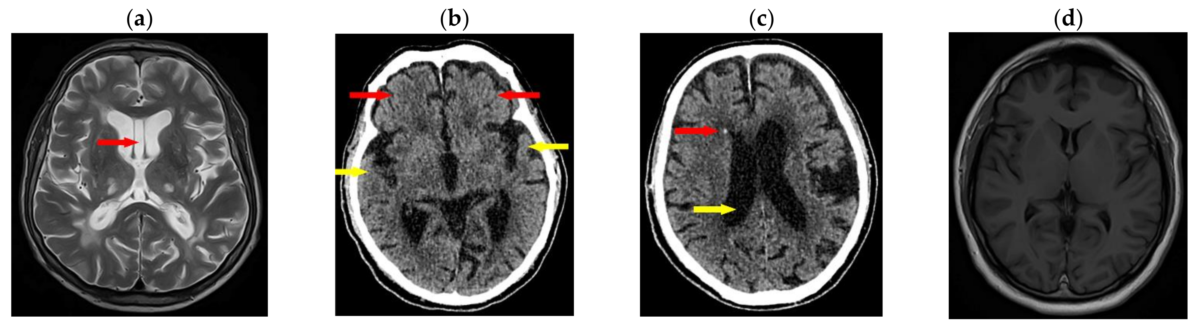
Medical Background
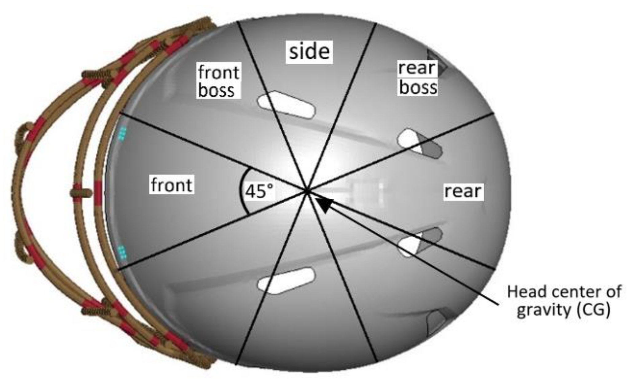
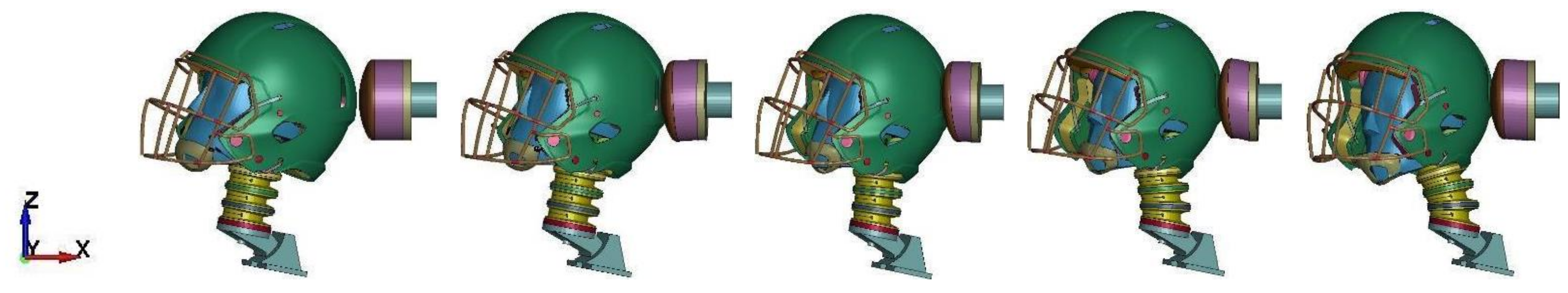
2. Materials and Methods
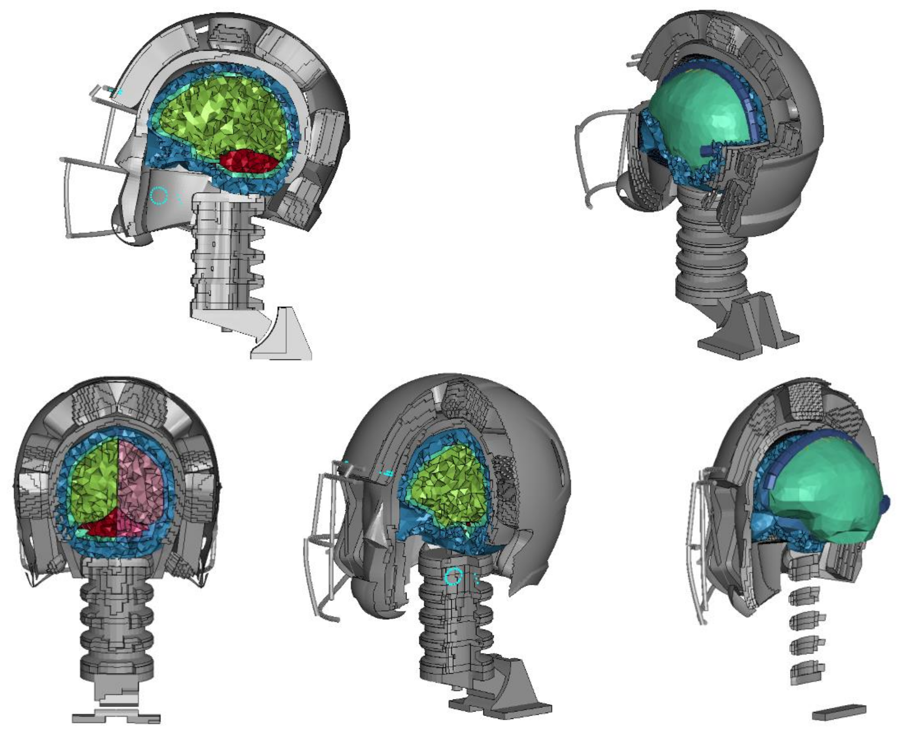
2.1. Model Discretization
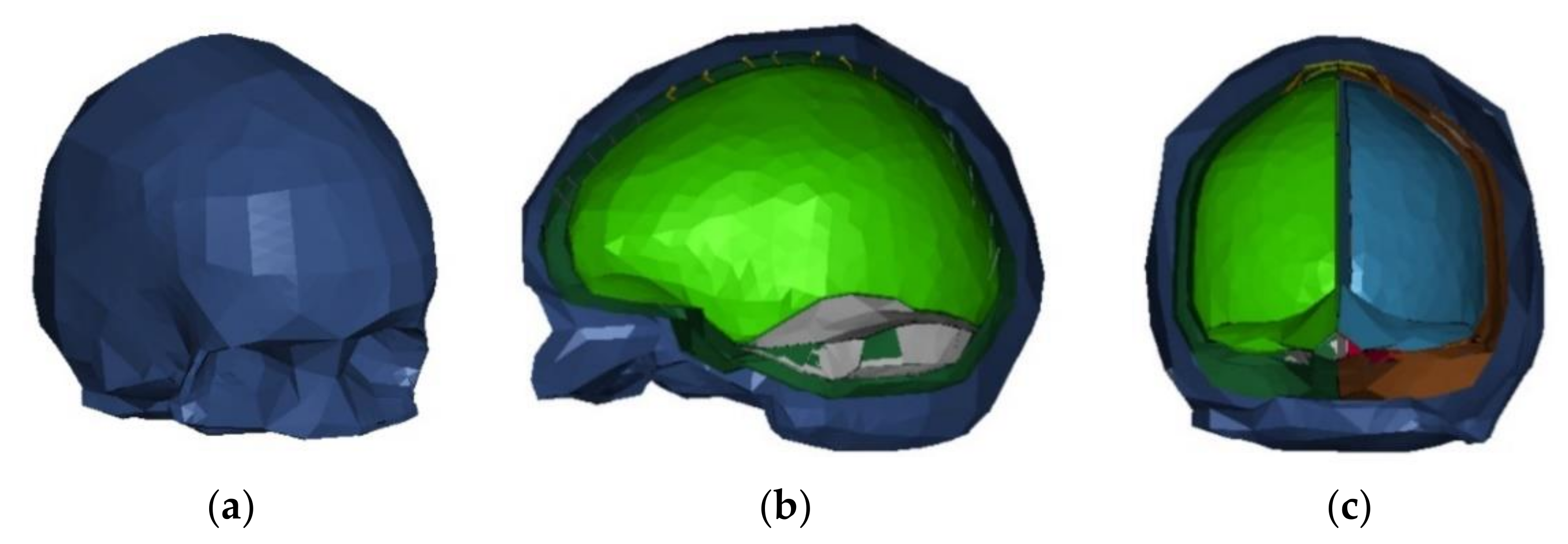
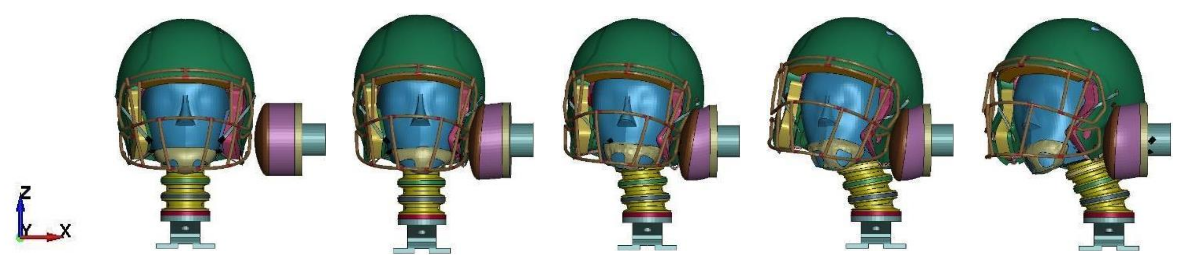
2.1.1. αHEAD Finite Element Head Model
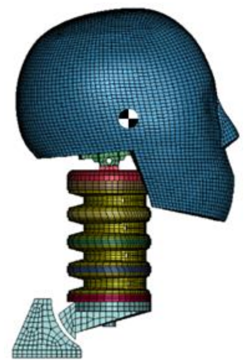
2.1.2. Hybrid III Head-Neck Model
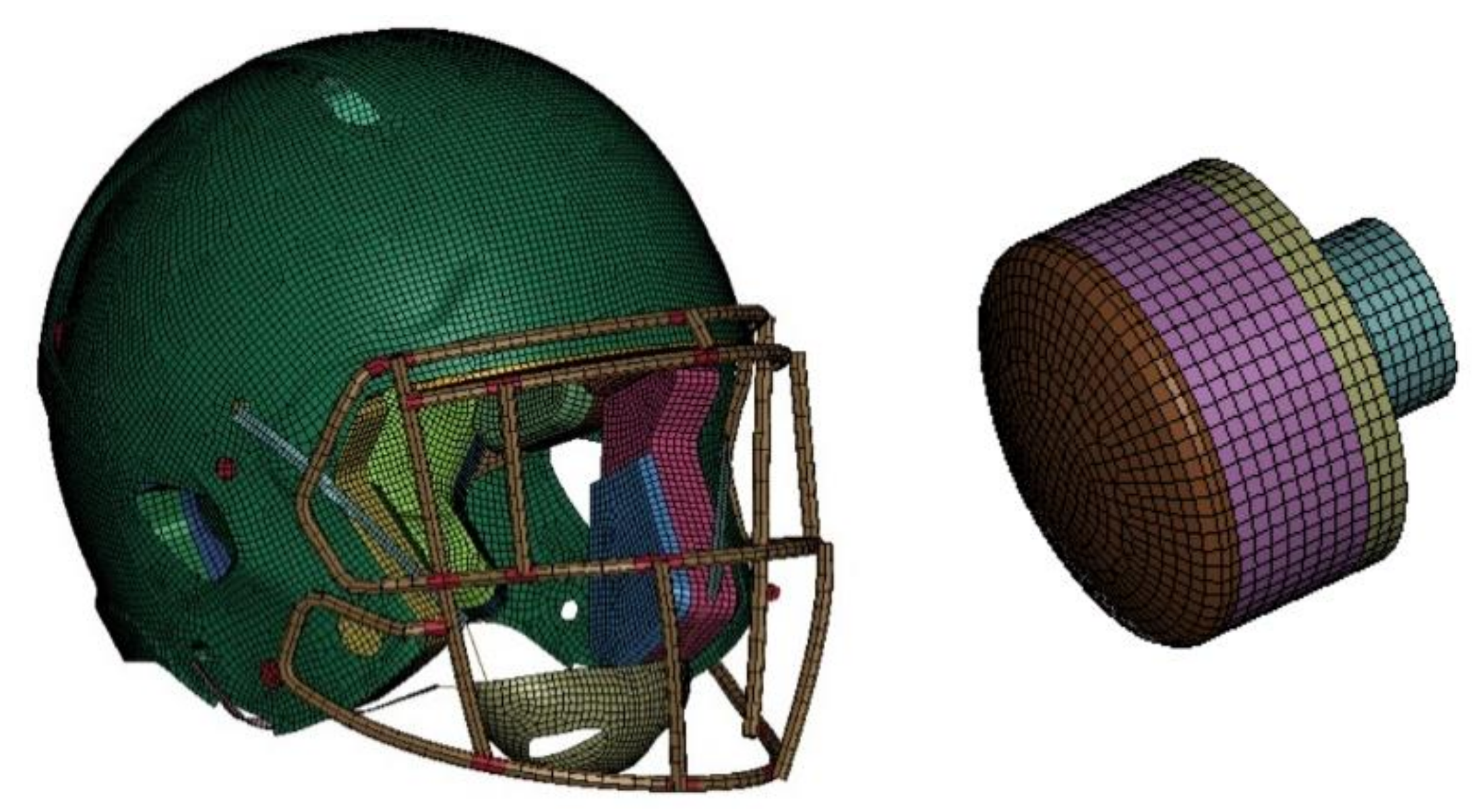
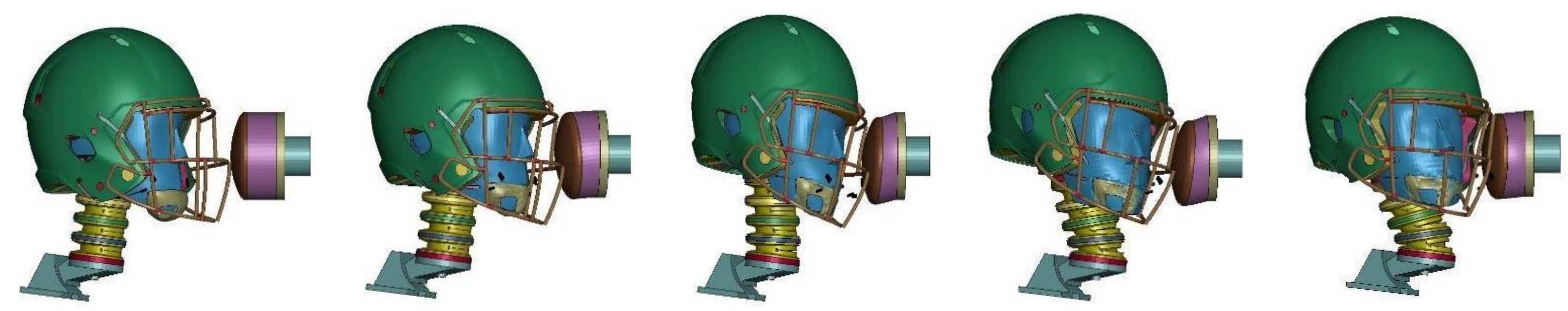
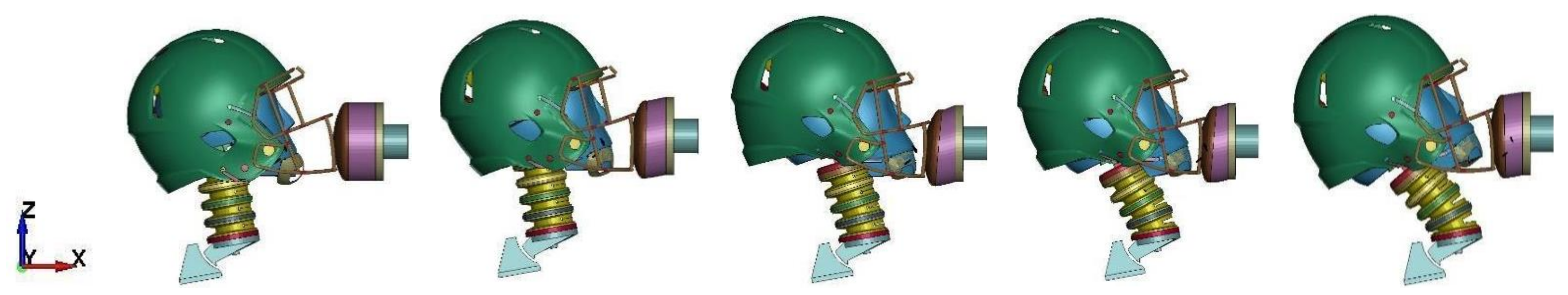
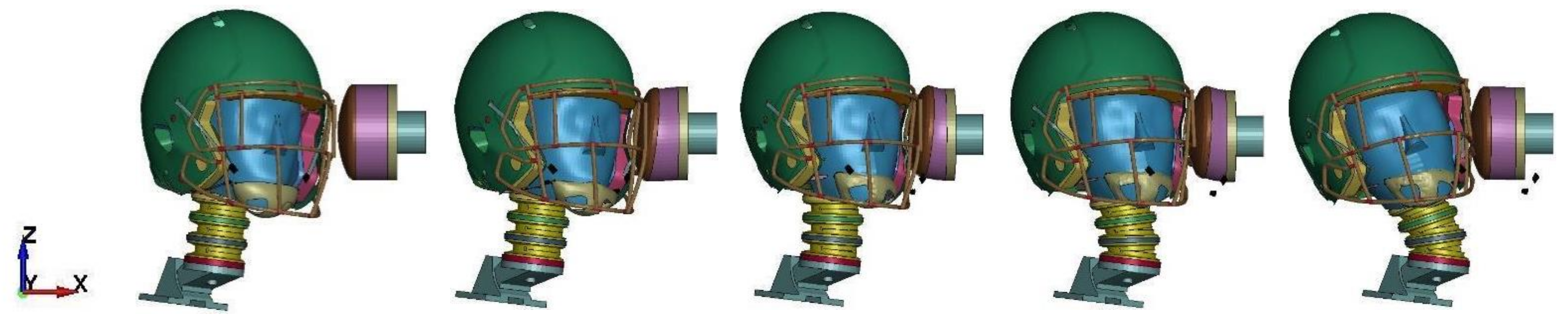
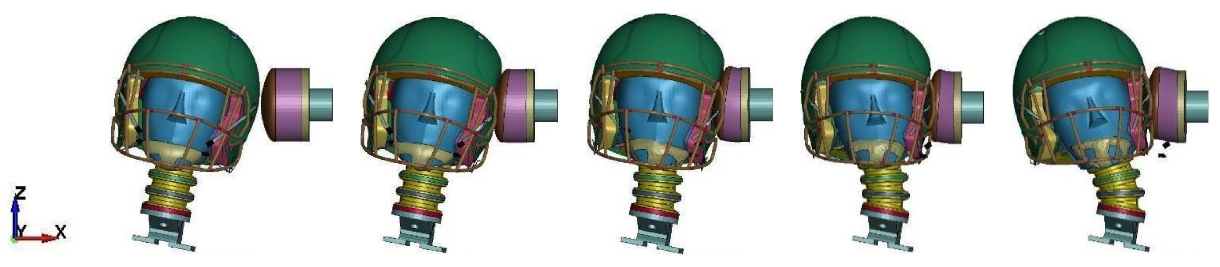
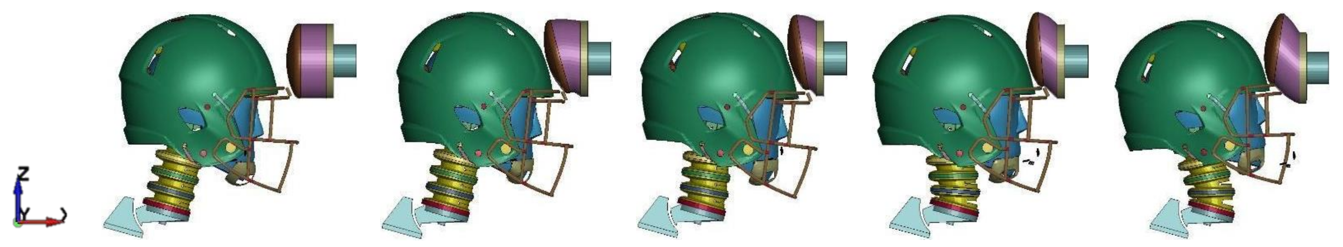
2.1.3. Helmet and Impactor Model
- In the real model, the padding is covered in plastic covers that can be pumped with air. In the discrete model, only a plastic sheet is modelled at the back of the pads. During component validation, the difference was found to be minimal.
- The comfort foam stiffness was increased by a factor of 2.5 compared to the experimental results to prevent instabilities. However, when evaluating the two stiffnesses for some linear impactor simulations, this difference was negligible.
- The foam material can be sensitive to weather conditions such as temperature or penetrating trauma.
2.2. Simulation Setup
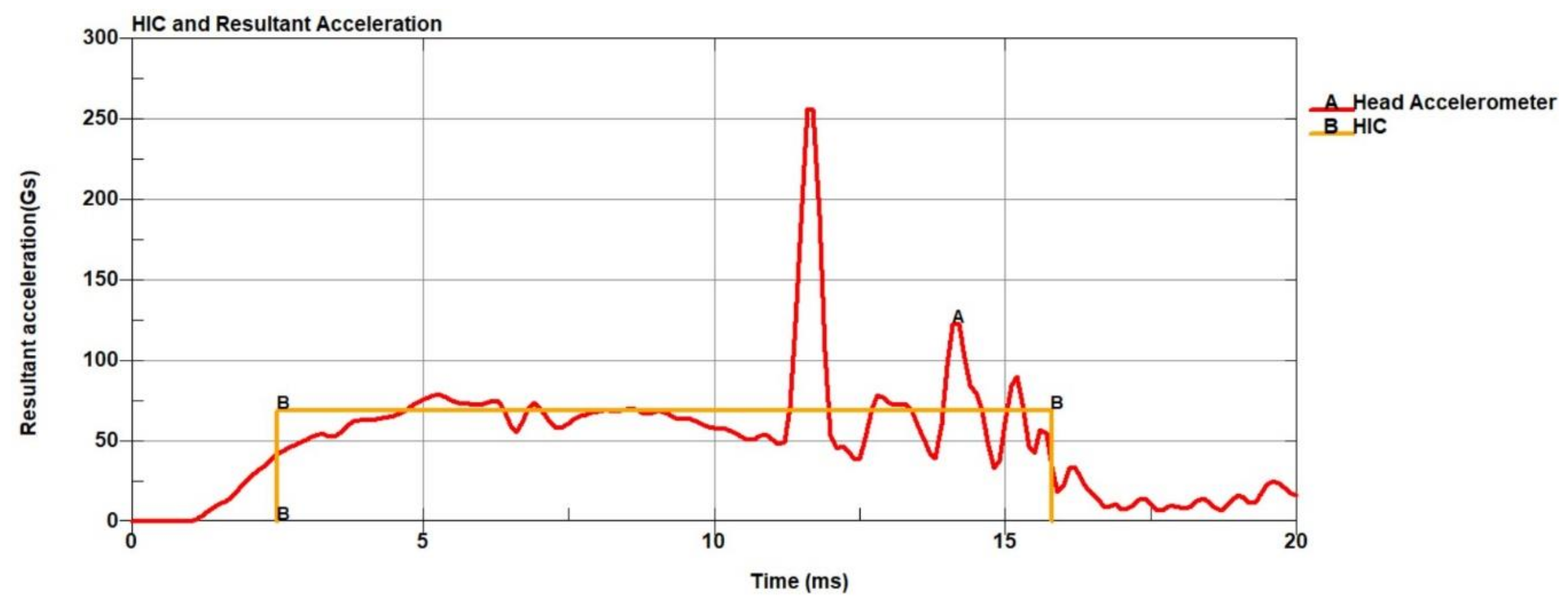
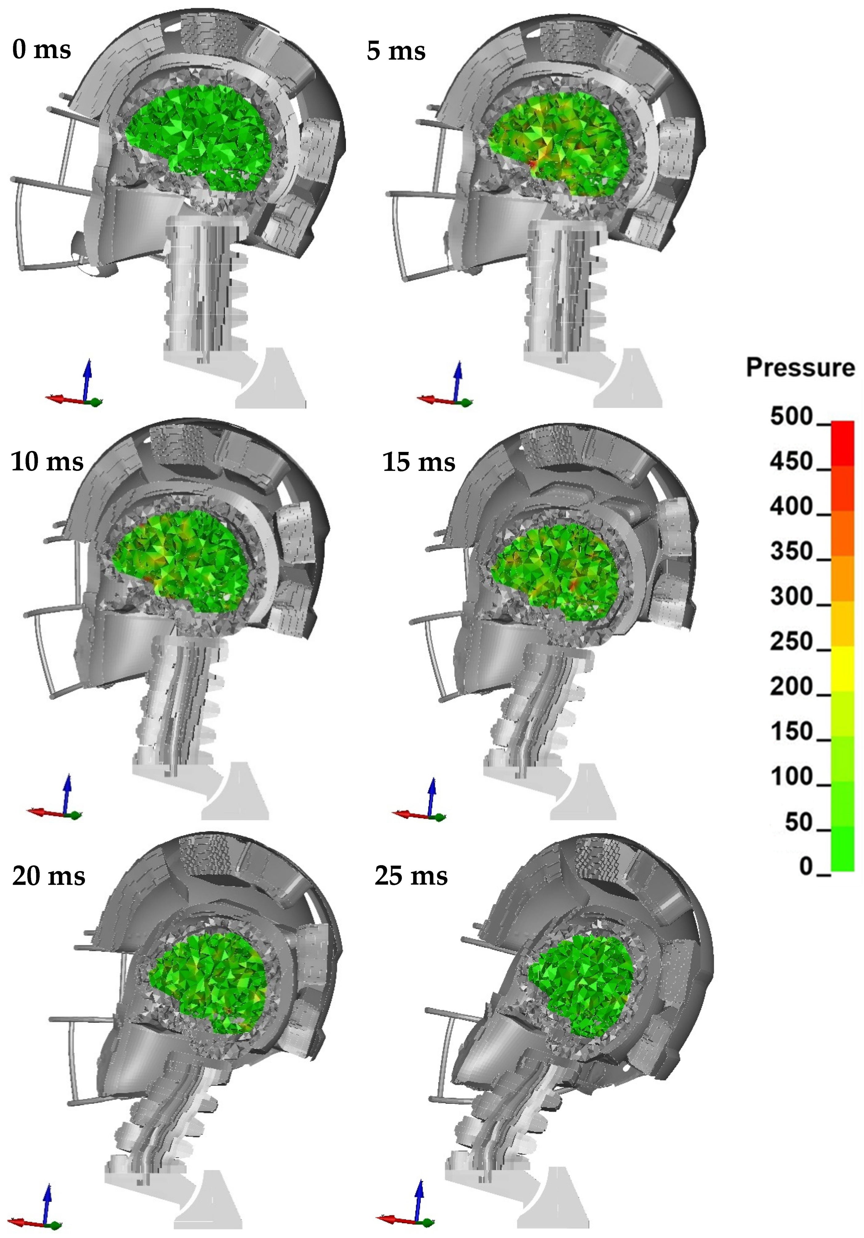
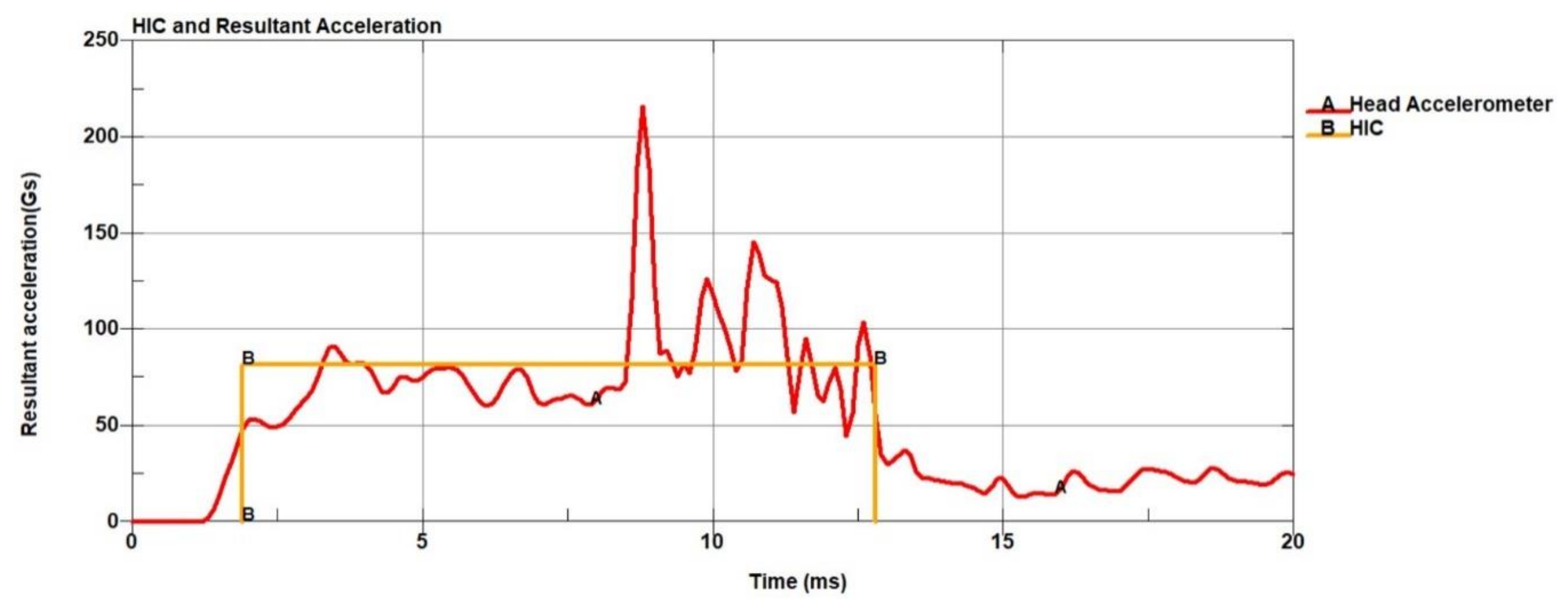
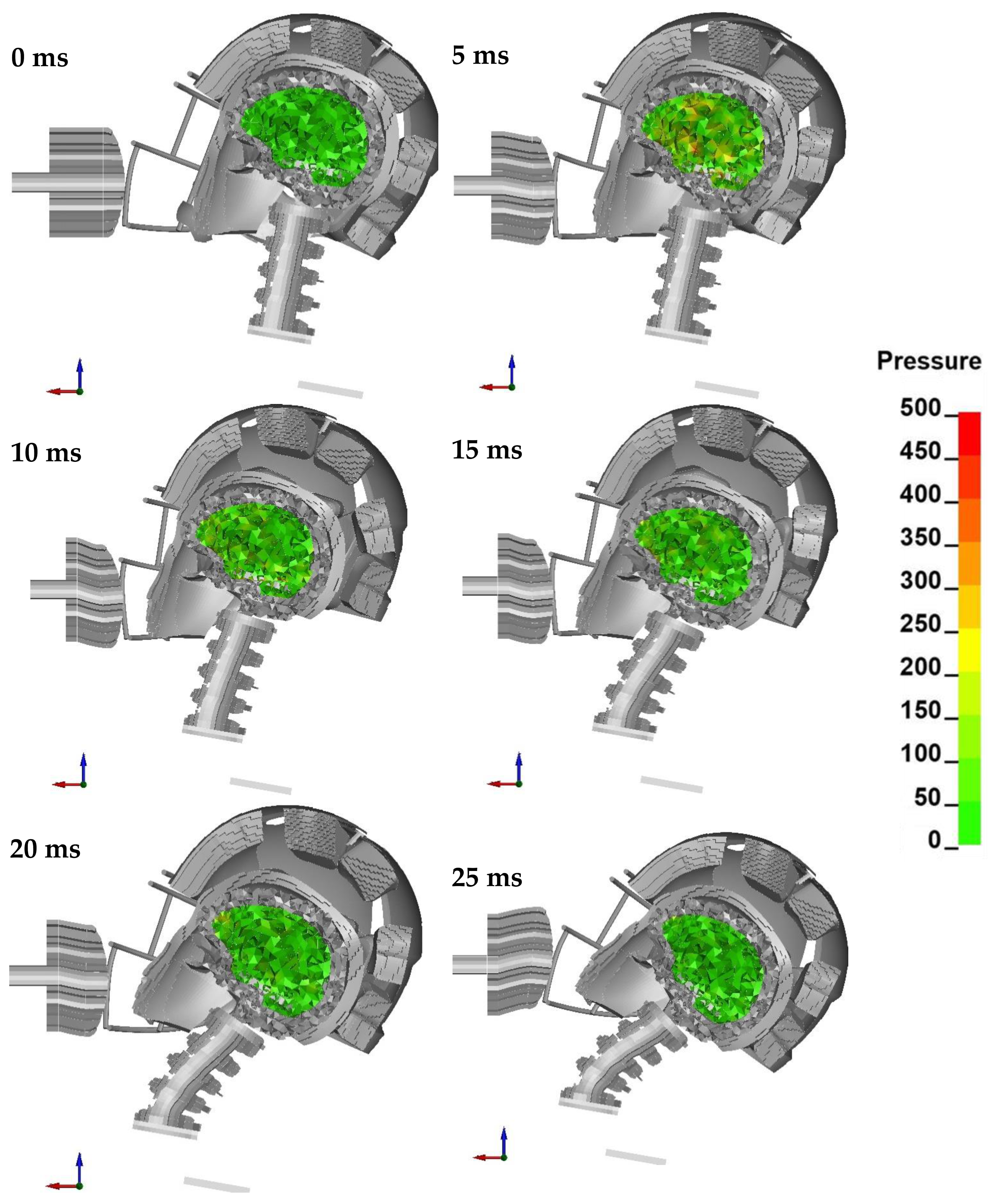
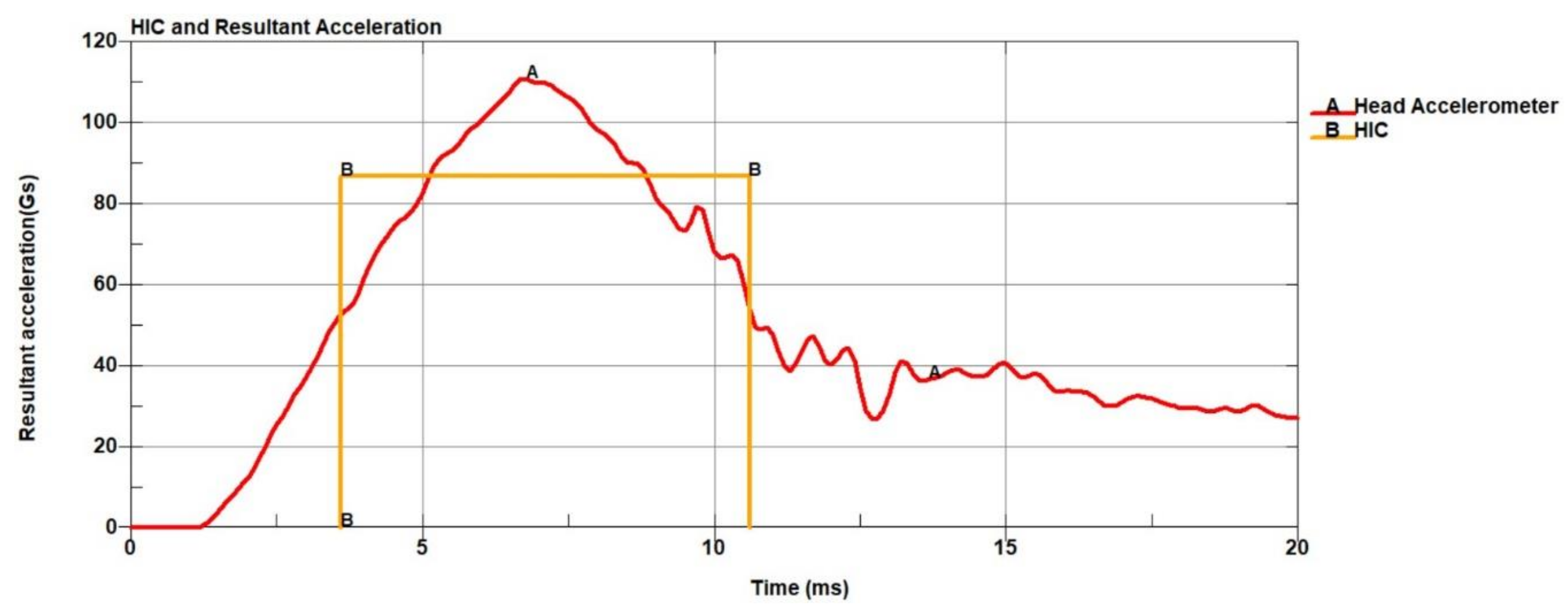
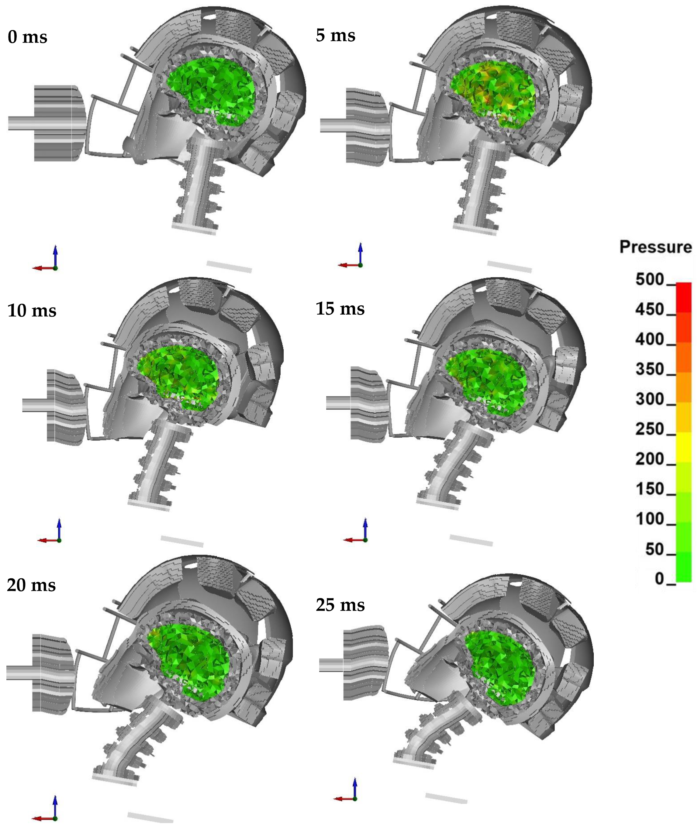
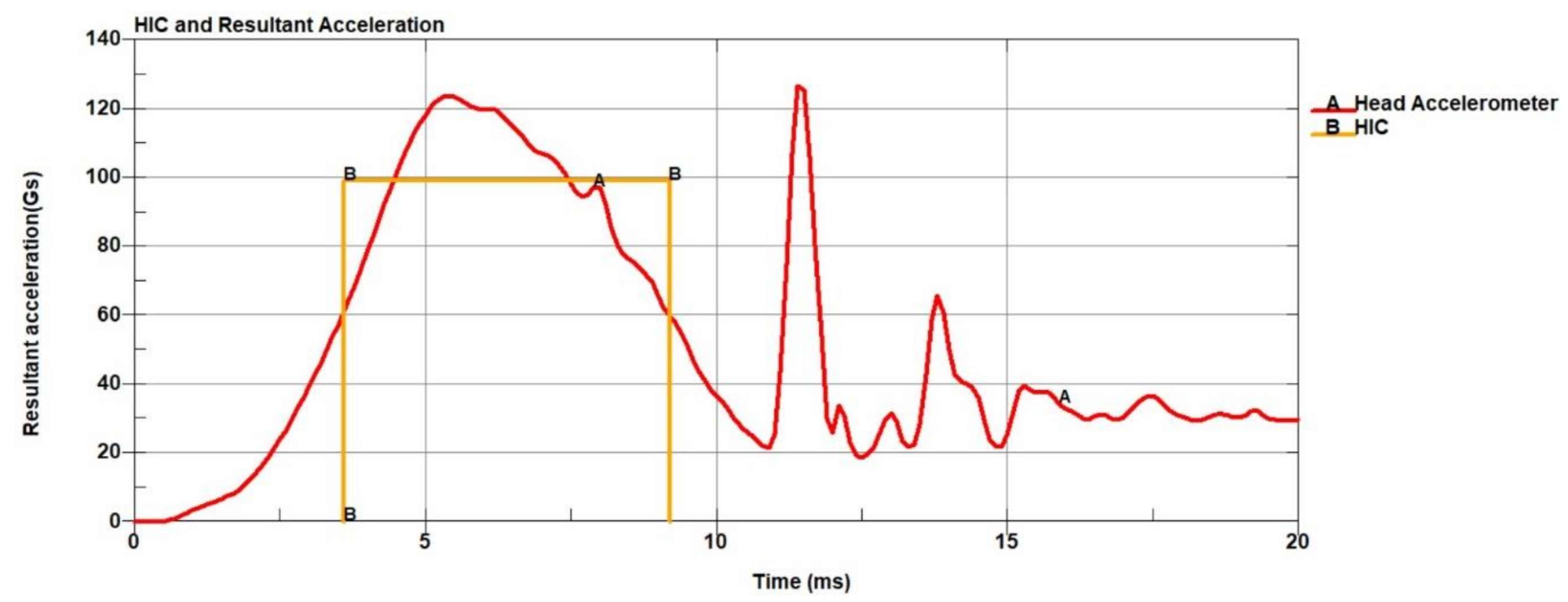
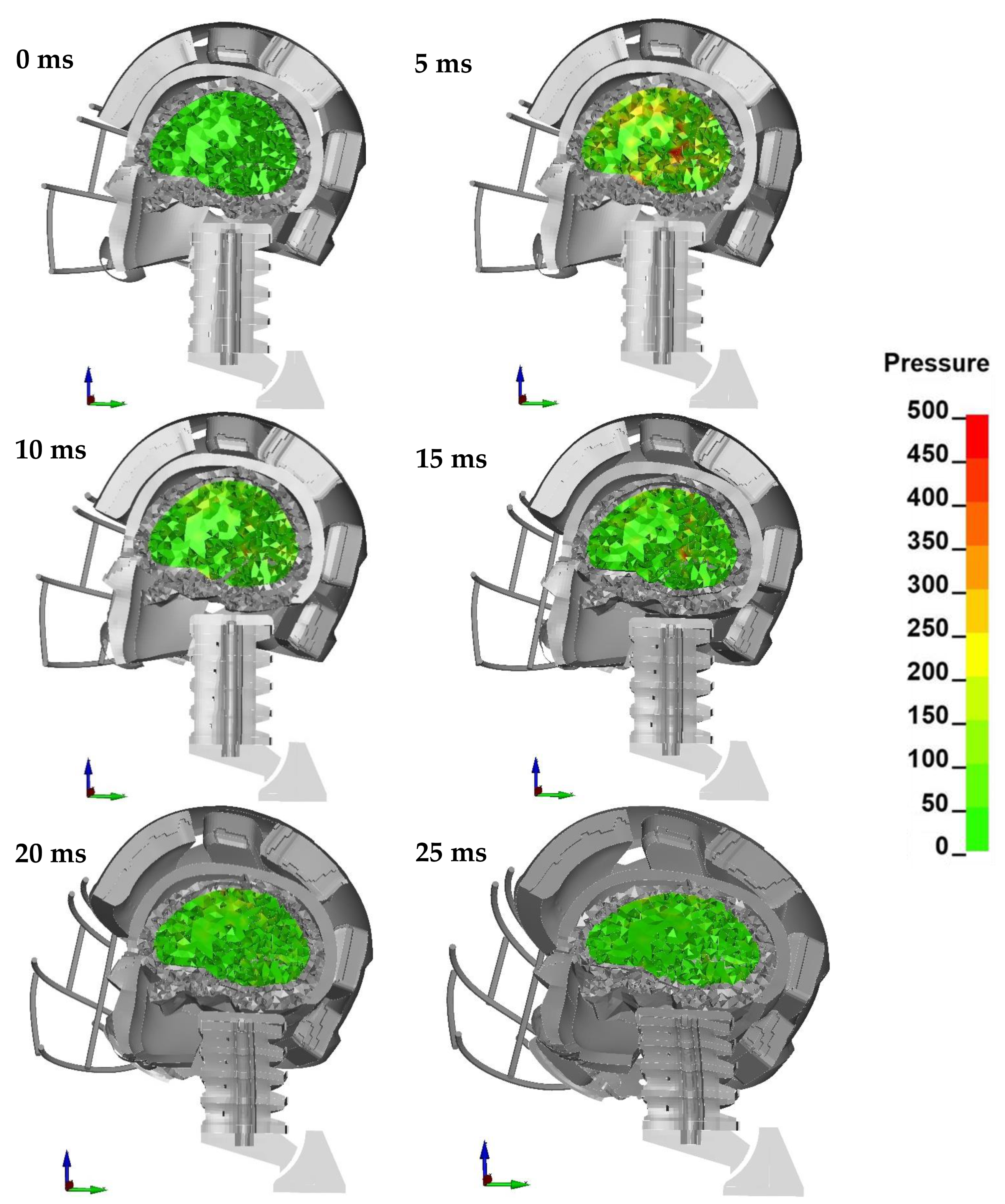
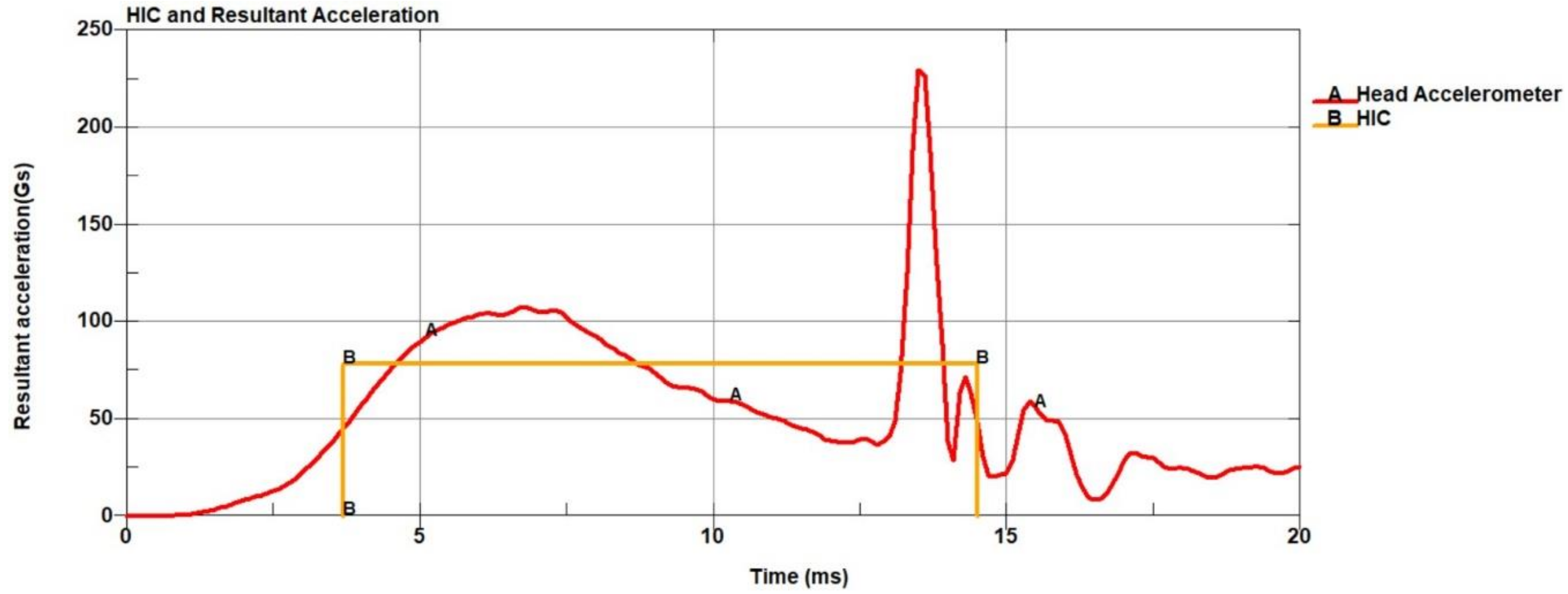
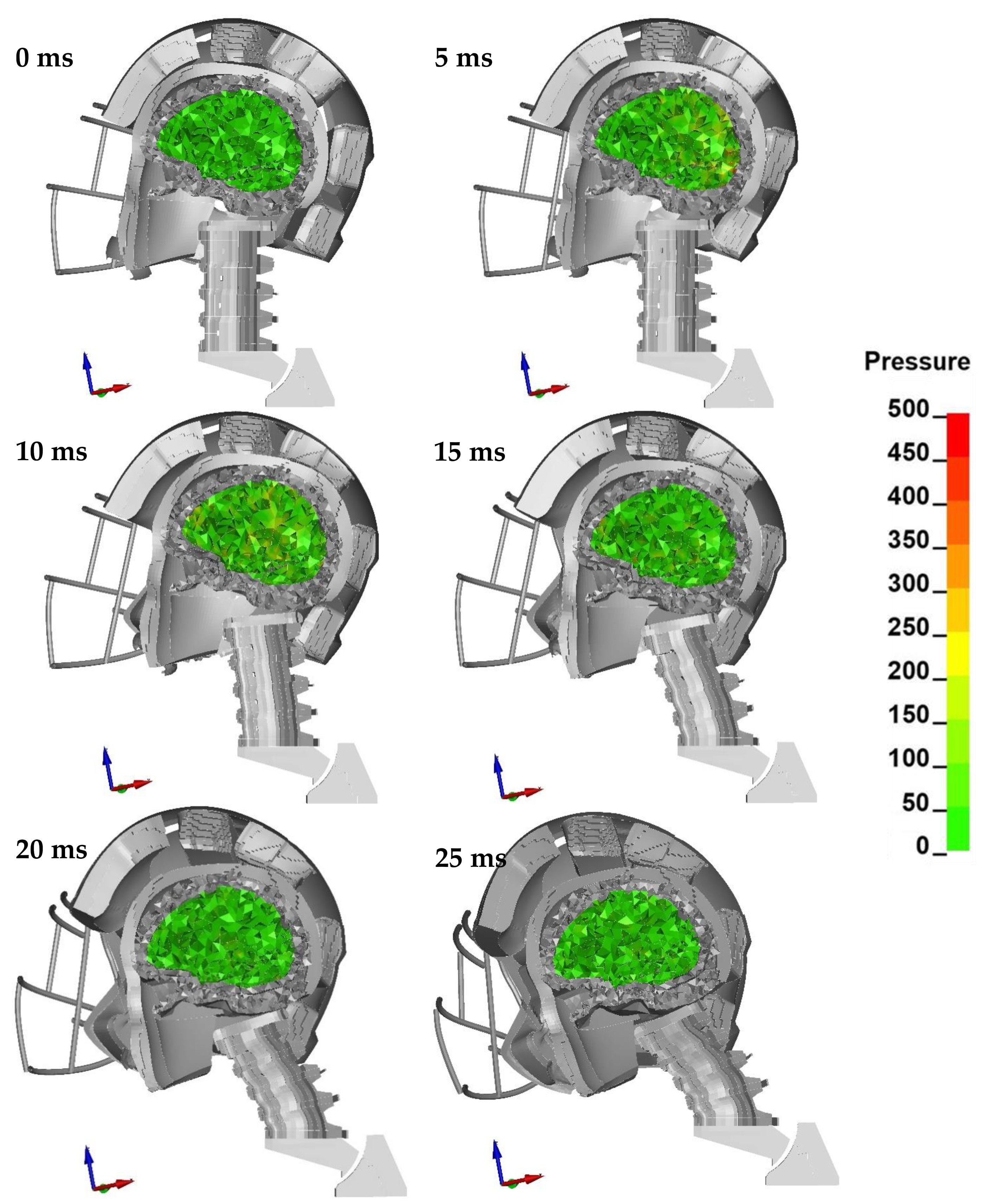
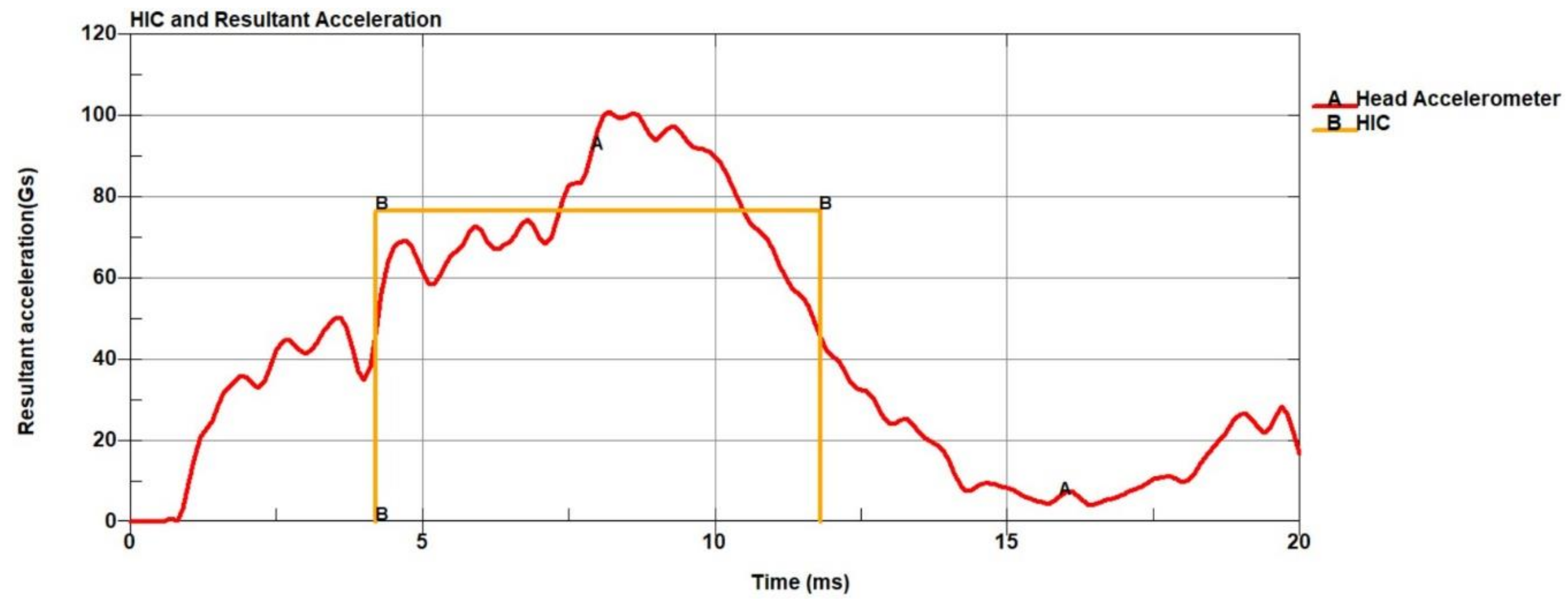
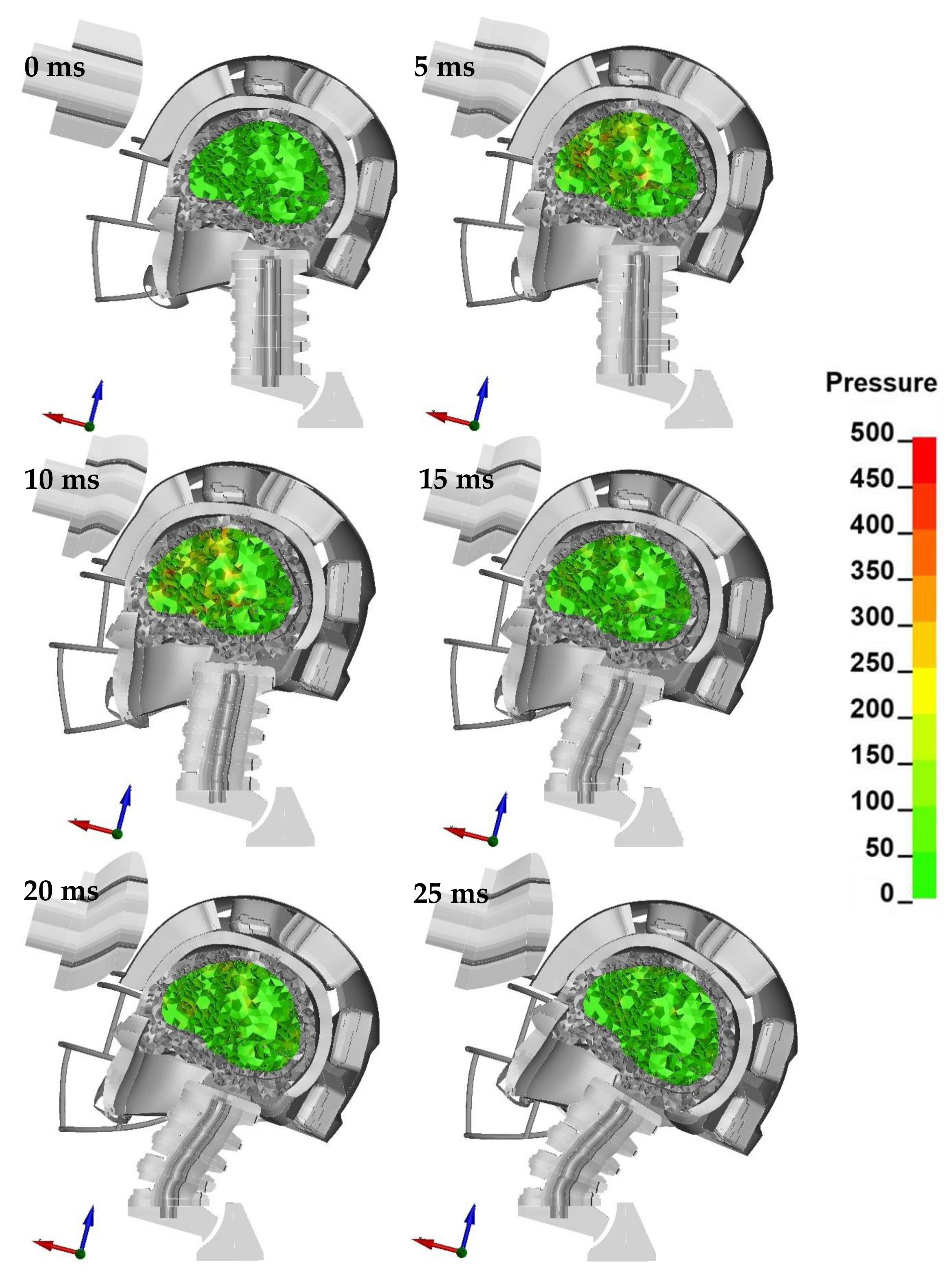
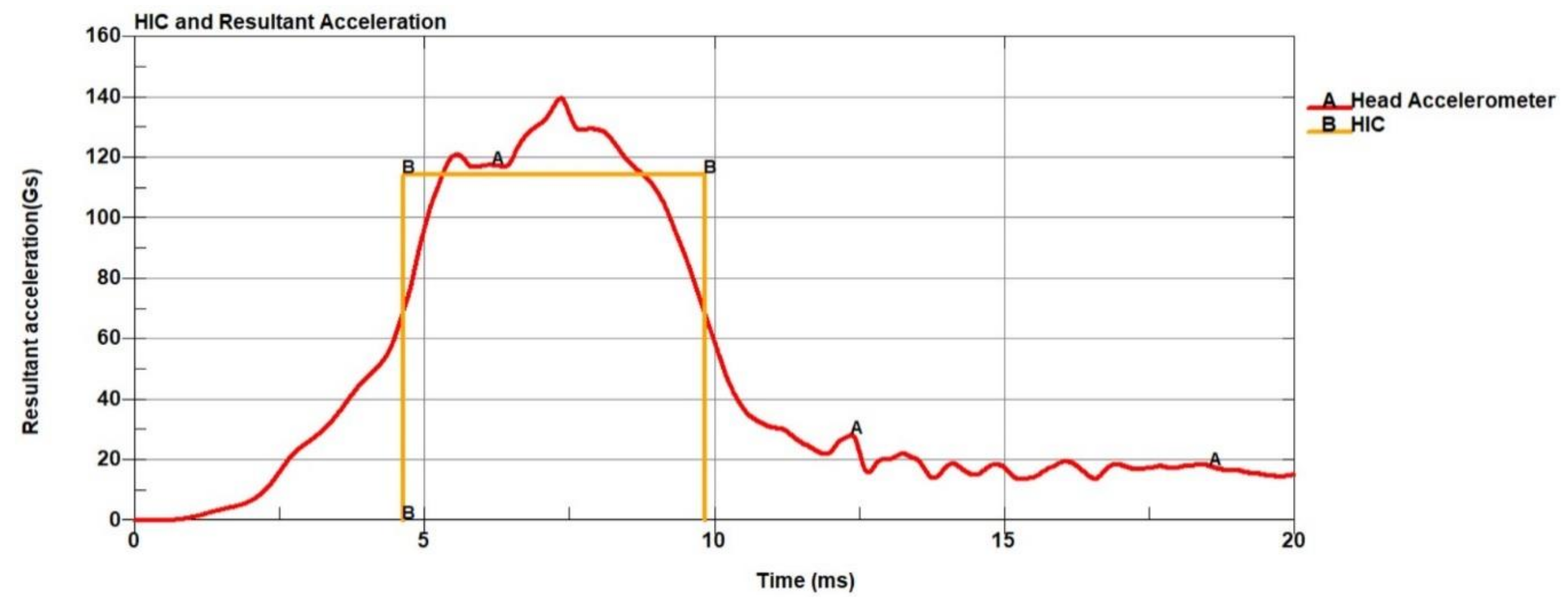
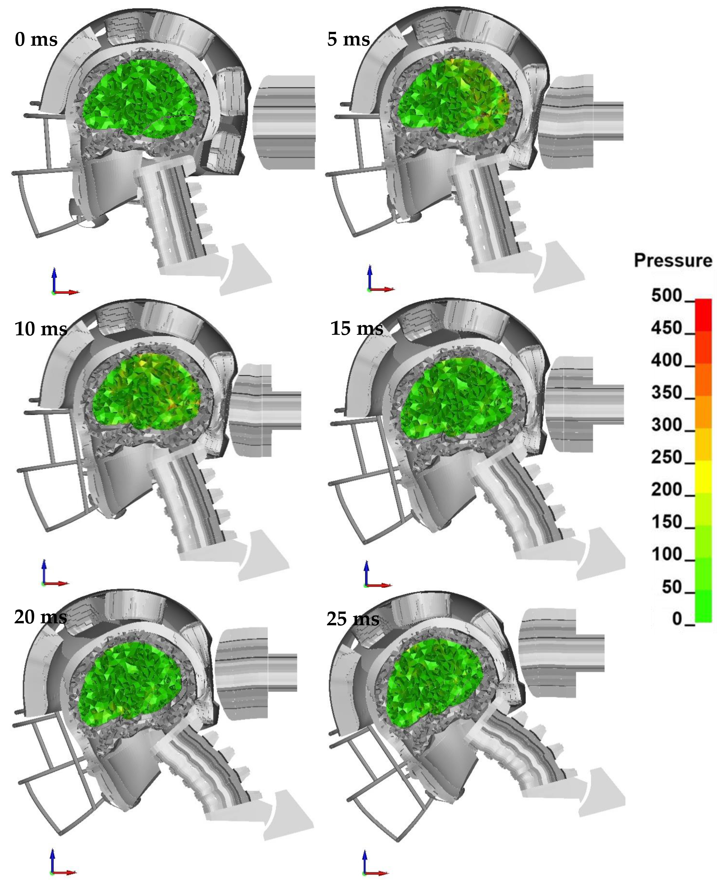
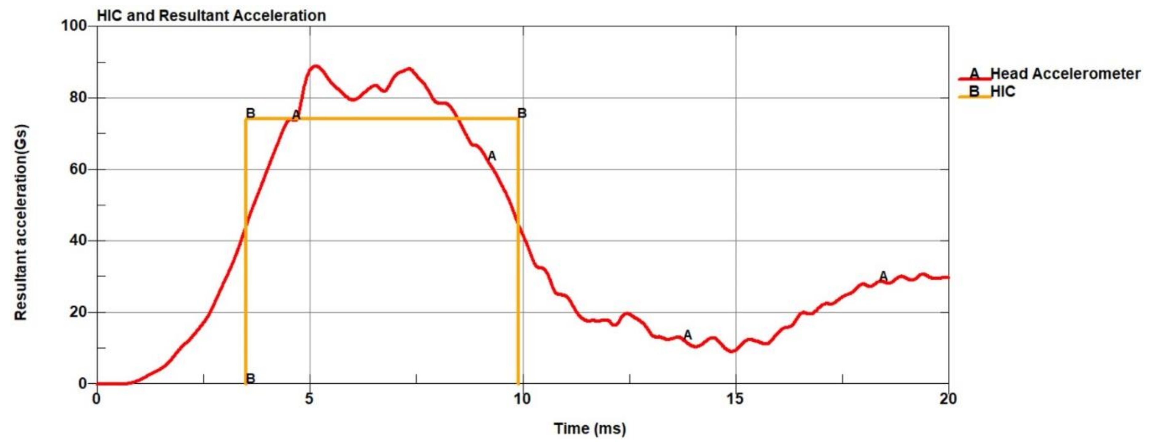
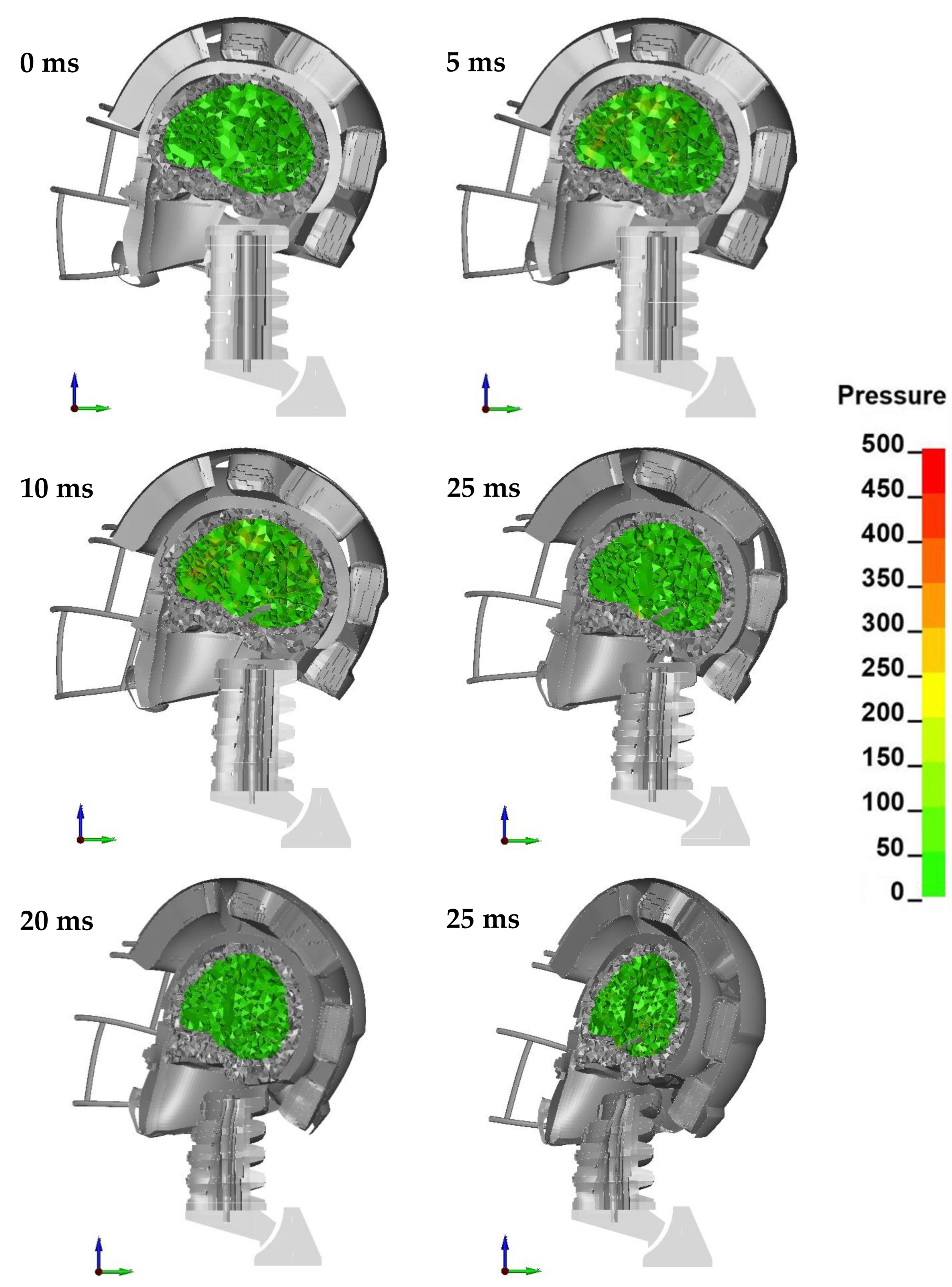
3. Results
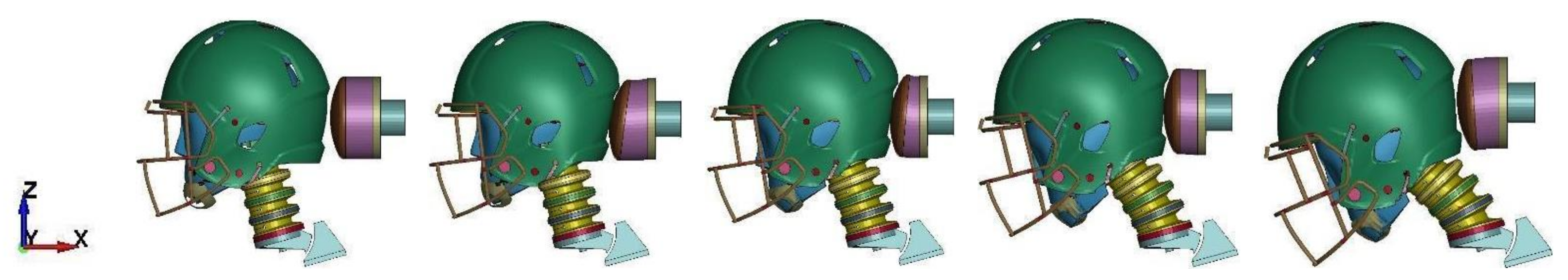
Configuration A
4. Discussion
5. Conclusions
Author Contributions
Funding
Institutional Review Board Statement
Informed Consent Statement
Data Availability Statement
Acknowledgments
Conflicts of Interest
Appendix A
Appendix A.1. Configuration AP


Appendix A.2. Configuration B


Appendix A.3. Configuration C


Appendix A.4. Configuration D


Appendix A.5. Configuration F


Appendix A.6. Configuration R


Appendix A.7. Configuration UT


References
- Saal, J.A. Common American Football Injuries. Sport. Med. 1991. [Google Scholar] [CrossRef]
- Kucera, K.L.; Yau, R.K.; Register-Mihalik, J.; Marshall, S.W.; Thomas, L.C.; Wolf, S.; Cantu, R.C.; Mueller, F.O.; Guskiewicz, K.M. Traumatic Brain and Spinal Cord Fatalities Among High School and College Football Players—United States, 2005–2014. MMWR. Morb. Mortal. Wkly. Rep. 2017, 65, 1465–1469. [Google Scholar] [CrossRef] [PubMed]
- Casson, I.R.; Viano, D.C.; Powell, J.W.; Pellman, E.J. Twelve Years of National Football League Concussion Data. Sport. Health A Multidiscip. Approach 2010, 2, 471–483. [Google Scholar] [CrossRef] [PubMed]
- Boden, B.P.; Tacchetti, R.L.; Cantu, R.C.; Knowles, S.B.; Mueller, F.O. Catastrophic head injuries in high school and college football players. Am. J. Sports Med. 2007. [Google Scholar] [CrossRef] [PubMed]
- Gessel, L.M.; Fields, S.K.; Collins, C.L.; Dick, R.W.; Comstock, R.D. Concussions among United States high school and collegiate athletes. J. Athl. Train. 2007, 42, 495–503. [Google Scholar] [PubMed]
- Liu, Y.; Domel, A.G.; Yousefsani, S.A.; Kondic, J.; Grant, G.; Zeineh, M.; Camarillo, D.B. Validation and Comparison of Instrumented Mouthguards for Measuring Head Kinematics and Assessing Brain Deformation in Football Impacts. Ann. Biomed. Eng. 2020, 48, 2580–2598. [Google Scholar] [CrossRef] [PubMed]
- Pellman, E.J.; Viano, D.C. Concussion in professional football. Neurosurg. Focus 2006, 21, 1–10. [Google Scholar] [CrossRef]
- Arnason, A.; Sigurdsson, S.B.; Gudmundsson, A.; Holme, I.; Engebretsen, L.; Bahr, R. Risk Factors for Injuries in Football. Am. J. Sports Med. 2004, 32, 5–16. [Google Scholar] [CrossRef]
- McIntosh, A.S.; McCrory, P. Preventing head and neck injury. Br. J. Sports Med. 2005, 39, 314–318. [Google Scholar] [CrossRef]
- Fernandes, F.A.O.; Alves de Sousa, R.J.; Ptak, M.; Wilhelm, J. Certified Motorcycle Helmets: Computational Evaluation of the Efficacy of Standard Requirements with Finite Element Models. Math. Comput. Appl. 2020, 25, 12. [Google Scholar] [CrossRef]
- Jamroziak, K.; Bajkowski, M.; Bocian, M.; Polak, S.; Magier, M.; Kosobudzki, M.; Stepien, R. Ballistic Head Protection in the Light of Injury Criteria in the Case of the Wz.93 Combat Helmet. Appl. Sci. 2019, 9, 2702. [Google Scholar] [CrossRef]
- Wilhelm, J.; Ptak, M.; Rusiński, E. Simulated depiction of head and brain injuries in the context of cellularbased materials in passive safety devices. Sci. Journals Marit. Univ. Szczecin 2017, 50, 98–104. [Google Scholar]
- Varela, M.M.; Fernandes, F.A.O.; Alves de Sousa, R.J. Development of an Eco-Friendly Head Impact Protection Device. Appl. Sci. 2020, 10, 2492. [Google Scholar] [CrossRef]
- Kraft, R.H.; Mckee, P.J.; Dagro, A.M.; Grafton, S.T. Combining the Finite Element Method with Structural Connectome-based Analysis for Modeling Neurotrauma: Connectome Neurotrauma Mechanics. PLoS Comput. Biol. 2012, 8, e1002619. [Google Scholar] [CrossRef]
- Aare, M.; Kleiven, S.; Halldin, P. Injury criteria for oblique helmet impacts. Int. J. Crashworthiness 2004, 9, 15–23. [Google Scholar] [CrossRef]
- Prasad, P.; Mertz, H.J. The position of the United States delegation to the ISO Working Group 6 on the use of HIC in the automotive environment. SAE Trans. 1985, 94, 106–116. [Google Scholar]
- Marjoux, D.; Baumgartner, D.; Deck, C.; Willinger, R. Head injury prediction capability of the HIC, HIP, SIMon and ULP criteria. Accid. Anal. Prev. 2008, 40, 1135–1148. [Google Scholar] [CrossRef]
- Takhounts, E.G.; Craig, M.J.; Moorhouse, K.; McFadden, J.; Hasija, V. Development of brain injury criteria (BrIC). Stapp Car Crash J. 2013, 57, 243. [Google Scholar]
- Post, A.; Kendall, M.; Cournoyer, J.; Karton, C.; Oeur, R.A.; Dawson, L.; Hoshizaki, T.B. Brain tissue analysis of impacts to American football helmets. Comput. Methods Biomech. Biomed. Engin. 2018, 21, 264–277. [Google Scholar] [CrossRef] [PubMed]
- Darling, T.; Muthuswamy, J.; Rajan, S.D. Finite element modeling of human brain response to football helmet impacts. Comput. Methods Biomech. Biomed. Engin. 2016, 19, 1432–1442. [Google Scholar] [CrossRef]
- Barker, J.; Corrales, M.; Gierczycka, D.; Bruneau, D.; Bustamante, M.C.; Cronin, D. Contribution of Energy-Absorbing Structures to Head Kinematics in Football Helmet Impacts. In Proceedings of the International Research Conference on the Biomechanics of Impacts (IRCOBI), Bern, Switzerland, 17–19 September 2008. [Google Scholar]
- Corrales, M.A.; Gierczycka, D.; Barker, J.; Bruneau, D.; Bustamante, M.C.; Cronin, D.S. Validation of a Football Helmet Finite Element Model and Quantification of Impact Energy Distribution. Ann. Biomed. Eng. 2020, 48, 121–132. [Google Scholar] [CrossRef]
- Lamb, L.; Hoshizaki, T.B. Deformation mechanisms and impact attenuation characteristics of thin-walled collapsible air chambers used in head protection. Proc. Inst. Mech. Eng. Part H J. Eng. Med. 2009, 223, 1021–1031. [Google Scholar] [CrossRef] [PubMed]
- Dean, S.W.; Hoshizaki, T.B.; Post, A. Impact Attenuation Characteristics of Thin Walled Collapsible Air Chambers for Use in Protective Helmets. J. ASTM Int. 2009, 6, 101883. [Google Scholar] [CrossRef]
- Schwarz, A. Helmet Design Absorbs Shock in a New Way. The New York Times. Available online: https://www.nytimes.com/2007/10/27/sports/football/27helmets.html (accessed on 12 October 2020).
- Sybilski, K.; Małachowski, J. Sensitivity study on seat belt system key factors in terms of disabled driver behavior during frontal crash. Acta Bioeng. Biomech. 2019, 21, 169–180. [Google Scholar] [CrossRef] [PubMed]
- Hazay, M.; Bakos, B.; Toth, P.J.; Buki, A.; Bojtar, I. Optimization of decompressive craniectomy based on finite element simulations. Acta Polytech. CTU Proc. 2018, 18, 6. [Google Scholar] [CrossRef][Green Version]
- Cobb, B.R.; Zadnik, A.M.; Rowson, S. Comparative analysis of helmeted impact response of Hybrid III and National Operating Committee on Standards for Athletic Equipment headforms. Proc. Inst. Mech. Eng. Part P J. Sport. Eng. Technol. 2016, 230, 50–60. [Google Scholar] [CrossRef]
- Viano, D.C.; Withnall, C.; Halstead, D. Impact Performance of Modern Football Helmets. Ann. Biomed. Eng. 2012, 40, 160–174. [Google Scholar] [CrossRef] [PubMed]
- Johnston, J.M.; Ning, H.; Kim, J.-E.; Kim, Y.-H.; Soni, B.; Reynolds, R.; Cooper, L.; Andrews, J.B.; Vaidya, U. Simulation, fabrication and impact testing of a novel football helmet padding system that decreases rotational acceleration. Sport. Eng. 2015, 18, 11–20. [Google Scholar] [CrossRef]
- Dymek, M.; Peliński, J. Numerical simulation of professional American football helmet with head model. Aktual. Probl. Biomech. 2020, 13–20. [Google Scholar]
- King, A.I.; Yang, K.H.; Zhang, L.; Hardy, W. Is head injury caused by linear or angular acceleration? In Proceedings of the International Research Conference on the Biomechanics of Impacts (IRCOBI), Lisbon, Portugal, 25–26 September 2003. [Google Scholar]
- Wu, L.C.; Kuo, C.; Loza, J.; Kurt, M.; Laksari, K.; Yanez, L.Z.; Senif, D.; Anderson, S.C.; Miller, L.E.; Urban, J.E.; et al. Detection of American Football Head Impacts Using Biomechanical Features and Support Vector Machine Classification. Sci. Rep. 2018, 8, 1–14. [Google Scholar]
- Hernandez, F.; Giordano, C.; Goubran, M.; Parivash, S.; Grant, G.; Zeineh, M.; Camarillo, D. Lateral impacts correlate with falx cerebri displacement and corpus callosum trauma in sports-related concussions. Biomech. Model. Mechanobiol. 2019, 18, 631–649. [Google Scholar] [CrossRef] [PubMed]
- Tierney, G.J.; Kuo, C.; Wu, L.; Weaving, D.; Camarillo, D. Analysis of head acceleration events in collegiate-level American football: A combination of qualitative video analysis and in-vivo head kinematic measurement. J. Biomech. 2020, 110, 109969. [Google Scholar] [CrossRef] [PubMed]
- Bailey, A.M.; Sanchez, E.J.; Park, G.; Gabler, L.F.; Funk, J.R.; Crandall, J.R.; Wonnacott, M.; Withnall, C.; Myers, B.S.; Arbogast, K.B. Development and Evaluation of a Test Method for Assessing the Performance of American Football Helmets. Ann. Biomed. Eng. 2020. [Google Scholar] [CrossRef] [PubMed]
- Bailey, A.M.; McMurry, T.L.; Cormier, J.M.; Funk, J.R.; Crandall, J.R.; Mack, C.D.; Myers, B.S.; Arbogast, K.B. Comparison of Laboratory and On-Field Performance of American Football Helmets. Ann. Biomed. Eng. 2020, 1–11. [Google Scholar] [CrossRef] [PubMed]
- Giudice, J.S.; Caudillo, A.; Mukherjee, S.; Kong, K.; Park, G.; Kent, R.; Panzer, M.B. Finite Element Model of a Deformable American Football Helmet Under Impact. Ann. Biomed. Eng. 2020, 48, 1524–1539. [Google Scholar] [CrossRef] [PubMed]
- Tierney, G.J.; Simms, C. Predictive Capacity of the MADYMO Multibody Human Body Model Applied to Head Kinematics During Rugby Union Tackles. Appl. Sci. 2019, 9, 726. [Google Scholar] [CrossRef]
- Hasegawa, Y.; Kawasaki, T.; Miyazaki, Y.; Sobue, S.; Kaketa, T.; Gonda, Y.; Kaneko, K. Changes of the Cervical Spine in Response to Head-first Impact in Rugby: A Finite Element Analysis. Juntendo Med. J. 2020, 66, 507–511. [Google Scholar] [CrossRef]
- Cogoluenhes, L.; Evin, M.; Forodighasemabadi, A.; Wei, W.; Thollon, L.; Llari, M. A modelisation of quantification of head and neck risks associated with tackles in rugby union. Comput. Methods Biomech. Biomed. Engin. 2019, 22, S273–S275. [Google Scholar] [CrossRef]
- O’Keeffe, E.; Kelly, E.; Liu, Y.; Giordano, C.; Wallace, E.; Hynes, M.; Tiernan, S.; Meagher, A.; Greene, C.; Hughes, S.; et al. Dynamic Blood–Brain Barrier Regulation in Mild Traumatic Brain Injury. J. Neurotrauma 2020, 37, 347–356. [Google Scholar] [CrossRef]
- Giudice, J.S.; Park, G.; Kong, K.; Bailey, A.; Kent, R.; Panzer, M.B. Development of Open-Source Dummy and Impactor Models for the Assessment of American Football Helmet Finite Element Models. Ann. Biomed. Eng. 2019, 47, 464–474. [Google Scholar] [CrossRef]
- Bustamante, M.C.; Bruneau, D.; Barker, J.B.; Gierczycka, D.; Coralles, M.A.; Cronin, D.S. Component-Level Finite Element Model and Validation for a Modern American Football Helmet. J. Dyn. Behav. Mater. 2019, 5, 117–131. [Google Scholar] [CrossRef]
- Ptak, M. Method to Assess and Enhance Vulnerable Road User Safety during Impact Loading. Appl. Sci. 2019, 9, 1000. [Google Scholar] [CrossRef]
- Schoell, S.L.; Weaver, A.A.; Urban, J.E.; Jones, D.A.; Stitzel, J.D.; Hwang, E.; Reed, M.P.; Rupp, J.D.; Hu, J. Development and Validation of an Older Occupant Finite Element Model of a Mid-Sized Male for Investigation of Age-related Injury Risk. Stapp Car Crash J. 2015, 59, 359–383. [Google Scholar]
- Greenberg, M. Greenberg Handbook of Neurosurgery; Thieme Medical Publishers: New York, NY, USA, 2019; Volume 53. [Google Scholar]
- Mondello, S.; Guedes, V.A.; Lai, C.; Czeiter, E.; Amrein, K.; Kobeissy, F.; Mechref, Y.; Jeromin, A.; Mithani, S.; Martin, C.; et al. Circulating Brain Injury Exosomal Proteins following Moderate-to-Severe Traumatic Brain Injury: Temporal Profile, Outcome Prediction and Therapy Implications. Cells 2020, 9, 977. [Google Scholar] [CrossRef]
- Champagne, A.A.; Coverdale, N.S.; Fernandez-Ruiz, J.; Mark, C.I.; Cook, D.J. Compromised resting cerebral metabolism after sport-related concussion: A calibrated MRI study. Brain Imaging Behav. 2020. [Google Scholar]
- Russman, A.; Figler, R.; Oh, S.-H.; Lowe, M.; Jones, S. 7 Tesla Brain MRI Characteristics Among Concussion Patients (P7.162). Neurology 2015, 84, 1526-632x. [Google Scholar]
- Ken Belson New York Times Aaron Hernandez Had Severe C.T.E. When He Died at Age 27. Available online: https://www.nytimes.com/2017/09/21/sports/aaron-hernandez-cte-brain.html (accessed on 12 October 2020).
- Boston University Research: CTE Center. Frequently Asked Questions about CTE. Available online: https://www.bu.edu/cte/about/frequently-asked-questions/ (accessed on 12 October 2020).
- Mez, J.; Daneshvar, D.H.; Kiernan, P.T.; Abdolmohammadi, B.; Alvarez, V.E.; Huber, B.R.; Alosco, M.L.; Solomon, T.M.; Nowinski, C.J.; McHale, L.; et al. Clinicopathological Evaluation of Chronic Traumatic Encephalopathy in Players of American Football. JAMA 2017, 318, 360. [Google Scholar] [CrossRef] [PubMed]
- Fernandes, F.A.O.; Sousa, R.J.A.D. Head injury predictors in sports trauma—A state-of-the-art review. Proc. Inst. Mech. Eng. Part H J. Eng. Med. 2015, 229. [Google Scholar] [CrossRef]
- Ji, S.; Zhao, W.; Ford, J.C.; Beckwith, J.G.; Bolander, R.P.; Greenwald, R.M.; Flashman, L.A.; Paulsen, K.D.; McAllister, T.W. Group-Wise Evaluation and Comparison of White Matter Fiber Strain and Maximum Principal Strain in Sports-Related Concussion. J. Neurotrauma 2015, 32, 441–454. [Google Scholar] [CrossRef] [PubMed]
- Ghazi, K.; Wu, S.; Zhao, W.; Ji, S. Instantaneous Whole-brain Strain Estimation in Dynamic Head Impact. J. Neurotrauma 2020. [Google Scholar] [CrossRef]
- Echemendia, R.J.; Meeuwisse, W.; McCrory, P.; Davis, G.A.; Putukian, M.; Leddy, J.; Makdissi, M.; Sullivan, S.J.; Broglio, S.P.; Raftery, M.; et al. The Sport Concussion Assessment Tool 5th Edition (SCAT5). Br. J. Sports Med. 2017, 51, 848–850. [Google Scholar] [CrossRef]
- Saunders, R.L. The Second Impact in Catastrophic Contact-Sports Head Trauma. JAMA J. Am. Med. Assoc. 1984, 252, 538. [Google Scholar] [CrossRef]
- Jordan, B.D.; Jahre, C.; Hauser, W.A.; Zimmerman, R.D.; Zarrelli, M.; Lipsitz, E.C.; Johnson, V.; Warren, R.F.; Tsairis, P.; Folk, F.S. CT of 338 active professional boxers. Radiology 1992, 185, 509–512. [Google Scholar] [CrossRef] [PubMed]
- Ratajczak, M.; Ptak, M.; Chybowski, L.; Gawdzińska, K.; Będziński, R. Material and Structural Modeling Aspects of Brain Tissue Deformation under Dynamic Loads. Materials 2019, 12, 271. [Google Scholar] [CrossRef] [PubMed]
- Costa, J.M.C.; Fernandes, F.A.O.; Alves de Sousa, R.J. Prediction of subdural haematoma based on a detailed numerical model of the cerebral bridging veins. J. Mech. Behav. Biomed. Mater. 2020, 111, 103976. [Google Scholar] [CrossRef] [PubMed]
- Fernandes, F.A.O.; Tchepel, D.; Alves de Sousa, R.J.; Ptak, M. Development and validation of a new finite element human head model: Yet another head model (YEAHM). Eng. Comput. 2018, 35, 477–496. [Google Scholar] [CrossRef]
- Wilhelm, J.; Ptak, M.; Fernandes, F.A.O.; Kubicki, K.; Kwiatkowski, A.; Ratajczak, M.; Sawicki, M.; Szarek, D. Injury Biomechanics of a Child’s Head: Problems, Challenges and Possibilities with a New aHEAD Finite Element Model. Appl. Sci. 2020, 10, 4467. [Google Scholar] [CrossRef]
- Giudice, J.; Sebastian, K.; Kong, A.; Caudillo, S.; Mukherjee, M.B.P. Finite Element Models of Helmet Assessment Tools 2018. Available online: https://biocorellc.com/finite-element-models/ (accessed on 12 October 2020).
- Kleiven, S.; Von Holst, H. Consequences of head size following trauma to the human head. J. Biomech. 2002, 35, 153–160. [Google Scholar] [CrossRef]
- Kleiven, S. Finite Element Modeling of the Human Head. Ph.D. Thesis, KTH Royal Institute of Technology, Stockholm, Sweden, 2002. [Google Scholar]
- Horanin-Dusza, M. The Analysis of the Biomechanical and Histological Properties of Cerebral Bridging Veins in Alcoholics and Nonalcoholics—The Importance in the Subdural Hematomas Etiology (in Polish). Ph.D. Thesis, Medical University, Wrocław. Poland, 2009. [Google Scholar]
- M. Fahlstedt, M.; Arnesen, E.; Jungstedt, P.H. Finite Element Model of 2016 Riddell Speed Classic (Safety Equipment Institute model R41179) 2018. Available online: https://biocorellc.com/finite-element-models/ (accessed on 12 October 2020).
- Standard Performance Specification for Newly Manufactured Football Helmets; National Operating Committee on Standards for Athletic Equipment: Overland Park, KS, USA, 2019.
- Gwin, J.T.; Diamond, S.G.; Halstead, P.D.; Greenwald, R.M. An Investigation of the NOCSAE Linear Impactor Test Method Based on In Vivo Measures of Head Impact Acceleration in American football. J. Biomech. Eng. 2010, 132, 011006. [Google Scholar] [CrossRef]
- Zhang, L.; Yang, K.H.; King, A.I. Comparison of brain responses between frontal and lateral impacts by finite element modeling. J. Neurotrauma 2001, 18, 21–30. [Google Scholar] [CrossRef]


| Element | Young’s (E) or Bulk Modulus (K) [MPa] | Density [kg/m3] | Poisson’s Ratio |
|---|---|---|---|
| Skull | E = 15,000.0 | 2000 | 0.22 |
| Dura mater | E = 31.5 | 1130 | 0.45 |
| Cerebrospinal fluid | K = 2200.0 | 1000 | 0.49 |
| Superior sagittal sinus | E = 28.2 | 1040 | 0.45 |
| Falx cerebri and cerebellar tentorium | E = 31.5 | 1130 | 0.45 |
| Brain tissue | K = 1130.0 | 1040 | not applicable |
| Bridging Veins Region | Young’s Modulus [MPa] | Density [kg/m3] | Poisson’s Ratio |
|---|---|---|---|
| Frontal | 56.45 | 1130 | 0.45 |
| Parietal | 94.09 | 1130 | 0.45 |
| Occipital | 97.21 | 1130 | 0.45 |
| Part | Young’s Modulus [MPa] | Poisson’s Ratio | Material Model in LS-DYNA | |
|---|---|---|---|---|
| Helmet | ||||
| FACEMASK | 8546.0 | 210,000 | 0.3 | ELASTIC |
| SHELL | 1095.0 | 1565 | 0.3 | ELASTIC |
| PADDING (FRONT) | 170.5 | 3 | - | FU_CHANG_FOAM_LOG_INTERPOLATION |
| PADDING (TOP, SIDES) | 70.0 95.0 | 20 | - | FU_CHANG_FOAM_LOG_INTERPOLATION |
| PADDING (BACK) | 100.0 | 200 | - | FU_CHANG_FOAM_LOG_INTERPOLATION |
| Impactor | ||||
| NYLON END CAP | 1140.0 | 2410 | 0.4 | ELASTIC |
| VINYL NITRILE | 122.6 | 1000 | - | FU_CHANG_FOAM_LOG_INTERPOLATION |
| BACKING PLATE | 6899.0 | 200,000 | 0.3 | RIGID |
| RAM | 140,700 | 200,000 | 0.3 | RIGID |
| Configuration | Simulation | Configu-Ration | Simulation |
|---|---|---|---|
| A |  | D |  |
| AP |  | F |  |
| B |  | R |  |
| C |  | UT |  |
| Configuration | HIC Score (HIII Model) | Hydrostatic Pressure [kPa] at 6 ms after Impact [ms] (αHEAD Model) | Ratio of Finite Elements Exceeding Threshold Criterion (237 kPa) | |
|---|---|---|---|---|
| A | 536 |  |  | 29.18% |
| P | 650 |  | 29.62% | |
| B | 449 |  | 24.20% | |
| C | 557 |  | 21.60% | |
| D | 594 |  | | 20.64% |
| F | 403 |  | 25.55% | |
| R | 731 |  | 25.42% | |
| UT | 304 |  | 25.66% | |
Publisher’s Note: MDPI stays neutral with regard to jurisdictional claims in published maps and institutional affiliations. |
© 2021 by the authors. Licensee MDPI, Basel, Switzerland. This article is an open access article distributed under the terms and conditions of the Creative Commons Attribution (CC BY) license (http://creativecommons.org/licenses/by/4.0/).
Share and Cite
Dymek, M.; Ptak, M.; Ratajczak, M.; Fernandes, F.A.O.; Kwiatkowski, A.; Wilhelm, J. Analysis of HIC and Hydrostatic Pressure in the Human Head during NOCSAE Tests of American Football Helmets. Brain Sci. 2021, 11, 287. https://doi.org/10.3390/brainsci11030287
Dymek M, Ptak M, Ratajczak M, Fernandes FAO, Kwiatkowski A, Wilhelm J. Analysis of HIC and Hydrostatic Pressure in the Human Head during NOCSAE Tests of American Football Helmets. Brain Sciences. 2021; 11(3):287. https://doi.org/10.3390/brainsci11030287
Chicago/Turabian StyleDymek, Mateusz, Mariusz Ptak, Monika Ratajczak, Fábio A. O. Fernandes, Artur Kwiatkowski, and Johannes Wilhelm. 2021. "Analysis of HIC and Hydrostatic Pressure in the Human Head during NOCSAE Tests of American Football Helmets" Brain Sciences 11, no. 3: 287. https://doi.org/10.3390/brainsci11030287
APA StyleDymek, M., Ptak, M., Ratajczak, M., Fernandes, F. A. O., Kwiatkowski, A., & Wilhelm, J. (2021). Analysis of HIC and Hydrostatic Pressure in the Human Head during NOCSAE Tests of American Football Helmets. Brain Sciences, 11(3), 287. https://doi.org/10.3390/brainsci11030287