Article

Quillaja saponaria Saponins with Potential to Enhance the Effectiveness of Disinfection Processes in the Beverage Industry

1
Institute of Fermentation Technology and Microbiology, Faculty of Biotechnology and Food Science, Lodz University of Technology, 171/173 Wolczanska, 90-924 Lodz, Poland
2
Center of Molecular and Macromolecular Studies, Polish Academy of Sciences, Sienkiewicza 112, 90-363 Lodz, Poland
*
Author to whom correspondence should be addressed.
Appl. Sci. 2018, 8(3), 368; https://doi.org/10.3390/app8030368
Submission received: 3 January 2018 / Revised: 15 February 2018 / Accepted: 28 February 2018 / Published: 3 March 2018
(This article belongs to the Section Chemical and Molecular Sciences)

Abstract

:

Featured Application

The extract of Q. saponaria can be used as a pretreatment agent to increase the sensibility of microorganisms to disinfectants. The results can be used to develop a strategy for the control of biofilms in beverage industry.

Abstract

This study examines the in vitro effect of Quillaja saponaria extracts on Asaia spp. planktonic cells and biofilms, in comparison and combination with two disinfectants: peracetic acid and N-ethyl-N,N-dimethylhexadecylammonium bromide. The growth of six bacterial strains was evaluated spectrophotometrically. Biofilm eradication was determined using the plate count method and luminometry. The planktonic cells were characterized by relatively high resistance to peracetic acid and higher sensitivity to N-ethylo-N,N-dimethylohexadecylioamonium bromide. In almost all the tested strains, growth was inhibited by 0.125% (v/v) peracetic acid and 0.0313% (w/v) quaternary ammonium compound. However, combinations of cell pretreatment using saponin and peracetic acid action were the most efficient against both planktonic and biofilm cells. The minimum inhibitory concentrations for peracetic acid were 4–8 times lower than those for bacterial strains without preliminary saponin action. Eradication of Asaia spp. biofilms reduced the number of living cells by 4–5 logarithmic units. These results demonstrate the synergetic action of saponin extract and disinfectant, and could be useful in the development of industrial strategies against Asaia spp. biofilms.

1. Introduction

Since Moore et al. first detected the presence of Asaia spp. in fruit-flavored bottled water in 2002, it has become recognized as a common bacterial spoilage in the beverage industry. It is difficult to determine the origin of Asaia spp. contamination in soft drinks. However, flowers and fruits from Asia have been suggested as the source [1,2,3,4]. Asaia spp. have also been isolated from grapes grown in South Australian vineyards and from insects native to tropical climates [5,6]. These bacteria exhibit unique adaptive abilities [7]. In the soft drinks environment, Asaia spp. are able to grow at pH 3.45, in the presence of sorbate, benzoate, and dimethyldicarbonate at concentrations of 1.5 mmol/L and higher [2]. The resistance of Asaia spp. to common preservatives limits not only the options for preventing the spoilage of soft drinks, but also the effectiveness of sanitation procedures in the beverage industry. These bacteria exhibit polysaccharide encapsulation, as well as very rigid biofilm formation on surfaces used commonly in the beverage industry: glass, polyethylene terephthalate, and polypropylene in the presence of sucrose [1,8,9]. Its removal may require additional forcing media (e.g., hot sodium hydroxide and detergent solutions), and in the case of difficult to access areas (e.g., pipe bends, branches, connections, valves) procedures based on mechanical treatment [2].
Microorganisms in biofilms can have up to 1000-times greater resistance to antimicrobial agents, such as disinfectants and sanitizers, than planktonic cells [10,11]. For this reason, novel biofilm control strategies use both conventional disinfectants (such as peracetic acid or quaternary ammonium compounds) and more novel approaches for reducing microbial adhesion abilities and increasing their sensitivity to antimicrobial agents. For example, substances with biocidal and/or antiadhesive properties—such as organosilanes or nanostructured silver multilayers—may be coated on the internal surfaces of industrial installations [12,13,14]. Various plant extracts and juices with antimicrobial properties can be used as soft drink additives [1,15,16,17]. Natural detergents can increase the permeability of microbial cell membranes, so enhancing their susceptibility to disinfection.
One group of natural detergents are saponins—classified as triterpenoid or steroid glycosides. Saponins show a wide range of important properties, and are used in medicine as well as in the beverage and cosmetics industries. They are non-toxic and environmentally friendly. Their amphipathic properties enable them to penetrate cellular membranes [18]. The beverage industry commonly uses extracts from Quillaja saponaria, mainly as a foaming agent and stabilizer in soft drinks or emulsion-based products. Saponins are stable in a wide range of environmental conditions (pH, ionic strength, temperature). This fact makes Q. saponaria saponins suitable for a wide range of applications in food products. According to the Codex Alimentarius Commission, Q. saponaria extracts (QEs) may be used as a foaming agent in soft drinks at doses of up to 200 mg/kg [19]. In the European Union, QEs appear on labels as E999, classified as a foaming agent for use in water-based, flavored, and non-alcoholic drinks (dose 200 mg/L calculated as anhydrous extract) [20,21].
Saponins in general show significant antifungal activity. Recent research on Quillaja saponins conducted in Poland found that saponin treatment leads to increased cell membrane permeability in different yeast strains [22,23]. The antibacterial activity of saponins is usually weak; however, such activity may occur in the case of Gram-negative bacteria. In Gram-negative bacteria, the outer membranes of the cell walls are covered in lipopolysaccharides. Q. saponaria extracts interact with the lipid part of cell membrane and increase the permeability of the bacterial cells. Sublethally injured or weakened microbial cells may thereby become more susceptible to conventional disinfectants. The purpose of the current study was therefore to investigate Quillaja saponaria saponins as a potential agent for enhancing the effectiveness of disinfection processes in the beverage industry, using as the model organism the spoilage bacteria, Asaia spp.

2. Materials and Methods

2.1. Bacterial Strains

Six strains of acetic acid bacteria belonging to the genus Asaia were used in this study. The bacteria were isolated from commercial soft drinks: Asaia bogorensis ISD1 (GenBank KP234014), As. bogorensis ISD2 (GenBank KP234015), As. bogorensis FFMW (GenBank KC756841), As. lannensis IFCW (GenBank KP234011), As. lannensis IFMW (GenBank KP234012), and As. lannensis FMW1 (GenBank HQ917850). Tested strains were isolated from strawberry-flavored mineral water, lemon-flavored mineral water, and isotonic drinks and then identified using morphological, physiological, and genetic methods described in our previous studies [8,9,15,16]. The isolates were maintained on GC agar slants (2% (w/v) glucose, 0.3% (w/v) peptone, 0.3% (w/v) yeast extract, 0.7% (w/v) CaCO3, 2% (w/v) agar) at 4 °C. Bacterial suspensions in phosphate-buffered saline (PBS, pH 7.4) were standardized using a DEN-1 densitometer (Merck-Millipore, Darmstadt, Germany) to final inoculum concentrations of 105–106 cells per mL. Rectangular glass slides (Knittel Glass, Braunschweig, Germany) measuring 76 × 26 mm were used as biofilm supports.

2.2. Antimicrobial Compounds

Quillaja saponaria saponin extract (QE) was purchased from Sigma Aldrich (Saint Louis, MO, USA). The extract is a heterogenous mixture of molecules, with sapogenin, the main aglycon, comprising 20–35%. A working solution of QEs 15% (w/v) was sterilized by membrane filtration (0.45-µm filters, Merck-Millipore, Darmstadt, Germany). N-ethyl-N,N-dimethylhexadecylammonium bromide (quaternary ammonium compound, QAC) was synthesized by the Center of Molecular and Macromolecular Studies of the Polish Academy of Science (Lodz, Poland). Peracetic acid (PAA) was purchased from Envolab Fine Chemicals (Czestochowa, Poland). The solutions of disinfectants (4%) were prepared in sterile deionized water immediately before use.

2.3. Minimal Inhibitory Concentration (MIC)

The effect of QAC and PAA on the growth of Asaia spp. was investigated in liquid minimal medium (0.3% (w/v) (NH4)2PO4, 0.3% (w/v) KH2PO4, 0.3% (w/v) MgSO4·7H2O, and 0.05% (w/v) yeast extract) with 2% (w/v) glucose placed in 96-well plates. For this purpose, 150 µL of 4% (w/v) for QAC or 4% (v/v) for PAA working solutions of each disinfectant was added to 150 µL of minimal medium and mixed; 150 µL was then transferred to the next cell with 150 µL of minimal medium. After preparing this series of dilutions, each microplate cell was inoculated with 150 µL of the standardized bacterial suspension. The final concentration of bacterial cells in the standardized suspension was approximately 105–106 cells per mL. The control sample was inoculated minimal medium without disinfectant. Multiplates were incubated using a UV–vis MULTISKAN GO spectrophotometer (Thermo Fisher Scientific, Waltham, MA, USA) at 25 °C for 24 h. Measurements of absorbance were carried out automatically every 1 h at a wavelength of 540 nm. The MICs were defined as the lowest concentrations of disinfectants at which there was no growth of Asaia spp.

2.4. Pretreatment of Planktonic Bacterial Cells with Saponin Extract

The tested strains of As. lannensis and As. bogorensis were pre-cultured in 20 mL of liquid GC medium at 25 °C for 48 h. The bacterial cells were then harvested by centrifugation (Eppendorf 5417R, Hamburg, Germany) at 6500 rpm for 10 min at 4 °C, washed with phosphate-buffered saline (PBS, pH 7.4) and resuspended in 20 mL of 1% (w/v) solution of QE. This concentration of QE was chosen on the base of our previous experiments (data not published; Polish patent application P. 423143). Bacterial suspensions were incubated at 25 °C for 1 h on a laboratory shaker (Unimax 1010, Heidolph, Germany) at 130 rpm. After pretreatment, the bacterial cells were harvested by centrifugation (Eppendorf 5417R, Hamburg, Germany) at 6500 rpm for 10 min at 4 °C, washed and resuspended in phosphate-buffered saline (PBS, pH 7.4) to obtain a final concentration of 105–106 cells per mL. The bacterial cells were then treated with PAA or QAC, as described above.

2.5. Biofilm Eradication

Six-day biofilms of Asaia spp. on glass carriers were obtained by bacterial cultivation in Erlenmeyer flasks with 20 mL of liquid minimal medium (0.3% (w/v) (NH4)2PO4, 0.3% (w/v) KH2PO4, 0.3% (w/v) MgSO4·7H2O, and 0.05% (w/v) yeast extract) with 2% (w/v) sucrose and glass carriers placed vertically. Bacterial cultures were incubated at 25 °C for six days on a laboratory shaker (Unimax 1010, Heidolph, Germany) at 130 rpm. The mature biofilms were transferred to 25 mL Erlenmeyer flasks containing 20 mL of the appropriate disinfection agent and incubated at 25 °C for 1 h on a laboratory shaker at 130 rpm. The glass carriers were then removed and washed with distilled water.
Analysis of the bacterial biofilms on the glass carriers was performed using luminometric measurements, the colony count method and microscopic observations followed by a qualitative assessment of the density of the bacteria covering the supports. For luminometric tests, the glass carrier with biofilm was rinsed with sterile distilled water and swabbed using HY-LiTE2 sampling pens (Merck, Darmstadt, Germany) for surface testing. The measurements were performed in relative light units (RLU) using a HY-LiTE2 luminometer [8]. For the colony count, the glass carrier plate was rinsed with sterile distilled water and swabbed with sterile swabs for surface testing. The bacterial suspensions were transferred onto GC agar medium and after incubation (25 °C, 72 h) colonies were counted. The number of colonies forming units per cm2 of the carrier was calculated. In microscopic studies, glass carriers were stained with basic fuchsin (0.5% w/v) and observed using a light microscope (OLYMPUS type BX41). Images were captured with a digital camera and analyzed using the UTHSCA ImageTool program available from http://imagetool.software.informer.com.
Analysis of the effect of Q. saponaria saponins on biofilm eradication was performed analogously, but before application of the disinfectant, the bacterial biofilms were treated with QE at a concentration of 1% (w/v) at 25 °C for 1 h on a rotary shaker at 130 rpm (shaker Unimax 1010, Heidolph, Germany).

2.6. Statistics

Mean values and standard deviations (SD) were obtained from three independent experiments. Statistical differences between the results were compared using a one-way repeated measures analysis of variance (ANOVA; OriginPro 9.2.214, OriginLab Corp., Northampton, MA, USA). Statistical significance was set at 5% (p < 0.05).

3. Results and Discussion

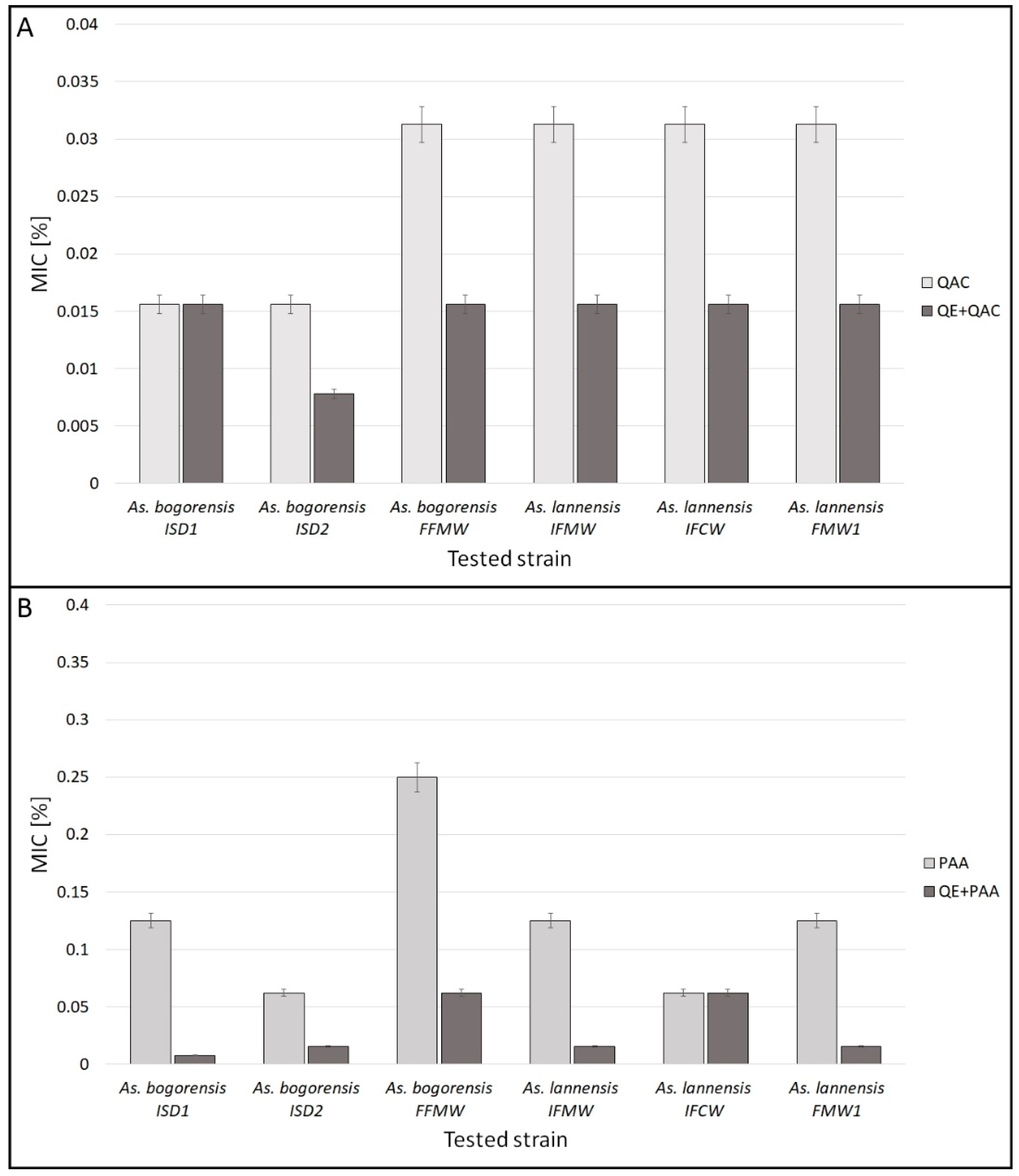
3.1. Minimal Inhibitory Concentration

MIC was used as a comparative measure of the effectiveness of two different disinfectants: N-ethyl-N,N-dimethylhexadecylammonium bromide (QAC) and peracetic acid (PAA) against Asaia spp. Figure 1 presents the MICs of QAC (Figure 1A) and PAA (Figure 1B) for six tested bacterial strains. The sensitivity of Asaia spp. to disinfectants was strain-dependent. The bacteria were characterized by similar sensitivity to QAC, but differentiation was noticeably more pronounced in the case of sensitivity to PAA. In general, higher sensitivity was observed for QAC, with MIC values ranging from 0.0156% to 0.0313% (w/v). For PAA, these values ranged from 0.0625 to 0.250% (v/v). The strain As. bogorensis FFMW was the most resistant to PAA and was also characterized by high resistance to QAC. It is worth noting that the MICs for QAC were almost 10 times lower in comparison to those obtained for PAA. Koziróg and co-workers obtained MIC results for dodecyl(trimethyl) ammonium bromide against As. lannensis FMW1 of 0.12–0.25 µM, dependent on both the type of carbon source and the availability of nutrients in the environment. They conclude that culture media with highly complex components may protect microorganisms from the presence of antimicrobial compounds [24]. The activity of PAA may be dependent not only on the kind of bacterial strain, but also on its metabolic activity. In a study conducted by Spoering and Lewis, the minimal inhibitory concentration of PAA against expotentially-growing Pseudomonas aeruginosa cells was 100 µg/mL, while for cells from stationary phase the concentration was four times higher. The authors suggest that the resistance of P. aeruginosa cells harvested from stationary phase was comparable to that of bacterial cells forming biofilms [25].
PAA is considered to be a strong oxidizing agent, with effective action against a wide spectrum of microorganisms [26]. The antimicrobial activity of this compound is based on the disruption of membranes, oxidation of thiol groups in proteins, and damage to DNA [27]. Despite the widespread use of PAA as an antimicrobial agent, the mechanisms of microbial tolerance to PAA are not fully understood. The QAC group includes both cationic surfactants and antimicrobial agents showing particular activity against yeasts and molds. However, they are also commonly used against Gram-positive and Gram-negative bacteria. The antimicrobial activity of these substances depends mainly on the length of the N-alkyl chain [28]. Disinfectants based on QAC are used widely in the pharmaceutical, cosmetic, and food industries [29]. Generally, the antimicrobial activity of QAC is based on interaction with cell membranes, disruption of membrane integrity and leakage of intracellular content [30]. The mechanism of antimicrobial activity involves the association of positively-charged quaternary nitrogen with negatively-charged head groups of acidic phospholipids in the bacterial membranes. The hydrophobic tail of the quaternary ammonium compound integrates into the microbial hydrophobic membrane core. The activity of QAC may also involve the disruption and denaturation of proteins and enzymes, as well as induction of dose- and time-dependent ultrastructural changes [28].
At low concentrations, the activity of QAC results in the loss of osmoregulatory capability and leakage of potassium ions and protons. Intermediate levels of QAC in turn disrupt membrane-association processes (respiration, transport, cell wall biosynthesis) [31]. However, it is well known that Gram-negative bacteria exhibit particular resistance to numerous biocides, including quaternary ammonium compounds. One of the reasons for this is the long-term use of the same types of disinfectant in the food industry. Loss of sensitivity to certain disinfectants can be caused by long-term use of the same or similar types of antimicrobial agent, by sustained exposure to sublethal concentrations of biocide, and by the cross-resistance of microbial cells [32].
The pretreatment of bacterial cultures in 1% (w/v) saponin solution led to significant increases in the sensitivity of most tested strains to the disinfectants. The MIC values for QAC were half those observed for cells not treated with saponin solution. In the case of PAA, the difference was even more significant. The MICs were 4–8 times lower than those for bacterial strains without saponin action. After 60 min of treatment, As. bogorensis FFMW, the most resistant strain to PAA, became four times more sensitive to this disinfectant. It may be concluded that such pretreatment of planktonic bacterial cells before disinfection can reduce the need for high doses of the tested antimicrobials.

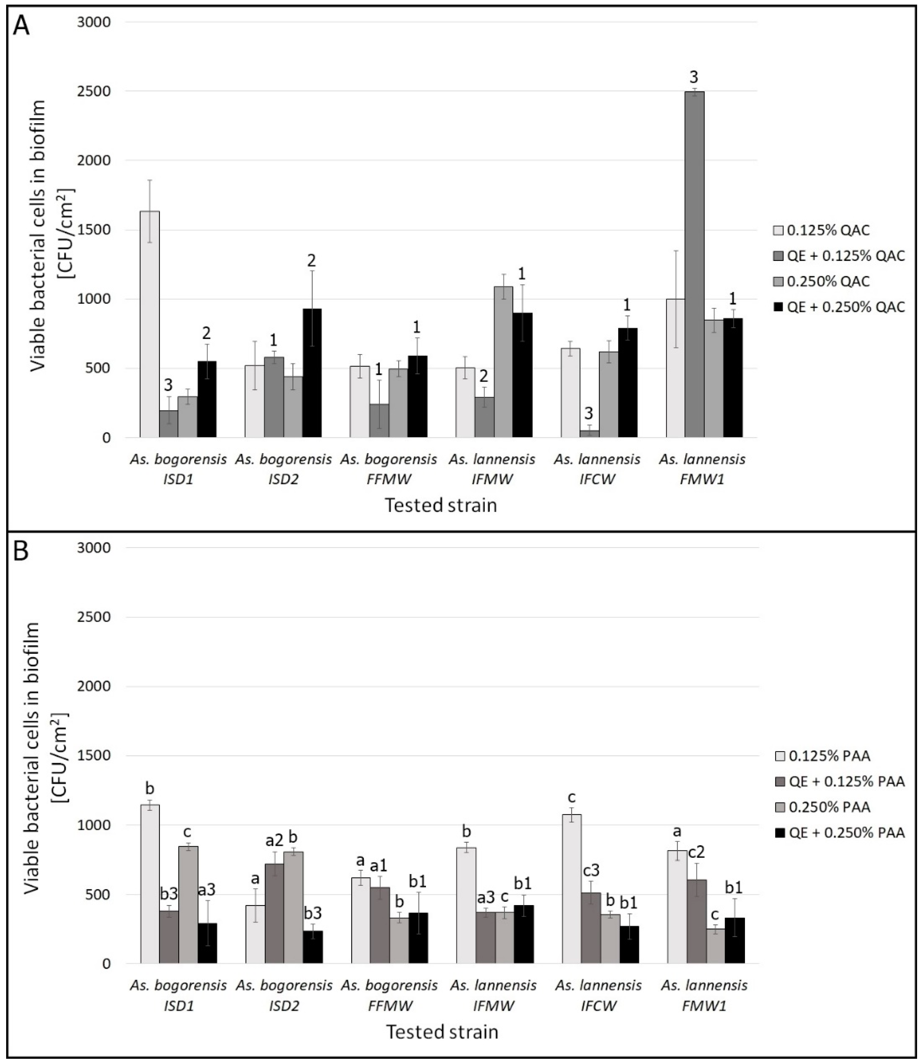
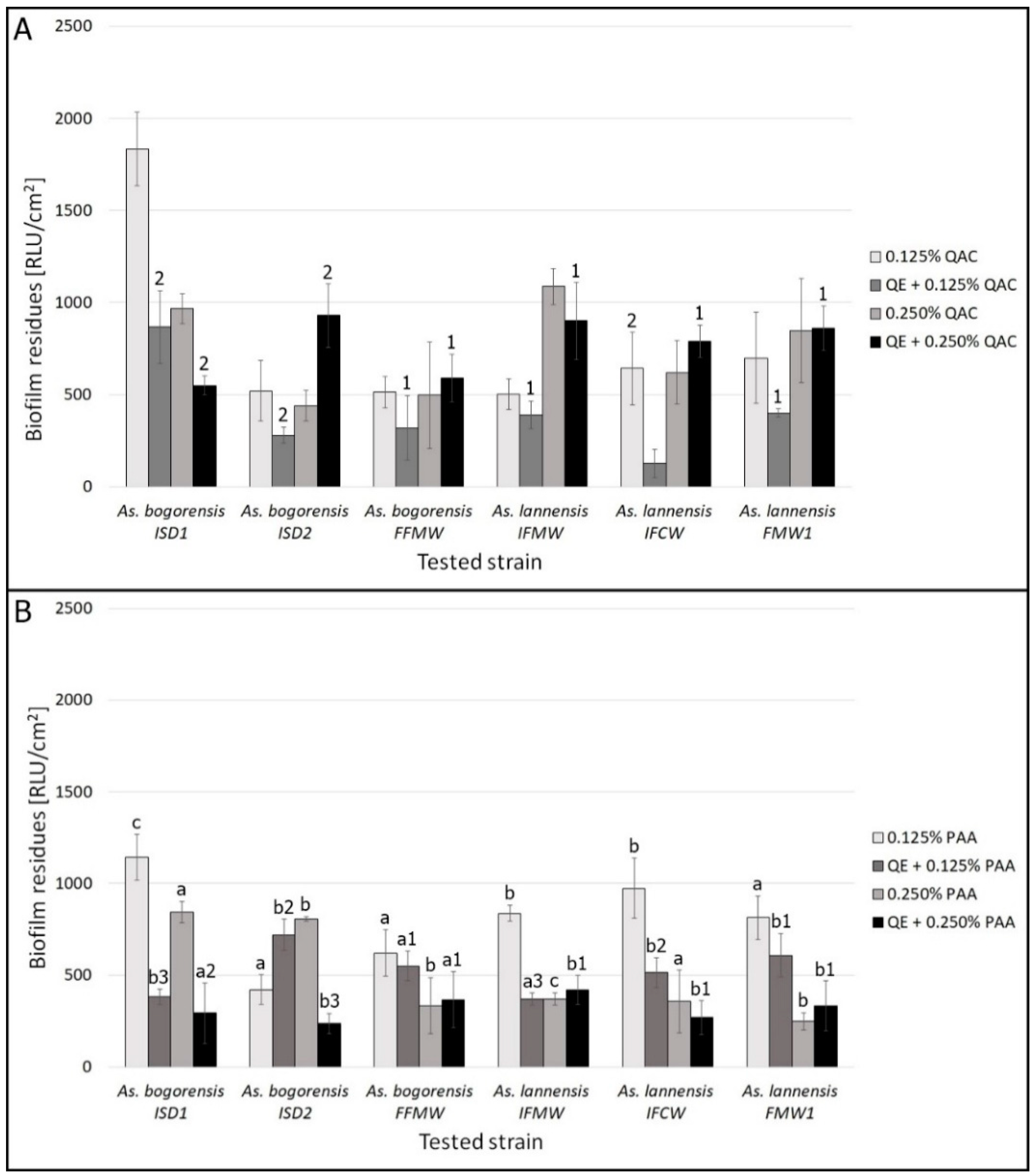
3.2. Biofilm Eradication

Biofilms are a serious problem in the beverage industry, and can be up to 1000 times more resistant to disinfectants than planktonic cells [10]. Diffusion through biofilm structures is one of the main mechanisms explaining their low sensitivity to antimicrobial agents. An essential step to improve the disinfection process is therefore the loosening or eradication of biofilm structures. One common and useful method is the mechanical disruption of biofilm matrices [33]. However, this method is effective only on easily-accessible surfaces. An enzyme-based strategy is also commonly used to improve the cleaning process. However, its effectiveness depends on the EPS composition of the microbial biofilms [34].
In this study, we investigated the influence of 1% (w/v) saponin pretreatment on the eradication of mature bacterial biofilms. The effectiveness of QE was evaluated using three methods: (i) the plate count method, for detecting the number of living cells on the tested surface; (ii) luminometry, for measuring ATP levels in the biofilm structures; and (iii) light microscopy, for visualizing the bacterial cells in biofilm.
Figure 2 shows the effect of QE solution (1% w/v) on the eradication of six-day bacterial biofilms treated with (Figure 2A) N-ethylo-N,N-dimethylohexadecylioamonium bromide (QAC) or (Figure 2B) peracetic acid (PAA) at two different concentrations (0.125% and 0.250%). These concentrations were chosen on the basis of MIC results for less active PAA and planktonic cells. Biofilms are considered to be highly resistant to antimicrobial agents; therefore, the higher MIC values were used in these experiments. The results were obtained using the plate count method. The initial results for viable bacterial cells forming mature biofilms ranged from 5.0 × 10⁷ CFU/cm2 for As. bogorensis FFMW to 2.3 × 10⁸ CFU/cm2 for As. bogorensis ISD2. Eradication of Asaia spp. biofilms led to a reduction in the number of living cells in the biofilms of around 4–5 logarithmic units.
Generally, synergistic effects were observed between QE and QAC when the QAC concentration was lower; however, the final effect depended on the bacterial strain. The reason for this synergy is unclear. It may be due to changes in the permeability of bacterial cell walls and loosening of the EPS structure, which are known to occur as a result of surfactant action. However, the mechanisms of action of nonionic detergent QE and cationic surfactants such as QAC are generally different [35]. Due to obtained differences in the plate count method, luminometric measurement was used as the second method.
Treatment with QE and PAA was more effective than eradication performed with QE and QAC. The reason for this was clearly related to the concentration of disinfectant used. It was thereby confirmed that cells forming biofilms are much less susceptible to biocidal than to planktonic action. The higher degree of eradication may have also depended on the type of action performed by the disinfectants. Both QE and QAC are surfactants with similar effects, whereas the action of PAA and QE is the combined effect of the detergent and the oxidizing agent.
Figure 3 presents the results of luminescence testing (in RLU/cm2) for mature biofilms treated with disinfectants, QAA (Figure 3A) and PAA (Figure 3B), alone or in combination with 1% (w/v) QE. The initial results for six-day biofilms formed by Asaia spp. strains varied, from 12,500 to 15,500 RLU/cm2. Eradication of Asaia spp. biofilms by antimicrobials led to as high as 10-fold reduction in RLU levels, and this effect was stronger in the case of combined treatment, especially with QE and PAA. In spite of general reduction of the obtained values of RLU/cm2, for biofilms formed by As. lannensis IFMW and As. lannensis FMW1, the results were higher for pretreatment by 0.250% QAC in comparison to 0.125% QAC. What is more, in some cases, results were higher for samples pretreated by QE. The action of QE and QAC is generally based on the interactions with cell membranes, disruption of membrane integrity, and leakage of intracellular content, including ATP [28,30]. Due to the fact that the bioluminescence measurement is based on the ATP determination, the higher results of RLU/cm2 may be caused by higher levels of ATP releasing from cells after treatment by QAC at higher concentrations. The results obtained using the conventional plate count method and luminometry were positively correlated (r = 0.84), but this relationship was not very strong. This fact may be due to the differences between the two detection methods. The plate count procedure evaluates the number of living bacterial cells able to grow on agar media, while luminometry detects the ATP levels. Of course, there is no perfect test method to detect biofilms on tested surfaces. Therefore, we used these two different methods to estimate biological material on the test surfaces [8].
Figure 4 shows exemplary images of Asaia spp. biofilm before (Figure 4A) and after treatment with QE and PAA (Figure 4B). Microscopic observations confirmed a significant reduction in the number of adhered Asaia cells. Regular cell adhesion to the glass surface was observed before treatment. After QE and PAA action, bacterial cells covered only around 10–20% of the total surface area. The synergistic action of antimicrobials against bacterial biofilms is well known. Changes in strain resistance and possible antibiotic synergy in combination with significant killing effects have also been noted by Monzón et al. for Staphylococcus epidermidis and by Anderson et al. for Pseudomonas aeruginosa biofilms [36,37]. Other agents that stimulate the action of antibiotics or disinfectants include enzymes, sodium salts, metal nanoparticles, acids, chitosan derivatives, and plant extracts. These combinations increase antimicrobial activity and reduce the concentration of biocides required. The in vitro antibacterial and antibiofilm activity of various plant extracts in synergy with erythromycin against erythromycin-resistant group A streptococci was also evaluated [38]. These various antimicrobial substances influence biofilm structure via various mechanisms and with different efficiencies [11]. Saponins from Q. saponaria also belong to this group, as active compounds facilitating the action of disinfectants commonly used in the food industry.

4. Conclusions

Based on the results presented in this study, it can be concluded that pretreatment with Q. saponaria saponins prior to the application of disinfectants led to more effective elimination of the spoilage bacteria Asaia spp. in both planktonic and biofilm form. The best results were obtained using two antimicrobial agents with different mechanisms of action: saponins as a natural detergent and peracetic acid as an oxidizing agent. These findings are of great interest not only with a view to increasing the antibacterial force of disinfectants, but also in terms of protecting the natural environment. Q. saponaria saponins are natural, environmentally friendly compounds [39]. High concentrations of PAA and QAC, despite their biodegradability, can have a significant effect on the biological processes of wastewater treatment [40,41]. Therefore, the benefits of reduced doses of antimicrobials in cleaning and disinfection processes are measurable in not only economic but also environmental terms.

5. Patents

This use of 1% (w/v) Q. saponaria saponins extract as a pretreatment agent before disinfection is the subject of the Polish patent application P. 423143.

Acknowledgments

This work was financially supported by the National Science Center, Poland, Project no. 2015/17/N/NZ9/03635.

Author Contributions

Hubert Antolak, Anna Otlewska, and Dorota Kręgiel conceived and designed the experiments; Hubert Antolak performed microbiological analysis; Hubert Antolak performed statistical analysis; Urszula Mizerska synthetized N-ethyl-N,N-dimethylhexadecylammonium bromide; Joanna Berłowska contributed analytical tools; Hubert Antolak and Dorota Kręgiel wrote the article.

Conflicts of Interest

The authors declare no conflict of interest. The founding sponsors had no role in the design of the study; in the collection, analyses, or interpretation of data; in the writing of the manuscript, or in the decision to publish the results.

References

  1. Antolak, H.; Kręgiel, D.; Czyżowska, A. Adhesion of Asaia bogorensis to glass and polystyrene in the presence of cranberry juice. J. Food Prot. 2015, 78, 1186–1190. [Google Scholar] [CrossRef] [PubMed]
  2. Horsáková, I.; Voldřich, M.; Čeřovskỳ, M.; Sedláčková, P.; Šicenrová, P.; Ulbrich, P. Asaia sp. as a bacterium decaying the packaged still fruit beverages. Czech J. Food Sci. 2009, 27, 362–365. [Google Scholar] [CrossRef]
  3. Kręgiel, D.; Rygała, A.; Libudzisz, Z.; Walczak, P.; Ołtuszak-Walczak, E. Asaia lannensis—The spoilage acetic acid bacteria isolated from strawberry-flavored bottled water in Poland. Food Control 2012, 26, 147–150. [Google Scholar] [CrossRef]
  4. Moore, J.E.; McCalmont, M.; Xu, J.; Millar, B.C.; Heaney, N. Asaia sp., an unusual spoilage organism of fruit-flavoured bottled water. Appl. Environ. Microbiol. 2002, 68, 4130–4131. [Google Scholar] [CrossRef] [PubMed]
  5. Chouaia, B.; Gaiarsa, S.; Crotti, E.; Comandatore, F.; Degli Esposti, M.; Ricci, I.; Alma, A.; Favia, G.; Bandi, C.; Daffonchio, D. Acetic acid bacteria genomes reveal functional traits for adaptation to life in insect guts. Genome Biol. Evol. 2014, 6, 912–920. [Google Scholar] [CrossRef] [PubMed]
  6. Mateo, E.; Torija, M.J.; Mas, A.; Bartowsky, E.J. Acetic acid bacteria isolated from grapes of South Australian vineyards. Int. J. Food Microbiol. 2014, 178, 98–106. [Google Scholar] [CrossRef] [PubMed]
  7. Antolak, H.; Kręgiel, D. Bakterie kwasu octowego Asaia sp. i ich unikalne zdolności adaptacyjne. Postep. Mikrobiol. 2016, 55, 392–397. (In Polish) [Google Scholar]
  8. Kręgiel, D. Attachment of Asaia lannensis to materials commonly used in beverage industry. Food Control 2013, 32, 537–542. [Google Scholar] [CrossRef]
  9. Kręgiel, D.; James, S.A.; Rygała, A.; Berłowska, J.; Antolak, H.; Pawlikowska, E. Consortia formed by yeasts and acetic acid bacteria Asaia spp. in soft drinks. Antonie Leeuwenhoek 2017. [Google Scholar] [CrossRef] [PubMed]
  10. Garrett, T.R.; Bhakoo, M.; Zhang, Z. Bacterial adhesion and biofilms on surfaces. Prog. Nat. Sci. Mater. Int. 2008, 18, 1049–1056. [Google Scholar] [CrossRef]
  11. Taraszkiewicz, A.; Fila, G.; Grinholc, M.; Nakonieczna, J. Innovative strategies to overcome biofilm resistance. BioMed Res. Int. 2013, 2013, 150653. [Google Scholar] [CrossRef] [PubMed]
  12. Kruk, T.; Szczepanowicz, K.; Kręgiel, D.; Szyk-Warszyńska, L.; Warszyński, P. Nanostructured multilayer polyelectrolyte films with silver nanoparticles as antibacterial coatings. Colloids Surf. B. Biointerfaces 2016, 137, 158–166. [Google Scholar] [CrossRef] [PubMed]
  13. Kręgiel, D. Advances in biofilm control for food and beverage industry using organo-silane technology: A review. Food Control 2014, 40, 32–40. [Google Scholar] [CrossRef]
  14. Kręgiel, D.; Berłowska, J.; Mizerska, U.; Fortuniak, W.; Chojnowski, W.; Ambroziak, W. Chemical modification of polyvinyl chloride and silicone elastomer in inhibiting adhesion of Aeromonas hydrophila. World J. Microbiol. Biotechnol. 2013, 29, 1197. [Google Scholar] [CrossRef] [PubMed]
  15. Antolak, H.; Czyżowska, A.; Kręgiel, D. Black currant (Ribes nigrum L.) and bilberry (Vaccinium myrtillus L.) fruit juices inhibit adhesion of Asaia spp. BioMed Res. Int. 2016, 2016, 3671306. [Google Scholar] [CrossRef]
  16. Antolak, H.; Czyżowska, A.; Sakač, M.; Mišan, A.; Đuragić, O.; Kręgiel, D. Phenolic compounds contained in little-known wild fruits as antiadhesive agents against the beverage-spoiling bacteria Asaia spp. Molecules 2017, 22, 1256. [Google Scholar] [CrossRef] [PubMed]
  17. Antolak, H.; Oracz, J.; Otlewska, A.; Żyżelewicz, D.; Kręgiel, D. Identification of carotenoids and isoprenoid quinones from Asaia lannensis and Asaia bogorensis. Molecules 2017, 22, 1608. [Google Scholar] [CrossRef] [PubMed]
  18. Kręgiel, D.; Berłowska, J.; Witońska, I.; Antolak, H.; Proestos, C.; Babic, M.; Babic, L.; Zhang, B. Saponin-based, biological-active surfactants from plants. In Application and Characterization of Surfactants, 1st ed.; Najjar, E., Ed.; InTech: Rijeka, Croatia, 2017; pp. 183–205. [Google Scholar] [CrossRef]
  19. Food and Agriculture Organization of the United Nations. 61st JECFA. Quillaja Extracts. Available online: http://www.fao.org/food/food-safety-quality/scientific-advice/jecfa/jecfa-additives/detail/en/c/476/ http://www.fao.org/fileadmin/tem‐plates/agns/pdf/jecfa/cta/61/QUILLAIA.pdf (accessed on 20 December 2017).
  20. European Food Safety Authority. Call for Food Additives Usage Level and/or Concentration Data in Food and Beverages Intended for Human Consumption. Available online: https://www.efsa.europa.eu/en/data/call/160524 (accessed 20 December 2017).
  21. Regulation (EC) No 1333/2008 of the European Parliament and of the Council of 16 December 2008 on Food Additives. Available online: http://eur-lex.europa.eu/legal-content/EN/TXT/HTML/?uri=CELEX:32008R1333&from=EN (accessed 20 December 2017).
  22. Berłowska, J.; Dudkiewicz, M.; Kręgiel, D.; Czyżowska, A.; Witońska, I. Cell lysis induced by membrane-damaging detergent saponins from Quillaja saponaria. Enzyme Microb. Technol. 2015, 75–76, 44–48. [Google Scholar] [CrossRef] [PubMed]
  23. Berłowska, J.; Dudkiewicz-Kołodziejska, M.; Pawlikowska, E.; Pielech-Przybylska, K.; Balcerek, M.; Czyżowska, A.; Kręgiel, D. Utilization of post-fermentation yeasts for yeast extract production by autolysis: The effect of yeast strain and saponin from Quillaja saponaria. J. Inst. Brew. 2017, 123, 396–401. [Google Scholar] [CrossRef]
  24. Koziróg, A.; Kręgiel, D.; Brycki, B. Action of monomeric/gemini surfactants on free cells and biofilm of Asaia lannensis. Molecules 2017, 22, 2036. [Google Scholar] [CrossRef] [PubMed]
  25. Spoering, A.L.; Lewis, K. Biofilms and planktonic cells of Pseudomonas aeruginosa have similar resistance to killing by antimicrobials. J. Bacteriol. 2001, 183, 6746–6751. [Google Scholar] [CrossRef] [PubMed]
  26. Briñez, W.J.; Roig-Sagués, A.X.; Herrero, M.M.H.; López-Pedemonte, T.; Guamis, B. Bactericidal efficacy of peracetic acid in combination with hydrogen peroxide against pathogenic and non pathogenic strains of Staphylococcus spp., Listeria spp. and Escherichia coli. Food Control 2006, 17, 516–521. [Google Scholar] [CrossRef]
  27. Rasimus, S.; Kolari, M.; Rita, H.; Hoornstra, D.; Salkinoja-Salonen, M. Biofilm-forming bacteria with varying tolerance to peracetic acid from a paper machine. J. Ind. Microbiol. Biotechnol. 2011, 38, 1379–1390. [Google Scholar] [CrossRef] [PubMed]
  28. Buffet-Bataillon, S.; Tattevin, P.; Bonnaure-Mallet, M.; Jolivet-Gougeon, A. Emergence of resistance to antibacterial agents: The role of quaternary ammonium compounds—A critical review. Int. J. Antimicrob. Agents 2012, 39, 381–389. [Google Scholar] [CrossRef] [PubMed]
  29. Chaidez, C.; Lopez, J.; Castro-del Campo, N. Quaternary ammonium compounds: An alternative disinfection method for fresh produce wash water. J. Water Health 2007, 5, 329–333. [Google Scholar] [PubMed]
  30. Gilbert, P.; Moore, L.E. Cationic antiseptics: Diversity of action under a common epithet. J. Appl. Microbiol. 2005, 99, 703–715. [Google Scholar] [CrossRef] [PubMed]
  31. Ortega Morente, E.; Fernández-Fuentes, M.A.; Grande Burgos, M.J.; Abriouel, H.; Pérez Pulido, R.; Gálvez, A. Biocide tolerance in bacteria. Int. J. Food Microbiol. 2013, 162, 13–25. [Google Scholar] [CrossRef] [PubMed]
  32. Gadea, R.; Glibota, N.; Pulido, R.P.; Gálvez, A.; Ortega, W. Effects of exposure to biocides on susceptibility to essential oils and chemical preservatives in bacteria from organic foods. Food Control 2017, 80, 176–182. [Google Scholar] [CrossRef]
  33. Bridier, A.; Dubois-Brissonnet, F.; Greub, G.; Thomas, V.; Briandet, R. Dynamics of biocide action in Pseudomonas aeruginosa biofilms. Antimicrob. Agents Chemother. 2011, 55, 2648–2654. [Google Scholar] [CrossRef] [PubMed]
  34. Valentini, F.; Diamanti, A.; Carbone, M.; Bauer, E.M.; Palleschi, G. New cleaning strategies based on carbon nanomaterials applied to the deteriorated marble surfaces: A comparative study with enzyme based treatments. Appl. Surf. Sci. 2012, 258, 5965–5980. [Google Scholar] [CrossRef]
  35. Von Rybinski, W. Physical aspects of cleaning processes. In Handbook for Cleaning/Decontamination of Surfaces, 1st ed.; Johansson, I., Somasundaran, P., Eds.; Elsevier: Amsterdam, The Netherlands, 2007; pp. 1–55. ISBN 978-0-444-51664-0. [Google Scholar]
  36. Anderson, G.G.; Kenney, T.F.; Macleod, D.L.; Henig, N.R.; O’Toole, G.A. Eradication of Pseudomonas aeruginosa biofilms on cultured airway cells by a fosfomycin/tobramycin antibiotic combination. Pathog. Dis. 2013, 67, 39–45. [Google Scholar] [CrossRef] [PubMed]
  37. Monzón, M.; Oteiza, C.; Leiva, J.; Amorena, B. Synergy of different antibiotic combinations in biofilms of Staphylococcus epidermidis. J. Antimicrob. Chemother. 2001, 48, 793–801. [Google Scholar] [CrossRef] [PubMed]
  38. Padalia, H.; Moteriya, P.; Baravalia, Y.; Chanda, S. Antimicrobial and Synergistic effects of Some Essential Oils to Fight against Microbial Pathogens—A Review. Available online: http://www.microbiology5.org/microbiology5/book/34-45.pdf (accessed 20 December 2017).
  39. Pelah, D.; Abramovich, Z.; Markus, A.; Wiesman, Z. The use of commercial saponin from Quillaja saponaria bark as a natural larvicidal agent against Aedes aegypti and Culex pipiens. J. Ethnopharmacol. 2002, 81, 407–409. [Google Scholar] [CrossRef]
  40. Toniolo, P. Peracetic Acid: A good choice for wastewater reuse. In Wastewater and Biosolids Treatment and Reuse: Bridging Modeling and Experimental Studies; Santoro, D., Ed.; ECI Symposium Series; ECI: New York, NY, USA, 2014; Available online: http://dc.engconfintl.org/wbtr_i/23 (accessed 20 December 2017).
  41. Lai, Y.S.; Ontiveros-Valencia, A.; Ilhan, Z.E.; Zhou, Y.; Miranda, E.; Maldonado, J.; Krajmalnik-Brown, R.; Rittmann, B.E. Enhancing biodegradation of C16-alkyl quaternary ammonium compounds using an oxygen-based membrane biofilm reactor. Water Res. 2017, 123, 825–833. [Google Scholar] [CrossRef] [PubMed]
Figure 1. Values for minimal inhibitory concentration of: (A) N-ethyl-N,N-dimethylhexadecylammonium bromide (QAC) and (B) peracetic acid (PAA) against Asaia spp. in comparison to values obtained for saponin pretreated cells by Q. saponaria extracts (QE): QE + QAC and QE + PAA.
Applsci 08 00368 g001
Figure 2. Effect of mature biofilm eradication by disinfectants: (A) N-ethyl-N,N-dimethylhexadecylammonium bromide (QAC) and (B) peracetic acid (PAA) acting alone or in combination with Q. saponaria extracts (QE). Plate count method (CFU/cm2). Values with the different superscript lowercase signs are statistically different (p < 0.05). Lowercase letters for PAA results compared to those for QAC: a p > 0.05; b 0.001 < p < 0.05; c p < 0.001. Lowercase numbers for disinfectant results compared to those for QE+ disinfectant: 1 p > 0.05; 2 0.001 < p < 0.05; 3 p < 0.001.
Applsci 08 00368 g002
Figure 3. Effect of mature biofilm eradication by disinfectants: (A) N-ethyl-N,N-dimethylhexadecylammonium bromide (QAC) and (B) peracetic acid (PAA) acting alone or in a combined action with Q. saponaria extracts (QE). Luminometric method (RLU/cm2). Values with the different superscript lowercase signs are statistically different (p < 0.05). Lowercase letters for PAA results compared to those for QAC: a p > 0.05; b 0.001 < p < 0.05; c p < 0.001. Lowercase numbers for disinfectant results compared to those for QE+ disinfectant: 1 p > 0.05; 2 0.001 < p < 0.05; 3 p < 0.001.
Applsci 08 00368 g003
Figure 4. Effect of biofilm eradication by peracetic acid (PAA) (0.250% v/v) in combined action with Q. saponaria extracts (QE) (1% w/v). (A) Six-day biofilm of Asaia spp.; (B) biofilm eradication by QE and PAA treatment. Bar represents 20 µm.
Applsci 08 00368 g004

