
Creation of Optimal Frequency for Electrostatic Force Microscopy Using Direct Digital Synthesizer


Abstract
:
1. Introduction
2. Methods and Results
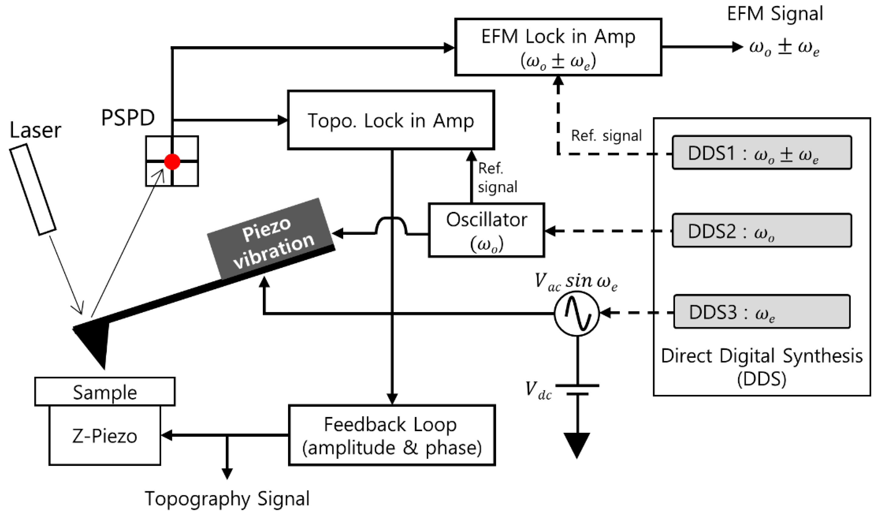
2.1. Materials and Set-Up
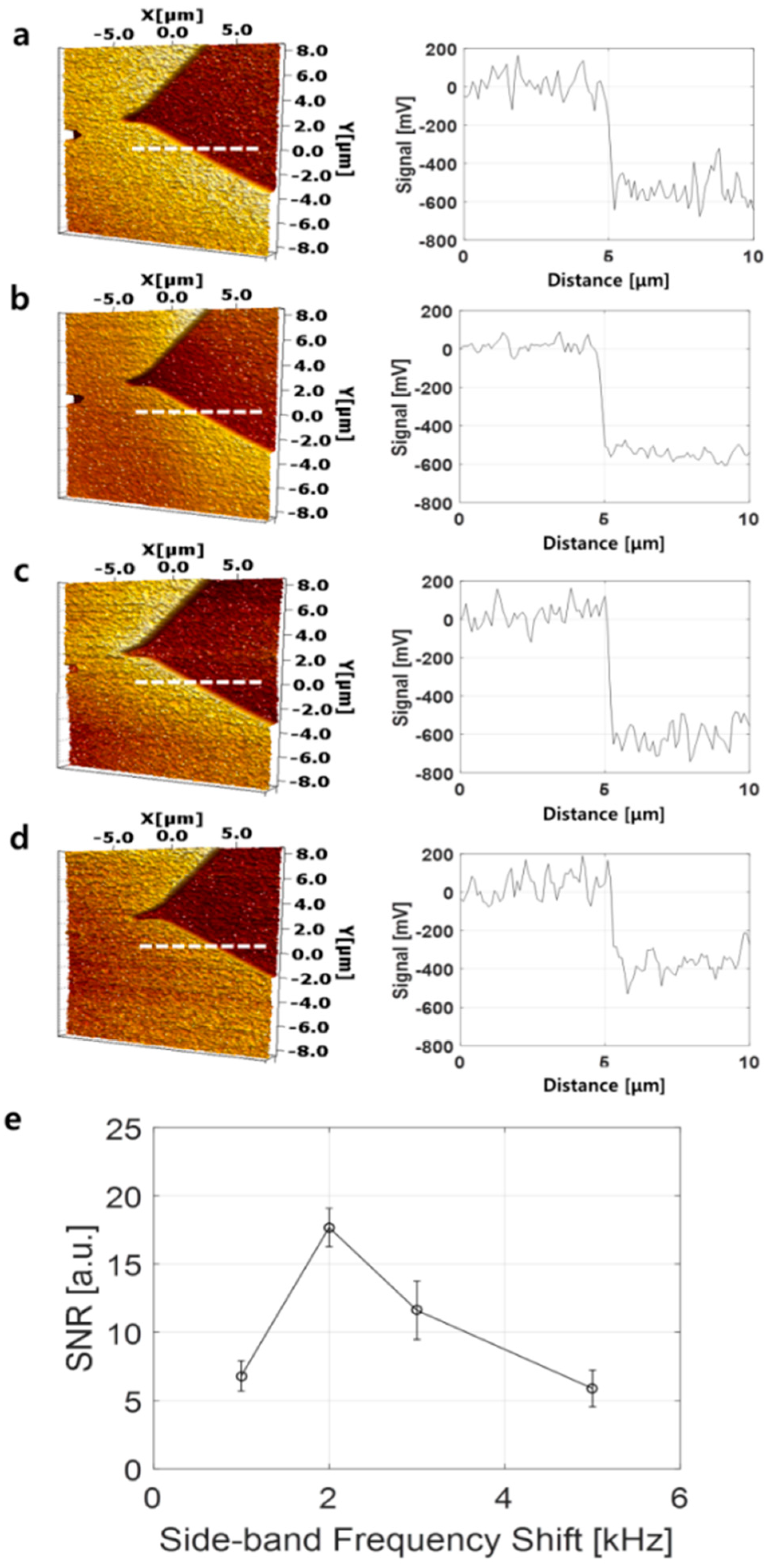
2.2. Optimal Frequency Detection for EFM Image
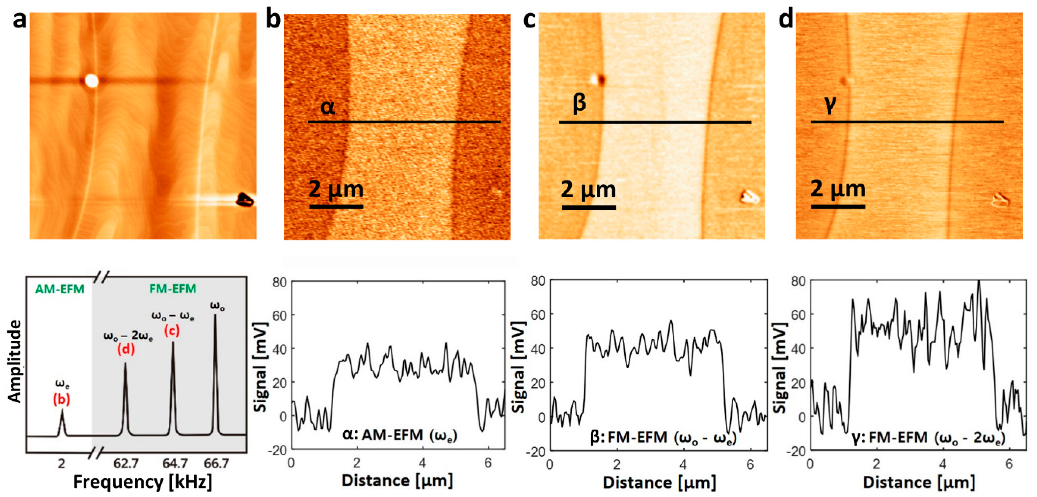
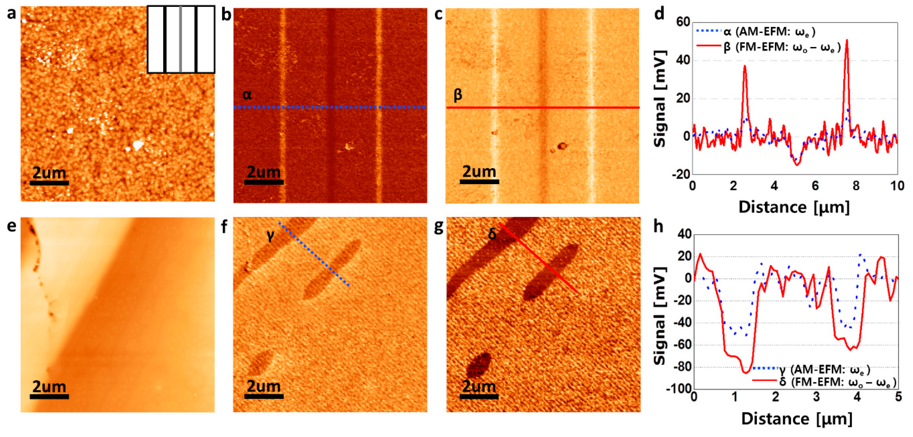
2.3. Applications of Optimized Side-Band Frequency
3. Discussion and Conclusions
Acknowledgments
Author Contributions
Conflicts of Interest
References
- Jacobs, H.; Knapp, H.; Müller, S.; Stemmer, A. Surface potential mapping: A qualitative material contrast in spm. Ultramicroscopy 1997, 69, 39–49. [Google Scholar] [CrossRef]
- Melitz, W.; Shen, J.; Kummel, A.C.; Lee, S. Kelvin probe force microscopy and its application. Surf. Sci. Rep. 2011, 66, 1–27. [Google Scholar] [CrossRef]
- Bottomley, L.A. Scanning probe microscopy. Anal. Chem. 1998, 70, 425–476. [Google Scholar] [CrossRef]
- Hansma, H.G.; Hoh, J.H. Biomolecular imaging with the atomic force microscope. Annu. Rev. Biophys. Biomol. Struct. 1994, 23, 115–140. [Google Scholar] [CrossRef] [PubMed]
- Albrecht, T.; Grütter, P.; Horne, D.; Rugar, D. Frequency modulation detection using high-q cantilevers for enhanced force microscope sensitivity. J. Appl. Phys. 1991, 69, 668–673. [Google Scholar] [CrossRef]
- Nonnenmacher, M.; O‘Boyle, M.; Wickramasinghe, H.K. Kelvin probe force microscopy. Appl. Phys. Lett. 1991, 58, 2921–2923. [Google Scholar] [CrossRef]
- Gaub, B.M.; Müller, D.J. Mechanical stimulation of piezo1 receptors depends on extracellular matrix proteins and directionality of force. Nano Lett. 2017, 17, 2064–2072. [Google Scholar] [CrossRef] [PubMed]
- Yin, F.; Park, S.-H.; Shin, H.-K.; Kwon, Y.-S. Study of hemoglobin-octadecylamine lb film formation and deposition by compressibility analyse, qcm and afm. Curr. Appl. Phys. 2006, 6, 728–734. [Google Scholar] [CrossRef]
- Kafle, K.; Xi, X.; Lee, C.M.; Tittmann, B.R.; Cosgrove, D.J.; Park, Y.B.; Kim, S.H. Cellulose microfibril orientation in onion (allium cepa l.) epidermis studied by atomic force microscopy (afm) and vibrational sum frequency generation (sfg) spectroscopy. Cellulose 2014, 21, 1075–1086. [Google Scholar] [CrossRef]
- Hong, J.; Park, S.-i.; Khim, Z. Measurement of hardness, surface potential, and charge distribution with dynamic contact mode electrostatic force microscope. Rev. Sci. Instrum. 1999, 70, 1735–1739. [Google Scholar] [CrossRef]
- Henning, A.K.; Hochwitz, T.; Slinkman, J.; Never, J.; Hoffmann, S.; Kaszuba, P.; Daghlian, C. Two-dimensional surface dopant profiling in silicon using scanning kelvin probe microscopy. J. Appl. Phys. 1995, 77, 1888–1896. [Google Scholar] [CrossRef]
- Jiang, J.; Krauss, T.D.; Brus, L.E. Electrostatic force microscopy characterization of trioctylphosphine oxide self-assembled monolayers on graphite. J. Phys. Chem. B 2000, 104, 11936–11941. [Google Scholar] [CrossRef]
- Shin, C.; Kim, K.; Kim, J.; Ko, W.; Yang, Y.; Lee, S.; Jun, C.S.; Kim, Y.S. Fast, exact, and non-destructive diagnoses of contact failures in nano-scale semiconductor device using conductive afm. Sci. Rep. 2013, 3, 2088. [Google Scholar] [CrossRef] [PubMed]
- Tanimoto, M.; Vatel, O. Kelvin probe force microscopy for characterization of semiconductor devices and processes. J. Vac. Sci. Technol. B Nanotechnol. Microelectron. 1996, 14, 1547–1551. [Google Scholar] [CrossRef]
- Rosenwaks, Y.; Shikler, R.; Glatzel, T.; Sadewasser, S. Kelvin probe force microscopy of semiconductor surface defects. Phys. Rev. B Cover. Condens. Matter Mater. Phys. 2004, 70, 085320. [Google Scholar] [CrossRef]
- Singh, A.; Guha, P.; Panwar, A.K.; Tyagi, P.K. Estimation of intrinsic work function of multilayer graphene by probing with electrostatic force microscopy. Appl. Surf. Sci. 2017, 402, 271–276. [Google Scholar] [CrossRef]
- Ziegler, D.; Gava, P.; Güttinger, J.; Molitor, F.; Wirtz, L.; Lazzeri, M.; Saitta, A.; Stemmer, A.; Mauri, F.; Stampfer, C. Variations in the work function of doped single-and few-layer graphene assessed by kelvin probe force microscopy and density functional theory. Phys. Rev. B Cover. Condens. Matter Mater. Phys. 2011, 83, 235434. [Google Scholar] [CrossRef]
- Filleter, T.; Emtsev, K.; Seyller, T.; Bennewitz, R. Local work function measurements of epitaxial graphene. Appl. Phys. Lett. 2008, 93, 133117. [Google Scholar] [CrossRef]
- Moores, B.; Hane, F.; Eng, L.; Leonenko, Z. Kelvin probe force microscopy in application to biomolecular films: Frequency modulation, amplitude modulation, and lift mode. Ultramicroscopy 2010, 110, 708–711. [Google Scholar] [CrossRef] [PubMed]
- Sinensky, A.K.; Belcher, A.M. Label-free and high-resolution protein/DNA nanoarray analysis using kelvin probe force microscopy. Nat. Nanotechnol. 2007, 2, 653–659. [Google Scholar] [CrossRef] [PubMed]
- Clack, N.G.; Salaita, K.; Groves, J.T. Electrostatic readout of DNA microarrays with charged microspheres. Nat. Biotechnol. 2008, 26, 825–830. [Google Scholar] [CrossRef] [PubMed]
- Drolle, E.; Bennett, W.; Hammond, K.; Lyman, E.; Karttunen, M.; Leonenko, Z. Molecular dynamics simulations and kelvin probe force microscopy to study of cholesterol-induced electrostatic nanodomains in complex lipid mixtures. Soft Matter 2017, 13, 355–362. [Google Scholar] [CrossRef] [PubMed]
- Stein, D.; Kruithof, M.; Dekker, C. Surface-charge-governed ion transport in nanofluidic channels. Phys. Rev. Lett. 2004, 93, 035901. [Google Scholar] [CrossRef] [PubMed]
- Vlassiouk, I.; Siwy, Z.S. Nanofluidic diode. Nano Lett. 2007, 7, 552–556. [Google Scholar] [CrossRef] [PubMed]
- Karnik, R.; Duan, C.; Castelino, K.; Daiguji, H.; Majumdar, A. Rectification of ionic current in a nanofluidic diode. Nano Lett. 2007, 7, 547–551. [Google Scholar] [CrossRef] [PubMed]
- Sparreboom, W.; Van Den Berg, A.; Eijkel, J. Principles and applications of nanofluidic transport. Nat. Nanotechnol. 2009, 4, 713–720. [Google Scholar] [CrossRef] [PubMed]
- Cheng, L.-J.; Guo, L.J. Nanofluidic diodes. Chem. Soc. Rev. 2010, 39, 923–938. [Google Scholar] [CrossRef] [PubMed]
- Guan, W.; Fan, R.; Reed, M.A. Field-effect reconfigurable nanofluidic ionic diodes. Nat. Commun. 2011, 2, 506. [Google Scholar] [CrossRef] [PubMed]
- Glatzel, T.; Sadewasser, S.; Lux-Steiner, M.C. Amplitude or frequency modulation-detection in kelvin probe force microscopy. Appl. Surf. Sci. 2003, 210, 84–89. [Google Scholar] [CrossRef]
- Zerweck, U.; Loppacher, C.; Otto, T.; Grafström, S.; Eng, L.M. Accuracy and resolution limits of kelvin probe force microscopy. Phys. Rev. B Cover. Condens. Matter Mater. Phys. 2005, 71, 125424. [Google Scholar] [CrossRef]
- Miyahara, Y.; Topple, J.; Schumacher, Z.; Grutter, P. Kelvin probe force microscopy by dissipative electrostatic force modulation. Phys. Rev. Appl. 2015, 4, 054011. [Google Scholar] [CrossRef]
- Li, C.; Minne, S.; Hu, Y.; Ma, J.; He, J.; Mittel, H.; Kelly, V.; Erina, N.; Guo, S.; Mueller, T. Peakforce Kelvin Probe Force Microscopy. Available online: https://www.bruker.com/fileadmin/user_upload/8-PDF-Docs/SurfaceAnalysis/AFM/ApplicationNotes/AN140-RevA1-PeakForce_KPFM-AppNote.pdf (accessed on 7 July 2017).
- Park, H.; Jung, J.; Min, D.-K.; Kim, S.; Hong, S.; Shin, H. Scanning resistive probe microscopy: Imaging ferroelectric domains. Appl. Phys. Lett. 2004, 84, 1734–1736. [Google Scholar] [CrossRef]
- Bruno, E.; De Santo, M.; Castriota, M.; Marino, S.; Strangi, G.; Cazzanelli, E.; Scaramuzza, N. Morphological and electrical investigations of lead zirconium titanate thin films obtained by sol-gel synthesis on indium tin oxide electrodes. J. Appl. Phys. 2008, 103, 064103. [Google Scholar] [CrossRef]
- Likodimos, V.; Orlik, X.; Pardi, L.; Labardi, M.; Allegrini, M. Dynamical studies of the ferroelectric domain structure in triglycine sulfate by voltage-modulated scanning force microscopy. J. Appl. Phys. 2000, 87, 443–451. [Google Scholar] [CrossRef]
- Lee, J.-W.; Park, G.-T.; Park, C.-S.; Kim, H.-E. Enhanced ferroelectric properties of pb (zr, ti) o 3 films by inducing permanent compressive stress. Appl. Phys. Lett. 2006, 88, 072908. [Google Scholar] [CrossRef]
- Gainutdinov, R.; Belugina, N.; Tolstikhina, A.; Lysova, O. Multimode atomic force microscopy of triglycine sulfate crystal domain structure. Ferroelectrics 2008, 368, 42–48. [Google Scholar] [CrossRef]
- Murphy, E.; Slattery, C. Direct Digital Synthesis (dds) Controls Waveforms in Test, Measurement, and Communications. Available online: http://www.analog.com/en/analog-dialogue/articles/dds-controls-waveforms-in-test.html (accessed on 7 July 2017).
- Ren, X. Large electric-field-induced strain in ferroelectric crystals by point-defect-mediated reversible domain switching. Nat. Mater. 2004, 3, 91–94. [Google Scholar] [CrossRef] [PubMed]
- Hong, J.; Noh, K.; Park, S.-I.; Kwun, S.; Khim, Z. Surface charge density and evolution of domain structure in triglycine sulfate determined by electrostatic-force microscopy. Phys. Rev. B Cover. Condens. Matter Mater. Phys. 1998, 58, 5078. [Google Scholar] [CrossRef]

© 2017 by the authors. Licensee MDPI, Basel, Switzerland. This article is an open access article distributed under the terms and conditions of the Creative Commons Attribution (CC BY) license (http://creativecommons.org/licenses/by/4.0/).
Share and Cite
Moon, S.; Kang, M.; Kim, J.-H.; Park, K.-R.; Shin, C. Creation of Optimal Frequency for Electrostatic Force Microscopy Using Direct Digital Synthesizer. Appl. Sci. 2017, 7, 704. https://doi.org/10.3390/app7070704
Moon S, Kang M, Kim J-H, Park K-R, Shin C. Creation of Optimal Frequency for Electrostatic Force Microscopy Using Direct Digital Synthesizer. Applied Sciences. 2017; 7(7):704. https://doi.org/10.3390/app7070704
Chicago/Turabian StyleMoon, Seunghyun, Mingyu Kang, Jung-Hwan Kim, Kyeo-Reh Park, and ChaeHo Shin. 2017. "Creation of Optimal Frequency for Electrostatic Force Microscopy Using Direct Digital Synthesizer" Applied Sciences 7, no. 7: 704. https://doi.org/10.3390/app7070704
APA StyleMoon, S., Kang, M., Kim, J.-H., Park, K.-R., & Shin, C. (2017). Creation of Optimal Frequency for Electrostatic Force Microscopy Using Direct Digital Synthesizer. Applied Sciences, 7(7), 704. https://doi.org/10.3390/app7070704