Article

Impact of Professional Hygiene Instruments on the Roughness of Implant Surfaces: An In Vitro Analysis

Division of Implant Prosthodontics, Department of Surgical Sciences (DISC), University of Genova, Largo R. Benzi 10, 16132 Genoa, Italy
*
Author to whom correspondence should be addressed.
Appl. Sci. 2025, 15(23), 12598; https://doi.org/10.3390/app152312598
Submission received: 3 September 2025 / Revised: 11 November 2025 / Accepted: 23 November 2025 / Published: 28 November 2025

Abstract

Introduction: This study highlights the importance of maintaining dental implants, particularly in the context of peri-implantitis. It emphasizes the need for dental hygienists to choose appropriate instruments that will not damage implant surfaces while effectively cleaning them. Materials and Methods: The research involved in vitro tests using 4 ultrasonic inserts for peri-implant mechanical instrumentation on one machined and one etched healing abutment, with a focus on how these instruments affect surface roughness. For each insert, four surface roughness measurements were recorded on each abutment. The data were then analyzed in two separate designs, one for the machined abutments and one for the etched abutments. The significance of the factors was determined by analyzing them using an ANOVA test. Results: The study found significant effects of surface treatment and instrument type on surface roughness parameters. Instrumentation tended to alter the roughness of machined surfaces more than etched ones, with notable differences in performance among the various inserts. Discussion: The results suggest that surface treatment has a more substantial impact on roughness than the choice of instrument. Future studies are encouraged to explore other parameters related to bacterial biofilm retention and the potential release of material from non-metallic inserts. Conclusions: Key findings include that surface treatment significantly influences surface roughness and that specific instruments can either increase or decrease roughness based on the type of surface.

1. Introduction

The widespread use of dental implants has improved patients’ quality of life [1,2]. However, significant issues regarding implant maintenance, related to peri-implantitis, have emerged over the years [3,4,5].
Therefore, updating professional hygiene techniques is crucial for long-term implant maintenance.
When managing implant-prosthetic patients, it is essential to make an informed choice of instruments for professional hygiene of peri-implant sites, both for the treatment of peri-implant disease and for maintaining healthy implants [6,7].
With healthy implants, the hygienist will often have to deal directly with machined surfaces.
In clinical practice, however, the increasing incidence of peri-implant disease forces us to focus attention on the treatment of surfaces that should not normally be exposed in the oral cavity.
In fact, modifications to the implant surface aimed at promoting better osseointegration through increased roughness (modification of the microtopography) and chemical passivation (modification of the chemical structure) of the implant surface are now the norm [8,9]. However, if exposed, these surfaces can more easily lead to plaque accumulation and consequent loss of peri-implant tissue volumes. It is therefore necessary to know the surfaces to be treated and the most suitable instruments to do so [10], as is already the case for the treatment of prosthetic surfaces [11].
Based on the type of implant surface to be treated and the health of the implant, the dental hygienist must therefore choose the most suitable instrument for professionally cleaning that implant site. The suitability of the instrument depends not only on its ability to remove soft and hard deposits from the implant site, but also on its degree of invasiveness towards the peri-implant tissues and the implant surfaces to be treated [12,13].
Over time, both manual and mechanical techniques have been proposed for treating implant surfaces, with different instruments and materials being proposed.
The literature consistently establishes that instruments for professional peri-implant hygiene should be made of a working insert material other than steel, in order to avoid profound alteration of the implant surface, which would lead to greater retention of bacterial biofilm, resulting in a greater risk of peri-implant disease [14,15,16,17].
Today’s market offers multiple options for mechanical and manual instruments, both for maintenance and for the treatment of pathological sites.
While it is known that metal instruments damage implants, the comparative effects of newer polymer inserts on different surface topographies are not well understood.
The aim of the study is to compare in vitro the degree of aggressiveness of some of the main instruments used for professional implant surface hygiene by evaluating the degree of alteration in the roughness of the treated surfaces, both machined and roughened.

2. Materials and Methods

2.1. Materials

In vitro instrumentation was performed by a single operator using Healing Abutments (HAs) specifically designed for this research, both machined and etched (Megagen, Seoul, Republic of Korea), and applied to a simulation Typodont (Typodont, Kavo Dental, Biberach, Germany) (Figure 1). This simulated the need to work on both healthy implant surfaces and those with tissue loss.
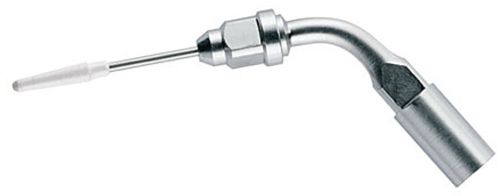
In particular, four inserts for peri-implant mechanical instrumentation mounted on ultrasonic devices (Mectron CombiTouch, Mectron, Carasco, Italy) were analyzed:
  • Acteon Satelec perioSoft (autoclavable carbon composite) (Figure 2);
  • EMS PI insert (autoclavable PEEK) (Figure 3);
  • Mectron Tip IC1 (autoclavable PEEK tip on ICS insert) (Figure 4);
  • Cavitron SofTip (disposable plastic tip) (Figure 5).
For each insert, two healing abutments were instrumented: one machined and one etched.

2.2. Working Protocol

The protocol used in this research was as follows:
  • Screwing of the healing abutment (HA) onto a model reproducing a lower dental arch (Typodont);
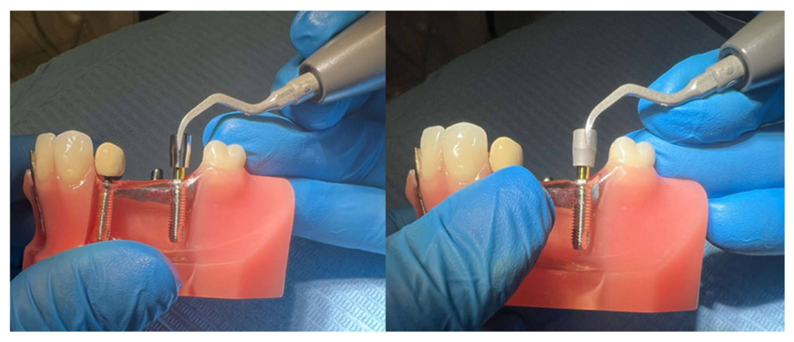
  • Continuous circumferential instrumentation for 1 min over the entire surface of the HA (two HAs with each insert, first the machined and then the etched). The ultrasonic device was set to perio-scaler mode, power 3, irrigation 3 (Figure 6).
  • Immediate, individual, and manipulation-free packaging of all instrumented HAs;
  • Shipping to the DIME (Department of Mechanical, Energy, Management, and Transport Engineering) of the University of Genoa.
The samples for analysis arrived at the laboratory properly packaged and were removed from the bags immediately before analysis, without the need for preparation, thus minimizing handling and contact with the surfaces.
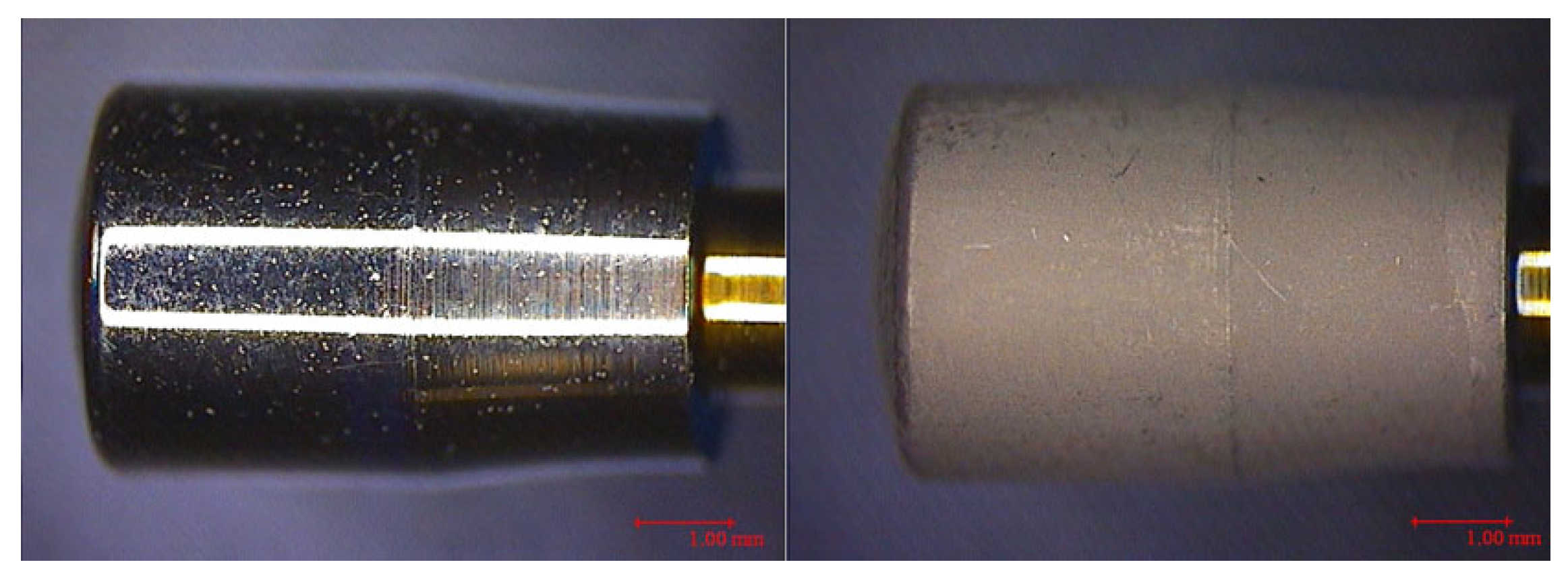
A preliminary analysis of both the machined HAs and an etched HA was performed using a stereoscopic optical microscope (MZ6, Leica, Wetzlar, Germany), obtaining images at 20× magnification.
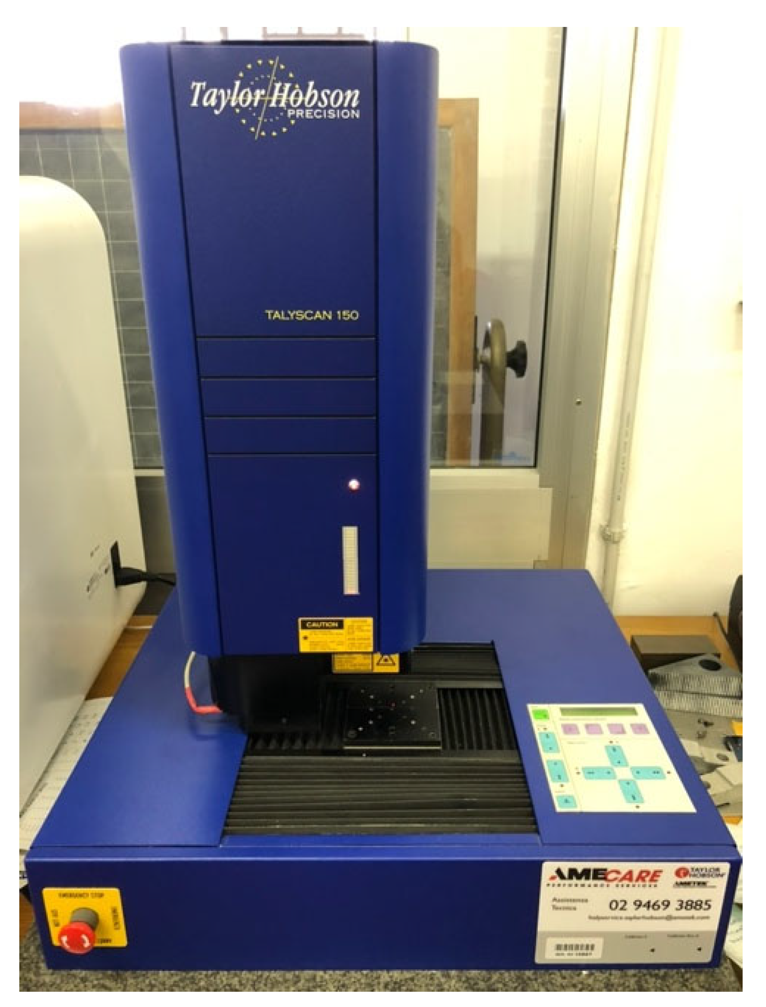
The roughness parameters for evaluating the degree of alteration of the implant surface following instrumentation were detected using the TalyScan 150 profilometer (Taylor Hobson Ltd., Leicester, UK) (Figure 7), a laser roughness meter equipped with automated software that calculates the surface elevation variations of the analyzed material through the reflection of the laser beam on the surface itself; in particular, four different surface portions of each HA were analyzed, detecting a point cloud with an area of 200 × 200 units, spaced 5 microns apart (total area 1 mm × 1 mm).
The roughness parameters detected and analyzed were:
  • Sa (arithmetic mean roughness);
  • Sq (mean square roughness);
  • Sp (maximum peak height);
  • Sv (maximum valley depth);
  • St (Sp + Sv);
  • SSk: Asymmetry of the density function with respect to the mean line, a parameter related to the height distribution according to the following values:
    -
    SSk = 0 (symmetry with respect to the midline, i.e., the same density of valleys and peaks);
    -
    SSk > 0 (deviation below the midline, i.e., a more open profile with a greater density of valleys);
    -
    SSk < 0 (deviation above the midline, i.e., a fuller profile with a greater density of peaks);
  • Sku: Kurtosis of the roughness profile, a parameter related to the sharpest/obtuse shape of the peaks and valleys according to the following values:
    -
    Sku = 3 (normal distribution);
    -
    Sku > 3 (acute height distribution);
    -
    Sku < 3 (obtuse height distribution);
  • Sz (maximum roughness averaged over 5 peaks and 5 valleys).

Data Analysis

An Excel table was created summarizing all the parameters measured on all HAs, i.e., on four surface portions each (Table 1).
Data analysis was performed in collaboration with the DIME Department of the University of Genoa using Design-Expert software (Stat-Ease, Minneapolis, MN, USA).
A number of experiments were conducted following the principle of Design of Experiments, which is a set of statistical techniques aimed at identifying the statistical significance of a set of factors of interest on one or more objective functions.
In this specific case, two factors of interest were identified:
  • Type of surface treatment of the healing abutments;
  • Type of insert used.
For each of these factors, different experimental levels were identified:
  • Two levels for the surface treatment: machined and etched;
  • Five levels for the insert:
    -
    Level 1: Non-instrumented HA (used as a control level);
    -
    Level 2: HA instrumented with Satelec perioSoft;
    -
    Level 3: HA instrumented with EMS PI;
    -
    Level 4: HA instrumented with a Mectron IC1;
    -
    Level 5: HA instrumented with a Cavitron sofTip.
The eight amplitude parameters measured by the profilometer (Sa, Sq, Sp, Sv, St, SSk, Sku, Sz) were considered as output variables (or objective functions).
Researchers began by conducting a two-factor factorial design using four replicates for each experimental combination to investigate the significance of the individual factors analyzed and any interaction (combined effect of the two factors on the experimental response).
The data were then analyzed in two separate designs: one for the machined treatment and one for the etched treatment.
The significance of the factors was determined by analyzing them using an ANOVA test.

3. Results

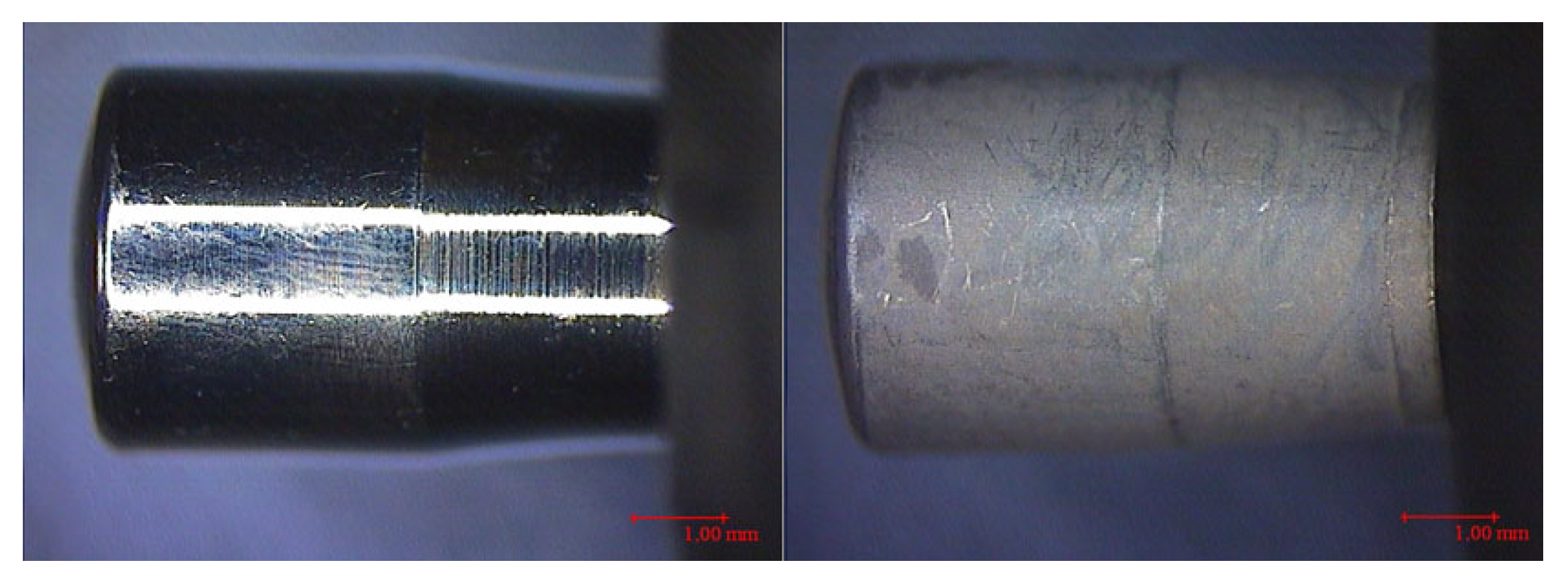
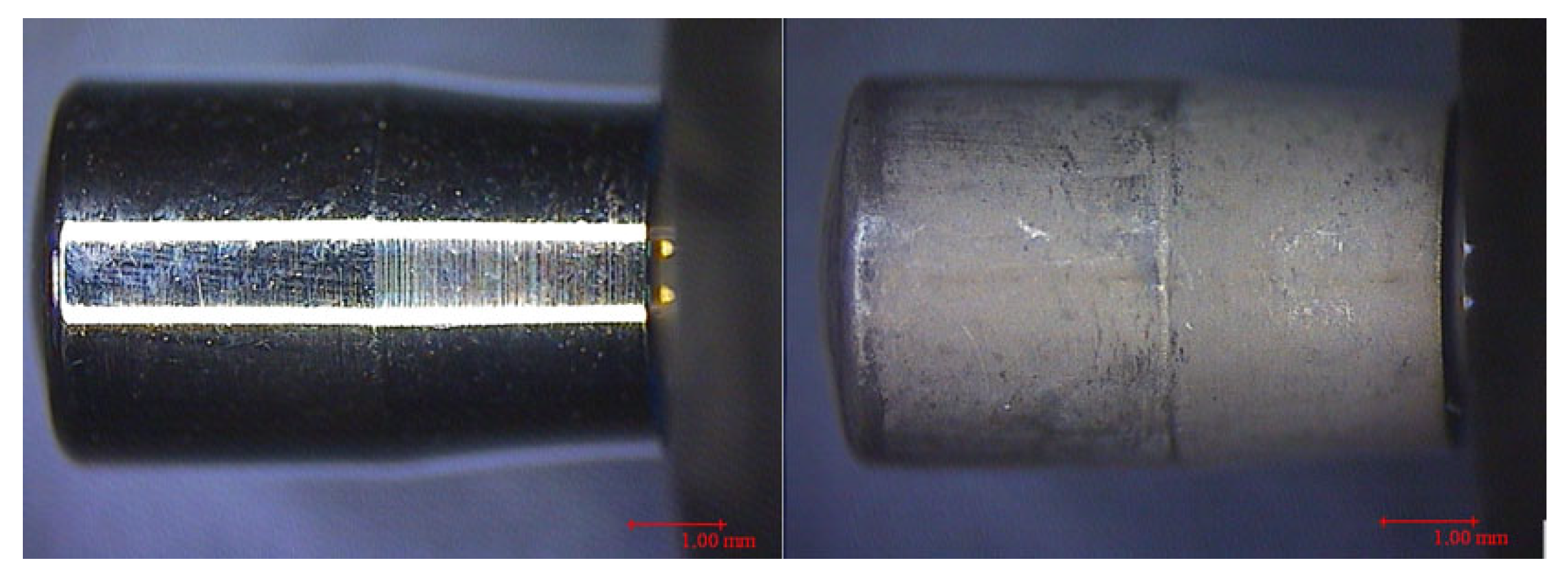
With the preliminary investigation using a stereoscopic optical microscope, the following 20× magnification images of the instrumented and non-instrumented HA were obtained (Figure 8, Figure 9, Figure 10, Figure 11 and Figure 12).
Statistical analysis revealed a significant effect of the two factors of interest (surface treatment and insert) on all the objective functions analyzed (parameters detected by the profilometer) with the exception of the Sp parameter, which was consequently omitted in subsequent study phases.
Among the functions that showed a significant effect of the factors, we focused on the analysis of the parameters Sa and Sq (arithmetic mean and quadratic mean roughness), as they are the most commonly used to define surface roughness.
A similar trend was found for these functions, which is why the results for the Sa objective function only are reported below, comparable to those obtained for Sq.
In the ANOVA table for the Sa objective function (Figure 13), the total variability (Core Total) is divided into variability explained by the factors (Model), a significantly larger share, and variability due to error (Pure Error).
The two-factor ANOVA revealed that surface treatment (machined vs. etched) was the most significant factor (p < 0.0001), accounting for the majority of the variance in Sa values (Figure 13).
In turn, the variability explained by the factors is divided between the two factors analyzed (A—insert; B—surface treatment) and their interaction (AB).
From this two-factor design, it is clear that the treatment factor (B) absorbs the majority of the explained variability; that is, it is the predominant factor in explaining the variability of the objective function under consideration (Sa), followed by the interaction (AB) and finally by the insert factor (A), which therefore present a weak significance compared to the treatment factor.
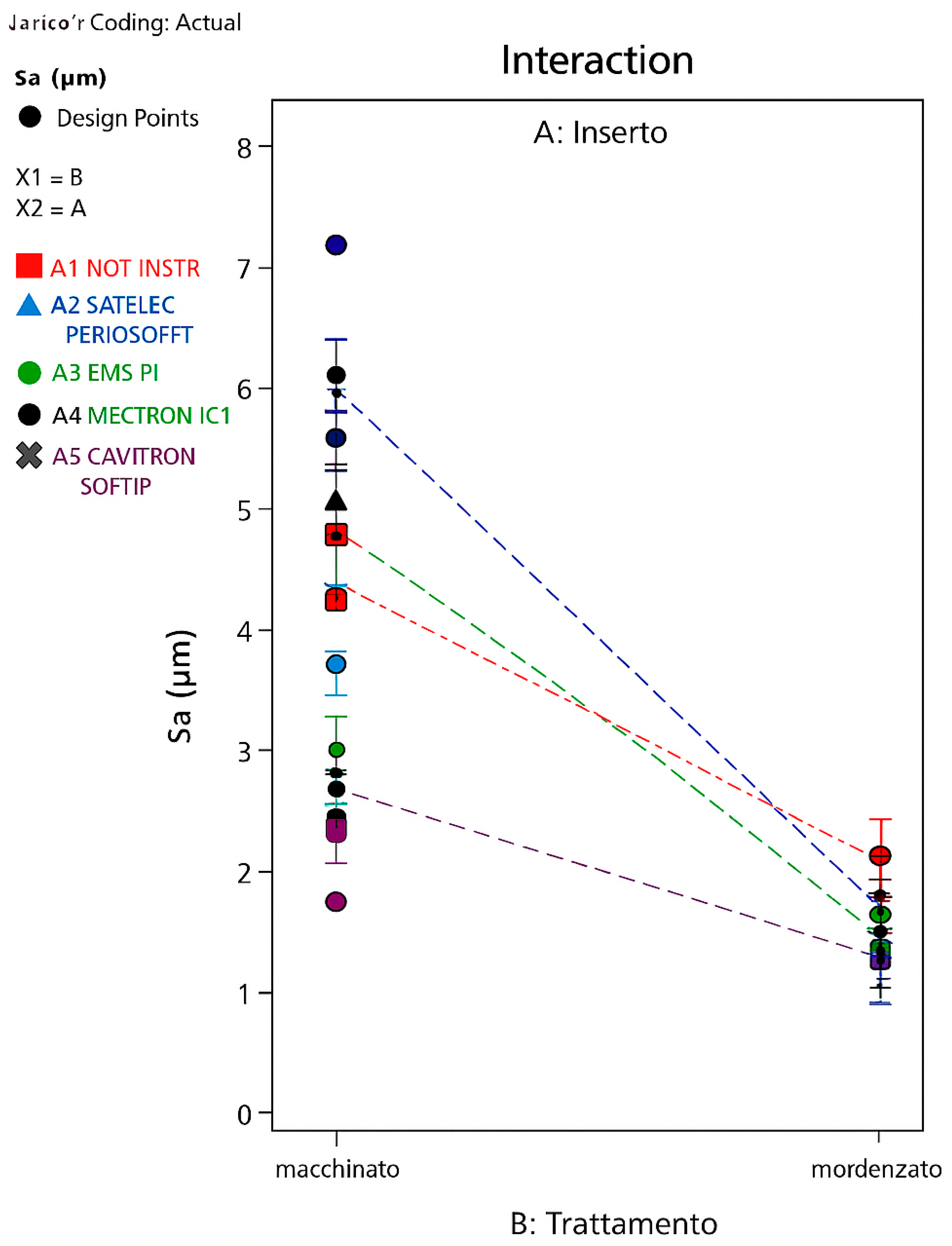
A graph generated by the software (DeltaLog, Genova, Italy) (Figure 14) showing the roughness scale (Sa) on the y-axis and the two levels of the treatment factor on the x-axis shows that the objective function Sa (as is also the case for the Sq parameter) exhibits significantly lower values when switching from machined to etched surface treatment, regardless of whether the HA is instrumented or not and regardless of the insert used for that instrumentation.
Only when using the Cavitron is the roughness of the machined surface made comparable to that of the etched surface; that is, the Cavitron sofTip insert reduces the roughness on the machined surface but not on the etched surface.
Another graph (Figure 15), showing the roughness scale (Sa) on the y-axis and the different levels of the insert factor on the x-axis, allows for a two-by-two comparison of the levels of the factors of interest.
It is therefore evident that the machined treatment (red line) shows a significant variation in roughness at some levels compared to the control level (uninstrumented HA).
In particular, it can be stated that levels 3 (HA instrumented with an EMS PI insert) and 5 (HA instrumented with a Cavitron sofTip insert) show a statistically significant difference compared to the control level, with an increase (the former) and a decrease (the latter) in the Sa objective function, respectively.
As for the etched treatment, however, this graph does not show any variability among the various levels of the insert factor; that is, it does not appear that the various inserts determine a significant variability in the Sa objective function compared to the control level (uninstrumented HA). However, it is important to remember that this graph reports the results of the two-factor design, in which the treatment factor is significantly more significant than the insert factor, which is why its significance could mask that of the less representative factor, namely the insert.
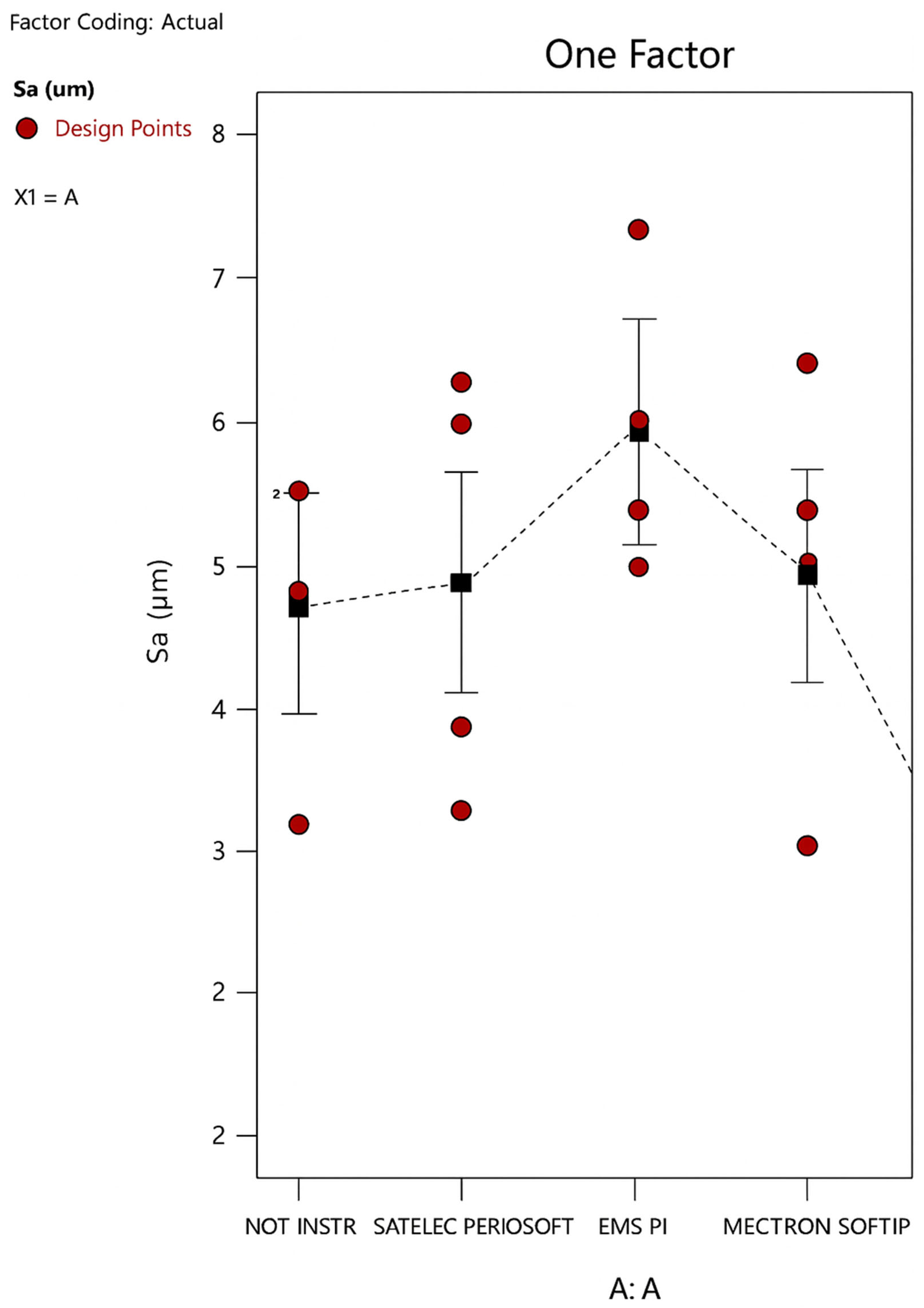
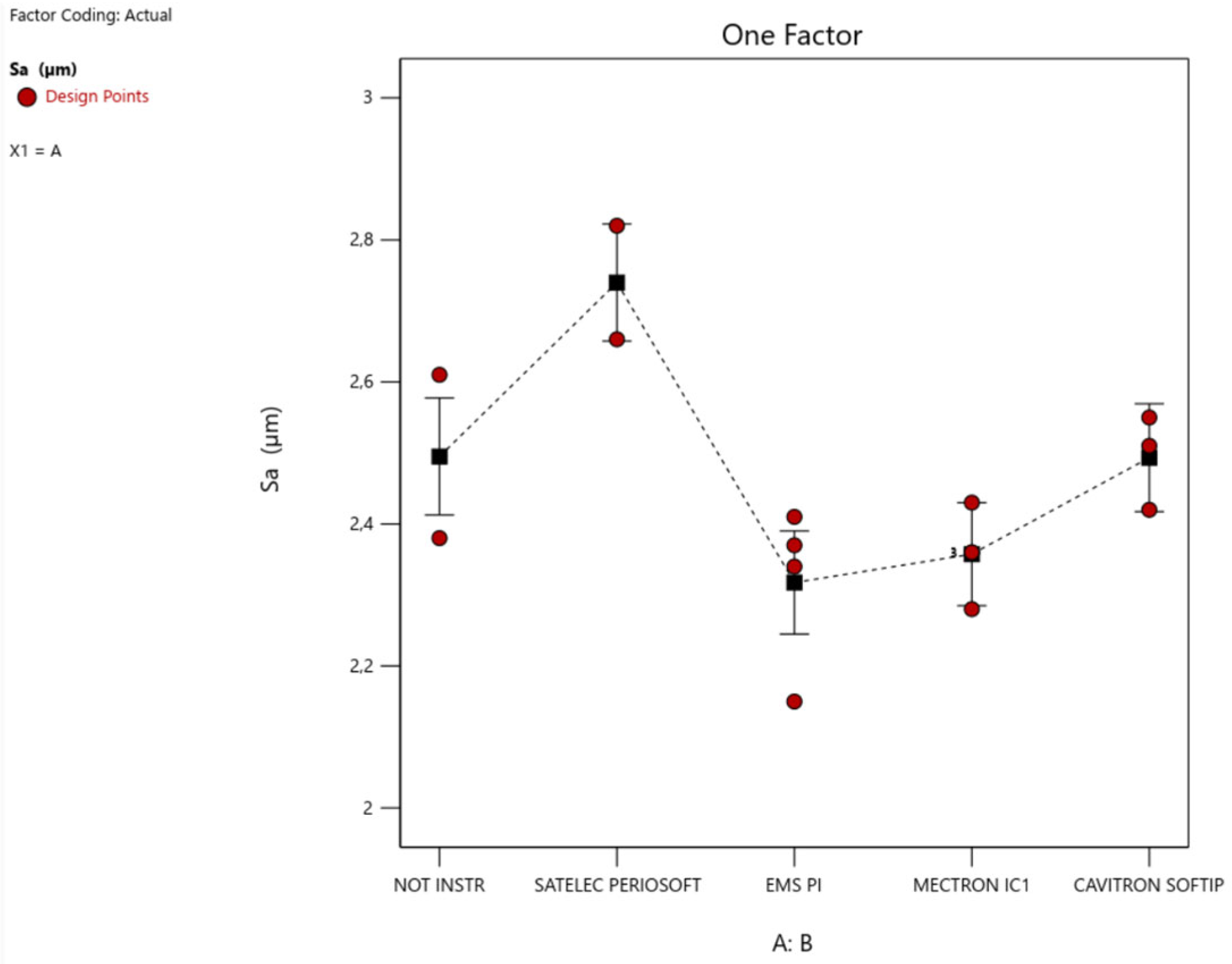
This concept is also evident from the different scales of Sa in the graphs for the two-factor design (approximately 2.5–7.5 microns) compared to those generated based on the single designs (approximately 2.1–2.8 microns (Figure 16 and Figure 17). These graphs, in fact, highlight a greater variability in the Sa parameter with changes in level (i.e., based on the instrumentation insert used) in the machined treatment than in the etched one. In other words, the machined surface is more susceptible to changes in the Sa and Sq parameters than the etched one.
For this reason, it is interesting to show the results obtained from the data analysis using the two separate designs for the machined and etched treatments.
The results of the single project on the machined treatment (Figure 16) confirm the same trend in variability for the two levels highlighted in the two-factor design, i.e., an increase in Sa with the use of EMS PI and a decrease in Sa with the use of Cavitron sofTip.
The significance of the insert factor in the etched treatment emerges in the single-factor design (Figure 17), which highlights:
  • A greater significance of the Satelec level compared to the others, with an increase in Sa; that is, the Satelec perioSoft insert would appear to determine an increase in the surface roughness of an etched HA;
  • A low but still significant significance of the EMS level, with a decrease in Sa; that is, the EMS PI insert would appear to determine a decrease in the surface roughness of an etched HA.

4. Discussion

The results obtained were compared both visually, using profilometer images, and numerically through statistical data analysis.
Preliminary analysis using an optical microscope showed that the uninstrumented HA analyzed originally exhibited turning grooves of uneven depth and imperfections transverse to the turning grooves themselves, or were generated by interference during the industrial manufacturing process. This evidence had already been observed in other similar in vitro studies and provides guidance for a better industrial protocol, aimed at producing surfaces that were as “smooth” and uniform as possible [18].
The study was conducted by an operator who, although experienced, inevitably produced a statistical bias, determined by the variation in inclination and pressure applied to the instruments. On the other hand, the use of an operator for the instrumentation allowed for obtaining conditions, and therefore results, more similar to those that actually characterize the phenomenon studied.
However, it is hoped that in the future, instrumented surfaces will be studied using a mechanical arm, which determines constant movement in terms of amplitude, inclination, and pressure, thus eliminating the human variable, so that the results can be compared with those obtained with an operator’s instrumentation.
It would also be useful to include a titanium insert in a future study, thus completing the range of materials currently on the market and providing a broader overview.
An important future research perspective arises from the need to find a microtopographic parameter suitable for measuring the degree of bacterial biofilm retention on an analyzed surface. Specifically, noting that the parameters considered in this study (Sa and Sq) varied in the opposite way to what was expected, we began to consider the other parameters detected by the profilometer, which, together with Sa and Sq, contribute to describing surface micromorphology.
The value of the SSk parameter, for example, defines a fuller surface profile (negative SSk), meaning one with a greater distribution of ridges compared to valleys, or a more empty surface profile (positive SSk), meaning one with a wider distribution of valleys. It is therefore hypothesized that the latter surface conformation may be more plaque-retentive, as it provides greater protected space (valleys) than the former, which features wider peaks where bacterial biofilm is less retentive and more exposed to the intraoral environment.
Another interesting future research hypothesis is to demonstrate, through chemical analysis of treated surfaces, the release of material particles from some non-metallic inserts.
Specifically, from the data collected in this research, we hypothesize that the two inserts that decreased surface roughness, or rather, may have released particles that filled the voids, resulting in a decrease in the Sa parameter, were:
  • Most significantly, the Cavitron sofTip insert on a machined surface, bringing the Sa parameter to a level comparable to that of an etched surface, which is essentially significantly lower than the machined ones;
  • Less significantly, but still significantly, the EMS PI insert on an etched surface.
We wondered whether this decrease was due to the filling of the wide, deep, long, and parallel valleys typical of the microtopography of machined surfaces in the case of the Cavitron sofTip insert, and the small and numerous valleys typical of the micromorphology of etched surfaces in the case of the EMS PI.
It can be hypothesized that the material composing the EMS PI tip (PEEK) has a greater capacity to modify the implant surface. Conversely, the material of the Cavitron SoftTip (disposable plastic tip) appears to be less aggressive toward the implant surface. This concept contrasts with the idea reported in the literature [19,20,21] that plastic instruments should not be used only on etched surfaces, while their use on machined surfaces is safe.
Although the literature [22,23] attributes greater plaque retention to a higher degree of surface roughness, the machined surface appears to have a greater roughness than the etched surface. We can hypothesize that surface roughness does not appear to be the discriminating parameter between a potentially more plaque-retentive surface and one that is less so, but this statement should be the subject of further in vivo evaluations related to the study of plaque accumulation on implant surfaces exposed in the oral cavity.

5. Conclusions

This research remains to be validated through clinical studies. However, we can draw some conclusions, which we summarize point by point for convenience:
(1)
Surface treatment (machined or etched) affects the variability of the average surface roughness (arithmetic and quadratic) to a much greater extent than the type of insert used for instrumentation.
(2)
Etching the surface after turning significantly decreases the average surface roughness, resulting in an average roughness of the etched surface half that of the machined surface, regardless of instrumentation, with the exception of instrumentation with the Cavitron sofTip, which makes the roughness of the machined surface comparable to that of the etched surface.
(3)
During instrumentation, the machined surface is more susceptible to modifications in terms of Sa/Sq parameters than the etched surface.
(4)
Instrumentation on machined HA significantly changes the average surface roughness only when using EMS PI inserts, which increases the surface roughness, and Cavitron SofTip, which decreases it, bringing it to values similar to those of an etched surface.
(5)
Instrumentation on etched HA significantly changes the average surface roughness only when using EMS PI inserts, which decreases the surface roughness, and Satelec perioSoft, which increases it.
We can hypothesize that clinically, based on the data from this study, the instruments that cause the least alteration to machined surfaces are Satelec PerioSoft and Mectron IC1. Meanwhile, for the treatment of rough surfaces, the least aggressive instrument appears to be the Cavitron SoftTip. Of course, further in vivo studies are needed to support these hypotheses.

Author Contributions

Conceptualization, D.B., F.B., P.G. and J.C.; methodology, D.B.; software, F.B.; validation, D.B. and J.C.; formal analysis, F.B.; investigation, G.M.; resources, D.B. and J.C.; data curation, G.M. and F.B.; writing—original draft preparation, G.M. and J.C.; writing—review and editing, J.C. and G.M.; visualization, F.B.; supervision, D.B.; project administration, J.C.; funding acquisition, D.B. All authors have read and agreed to the published version of the manuscript.

Funding

This research received no external funding.

Institutional Review Board Statement

Not applicable.

Informed Consent Statement

Not applicable.

Data Availability Statement

The research data are available upon request from the corresponding authors.

Conflicts of Interest

The authors declare no conflicts of interest.

References

  1. Butterworth, C.; McCaul, L.; Barclay, C. Restorative dentistry and oral rehabilitation: United Kingdom National Multidisciplinary Guidelines. J. Laryngol. Otol. 2016, 130, S41–S44. [Google Scholar] [CrossRef]
  2. Sanderson, W.C.; Scherbov, S. Prospective Longevity: A New Vision of Population Aging; Harvard University Press: Cambridge, MA, USA, 2019. [Google Scholar]
  3. Chapple, I.L.; Van der Weijden, F.; Doerfer, C.; Herrera, D.; Shapira, L.; Polak, D.; Madianos, P.; Louropoulou, A.; Machtei, E.; Donos, N.; et al. Primary prevention of periodontitis: Managing gingivitis. J. Clin. Periodontol. 2015, 42 (Suppl. S16), S71–S76. [Google Scholar] [CrossRef] [PubMed]
  4. Rajwani, A.R.; Hawes, S.N.D.; To, A.; Quaranta, A.; Rincon Aguilar, J.C. Effectiveness of Manual Toothbrushing Techniques on Plaque and Gingivitis: A Systematic Review. Oral Health Prev. Dent. 2020, 18, 843–854. [Google Scholar] [CrossRef] [PubMed] [PubMed Central]
  5. Sälzer, S.; Graetz, C.; Dörfer, C.E.; Slot, D.E.; Van der Weijden, F.A. Contemporary practices for mechanical oral hygiene to prevent periodontal disease. Periodontology 2020, 84, 35–44. [Google Scholar] [CrossRef] [PubMed]
  6. Baldi, D.; De Giorgis, L.; Menini, M.; Motta, F.; Colombo, J. Efficacy of Instruments for Professional Oral Hygiene on Dental Implants: A Systematic Review. Appl. Sci. 2022, 12, 26. [Google Scholar] [CrossRef]
  7. Abushahba, F.; Algahawi, A.; Areid, N.; Vallittu, P.K.; Närhi, T. Efficacy of biofilm decontamination methods of dental implant surfaces: A systematic review of in vitro studies. Eur. J. Oral Sci. 2025, 133, e70005. [Google Scholar] [CrossRef] [PubMed]
  8. Baldi, D.; Colombo, J.; Verardi, S.; Rebaudi, A.; Rebaudi, F.; Makary, C. Clinical osseointegration of bone level implants with conical shape and textured surface with low primary stability. Minerva Stomatol. 2020, 69, 8–13. [Google Scholar] [CrossRef] [PubMed]
  9. Alexander, R.; Liu, X. Soft tissue integration around dental implants: A pressing priority. Biomaterials 2026, 324, 123491. [Google Scholar] [CrossRef] [PubMed] [PubMed Central]
  10. Fiorellini, J.P.; Mojaver, S.; Sarmiento, H.; Aghaloo, T. Clinical Translation of the 2024 AO/AAP Consensus on Prevention and Management of Peri-implant Diseases and Conditions. Int. J. Periodontics Restor. Dent. 2025, 45, S1–S23. [Google Scholar] [CrossRef] [PubMed]
  11. Baldi, D.; Colombo, J.; Gavoglio, P.; De Giorgis, L.; Motta, F.; Lugas, A.; Lertora, E.; Schierano, G. Roughness and SEM Analysis of Manual and Ultrasonic Instrumentation over Different Crown Materials for Dental Implants Restorations. Materials 2022, 15, 1159. [Google Scholar] [CrossRef] [PubMed] [PubMed Central]
  12. Kim, Y.J.; Song, Y.W.; Park, S.Y.; Cha, J.K.; Lee, H.J.; Yang, S.M.; Park, J.B.; Koo, K.T. Current understanding of the etiology, diagnosis, treatment, and management of peri-implant diseases: A narrative review for the consensus report of the Korean Academy of Periodontology. J. Periodontal Implant. Sci. 2024, 54, 377–392. [Google Scholar] [CrossRef] [PubMed] [PubMed Central]
  13. Romito, G.A.; Hassan, M.A.; do Amaral, G.C.L.S.; Villar, C.C. Decision-making on peri-implant mucositis management and treatment approaches. Br. Dent. J. 2024, 236, 797–801. [Google Scholar] [CrossRef] [PubMed]
  14. Maiorani, C.; Butera, A.; Pérez-Albacete Martínez, C.; Pascadopoli, M.; Sabatini, S.; Nardi, G.M.; Scribante, A. Effectiveness of Erythritol-Based Air Polishing and Ultrasonic Instrumentation with PEEK Inserts in Peri-Implant Maintenance: A Randomized Clinical Trial Including Different Prosthetic Materials. Dent. J. 2025, 13, 235. [Google Scholar] [CrossRef] [PubMed] [PubMed Central]
  15. Iommiello, A.M.; Pesce, P.; Sanavia, C.; Arata, F.; Caponio, V.C.; Baldi, D.; Migliorati, M.; Menini, M. The use of water flosser in implant-supported fixed prosthesis: A narrative review and the opinion of Italian dental hygienists. Minerva Dent. Oral Sci. 2025, 74, 257–265. [Google Scholar] [CrossRef] [PubMed]
  16. Delucchi, F.; Ingegnieros, L.; Pesce, P.; Baldi, D.; Canullo, L.; Bagnasco, F.; Zunino, P.; Menini, M. Efficacy and safety of erythritol air-polishing in implant dentistry: A systematic review. Int. J. Dent. Hyg. 2024, 23, 44–62. [Google Scholar] [CrossRef] [PubMed]
  17. Taymour, N.; Abd El-Fattah, A.; Kandil, S.; Fahmy, A.E.; Al-Qahtani, N.H.; Khaled, A.; Al-Dulaijan, Y.A.; Gepreel, M.A.-H. Revolutionizing Dental Polymers: The Versatility and Future Potential of Polyetheretherketone in Restorative Dentistry. Polymers 2024, 17, 80. [Google Scholar] [CrossRef]
  18. Ozgu, I.; Ustun, K. Effects of Mechanical Methods Used in Peri-implantitis Treatment on Implant Surface Decontamination and Roughness. J. Vis. Exp. 2025, 217, e67778. [Google Scholar] [CrossRef]
  19. Hempton, T.; Lancaster, D.; Pechter, J. Implant Maintenance; Technique and tools for effective debridement of artificial anatomy. Dimens. Dent. Hyg. 2011, 9, 58–61. [Google Scholar]
  20. Pattison, A.M.; Sumi, J.Y. Post-surgical implant care. Dimens. Dent. Hyg. 2015, 13, 24–29. [Google Scholar]
  21. Salvi, G.E.; Persson, G.R.; Heitz-Mayfield, L.J.; Frei, M.; Lang, N.P. Adjunctive local antibiotic therapy in the treatment of peri-implantitis II: Clinical and radiographic outcomes. Clin. Oral Implant. Res. 2007, 18, 281–285. [Google Scholar] [CrossRef] [PubMed]
  22. Baldi, D.; Menini, M.; Pera, F.; Ravera, G.; Pera, P. Plaque accumulation on exposed titanium surfaces and peri-implant tissue behavior. A preliminary 1-year clinical study. Int. J. Prosthodont. 2009, 22, 447–455. [Google Scholar] [PubMed]
  23. Blasi, A.; Iorio-Siciliano, V.; Pacenza, C.; Pomingi, F.; Matarasso, S.; Rasperini, G. Biofilm removal from implants supported restoration using different instruments: A 6-month comparative multicenter clinical study. Clin. Oral Implant. Res. 2014, 27, e68–e73. [Google Scholar] [CrossRef] [PubMed]
Figure 1. Simulation typodont with smooth and etched healing abutments.
Applsci 15 12598 g001
Figure 2. Acteon Satelec perioSoft.
Applsci 15 12598 g002
Figure 3. EMS PI insert.
Applsci 15 12598 g003
Figure 4. Mectron Tip IC1.
Applsci 15 12598 g004
Figure 5. Cavitron SofTip.
Applsci 15 12598 g005
Figure 6. Subsequent instrumentation of the machined and etched healing abutments.
Applsci 15 12598 g006
Figure 7. Talyscan 150 Profilometer.
Applsci 15 12598 g007
Figure 8. Machined (left) and etched (right) non-instrumented healing abutments.
Applsci 15 12598 g008
Figure 9. Machined (left) and etched (right) Has instrumented with Satelec perioSoft.
Applsci 15 12598 g009
Figure 10. Machined (left) and etched (right) HAs instrumented with EMS PI.
Applsci 15 12598 g010
Figure 11. Machined (left) and etched (right) HAs instrumented with Mectron IC1.
Applsci 15 12598 g011
Figure 12. Machined (left) and etched (right) HAs instrumented with Cavitron SofTip.
Applsci 15 12598 g012
Figure 13. ANOVA table referring to the objective function Sa (2-factor design).
Applsci 15 12598 g013
Figure 14. Graph showing the roughness scale (Sa) on the y-axis and the two levels of the surface treatment factor on the x-axis (machined and etched).
Applsci 15 12598 g014
Figure 15. Graph of Sa behavior in the two-factor design.
Applsci 15 12598 g015
Figure 16. Behavior of Sa in the single-factor machined design.
Applsci 15 12598 g016
Figure 17. Behavior of Sa in the single-factor etched design.
Applsci 15 12598 g017
Table 1. Roughness parameters measured on all HAs surfaces.
Healing Abutment (HA) CODE SURFACES PARAMETERS
Sa (µm) Sq (µm) Sp (µm) Sv (µm) St (µm) SSk Sku Sz (µm)
HA machined not instrumented15.667.5215.224.439.6−1.13.9937.9
25.056.8419.720.740.5−0.2733.6939.2
35.687.672223.545.5−0.1793.4843.7
43.294.491216.428.4−0.6584.0127.4
HA etched not instrumented12.613.313.914.328.20.03853.225.8
23.083.9424.314.939.20.9055.123.9
32.974.0728.113.341.40.8997.3728.6
42.382.959.6412.321.9−0.1642.8721.1
HA machined SATELEC PERIOSOFTAL16.468.8625.831.457.1−0.8994.4855.9
23.564.6916.116.232.3−0.1443.4629.6
36.177.7519.825.2450.2222.7340.1
44.115.2812.617.430−0.783.3828.9
HA etched SATELEC PERIOSOFTAM12.012.5711.211.722.80.09593.4320.3
22.663.4522.213.735.80.3054.4530.8
35.736.7121.218.940.1−0.1182.0735.5
42.823.5917.91633.90.133.4229.4
HA machined EMS PIBL15.247.1213.62538.5−1.294.5837
27.4310.324.934.259.1−0.9274.2454.8
35.67.3513.525.939.4−1.184.3637.2
46.198.6422.332.254.4−0.6874.4150.4
HA etched EMS PIBM12.413.0210.614.525.1−0.001483.0422.8
22.342.9713.312.125.40.1623.2623.2
32.152.7310.914.124.90.01393.3221.9
42.373.0111.91223.9−0.1953.1722
HA machined MECTRON IC1CL15.247.251925.244.1−1.014.5340
25.67.1815.737.853.4−0.7213.2634.4
36.68.429.425.454.8−0.2582.8548.6
43.093.9719.215.734.9−0.002883.5232
HA etched MECTRON IC1CM12.362.9614.413.628.10.08493.123.2
22.282.8412.310.422.70.05553.0420.1
32.362.9711.813.725.50.008163.0722.7
42.433.0411.611.4230.06682.9521.8
HA machined CAVITRON SOFTIPDL12.483.1413.217.230.40.02743.3623.1
23.113.9220.714.134.70.2453.4828.6
33.234.1118.821.240−0.0673.534.5
44.556.420.122.542.7−0.3774.1739.6
HA etched CAVITRON SOFTIPDM12.513.1412.212.624.8−0.1282.9822.4
22.423.0613.312.625.90.3343.1821.3
32.032.5910.610.721.40.2093.2719.3
42.553.1411.510.622.10.1612.7821
Disclaimer/Publisher’s Note: The statements, opinions and data contained in all publications are solely those of the individual author(s) and contributor(s) and not of MDPI and/or the editor(s). MDPI and/or the editor(s) disclaim responsibility for any injury to people or property resulting from any ideas, methods, instructions or products referred to in the content.