Article

Radiological and Neuroradiological Features in Pediatric Mucopolysaccharidoses: A Retrospective Case Series from the Emilia-Romagna Regional Referral Center

1
Specialty School of Pediatrics, Alma Mater Studiorum—University of Bologna, 40126 Bologna, Italy
2
Pediatric Unit, IRCCS Azienda Ospedaliero-Universitaria di Bologna, 40138 Bologna, Italy
3
Department of Medical and Surgical Sciences, Alma Mater Studiorum—University of Bologna, Via Massarenti, 40126 Bologna, Italy
4
Pediatric and Adult Cardiothoracic and Vascular, Oncohematologic and Emergency Radiology Unit, IRCCS Azienda Ospedaliero-Universitaria di Bologna, 40138 Bologna, Italy
5
Neuroradiology Unit, IRCCS Istituto Delle Scienze Neurologiche di Bologna, 40138 Bologna, Italy
6
Pediatrics and Neonatology Unit, Guglielmo da Saliceto Hospital, 29121 Piacenza, Italy
7
Department of Medicine and Surgery, University of Parma, 43126 Parma, Italy
*
Authors to whom correspondence should be addressed.
Appl. Sci. 2025, 15(16), 9093; https://doi.org/10.3390/app15169093
Submission received: 15 July 2025 / Revised: 6 August 2025 / Accepted: 14 August 2025 / Published: 18 August 2025

Abstract

Background: Mucopolysaccharidoses (MPSs) are rare lysosomal storage disorders characterized by multisystem involvement; notably, skeletal abnormalities known as dysostosis multiplex and varying degrees of central nervous system impairment. Accurate radiological evaluation is crucial for accurate diagnosis and effective planning. This study aims to describe the clinical and radiological features of patients with MPS managed at our tertiary care center. Methods: We retrospectively reviewed clinical and radiological data from eight patients with confirmed MPS treated at S. Orsola University Hospital (Bologna, Italy) since 2000. Imaging included conventional radiography, supplemented by MRI and CT. The findings were analyzed by MPS subtype and correlated with clinical evolution and therapeutic interventions. A literature review complemented the analysis. Results: The cohort included one patient with MPS I, two with MPS II, one with MPS III, and four with MPS IV. Common skeletal findings were vertebral deformities, hip dysplasia, and shortening of long bones. Patients with MPS IV showed the most severe bone involvement, including pronounced platyspondyly and odontoid hypoplasia. Follow-up imaging demonstrated progression of bone and CNS pathology despite enzyme replacement therapy (ERT). Conclusions: Our findings underscore the pivotal role of imaging in MPS management. Tailored radiological protocols and multidisciplinary care are crucial for optimizing diagnosis and monitoring disease progression.

1. Introduction

Mucopolysaccharidoses (MPSs) are a group of rare inherited lysosomal storage disorders caused by mutations in genes encoding various lysosomal hydrolases involved in the breakdown of glycosaminoglycans (GAGs) [1]. These enzymatic defects lead to intracellular accumulation of partially undegraded GAGs, resulting in cellular dysfunction and subsequent multisystemic damage. Seven distinct clinical forms of MPS, along with numerous subtypes, have been described [2]. Although MPSs are considered a heterogeneous category, some key clinical features are shared by most types, including coarse facial features, growth failure, inguinal or abdominal hernias, recurrent upper respiratory tract infections, heart valve disease, and neurological involvement. The different forms of MPS also exhibit significant musculoskeletal involvement, likely due to the progressive accumulation of partially degraded GAGs in connective tissue cells, resulting in subsequent tissue inflammation. This phenomenon affects the joints, leading to stiffness and contractures in nearly all forms; the growth plates, causing disproportional short stature; and the articular cartilages, resulting in a wide range of skeletal deformities commonly referred to as “dysostosis multiplex” [2,3].
The main objective of this article is to describe the skeletal and neuroradiological abnormalities found in our cohort of patients in the Regional Reference Clinical Center for Newborn Screening of Endocrine and Metabolic Diseases at the S. Orsola University Hospital in Bologna (Italy). Moreover, we evaluate whether our results are consistent with those described in the literature. In particular, this study aims to identify early radiological and neuroradiological signs suggestive of mucopolysaccharidosis in pediatric patients, in order to support earlier clinical recognition and timely referral to specialized care.

2. Materials and Methods

This study is a single-center, retrospective descriptive analysis conducted at the Regional Reference Clinical Center for Newborn Screening of Endocrine and Metabolic Diseases at the S. Orsola University Hospital in Bologna (Italy).
We conducted a retrospective analysis of the medical and radiological records of all patients with confirmed MPS diagnosed and treated in our center since January 2000, without applying exclusion criteria based on the extent or completeness of radiological documentation. This approach was intended to reflect the real-world clinical heterogeneity of MPS presentations and diagnostic workups. In cases where certain imaging modalities were not performed (e.g., neuroimaging in patient 6), the decision was based on clinical considerations, as detailed in the Section 3 and Section 4.
All patients underwent standard bone radiological examinations, which included anteroposterior and lateral cranial bone position, full spinal anteroposterior and lateral position (including cervical hyperflexion and hyperextension position), thorax anteroposterior position, pelvic anteroposterior position, hand anteroposterior position, and full-length anteroposterior position of both lower extremities. In patient 6, only total body radiographs performed at an adult age were available. Radiological assessments were eventually followed by MRI and CT scans. The radiological findings were classified according to the MPS subtype present in each patient.
Clinical data were analyzed to correlate skeletal and neuroradiological abnormalities with disease progression and the therapeutic interventions the patients had received.
In parallel, we conducted a narrative review of the literature on the radiological manifestations of MPS.

3. Results

We included in the study eight pediatric patients with diagnosis of MPS (five male, three female; range of age at diagnosis 1–17 years, with average age at diagnosis of 5 years and 11 months) distributed as follows: one patient with MPS I, two patients with MPS II, one patient with MPS III, and four patients with MPS IV.
The clinical and therapeutic characteristics for each diagnostic group of patients are reported in Table 1.
Short stature was observed in all patients (shown in Table 1), with a variable degree of severity (ranging from −10.61 to −0.94 SDS), even among individuals with the same MPS subtype.
Radiographic examination revealed skeletal abnormalities consistent with “dysostosis multiplex” in all patients (Table 2).
The patients in the cohort also showed neuroradiological abnormalities at both the cerebral and spinal levels. The main findings are summarized in Table 3.

3.1. MPS I

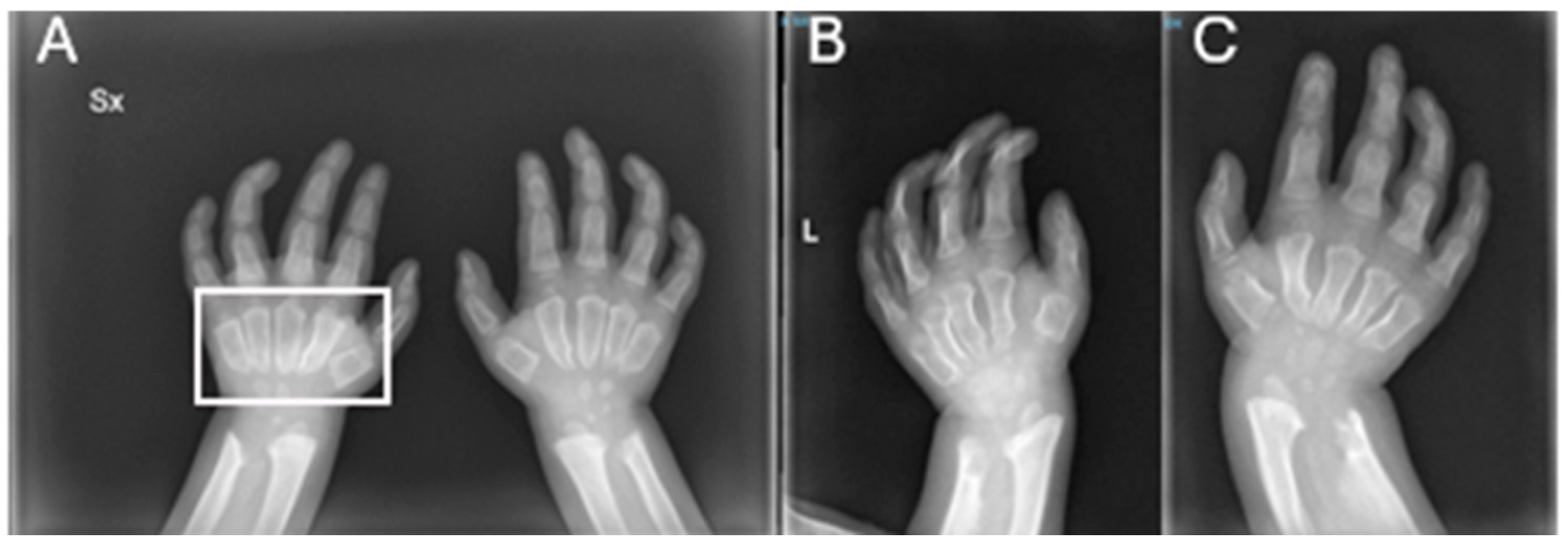
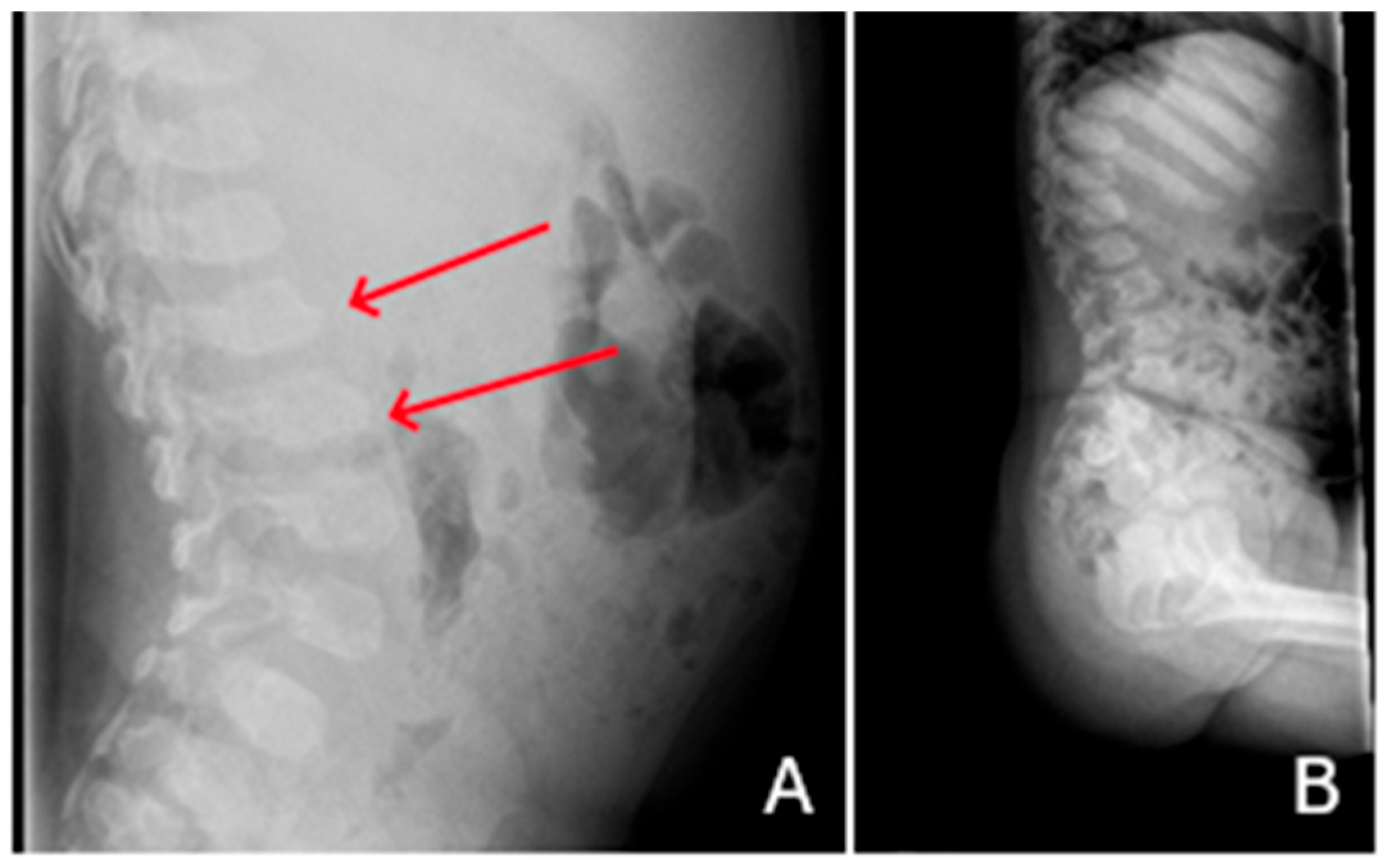
Patient 1, affected by a moderate form of MPS I (Hurler–Scheie), exhibited several bone abnormalities in the cranium, thorax, spine, ribs, long bones, pelvis, and extremities. Concerning the skull, the patient exhibited a ‘J-shaped’ sella turcica, macrocephaly, and dolicocephaly (Figure 1). One of the first clinical signs to appear was hip dysplasia (Figure 2A,B), with an onset within the first year of life. Hands also appeared dysmorphic; in particular, metacarpal bones were short, widened, with a tapered proximal end (Figure 3). Spinal abnormalities included mild kyphosis and infero-anterior beaking of thoracolumbar vertebrae (Figure 4A), which were documented even at the follow-up X-ray, taken after three years of ERT (Figure 4B).
The progression of skeletal lesions affecting the limbs, spine, and pelvis, despite the early initiation of ERT, is summarized in Table 4.
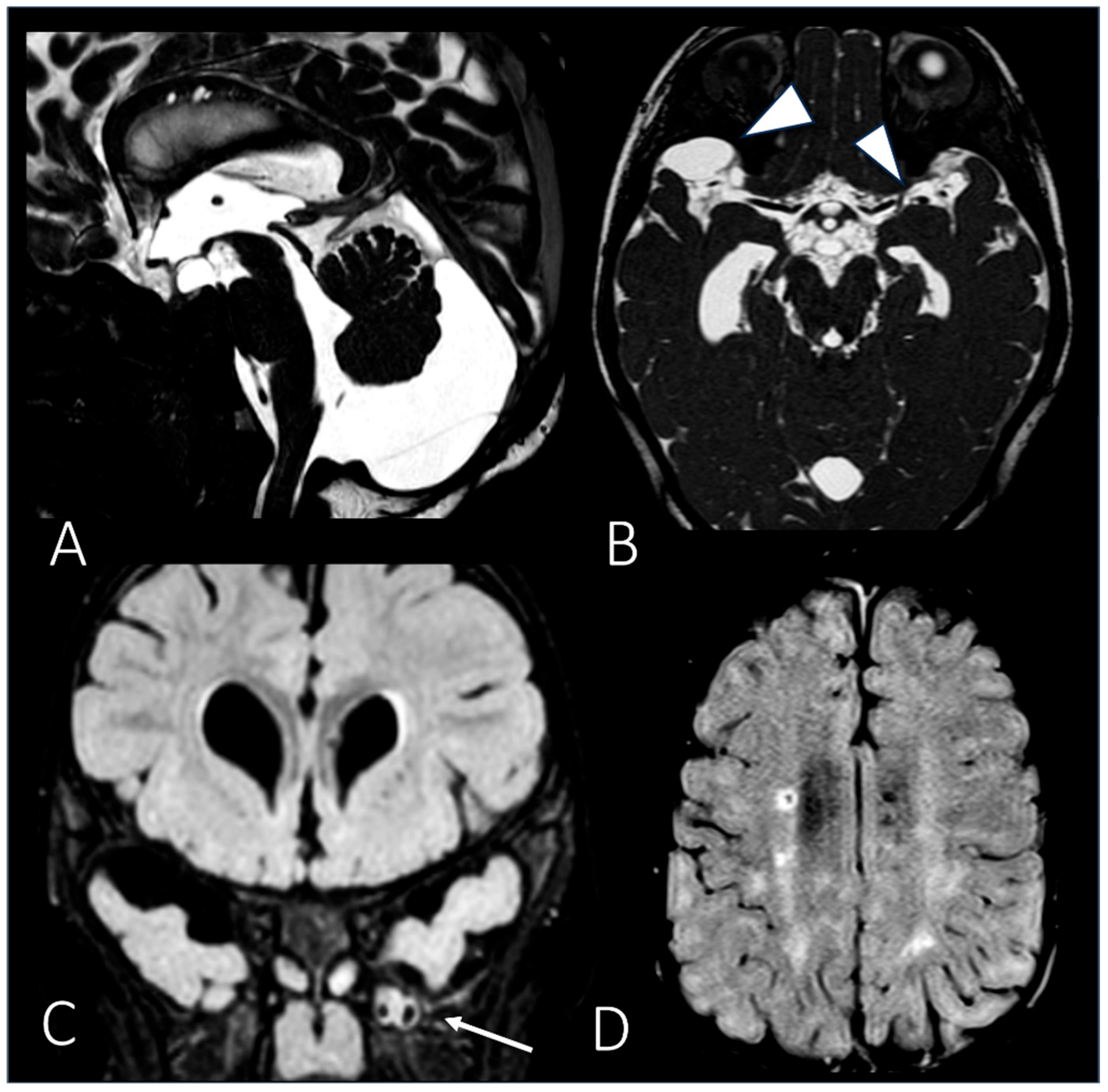
Brain MRI performed at 3 years of age revealed mild ventriculomegaly, mild white matter abnormalities, brain atrophy, enlargement of perivascular spaces, and arachnoid cysts (Figure 5A–D).
We present the skeletal radiological images of a female patient with MPS-1 (patient 1) at diagnosis and during the post-therapy follow-up.

3.2. MPS II

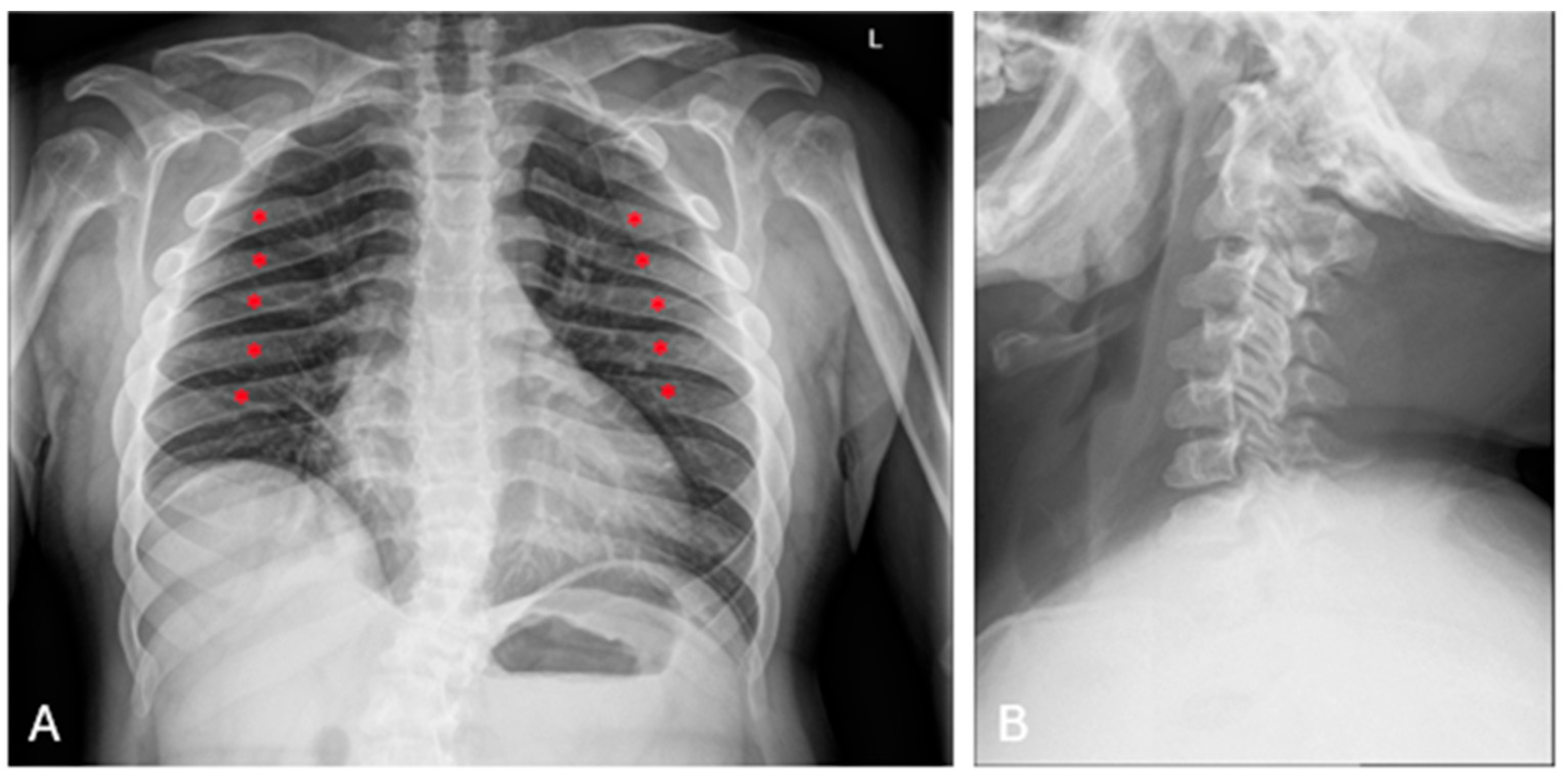
In both our patients affected by MPS II, dysostosis involved different major skeletal segments. They both displayed vertebral anomalies, specifically anterior beaking of vertebral bodies (Figure 6B), hip dysplasia, abnormal modeling of the long bones and short stature. Patient 2 initially exhibited overgrowth during early childhood, followed by a progressive decline in growth velocity, ultimately resulting in a final state of short stature. In the same patient, radiography also showed J-shaped sella turcica and kyphosis.
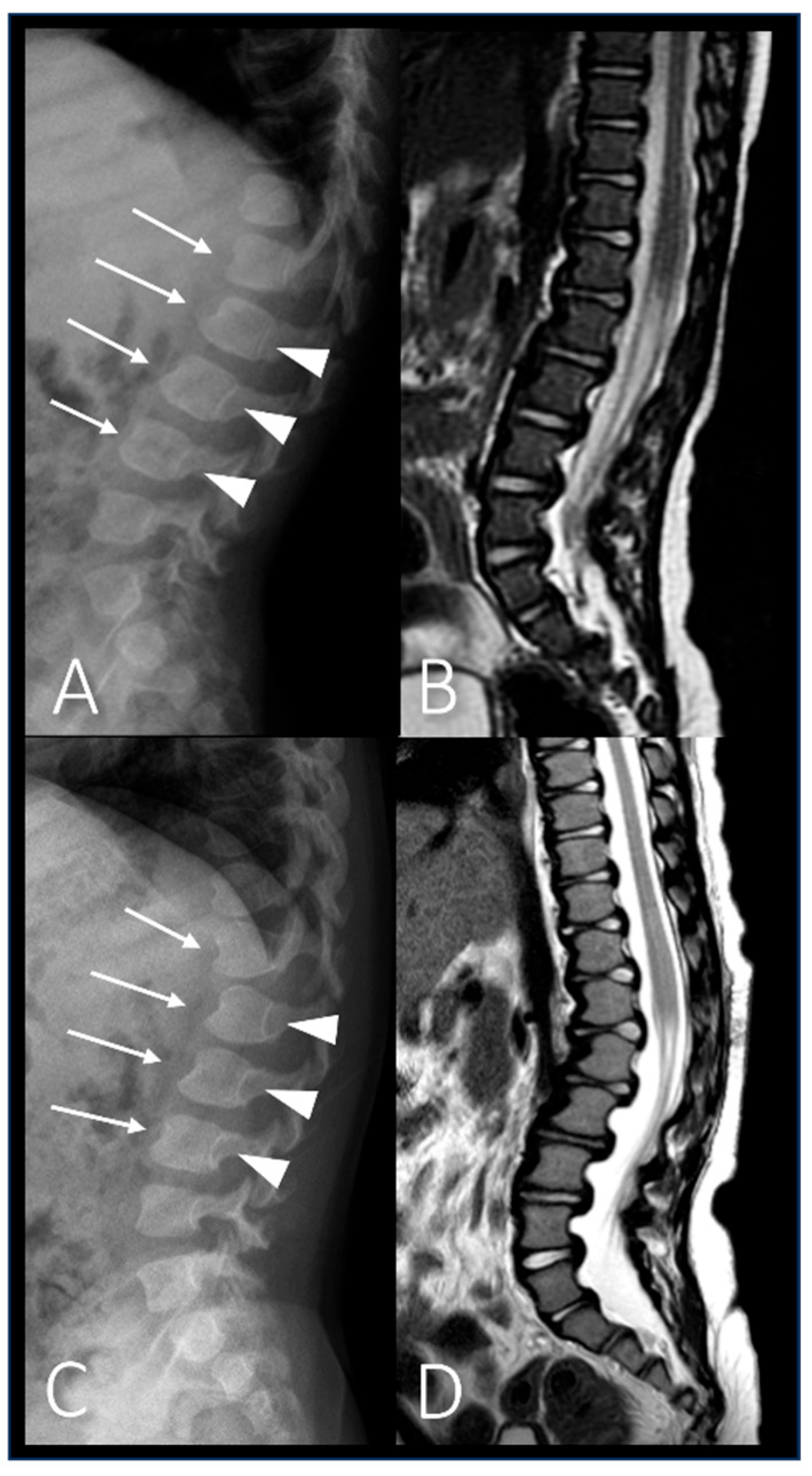
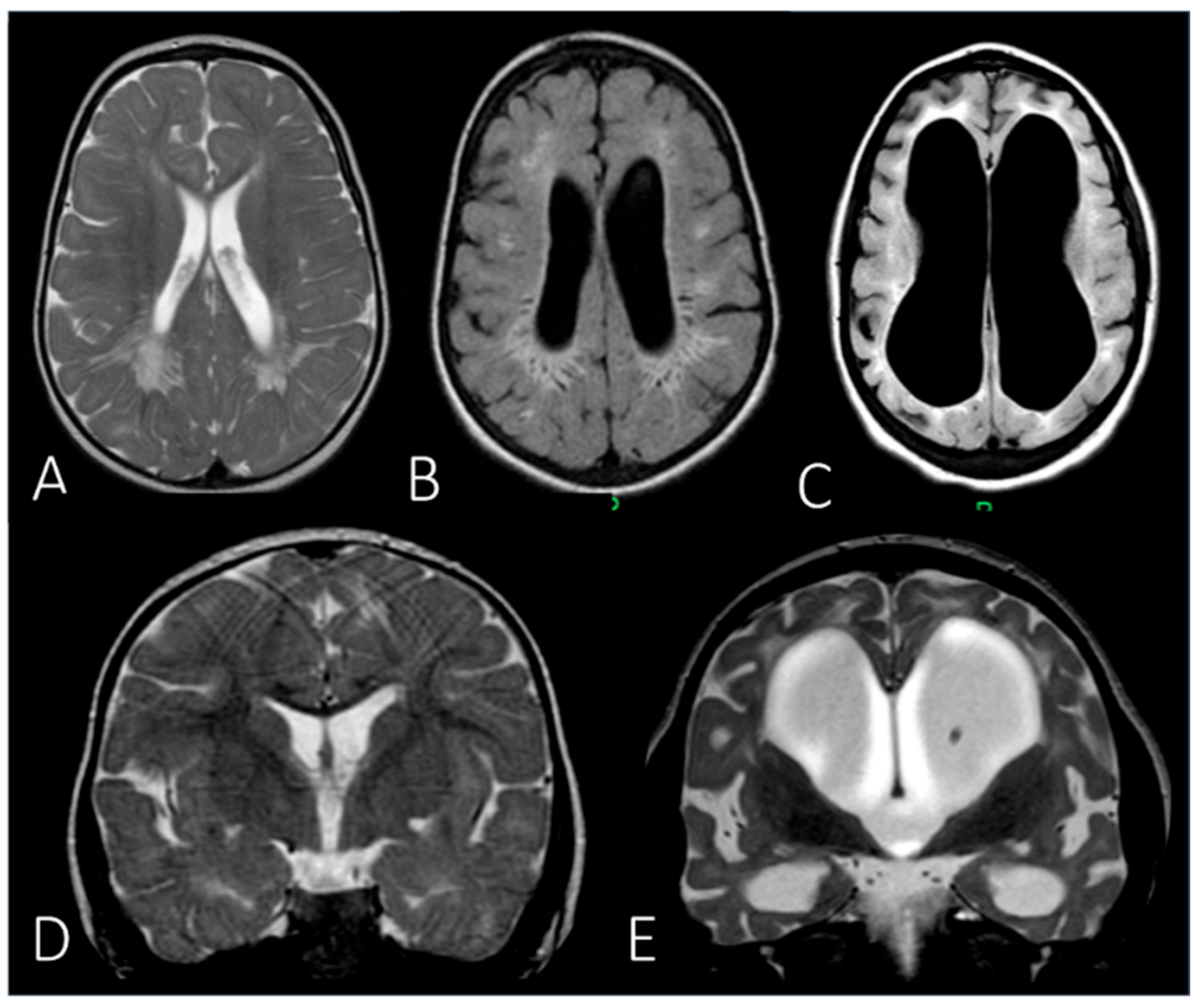
Spinal X-ray (Figure 7A,C), spinal MRI (Figure 7B,D), and brain MRI (Figure 8) were conducted at two different time points: at the age of one year, approximately corresponding to the time of diagnosis, and after 6 years of treatment with Elaprase. The progression of tissue damage in patient 2 is summarized in Table 4.
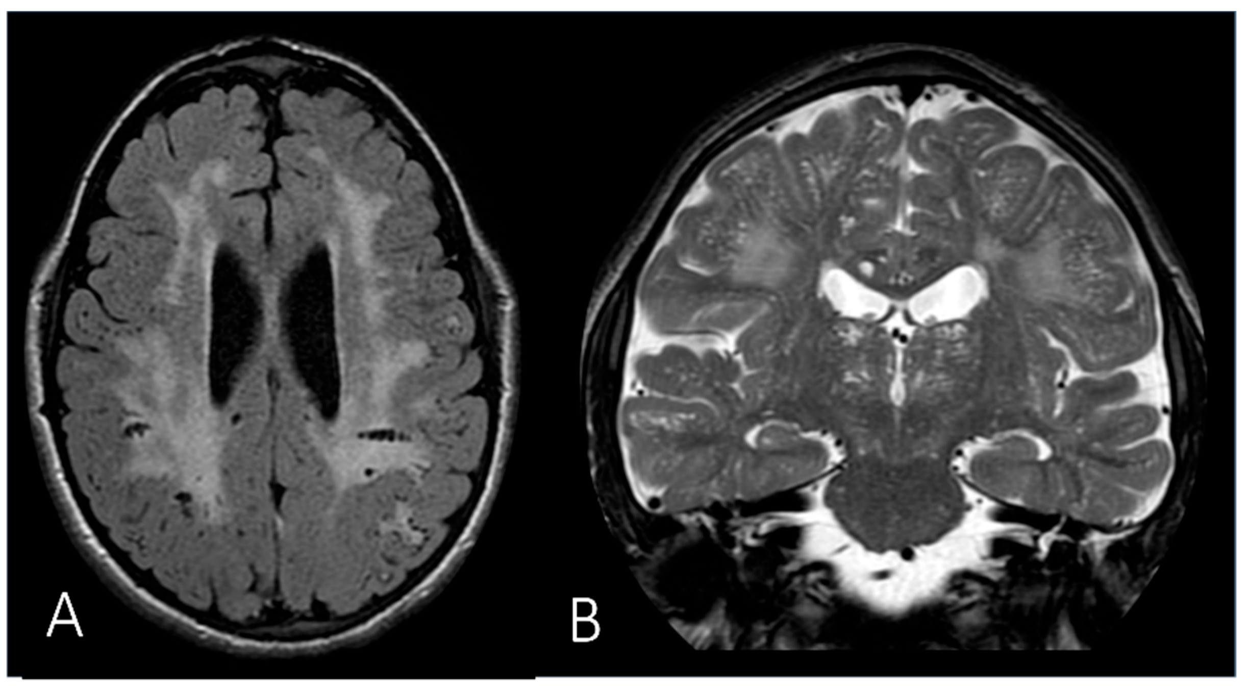
‘Oar-shaped’ ribs and scoliosis (Figure 6A) were found only in patient 3. In the same patient, a brain MRI (Figure 9) revealed enlarged perivascular spaces (PVSs) involving the cortico-subcortical junction, the basal ganglia, and the thalamus, as well as alterations in white matter signal.

3.3. MPS III

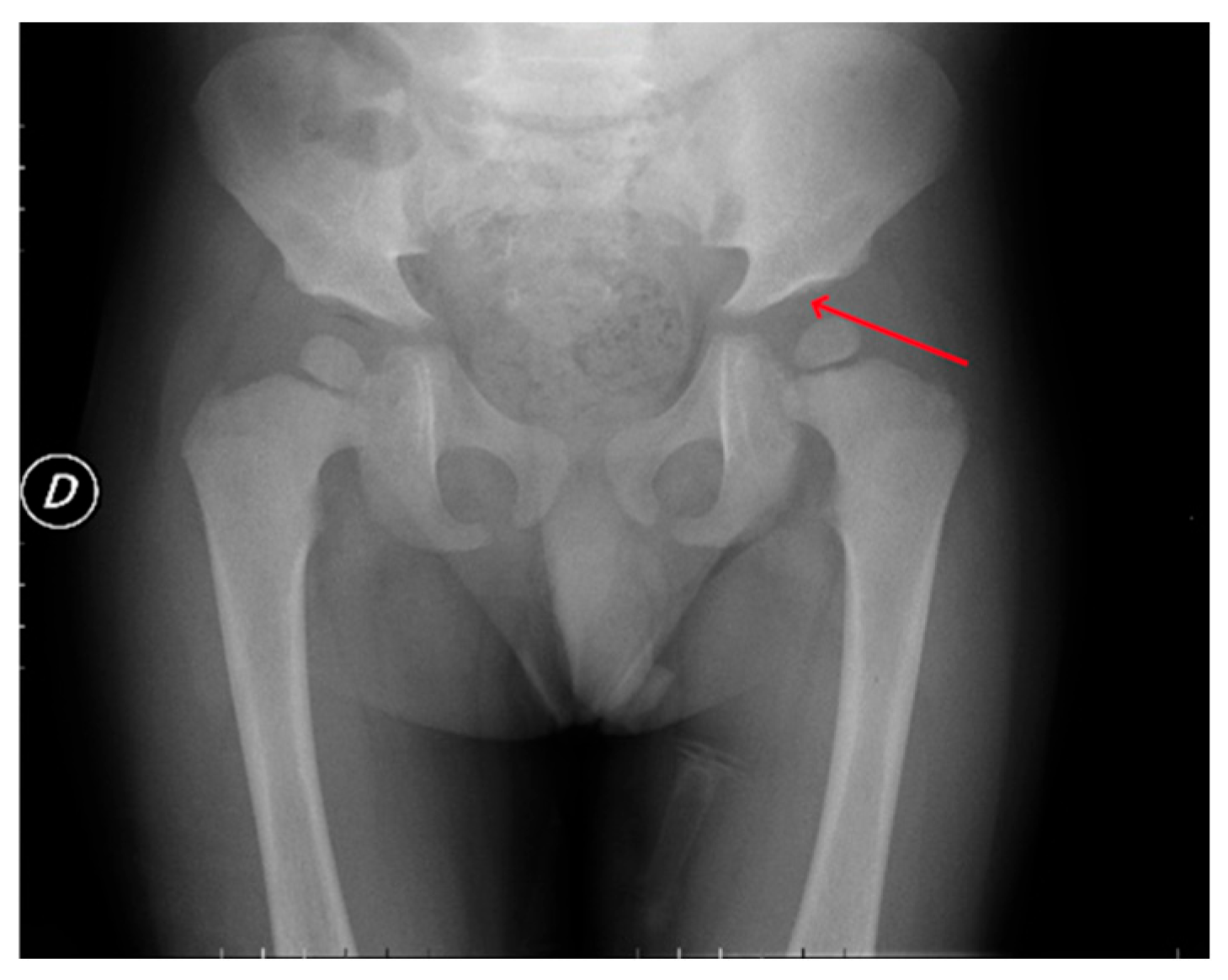
In patient 4, affected by MPS IIIB, dysostosis presented in a localized rather than generalized pattern. Pelvic imaging revealed impaired bone modeling and irregular configuration of the acetabular and proximal femoral epiphyses (Figure 10). Additionally, spinal X-ray demonstrated ovoid-shaped thoracolumbar vertebral bodies, associated with scoliosis and accentuated lumbar lordosis. No relevant skeletal deformities were identified in other anatomical districts.
Brain MRI was not performed in the MPS III patient due to a combination of factors, including the presence of an already advanced clinical phenotype, the limited expected diagnostic benefit, and the patient’s poor compliance with procedures requiring sedation, which posed additional risks.

3.4. MPS IV

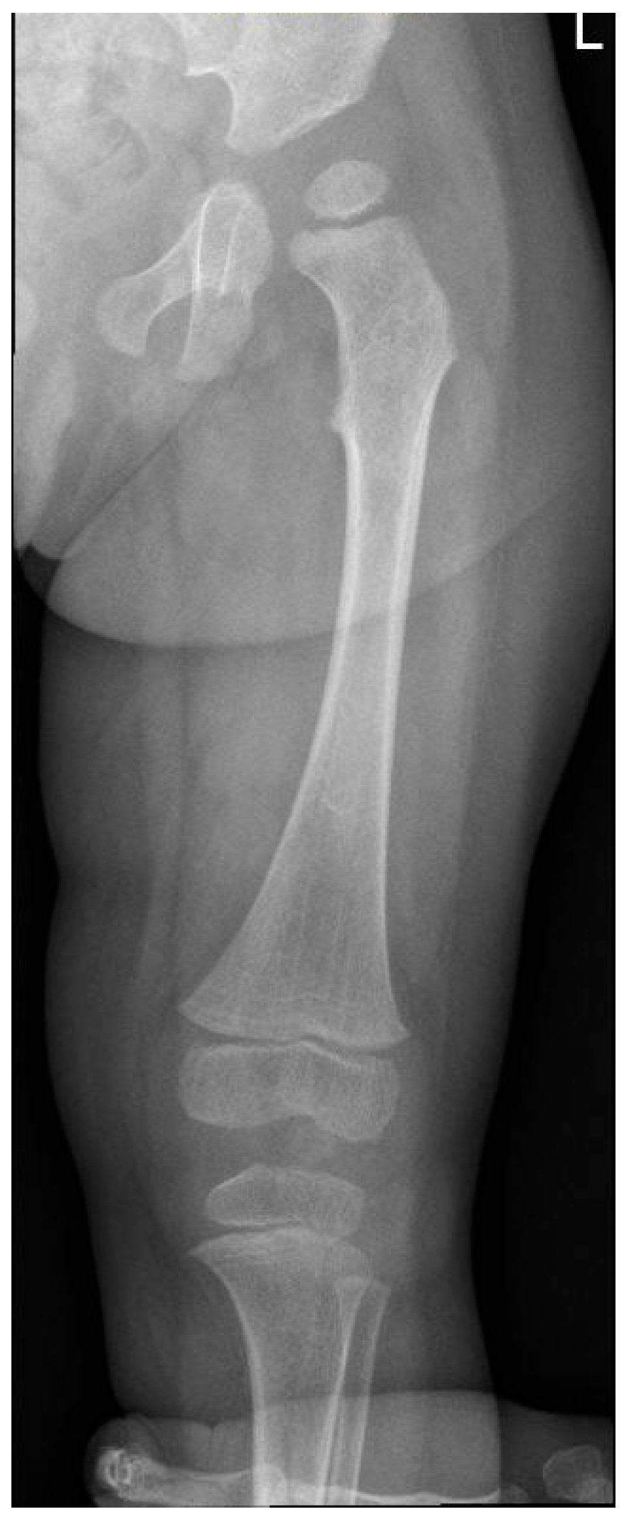
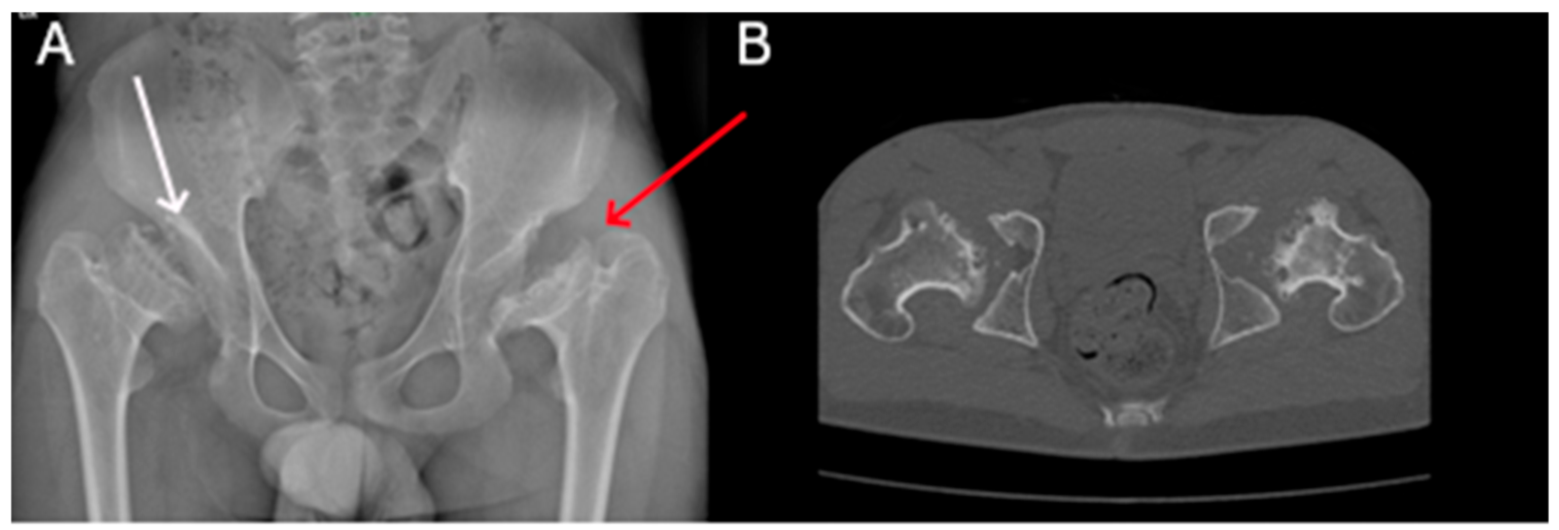
All four patients with MPS IVA included in our study exhibited disharmonic short stature with a short trunk. All also presented with pelvic bone deformities, kyphosis, pectus carinatum, genu valgum, and odontoid hypoplasia—the latter associated with atlanto-occipital instability in two cases (patients 7 and 8). Additionally, abnormal modeling of the long bones (Figure 11) was observed in patients 5, 7, and 8. Vertebral body deformities (platyspondyly) were present in all patients. In patient 6, these were accompanied by posterior bulging of the intervertebral discs, resulting in spinal canal impression at multiple levels. This patient also showed progressive pelvic involvement, as documented through follow-up imaging (Figure 12A,B); due to severe degeneration of both hip joints, he underwent hip replacement surgery and is currently a candidate for contralateral hip arthroplasty.
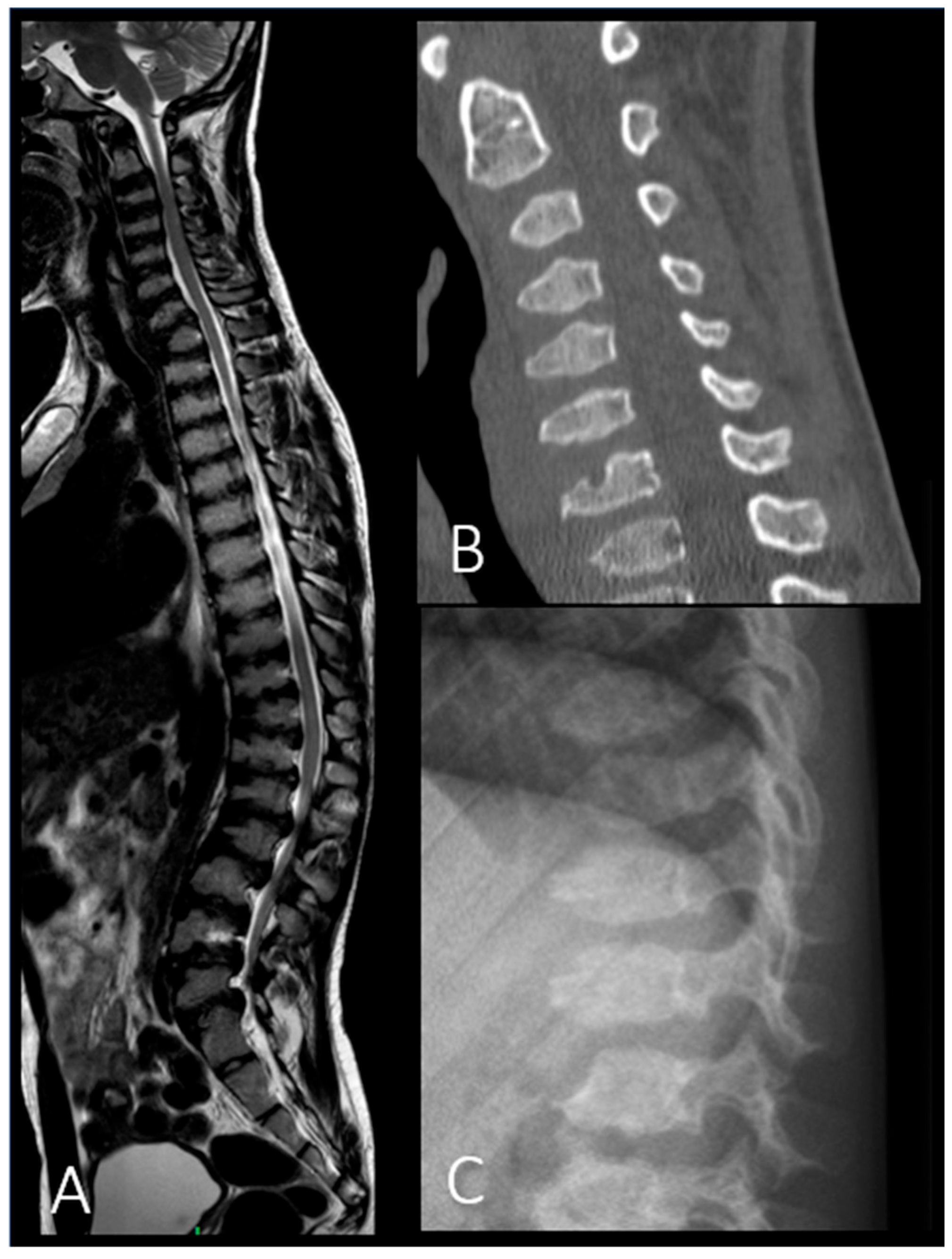
All patients exhibited stenosis of the foramen magnum and/or spinal canal (Figure 13). Scoliosis was documented in patients 6, 7, and 8 (Figure 14B), while lumbar hyperlordosis was noted exclusively in patient 6 (Figure 14A). Moreover, 50% of the cohort (patients 5 and 8) showed a J-shaped sella turcica. Patient 8 was the only individual who exhibited the characteristic “claw hand” deformity.
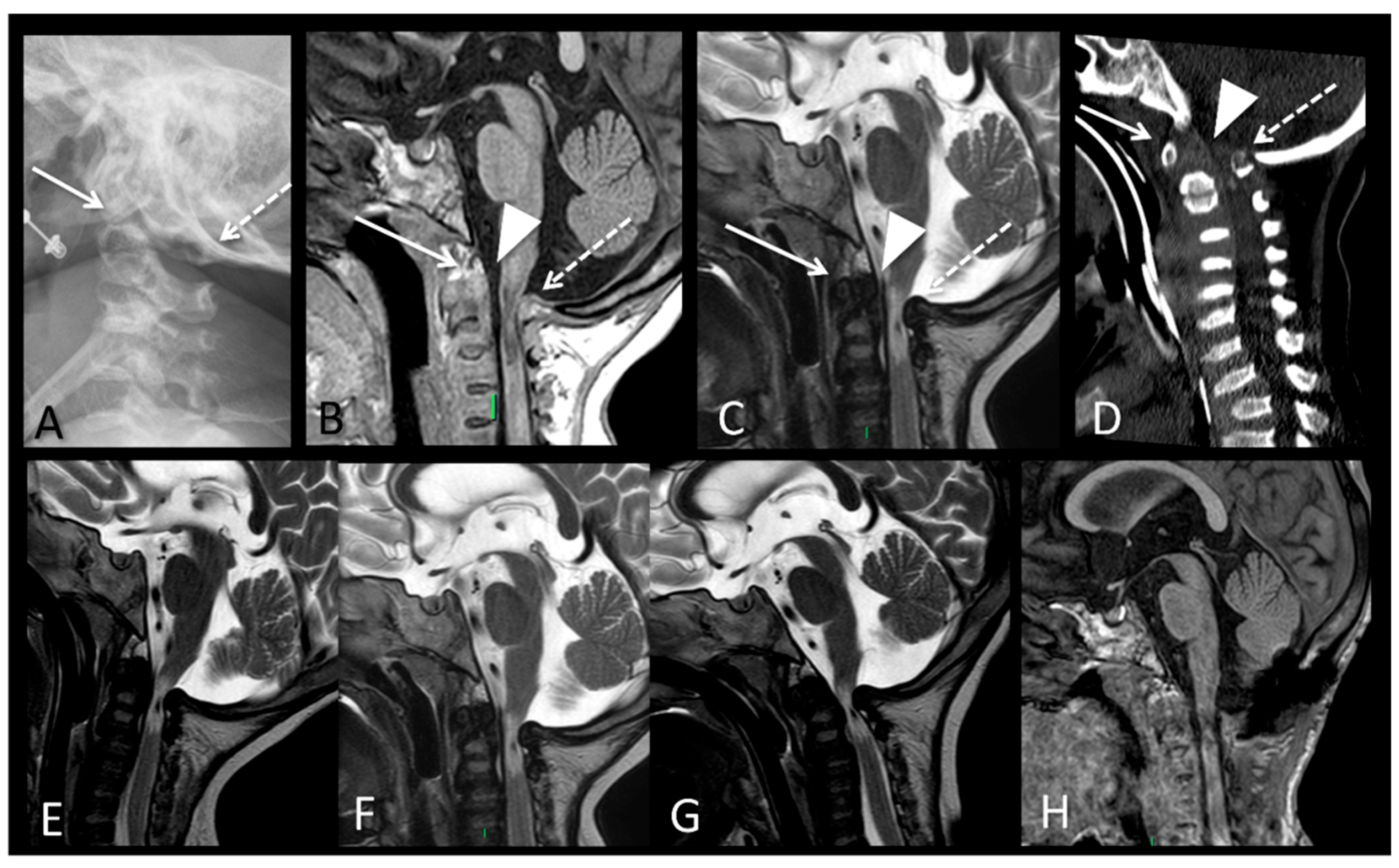
Additionally, cervical spinal cord signal alterations suggestive of compressive myelopathy were observed in three patients on spinal MRI (Figure 15). No evidence of cerebral involvement was detected in any of the cases.

4. Discussion

Dysostosis multiplex represents one of the main systemic features of different types of mucopolysaccharidosis [2,4]. Although many aspects of dysostosis are common across various types of MPS, distinct forms may present with slightly different musculoskeletal involvement.
Concerning cranial findings, a widened J-shaped sella turcica was observed in four patients belonging to three different MPS subtypes. This characteristic, which is due to a flattened tuberculum sellae and a rounded dorsum sellae, has been previously described in various MPS forms [5,6]. In contrast to the reports of other studies, none of our patients had a lack of pneumatization of mastoid cells and paranasal cavities [6].
Our findings highlight that the most characteristic thoracic deformities in MPS patients are the so-called ‘oar-shaped’ or ‘paddle-shaped’ ribs, characterized by widened anterior arcs and tapered posterior segments, resulting from progressive glycosaminoglycan (GAG) accumulation within bone tissue [2,6]. In addition, clavicles may be short and widened, as in our MPS I patient [6].
All patients displayed deformities of vertebral bodies commonly referred to as platyspondyly, already described as a prominent feature of dysostosis. Vertebral bodies are frequently flattened and rounded; at the thoracolumbar level in particular, they may exhibit an elongated anteroinferior margin, producing a characteristic ‘beaking’ appearance on lateral radiographs [6]. Thoracolumbar kyphosis (gibbus) and scoliosis also have a significant incidence within our cohort. These findings, secondary to vertebral deformities, have been widely described in MPS patients [5,6].
Deformities of pelvic bones were found in most of our patients, regardless of the MPS subtype. Pelvic radiography revealing several abnormalities widely described in the literature [2,4,5,6], such as rounded iliac wings, inferior tapering of the iliac bones, flattened acetabula, and dysplastic femoral heads, with poor development of the medial part of the proximal epiphysis, which may result in hip dysplasia.
Our data also underline that dysmorphisms of long bones are a common finding across different MPS types. As other research reported [6], long bones may show shortened diaphysis, curved in the distal part, irregular metaphysis, narrow epiphyses, as well as hypoplasia of the lateral tibial hemiplate, which can result in genu valgum.
In addition, our results outline a frequent involvement of the extremities, with deformities of the hands and feet already described by other authors [6]. In particular, carpal and tarsal bones might be irregular and hypoplastic; metacarpal bones can be short, broad, and proximally pointed.
In the following sections, we analyze the imaging findings associated with the different MPS subtypes included in this study.

4.1. MPS I

Our patient showed a complex musculoskeletal involvement. The clinical history revealed early-onset hip dysplasia and macrocephaly, which, as reported by De Ponti et al. [7], can be among the first clinical signs to emerge in the severe form of MPSI. Interestingly, our patient appears to have presented a particularly early onset of hip dysplasia compared with data reported in an international registry of patients with Hurler–Scheie, which documented a median age at diagnosis of 6.2 years [8]. Regarding the skull, our patient also showed dolichocephaly; this deformity, caused by premature closure of the sagittal suture, has been previously described as a frequent finding [6,9]. Even the dysmorphism of metacarpal bones, with the typical tapered proximal end, is frequently described [2]. Kyphosis was identified in our patient 1 after the age of two. Previous studies have reported that kyphosis is often a very early manifestation of MPS I in the severe variant [7,8], whereas in the moderate form, it tends to have a lower incidence and later onset [8,10]. For instance, in the cohort studied by Beck et al. [8], kyphosis/gibbus appeared at a median age of 4.6 years in Hurler–Scheie patients. Therefore, our result appeared to be consistent with those described in the literature.
As far as brain MRI is concerned, the abnormalities found in patient 1 were overall consistent with those reported in other studies [5,11], even though brain atrophy is reported as significantly less frequent in Hurler–Scheie disease than in the severe form [11]. It is worth mentioning that ventriculomegaly, enlargement of perivascular spaces, and white matter alterations reflect lysosomal storage in the central nervous system and the consequent disruption of tissue metabolism [5].
Therefore, we propose that the presence of macrocephaly and/or hip dysplasia in an infant—particularly when accompanied by other non-skeletal manifestations—should prompt clinical suspicion of MPS I.

4.2. MPS II

Both of our patients displayed vertebral anomalies, including anterior beaking of vertebral bodies, which represent an early radiographic marker of MPS II [12]. While in other patients of the cohort, vertebral dysmorphisms only affected the thoracolumbar segment, in both our MPS II patients, the whole spine, including the cervical segment, was affected by dysostosis. These data are consistent with those reported by Link et al. [13], with 93.8% of MPS II patients showing vertebral deformities and 68.8% with anomalies extended to the cervical segment. The same review [13] also reported a relatively low prevalence of spinal cord compression and craniocervical instability, both of which were absent in our two patients. Interestingly, neither of our patients presented claw hands, which has been reported as a common feature in this condition.
Remarkably, in patient 3, brain MRI (Figure 9) showed enlarged perivascular spaces (PVSs), with a distribution pattern frequently found in patients with MPS I and II, involving the cortico-subcortical junction, the basal ganglia, and the thalamus; unlike what other authors reported [5], PVSs were not found in the corpus callosum.
Moreover, one of the two patients exhibited a growth pattern characterized by overgrowth during the first 5–6 years of life, followed by a gradual deceleration in growth velocity, ultimately resulting in short stature. This interesting growth trend, which has been previously reported in the literature, is considered characteristic of MPS II and has not been described in other MPS subtypes [14].
Overall, the results that emerged confirm that musculoskeletal and brain involvement in MPS II has several similarities with that of MPS I, as previously outlined by other authors [3,15,16].

4.3. MPS III

The patient with MPS III enrolled in our study displayed a non-generalized pattern of dysostosis, with skeletal abnormalities limited mainly to the pelvic bones. Consistently, in a study on 18 patients with MPS III, White et al. [17] reported that hip abnormalities—particularly acetabular dysplasia and femoral head necrosis—were the most common orthopedic findings, observed in 10 out of 18 patients. The same research reported vertebral body deformities, and forearm bone involvement (radius and ulna) in fewer than 50% of cases. In contrast, cervical spine involvement and odontoid hypoplasia, both absent in our patient, were not detected in any of the subjects of the cohort. Our findings support the notion that MPS III is typically associated with a milder degree of dysostosis compared to most other MPS subtypes, with skeletal abnormalities confined to specific anatomical regions [3].

4.4. MPS IV

All four patients with MPS IVA in our cohort, including the one with a mild phenotype, presented with short stature. Consistently, in the MorCAP study, Harmatz et al. [18] reported short stature in 97% of 325 patients, with a median of −5.6 SDS. In addition, all of our patients showed widespread skeletal involvement with various abnormalities frequently described in Morquio A. Among them, it is worth mentioning pectus carinatum, genu valgum, kyphoscoliosis, and dysostosis of pelvic bones, reported in 97%, 93%, 85%, and 71% of cases, respectively, in the MorCAP cohort [18]. It should be noted that patient 6, although suffering from a mild form of the condition, has experienced a progressive degeneration of both coxofemoral joints despite the initiation of ERT. Other authors have reported that progression of skeletal deformities and the need for orthopedic surgery (including hip replacement) often occur in patients treated with ERT, even in subjects with non-severe phenotype [19]. The same patient also exhibited lumbar hyperlordosis, a condition that Harmatz and colleagues reported as a relatively frequent finding, affecting 56% of patients [18].
Two of our patients exhibited anterior–central beaking of the vertebral bodies, whereas another showed an anteroinferior deformity. Consistently, a previous study [5] reported that in Morquio disease, beaking is more often central than inferior.
Odontoid hypoplasia was detected in all the four patients, accompanied by cervical spinal cord compression in 75% of the cohort. These data are consistent with what is reported in the literature [9,18]: odontoid hypoplasia frequently leads to cervical spine instability, which in turn can cause compression and cervical myelopathy. Interestingly, odontoid hypoplasia and its complications were not detected in any of our patients affected by other forms of MPS. Other studies [5,9] have reported a higher frequency of compressive myelopathy in MPS IV and VI compared to different conditions. Even patient 6, despite being affected by a mild form of the disease, showed a complex spinal involvement with spinal canal impression at multiple levels. On the contrary, none of our patients showed any signs of cortical atrophy at brain MRI; consistently, intellectual disability is not described as a common feature of MPS IV, unlike other forms of the disease [5]. These results underscore the importance of MRI as a diagnostic tool in assessing patients with MPS. In particular, cervical and spinal MRI is essential for detecting critical skeletal abnormalities and their complications, such as odontoid hypoplasia, spinal canal stenosis, and spinal cord compression [20].
Two patients in our cohort were affected by joint hypermobility (one of them also exhibited contractures limited to inferior limbs), while only one patient showed joint stiffness. These data underline that Morquio disease is typically characterized by joint hyperlaxity, eventually associated with stiffness in other articular districts [18], whereas most MPS subtypes are characterized only by diffuse joint stiffness and contractures.
Our results confirm that dysostosis multiplex represents the most consistent radiological finding across various MPS subtypes and is often detectable in the early stages of the disease. In patients with suspected MPS, radiological imaging plays a crucial role in the initial diagnostic workup and, according to established diagnostic algorithms, should precede molecular testing [10,21]. In a study on MPS IVA, Wood et al. [21] suggested that radiography of multiple parts of the body should be performed even before urinary and blood tests in case of clinical suspicion.
Plain radiography is an immediate diagnostic modality which allows the detection of many features of dysostosis in different skeleton segments; therefore, it is particularly useful in supporting the diagnosis of MPS, as well as in assessing the severity and the extent of the skeletal involvement [2,4].
Also, the use of both MRI and CT is fundamental for the detection and comprehensive assessment of critical skeletal abnormalities, including odontoid hypoplasia, spinal canal stenosis, spinal cord compression, and femoral osteonecrosis [22,23].
On the other hand, brain MRI is essential for evaluating the effects of lysosomal storage in the central nervous system, revealing ventriculomegaly, enlargement of perivascular spaces, cortical atrophy, and white matter abnormalities [20].
Given the multisystemic nature of MPS, the comprehensive use of multiple imaging modalities is indispensable for the diagnosis, classification, follow-up monitoring, and treatment planning of MPS.
In all three patients, each affected by a different MPS subtype, serial imaging performed at different time points documented progressive worsening of skeletal involvement. These data confirm that ERT, which has demonstrated effectiveness in limiting the symptoms of MPS in various organs [7], has a very poor effect on dysostosis. Other authors have pointed out the limited impact of ERT on skeletal deformities, especially when they are already present at diagnosis, probably due to the poor vascular supply of bones and the subsequent limited biodistribution of ERT [24,25]. Nevertheless, some case reports involving siblings with MPS I, II, and VI have demonstrated promising results in patients treated with ERT from the very first weeks or months of life; in fact, the earlier treated siblings showed better growth and milder signs of dysostosis multiplex [26,27,28,29]. Furthermore, the severe progression of brain damage documented at follow-up investigations in patient 2 demonstrates the limited benefit of ERT in preventing and reducing the deterioration of the central nervous system. These conditions are therefore ideal candidates for neonatal screening, as they often remain clinically silent in early life and are typically diagnosed at an advanced stage, when the effectiveness of enzyme replacement therapy (ERT) is significantly reduced [30].
It is worth noting that only one patient in our cohort (Patient 5) initiated treatment before the age of two (at 23 months). In this case, the duration of enzyme replacement therapy at the time of evaluation was less than one year, making it premature to draw definitive conclusions regarding the potential impact of early treatment on skeletal disease progression
Notably, no significant clinical improvements in bone findings have been observed so far. A longer follow-up will be necessary to determine whether early initiation of ERT may have a protective effect on skeletal involvement over time.
As far as short stature is concerned, one of our MPS IVA patients (Patient 6), who was treated with ERT from the age of nine, experienced a milder growth failure compared to other subjects with the same condition who were not diagnosed until adolescence. However, this patient also showed a significant progression of skeletal disease over time. Ten years after starting ERT, he experienced a marked worsening of hip joint involvement and is currently awaiting a second hip replacement surgery. The clinical course of Patient 6, despite relatively early initiation of ERT, underscores the limited efficacy of enzyme replacement therapy on pre-existing skeletal abnormalities. Imaging studies revealed a rapid progression from early radiographic changes to advanced joint destruction on CT, ultimately requiring surgical intervention. This observation supports the view that ERT has little therapeutic effect on already formed bone lesions. Overall, while our observations suggest a limited effect of ERT on established skeletal abnormalities, this conclusion must be considered with caution due to the small number of treated patients, which prevents definitive evaluations.

5. Conclusions

Radiological imaging represents a fundamental tool in the diagnostic workup and monitoring of patients affected by MPS. Skeletal alterations associated with dysostosis multiplex are often highly suggestive of the diagnosis and can precede or support biochemical and molecular findings. Plain radiography enables the identification of characteristic bone abnormalities that underlie the musculoskeletal phenotype of MPS, allowing the clinician to differentiate between MPS subtypes. In our experience, ERT appears to have limited efficacy on dysostosis.
Moreover, MRI plays a key role in evaluating central nervous system involvement, highlighting brain abnormalities related to lysosomal storage as well as spinal complications.
Given the progressive nature of skeletal and neurological damage in MPS, radiological follow-up remains essential not only for assessing disease evolution but also for evaluating therapeutic response, especially in the context of enzyme replacement therapy or surgical planning. Our findings reinforce the pivotal role of imaging in the multidisciplinary management of MPS and underscore its value in guiding clinical decision-making throughout the course of the disease.

Author Contributions

Conceptualization, G.S., E.B., M.B., F.T., R.O. and E.C.; methodology, F.B.; software, G.S.; validation, M.L., G.B. and F.B.; formal analysis, F.B.; investigation, G.S., M.B. and F.T.; resources, M.B. and F.T.; data curation, R.O.; writing—original draft preparation, G.S., E.B., R.O. and E.C.; writing—review and editing, M.L., G.B. and F.B.; visualization, M.B. and F.T.; supervision, M.L. and G.B.; project administration, F.B.; funding acquisition, R.O. All authors have read and agreed to the published version of the manuscript.

Funding

This research received no external funding.

Institutional Review Board Statement

This retrospective study was conducted in compliance with Regulation (EU) 2016/679 (GDPR) and the Italian Privacy Code (Legislative Decree 196/2003, Art. 110, as amended by Legislative Decree 101/2018 and PNRR-bis, April 2024), which allow the use of anonymised or pseudonymised health data for retrospective scientific research without Ethics Committee approval. All patient data were processed in an anonymised form in accordance with the Italian Data Protection Authority guidelines.

Informed Consent Statement

Informed consent was obtained from all subjects involved in this study.

Data Availability Statement

All clinical data and materials are available in our Pediatric Unit.

Conflicts of Interest

The authors declare no conflict of interest.

Abbreviations

The following abbreviations are used in this manuscript:
MPSMucopolysaccharidosis
ERTEnzyme Replacement Therapy
GAGGlycosaminoglycan
MRIMagnetic Resonance Imaging
CTComputed Tomography
SDSStandard Deviation Score
PVSPerivascular Spaces
FLAIRFluid Attenuated Inversion Recovery
OSASObstructive Sleep Apnea Syndrome

References

  1. Filocamo, M.; Tomanin, R.; Bertola, F.; Morrone, A. Biochemical and molecular analysis in mucopolysaccharidoses: What a paediatrician must know. Ital. J. Pediatr. 2018, 44, 129. [Google Scholar] [CrossRef]
  2. Aldenhoven, M.; Sakkers, R.J.B.; Boelens, J.; De Koning, T.J.; Wulffraat, N.M. Musculoskeletal manifestations of lysosomal storage disorders. Ann. Rheum. Dis. 2009, 68, 1659–1665. [Google Scholar] [CrossRef]
  3. Galimberti, C.; Madeo, A.; Di Rocco, M.; Fiumara, A. Mucopolysaccharidoses: Early Diagnostic Signs in Infants and Children; BioMed Central Ltd.: London, UK, 2018. [Google Scholar] [CrossRef]
  4. Spina, V.; Barbuti, D.; Gaeta, A.; Palmucci, S.; Soscia, E.; Grimaldi, M.; Leone, A.; Manara, R.; Polonara, G. The role of imaging in the skeletal involvement of mucopolysaccharidoses. Ital. J. Pediatr. 2018, 44, 118. [Google Scholar] [CrossRef] [PubMed]
  5. Machnikowska-Sokołowska, M.; Myszczuk, A.; Wieszała, E.; Wieja-Błach, D.; Jamroz, E.; Paprocka, J. Mucopolysaccharidosis Type 1 among Children—Neuroradiological Perspective Based on Single Centre Experience and Literature Review. Metabolites 2023, 13, 209. [Google Scholar] [CrossRef]
  6. Palmucci, S.; Attinà, G.; Lanza, M.L.; Belfiore, G.; Cappello, G.; Foti, P.V.; Milone, P.; Di Bella, D.; Barone, R.; Fiumara, A.; et al. Imaging findings of mucopolysaccharidoses: A pictorial review. Insights Imaging 2013, 4, 443–459. [Google Scholar] [CrossRef]
  7. De Ponti, G.; Donsante, S.; Frigeni, M.; Pievani, A.; Corsi, A.; Bernardo, M.E.; Riminucci, M.; Serafini, M. MPSI Manifestations and Treatment Outcome: Skeletal Focus. Int. J. Mol. Sci. 2022, 23, 11168. [Google Scholar] [CrossRef] [PubMed]
  8. Beck, M.; Arn, P.; Giugliani, R.; Muenzer, J.; Okuyama, T.; Taylor, J.; Fallet, S. The natural history of MPS I: Global perspectives from the MPS I Registry. Genet. Med. 2014, 16, 759–765. [Google Scholar] [CrossRef]
  9. Blau, N.; Duran, M.; Gibson, K.M.; Vici, C.D. Physician’s Guide to the Diagnosis, Treatment, and Follow-Up of Inherited Metabolic Diseases; Springer International Publishing: Cham, Switzerland, 2022. [Google Scholar] [CrossRef]
  10. Baronio, F.; Zucchini, S.; Zulian, F.; Salerno, M.; Parini, R.; Cattoni, A.; Deodato, F.; Gaeta, A.; Bizzarri, C.; Gasperini, S.; et al. Proposal of an Algorithm to Early Detect Attenuated Type I Mucopolysaccharidosis (MPS Ia) among Children with Growth Abnormalities. Medicina 2022, 58, 97. [Google Scholar] [CrossRef] [PubMed]
  11. Zafeiriou, D.I.; Batzios, S.P. Brain and Spinal MR Imaging Findings in Mucopolysaccharidoses: A Review. Am. J. Neuroradiol. 2013, 34, 5–13. [Google Scholar] [CrossRef]
  12. Martin, R.; Beck, M.; Eng, C.; Giugliani, R.; Harmatz, P.; Muñoz, V.; Muenzer, J. Recognition and Diagnosis of Mucopolysaccharidosis II (Hunter Syndrome). Pediatrics 2008, 121, e377–e386. [Google Scholar] [CrossRef]
  13. Link, B.; de Camargo Pinto, L.L.; Giugliani, R.; Wraith, J.E.; Guffon, N.; Eich, E.; Beck, M. Orthopedic manifestations in patients with mucopolysaccharidosis type II (Hunter syndrome) enrolled in the Hunter Outcome Survey. Orthop. Rev. 2010, 2, e16. [Google Scholar] [CrossRef]
  14. Parini, R.; Jones, S.A.; Harmatz, P.R.; Giugliani, R.; Mendelsohn, N.J. The natural history of growth in patients with Hunter syndrome: Data from the Hunter Outcome Survey (HOS). Mol. Genet. Metab. 2016, 117, 438–446. [Google Scholar] [CrossRef]
  15. Mao, S.J.; Chen, Q.Q.; Dai, Y.L.; Dong, G.P.; Zou, C.C. The diagnosis and management of mucopolysaccharidosis type II. Ital. J. Pediatr. 2024, 50, 207. [Google Scholar] [CrossRef]
  16. Wraith, J.E.; Scarpa, M.; Beck, M.; Bodamer, O.A.; De Meirleir, L.; Guffon, N.; Meldgaard Lund, A.; Malm, G.; Van der Ploeg, A.T.; Zeman, J. Mucopolysaccharidosis type II (Hunter syndrome): A clinical review and recommendations for treatment in the era of enzyme replacement therapy. Eur. J. Pediatr. 2008, 167, 267–277. [Google Scholar] [CrossRef]
  17. White, K.K.; Karol, L.A.; White, D.R.; Hale, S. Musculoskeletal Manifestations of Sanfilippo Syndrome (Mucopolysaccharidosis Type III). J. Pediatr. Orthop. 2011, 31, 594–598. [Google Scholar] [CrossRef]
  18. Harmatz, P.; Mengel, K.E.; Giugliani, R.; Valayannopoulos, V.; Lin, S.P.; Parini, R.; Guffon, N.; Burton, B.K.; Hendriksz, C.J.; Mitchell, J.J.; et al. The Morquio a Clinical Assessment Program: Baseline results illustrating progressive, multisystemic clinical impairments in Morquio A subjects. Mol. Genet. Metab. 2013, 109, 54–61. [Google Scholar] [CrossRef]
  19. Sung, J.; Kim, I.; Im, M.; Ahn, Y.J.; Kim, S.-M.; Jang, J.-H.; Park, H.-D.; Jeon, T.Y.; Ko, K.R.; Park, S.-J.; et al. Long-term outcomes of enzyme replacement therapy from a large cohort of Korean patients with mucopolysaccharidosis IVA (Morquio A syndrome). Mol. Genet. Metab. Rep. 2025, 42, 101189. [Google Scholar] [CrossRef] [PubMed]
  20. Reichert, R.; Campos, L.G.; Vairo, F.; de Souza, C.F.M.; Pérez, J.A.; Duarte, J.Á.; Leiria, F.A.; Anés, M.; Vedolin, L.M. Neuroimaging Findings in Patients with Mucopolysaccharidosis: What You Really Need to Know. Radiographics 2016, 36, 1448–1462. [Google Scholar] [CrossRef] [PubMed]
  21. Wood, T.C.; Harvey, K.; Beck, M.; Burin, M.G.; Chien, Y.H.; Church, H.J.; D’Almeida, V.; van Diggelen, O.P.; Fietz, M.; Giugliani, R.; et al. Diagnosing mucopolysaccharidosis IVA. J. Inherit. Metab. Dis. 2013, 36, 293–307. [Google Scholar] [CrossRef] [PubMed]
  22. Solanki, G.A.; Martin, K.W.; Theroux, M.C.; Lampe, C.; White, K.K.; Shediac, R.; Lampe, C.G.; Beck, M.; Mackenzie, W.G.; Hendriksz, C.J.; et al. Spinal involvement in mucopolysaccharidosis IVA (Morquio-Brailsford or Morquio A syndrome): Presentation, diagnosis and management. J. Inherit. Metab. Dis. 2013, 36, 339–355. [Google Scholar] [CrossRef] [PubMed]
  23. Amanatullah, D.F.; Strauss, E.J.; Di Cesare, P.E. Current management options for osteonecrosis of the femoral head: Part 1, diagnosis and nonoperative management. Am. J. Orthop. 2011, 40, E186–E192. [Google Scholar] [PubMed]
  24. Concolino, D.; Deodato, F.; Parini, R. Enzyme replacement therapy: Efficacy and limitations. Ital. J. Pediatr. 2018, 44, 120. [Google Scholar] [CrossRef] [PubMed]
  25. Opoka-Winiarska, V.; Jurecka, A.; Emeryk, A.; Tylki-Szymańska, A. Osteoimmunology in mucopolysaccharidoses type I, II, VI and VII. Immunological regulation of the osteoarticular system in the course of metabolic inflammation. Osteoarthr. Cartil. 2013, 21, 1813–1823. [Google Scholar] [CrossRef]
  26. Tajima, G.; Sakura, N.; Kosuga, M.; Okuyama, T.; Kobayashi, M. Effects of idursulfase enzyme replacement therapy for Mucopolysaccharidosis type II when started in early infancy: Comparison in two siblings. Mol. Genet. Metab. 2013, 108, 172–177. [Google Scholar] [CrossRef]
  27. Tylki-Szymanska, A.; Jurecka, A.; Zuber, Z.; Rozdzynska, A.; Marucha, J.; Czartoryska, B. Enzyme replacement therapy for mucopolysaccharidosis II from 3 months of age: A 3-year follow-up. Acta Paediatr. 2012, 101, e42–e47. [Google Scholar] [CrossRef]
  28. McGill, J.J.; Inwood, A.C.; Coman, D.J.; Lipke, M.L.; De Lore, D.; Swiedler, S.J.; Hopwood, J.J. Enzyme replacement therapy for mucopolysaccharidosis VI from 8 weeks of age—A sibling control study. Clin. Genet. 2010, 77, 492–498. [Google Scholar] [CrossRef]
  29. Gabrielli, O.; Clarke, L.A.; Bruni, S.; Coppa, G.V. Enzyme-Replacement Therapy in a 5-Month-Old Boy With Attenuated Presymptomatic MPS I: 5-Year Follow-up. Pediatrics 2010, 125, e183–e187. [Google Scholar] [CrossRef]
  30. Montanari, G.; Candela, E.; Baronio, F.; Ferrari, V.; Biasucci, G.; Lanari, M.; Ortolano, R. Early-Onset Inherited Metabolic Diseases: When Clinical Symptoms Precede Newborn Screening-Insights from Emilia-Romagna (Italy). Children 2025, 12, 464. [Google Scholar] [CrossRef] [PubMed]
Figure 1. Patient 1 (MPS I) at 2.5 years of age: X-ray lateral view of the skull, which shows prevalence of neurocranium on splancnocranium, scafocephaly, thickened skull, and abnormal “J-shaped” sella turcica (red arrow).
Applsci 15 09093 g001
Figure 2. (Patient 1) (A) Pelvis X-ray taken at the age of 2.5 years, showing mild iliac wing hypoplasia and shifty acetabular roofs. Femoral heads appear relatively small for the age and are lateralized. (B) Pelvis X-ray taken at the age of 6 years, displaying progression of the deformative aspects of the bones; in particular, the acetabular roofs appeared to be more thickened and receding in appearance.
Applsci 15 09093 g002
Figure 3. (A) Hands X-ray of patient 1 at the age of 2.5 years: metacarpal bones appear markedly dysmorphic, with short, thickened bones with tapered proximal ends (white box); phalanges also show milder dysmorphisms. Radiographs of the left (B) and right (C) hand taken at 6 years of age, showing progression of the metacarpal deformities and the abnormal modeling of radius and ulna.
Applsci 15 09093 g003
Figure 4. (Patient 1) (A) Lateral X-ray of the thoracolumbar vertebrae taken at the age of 2.5 years: vertebral bodies appear dysmorphic, with the antero-inferior ‘beaking’ (red arrows). (B) Lateral X-ray of the spine taken at the age of 6 years: the dysmorphic aspect of thoracolumbar vertebral bodies is still evident.
Applsci 15 09093 g004
Figure 5. Brain MRI images of patient 1 at the age of 3 years: Sagittal (A) and axial (B) high-resolution, heavily T2-weighted images show mild dilation of the III ventricle and enlarged cisterna magna (A), along with tiny arachnoid cysts within the subarachnoid spaces of the basal cisterns (arrowheads in (B)). Coronal (C) and axial (D) 3D FLAIR T2-weighted reformatted images demonstrate a right temporopolar encephalocele (arrow in (C)), as well as mild white matter abnormalities and enlarged perivascular spaces. The latter are also depicted, as a specific finding of MPS, within the corpus callosum in (A).
Applsci 15 09093 g005
Figure 6. Radiographs of patient 3 (MPS II) at the age of 21 years. (A) Chest X-ray: ribs show the typical “oar shape” with widened anterior arches and tapered posterior ones (red asterisks); scoliosis is also evident. (B) Lateral cervical X-ray: vertebral bodies appear dysmorphic with the typical antero-inferior ‘beaking’.
Applsci 15 09093 g006
Figure 7. Patient 2. Lateral X-rays (A,C) and sagittal T2-weighted MRIs (B,D), acquired six years apart, between one and seven years of age, demonstrate progression of vertebral body deformities with posterior bulging of the intervertebral discs, leading to developing thoracolumbar kyphosis (posterior gibbus). Note the increasing severity of vertebral body changes, characterized by anterior beaking (arrows) and posterior scalloping (arrowheads).
Applsci 15 09093 g007
Figure 8. Patient 2. (AC): axial T2 (A) and FLAIR T2 ((B,C)) images acquired six years apart, between one and seven years of age, show periventricular white matter abnormalities associated with enlarged perivascular spaces, more clearly visible on the initial and intermediate MRI ((A,B)). This is followed by progressive extension of the white matter signal alterations, associated with severe dilation of the supratentorial portion of the ventricular system (C). (D,E) Coronal T2-weighted sequences demonstrate, along with hydrocephalus, a progressive widening of cerebral sulci and fissures, consistent with cerebral atrophy.
Applsci 15 09093 g008
Figure 9. Brain MRI of patient 3 at the age of 16 years: Axial FLAIR T2 (A) and coronal T2 (B) weighted images display enlarged perivascular spaces with a distribution in particular for MPS, at the cortico-subcortical junction, in the basal ganglia, and in the thalamic region. There is a concomitant diffuse alteration of the cerebral white matter.
Applsci 15 09093 g009
Figure 10. Pelvis X-ray of a 3-year-old male patient with MPS IIIB (patient 4): the acetabula (red arrow) and the femoral epiphyses appear hypoplastic and irregularly shaped; all pelvic bones appear poorly modeled.
Applsci 15 09093 g010
Figure 11. Femur X-ray of patient 5 at 18 months of age, showing a short and poorly modeled femur and valgism of the femoral neck.
Applsci 15 09093 g011
Figure 12. Patient 6 (MPS IVA): (A) Pelvis X-ray taken at the age of 16 years, showing flattening of the femoral epiphyses (red arrow) with irregular, jagged, and thickened margins and elongation of the cotyloid roofs (white arrow) with hypoplasia of the acetabular cavities, which poorly contain the femoral heads. (B) Pelvis CT scan in the axial plane, taken three years later: it displays progression of degenerative aspects with severe deformity of the femoral heads showing bone degeneration and subchondral thickening with almost complete loss of joint relationships.
Applsci 15 09093 g012
Figure 13. (Patient 6) Sagittal T2-weighted MRI taken at 16 years of age (A) shows deformities of the vertebral bodies and posterior bulging of the intervertebral discs, resulting in spinal canal impression at multiple levels. Reformatted CT of the cervical spine (B) and lateral X-ray of the thoracolumbar spine (C), both taken at the age of 8 (before the start of ERT), demonstrate flattened vertebral bodies (platyspondyly) in both cervical and thoracolumbar regions, along with anterior vertebral body beaking. A tendency toward gibbus deformity is also noted.
Applsci 15 09093 g013
Figure 14. Patient 6 at 19 years of age: Lateral (A) and anteroposterior (B) radiographs of the spine: The vertebral malformative alterations correlate with significant right-convex lumbar rotoscoliosis with accentuation of dorsal kyphosis and lumbar lordosis.
Applsci 15 09093 g014
Figure 15. Patient 7, 11 years old: Lateral X-ray (A), sagittal T2-weighted MRI (B), sagittal T1-weighted MRI (C), and sagittal reformatted CT (D) show odontoid dysplasia with a hypoplastic dens (arrows), best visualized on the X-ray (A), contributing to atlantoaxial instability, thus resulting in anterior displacement of the posterior arch of C1 (dashed lines). There is thickening of the dural and paraspinal ligaments (arrowheads), due to glycosaminoglycan (GAG) deposition and hypertrophy, the latter related to the aforementioned instability. These factors contribute to cervical spinal canal stenosis. At the C0–C2 level, a cervical myelomalacia is noted, with focal calcification. Sagittal T2-weighted images acquired in extension (E), neutral (F), and flexion (G) position demonstrate atlantoaxial instability with dynamic spinal canal narrowing, most pronounced during flexion. Sagittal T1-weighted image (H) shows the postoperative result of spinal canal decompression.
Applsci 15 09093 g015
Table 1. Significant clinical and therapeutic features of the eight patients in our cohort.
Subtype of MPSPatientSexAge at
Diagnosis
Current Clinical Signs and
Symptoms
Therapy
MPS I1F2.6 yearsCoarse facial features; Heart valve disease; Hepatosplenomegaly; OSAS; Umbilical hernia; Mild neurodevelopmental delay; Short stature (age 6: −0.94 SDS, −3.40 SDS from target height)Aldurazyme
(started at 3 years)
MPS II2M14 monthsDevelopmental delay; Overgrowth up to 6 years old; Hearing loss; Severe OSAS; Dysphagia; Mild mitral valve disease; Short stature (−3.43 SDS at 10, age of exitus)Elaprase
(started at 14 months)
MPS II3M4 yearsCoarse features; Cardiomyopathy, heart valve disease; Hearing loss; Hepatosplenomegaly; Joint contractures; Short stature (final height −4.32 SDS); Carpal tunnel syndrome; Mild neurodevelopmental delay;
OSAS
Elaprase
(started at 15 years)
MPS IIIB4M1 yearHepatosplenomegaly; Umbilical and inguinal hernia; Psychomotor regression; Agitation; Sleep disturbance; Dysphagia; Pes cavus; Recurrent otitis; Coarse facial features; Macroglossia; Short stature (−3.75 SDS at 14 years 6 months)No therapy
MPS IV5F1.5 yearsShort stature (−2.37 SDS at 2 years and 6 months); Normal psychomotor development; Facial asymmetry with left-sided hypoplasia; Short neck; Pectus carinatum; Limited elbow extension;Vimizim
(started at 23 months)
MPS IV6M8.9 yearsJoint stiffness; Flat feet; Mandibular laterodeviation; Hypermetropia and astigmatism; Short trunk; Pectus carinatum; Umbilical hernia; Short stature (final height: −3.45 SDS)Vimizim
(started at 8.9 years)
MPS IV7F11 yearsDevelopmental delay (onset 4–5 years); Coarse facial features; Corneal clouding; Joint hyperlaxity; Severe short stature (−7.81 SDS at 11) with a short trunk; Pectus carinatumNo therapy
MPS IV8M17 yearsDevelopmental delay (onset ~5 years); Coarse facial features; Corneal clouding; OSAS; Severe short stature (−10.61 SDS at 17) with a short trunk; Pectus carinatum; Short limbs; Joint hyperlaxity (except for contractures of inferior limbs); MacrobrachycephalyNo therapy
Table 2. Manifestations of dysostosis in the eight patients of the cohort. The (+) indicates the presence of the symptom in the patient.
Patient12345678
MPS TypeIIIIIIIIIVIVIVIV
Short stature++++++++
Overgrowth in the first years of life +
Claw hands +
Odontoid hypoplasia ++++
Craniocervical instability ++
Pectus carinatum ++++
Genu valgum ++++
Scoliosis ++ +++
Lumbar hyperlordosis + +
Hip dysplasia++++++++
Dysmorphic long bones+++ + ++
Radio–ulnar deformity+ + +
Oar-shaped ribs+ + ++++
Short, wide clavicles+
Platyspondyly with beaking of vertebral bodies++++++++
Macro and dolicocephaly+
Thickened skull+ +
J-shaped turcica++ + +
Kyphosis++ ++++
Table 3. Neuroimaging findings in the eight patients of our cohort. N.A. = Not available. The (+) indicates the presence of the symptom in the patient.
Patient12345678
MPS TypeIIIIIIIIIVIVIVIV
White matter abnormality+ (mild)++N.A.+
Brain atrophy++ N.A.
Hydrocephalus+ (mild)+ N.A.
Spinal canal/
foramen magnum stenosis
N.A.++++
Enlarged perivascular spaces+++N.A.
Odontoid hypoplasia N.A.++++
Compressive myelopathy N.A. +++
Table 4. Radiological evolution of bone lesions over time for patients 1, 2, and 6. The (+) indicates the presence of the symptom in the patient.
PatientTimepointImaging
Modality
Location of LesionsKey Findings
P1 (MPSI)BaselineRXPelvisMild iliac wing hypoplasia; shifty acetabular roofs.
RXHandsShort, thickened metacarpal bones; tapered proximal ends.
After 3 years of ERTRXPelvisThickening of the acetabular roofs.
RXHandsProgression of the metacarpal deformities; malformative aspects of radius and ulna.
P2 (MPSII)BaselineRX and MRISpineVertebral body deformities; posterior bulging of the intervertebral discs.
MRIBrainPeriventricular white matter abnormalities associated with enlarged perivascular spaces.
After 6 years of ERTRX and MRISpineThoracolumbar kyphosis; anterior beaking and posterior scalloping of vertebral bodies.
MRIBrainExtension of the white matter signal alterations; severe dilation of the ventricular system; cerebral atrophy.
P6
(MPSIV)
BaselineCTSpinePlatyspondyly in both cervical and thoracolumbar regions; anterior vertebral body beaking.
After 8 years of ERTMRISpineDeformities of the vertebral bodies and posterior bulging of the intervertebral discs; spinal canal impression.
Disclaimer/Publisher’s Note: The statements, opinions and data contained in all publications are solely those of the individual author(s) and contributor(s) and not of MDPI and/or the editor(s). MDPI and/or the editor(s) disclaim responsibility for any injury to people or property resulting from any ideas, methods, instructions or products referred to in the content.