Article

Differences in Lower Limb Muscle Activity and Gait According to Walking Speed Variation in Chronic Stroke

Department of Physical Therapy, Korea National University of Transportation, Jeungpyeong-gun 27909, Chungcheongbuk-do, Republic of Korea
*
Author to whom correspondence should be addressed.
Appl. Sci. 2025, 15(15), 8479; https://doi.org/10.3390/app15158479
Submission received: 9 July 2025 / Revised: 25 July 2025 / Accepted: 28 July 2025 / Published: 30 July 2025

Abstract

In this study, the effects of walking speed on lower limb muscle activity and gait parameters during over-ground walking were investigated in individuals with chronic stroke. Twenty-four patients with chronic stroke participated in a cross-sectional repeated-measures study, walking 20 m at three different speeds: slow (80% of self-selected speed), self-selected, and maximal speed. Surface electromyography was used to measure muscle activity in five paretic-side muscles (rectus femoris, biceps femoris, tibialis anterior, gastrocnemius, and gluteus medius), while gait parameters, including stride length, stance and swing phases, single-limb support time, and the gait asymmetry index were assessed using a triaxial accelerometer. As walking speed increased, activity in the rectus femoris, biceps femoris, and gastrocnemius muscles significantly increased during the stance and swing phases (p < 0.05), whereas the gluteus medius activity tended to decrease. Stride length on the paretic and non-paretic sides significantly increased with faster walking speed (p < 0.05); however, no significant improvements were observed in other gait parameters or gait asymmetry. These findings suggest that although increasing walking speed enhances specific muscle activities, it does not necessarily improve overall gait quality or symmetry. Therefore, rehabilitation programs should incorporate multidimensional gait training that addresses speed and neuromuscular control factors such as balance and proprioception.

1. Introduction

Gait disorder is a common complication after stroke, affecting over 80% of survivors [1]. Even after acute stroke rehabilitation, 18% are still unable to walk, 11% require assistance to walk, and only 50% are able to walk independently [2]. Particularly, reduced gait speed is an important indicator of dysfunction and is strongly associated with impaired mobility, restricted community participation, and lower quality of life [3,4]. Therefore, gait speed is generally used as a sensitive indicator of the level of gait function recovery in patients post-stroke [5,6].
Stroke survivors generally walk at a speed of 0.3–0.8 m/s, significantly slower than the average 1.4 m/s observed in healthy older adults [7]. A gait speed of 0.8–1.0 m/s is generally needed for safe daily ambulation, including crossing a street [6,8]. Furthermore, a gait speed greater than 0.83–1.0 m/s is recommended to optimize energy expenditure during walking [9].
Gait speed, an indicator of gait performance in patients who have had a stroke, is associated with many temporospatial gait parameters, including cadence, stride length, double support duration, paretic and nonparetic stance duration, and stride period [1]. Reports of previous studies show that progressive speed-dependent treadmill training improves cadence, step length, and stride length in patients post-stroke [10,11]. Recently, the attention given to the relationship between motor recovery and gait speed in stroke rehabilitation has increased. Furthermore, there has been emphasis on qualitative and quantitative analyses of gait patterns to better understand this correlation [9,12,13]. Louis et al. found that training focused on maximal gait speed was a crucial determinant of long-distance walking ability in stroke survivors [13]. Similarly, other research has shown that walking fast can significantly reduce energy expenditure during ambulation [14].
Many studies have explored the relationship between gait speed and motor recovery in patients post-stroke; however, most have focused on difference analysis for spatiotemporal parameters, joint angles, or other kinematic parameters between fast and comfortable walking speeds on a treadmill [14,15,16]. Biomechanical involvement of lower limb muscles, including the rectus femoris, biceps femoris, and gastrocnemius, is important in determining gait speed after stroke [15,17,18]. However, studies investigating how lower limb muscle activation varies with gait speed on over-ground surfaces are limited [19]. Particularly, studies on kinematics and lower limb muscle activation during slow gait in patients who have had a stroke are limited [20]. A more definitive investigation into the biomechanical involvement of lower limb muscles during slow walking is needed, as slowing down below a comfortable walking speed requires more complex motor control strategies [21].
Therefore, the aim of this study is to investigate how the over-ground walking speed variation (slow, preferred, and maximum) affects lower limb muscle activity and gait in patients post-stroke. We hypothesized that changes in walking speed would significantly influence lower limb muscle activity and gait parameters in patients post-stroke.

2. Materials and Methods
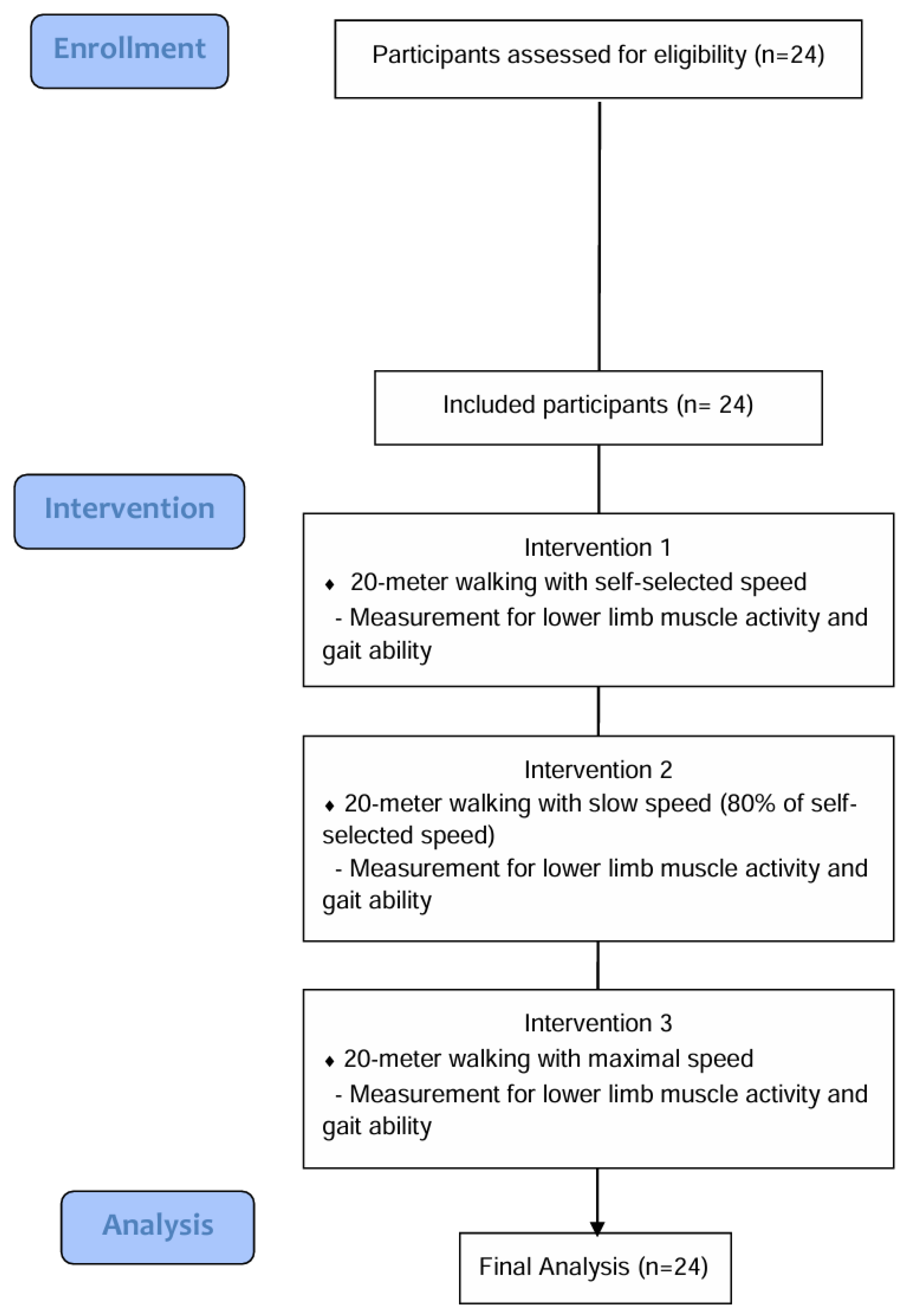
2.1. Study Design

This study was conducted at a neurological rehabilitation hospital in South Korea using a cross-sectional, repeated-measures design to examine the changes in lower limb muscle activity and gait ability according to differences in walking speed in patients who had had a stroke. Walking speed was set to three conditions: slow speed, self-selected speed, and maximum speed. Lower limb muscle activity and gait-related variables were measured in all participants under each walking condition.

2.2. Study Participants

Overall, 24 patients with chronic stroke (13 males; mean age: 66.50 years; mean time since stroke: 275.66 days) participated in this study. All participants participated in a standard rehabilitation program comprising 30 min of neurodevelopmental treatment performed by licensed physical therapists, 20 min of functional electrical stimulation, and 30 min of occupational therapy. No additional cognitive rehabilitation training was provided. Based on a previous study of stroke survivors, the required sample size of 24 participants was calculated using G*Power 3.1.9.3 [22] with an effect size of 0.607, power of 0.80, and alpha of 0.05. The inclusion criteria were the following: (1) time since stroke > 6 months; (2) ability to understand this study and provide informed consent; (3) independent walking ≥ 20 m with or without assistive devices; (4) Functional Ambulation Category (FAC) ≥ 2 [23]; (5) MMSE-K score ≥ 24 [24]. The exclusion criteria were the following: (1) diagnosed cardiovascular conditions contraindicating physical activity (uncontrolled hypertension, recent myocardial infarction); (2) musculoskeletal impairments affecting walking, such as severe lower limb contractures or advanced osteoarthritis; (3) diagnosed visual or vestibular disorders interfering with balance or mobility.
The Ethics Committee of Korea National University of Transportation (KNUT-2024-HR-12-28) approved this study. This study was conducted in accordance with the Declaration of Helsinki. All participants provided written informed consent.

2.3. Procedures

In this study, walking ability (FAC), lower extremity muscle strength (Manual Muscle Testing (MMT)) and muscle tone (Modified Ashworth Scale (MAS)), cognition (Mini-Mental State Examination—Korean version (MMSE-K)), balance ability (Berg Balance Scale (BBS)), and activities of daily living performance (Modified Barthel Index (MBI)) were evaluated to investigate the baseline clinical characteristics of the subjects. An investigation of changes in lower limb muscle activity and gait according to differences in walking speed was conducted in an unobstructed hallway 40 m in length and 5 m wide. Twenty-four patients who had suffered a stroke walked on 20 m over-ground at three different walking speeds (slow (80% of self-selected), self-selected, and maximal speeds), during which changes in lower limb muscle activity and gait were analyzed (Figure 1). In order to minimize potential bias, data on muscle activity and gait variables were collected by researchers who were not involved in this study and were unaware of the specific objectives of the study.
Lower limb muscle activity was measured using surface electromyography targeting five muscles (rectus femoris, biceps femoris, tibialis anterior, gastrocnemius, and gluteus medius) on the paretic side, and gait ability was measured using a gait analyzer for the stride length, stance and swing phases, and single limb support time on the paretic and non-paretic sides, and the gait asymmetry index was calculated based on the measured parameters. In this study, the self-selected speed was set to the participant’s usual walking speed, which was comfortable, and the slow speed was set to 80% of the self-selected speed [16]. The maximum speed was set to the fastest possible speed. All participants underwent the measurements in the following order: using self-selected, slow, and maximum walking speeds. Participants first walked 20 m at their usual comfortable (self-selected) speed. For the slow-speed condition, participants walked on a treadmill for 5 min at a walking speed adjusted to 80% of their self-selected walking speed. Subsequently, measurements were taken while they walked on the ground at the same speed as they did on the treadmill. For the maximum speed condition, participants were instructed to “walk as fast as possible” over 20 m. Each measurement was conducted three times, and the average was used for analysis. Two assistants ensured the participants’ safety throughout the experiments. Participants wore comfortable clothing and shoes and were given a 5 min break between trials, with additional rest upon request. Verbal encouragement was provided during the maximal speed trial to ensure maximal effort.

2.4. Muscle Activity and Gait Ability Measurements

Lower limb muscle activity on the paretic side was measured using surface electromyography (sEMG) (freeEMG1000, BTS Bioengineering, Milan, Italy) targeting five muscles (rectus femoris, biceps femoris, tibialis anterior, gastrocnemius, and gluteus medius). Disposable wireless bipolar Ag/AgCl surface electrodes (with an interelectrode distance of 20 mm) were placed on each muscle’s belly following the sEMG for non-invasive assessment of muscle guidelines [25]. The skin was prepared by shaving hair (if needed), lightly abrading it, and cleaning it with alcohol to reduce impedance before electrode placement. The EMG signals were recorded at a sampling rate of 1000 Hz. A band-pass filter (20–500 Hz) was applied in real time to attenuate movement artifacts and high-frequency noise. Collected EMG data were analyzed using the BTS EMG Analyzer software (version 2.9.37.0). The raw EMG signals were first band-pass-filtered (20–500 Hz) and then full-wave-rectified. We processed the rectified signals to calculate root-mean-square envelopes with a moving window of 50 ms, which provided a smoothed measure of muscle activation amplitude. For each muscle, the EMG values were normalized to a reference voluntary contraction (RVC) and expressed as a percentage of the RVC (%RVC). Normalization to maximal voluntary isometric contraction is often recommended for comparing muscle activity across individuals; nevertheless, the population with stroke may struggle to generate consistent maximal isometric contractions, owing to motor impairment, fatigue, and concerns over safety [26]. Thus, to obtain the %RVC of each muscle during the stance phase, the EMG signal recorded from the same muscle during standing without movement was used as a reference value. The lower limb muscle activity was measured three times, and the average value was used for the final statistical analysis.
Gait ability (paretic and non-paretic side stride length, stance and swing phases, and single-limb support time) was measured using a three-axis accelerometer (G-Walk; BTS Bioengineering, Milan, Italy). The three-axis accelerometer, which enclosed four inertial platforms and a Global Positioning System (GPS), was attached to the first sacral vertebral level of the participants using a special Velcro strap. The sampling frequency was set to 100 Hz, and data were acquired using G-Studio (BTS Bioengineering, Milano, Italy). The three-axis accelerometer sent data to a G-Studio connected via Bluetooth during the gait ability measurement. Gait parameters recorded during the test were displayed automatically. In addition, the gait asymmetry index was calculated using recorded gait parameters. A gait asymmetry index of 0% indicates perfect symmetry. Based on a previous study, the gait asymmetry index was calculated using the following formula [27].
G a i t a s y m m e t r y i n d e x = N o n p a r e t i c s i d e p a r a m e t e r P a r e t i c s i d e p a r a m e t e r N o n p a r e t i c s i d e p a r a m e t e r + P a r e t i c s i d e p a r a m e t e r × 100

2.5. Data Analysis

All statistical analyses were performed using SPSS v.27.0 (IBM Corp., Armonk, NY, USA). Descriptive statistics and frequency analyses were used to present the general characteristics of the participants, which were expressed as means, standard deviations, and frequencies. The Shapiro–Wilk test was used to determine the normality of the data. To analyze changes in paretic lower limb muscle activity and gait parameters according to differences in walking speed, a one-way repeated-measures analysis of variance (ANOVA) was conducted. When a significant difference was found, the Bonferroni post hoc test was used for pairwise comparisons. The level of statistical significance was set at p < 0.05.

3. Results

3.1. General Characteristics of the Participants

Table 1 shows the general characteristics of the participants. Lower extremity muscle strength, assessed using MMT, typically ranged from grades 3 to 4—sufficient to move against gravity—except for the ankle dorsiflexors and plantar flexors. Muscle tone in the ankle plantar flexors, assessed by the MAS, revealed mild to moderate spasticity. The BBS score was 42.16, suggesting a moderate risk of falls and balance impairment. The MBI score was 75.41, reflecting a fair level of functional independence in activities of daily living. Cognitive function was relatively preserved, with an MMSE-K score of 25.70, indicating that the participants had sufficient cognitive ability to follow study instructions. Walking ability, as assessed using the FAC, ranged from 2 to 4. This means that most participants required some assistance when walking.

3.2. Changes in Lower Limb Muscle Activity

The changes in lower limb muscle activity according to walking speed are shown in Table 2. Muscle activity was expressed as a percentage of reference voluntary contraction (%RVC). A one-way repeated-measures ANOVA with a Bonferroni post hoc test was conducted to compare the conditions. Patients who had a stroke showed increased muscle activity in the paretic lower limb, except for the gluteus medius, at faster walking speeds during the stance and swing phases. Rectus femoris muscle activity significantly increased from 659.95 to 1319.67 (99.97%, p < 0.05) in the stance phase. Significant increases were also observed in the rectus femoris (407.49 to 849.66, 108.51%), biceps femoris (604.73 to 1118.23, 84.91%), and gastrocnemius (329.56 to 576.36, 74.89%) (p < 0.05 for all) in the swing phase. Conversely, gluteus medius muscle activity showed a decreasing trend with increasing walking speed, but this change was not statistically significant (p > 0.05).

3.3. Changes in Gait Ability

Table 3 shows the changes in gait parameters based on walking speed. The analyzed parameters included paretic and non-paretic stride length, stance and swing phases, single-limb support time, and the GAI. A one-way repeated-measures ANOVA with a Bonferroni post hoc test was conducted to compare walking speeds. Stride length significantly increased with faster walking speeds on the paretic side (46.22 to 60.00 %height, 29.81%) and the non-paretic side (45.99 to 59.09 %height, 28.48%) (p < 0.05 for both). However, no statistically significant changes were observed in other gait parameters, including stance and swing phases, single-limb support time, and the GAI (p > 0.05).

4. Discussion

In this study, we analyzed changes in lower limb muscle activity and gait ability according to walking speed in patients post-stroke. Participants conducted 20 m over-ground walking under three conditions: slow speed, self-selected speed, and maximum speed. The patients who had had a stroke showed a tendency toward increased paretic-side lower limb muscle activity with faster walking speeds. Specifically, in the stance phase, rectus femoris activity significantly increased from 659.95 to 1319.67 %RVC (99.97%). In the swing phase, significant increases were observed in the rectus femoris (407.49 to 849.66, 108.51%), biceps femoris (604.73 to 1118.23, 84.91%), and gastrocnemius (329.56 to 576.36, 74.89%) (p < 0.05 for all). Conversely, gluteus medius activity decreased slightly as walking speed increased, but this change was not statistically significant (p > 0.05).
The increase in muscle activity in the paretic lower limb at higher walking speeds possibly reflects greater neuromuscular demand, as faster walking requires additional muscle recruitment. At maximal speed, participants might have increased activation of muscles associated with knee flexion, hip extension, and ankle push-off, potentially generating greater propulsive force. This pattern shows a compensatory strategy to support and move the limb more quickly. However, this is not a novel finding, as previous studies have reported similar results [1,28,29].
One interesting finding of this study was the tendency for gluteus medius muscle activity to decrease as walking speed increased, contrary to the overall pattern observed in other muscles. These results may be associated with the interaction between the functional role of the gluteus medius and the use of walking aids. During the single-limb support phase, the gluteus medius contributes primarily to pelvic stabilization and mediolateral weight shifting rather than to forward propulsion [30,31]. Previous studies reported that using walking aids can reduce gluteus medius activation by being substituted for weight-bearing demands, with the use of a cane reportedly decreasing muscle activity by approximately 22% [32,33]. In this study, 13 of the 24 participants (55.17%) used a mono-cane during all walking trials. The observed decrease in gluteus medius activity across speed conditions may therefore show a shift in load-bearing responsibility from the muscle to the cane.
In gait parameters, although both paretic (46.22 to 60.00 %height, 29.81%) and non-paretic (45.99 to 59.09 %height, 28.48%) stride lengths significantly increased with faster walking speeds (p < 0.05), other gait parameters and gait asymmetry did not show noticeable improvement with changes in walking speed. This suggests that patients post-stroke maintained their baseline asymmetric gait pattern despite significantly increasing their stride length to cope with faster speeds. Greater gait asymmetry has been associated with a higher risk of falls in people with chronic stroke and with increased metabolic energy cost during walking. Thus, restoration of gait asymmetry has important clinical implications in stroke [34]. Awad et al. found that increased walking speed did not necessarily indicate improved walking efficiency or symmetry [14]. In addition, another study reported that increased walking speed may not lead to improved symmetry, and faster walking may reinforce existing asymmetric gait patterns due to overuse of the non-paretic side [35].
Gait symmetry is more closely associated with neuromuscular factors such as balance, proprioception, and coordination than with simple elements such as muscle strength or walking speed [30,36]. Moreover, gait symmetry refers to the balance between the left and right sides of the body while walking, including parameters such as stride length, stance time, and swing phase. Without sufficient recovery in these complex domains, improvements in gait symmetry through increased walking speed alone may be limited. Some studies have reported that increasing walking speed can improve certain gait parameters in patients post-stroke [13,15,37]; nonetheless, other evidence highlights the importance of task-specific interventions, including weight shifting, trunk rotation, and dual-task training [36]. Particularly, cognitive dual-task training—involving walking while performing a simultaneous mental task—enhances gait and balance more effectively than walking alone [38]. Therefore, restoring qualitative aspects, such as gait symmetry, necessitates a more comprehensive consideration of various influencing parameters.
The results of the present study showed that the improvement of some lower extremity muscle activity and gait parameters by increasing walking speed may not affect the overall improvement of gait quality or symmetry; however, this study had some limitations that should be acknowledged. First, the number of participants was relatively small (24 patients who suffered chronic stroke), and the functional level was in the moderate category (average comfortable walking speed, 0.67 m/s; FAC, 2–4). This sample size limits the generalizability of the results to the broader stroke population, especially to those with mild impairment (who walk faster and more symmetrically) or severe impairment (who may not tolerate fast walking at all). Second, 55% of the participants in this study used a cane while walking. According to previous studies, patients who have had a stroke and use a cane tend to offload over 7% of their body weight onto the assistive device [39]. Therefore, using a cane may have introduced potential confounding effects on the study outcomes. Additionally, this study’s small sample size prevented us from conducting subgroup analyses by cane use. Third, this was a cross-sectional study that compared three walking speed conditions at a single measurement point, and thus repetitive training or changes over time were not considered. Therefore, it is not possible to confirm the effectiveness of fast or slow speed walking training. Finally, since this study’s gait analysis was conducted based on spatiotemporal indicators, kinematic or kinetic information that includes specific compensatory movements (including increased hip hiking or circumduction) could not be analyzed.
Future research should involve subgroup analyses (comparing walking with and without a cane) with sufficient sample sizes to examine the influence of assistive device usage. Additionally, longitudinal or randomized controlled trials are also needed to investigate how repeated gait training at different walking speeds (including progressive speed-increase protocols) influences muscle activity and gait patterns over time. Furthermore, using 3D motion analysis would permit a more comprehensive assessment of joint kinematics and muscle activation responses across different walking speeds.
In summary, this study revealed that increasing walking speed improved certain aspects of lower limb muscle activity and gait performance; however, it did not enhance overall gait quality or symmetry. Faster walking may improve biomechanical elements, including propulsion; nevertheless, it does not necessarily result in better gait symmetry. Gait symmetry depends more on complex neuromuscular factors such as balance, proprioception, and coordination than just speed. Therefore, clinical gait interventions for patients post-stroke should include multidimensional training approaches that target neuromuscular control, instead of focusing solely on speed enhancement.

5. Conclusions

We examined how different walking speeds influenced paretic lower limb muscle activity and gait performance in patients who had had a stroke by comparing three over-ground walking conditions. Fast walking led to increased stride length and significantly activated the rectus femoris, biceps femoris, and gastrocnemius. However, gluteus medius activity decreased, with no significant improvements in gait asymmetry being observed. Improving gait symmetry in patients post-stroke may depend more on complex neuromuscular factors than just speed.

Author Contributions

Conceptualization, K.H.C.; methodology, K.H.C.; formal analysis, Y.G.S.; investigation, Y.G.S.; writing—original draft preparation, Y.G.S. All authors have read and agreed to the published version of the manuscript.

Funding

This study was supported by the Changwon Medical-Bio Advanced Device Manufacturing Industry Promotion Project 2023. In addition, this work was supported by Chungcheongbuk-do RISE (Regional Innovation System & Education) grants funded by the Ministry of Education and Chungcheongbuk-do.

Institutional Review Board Statement

This study was approved by the Institutional Review Board of the Korea National University of Transportation (Approval No.: KNUT-2024-HR-12-28, Approval date: 23 September 2024).

Informed Consent Statement

Written informed consent was obtained from the patients to publish this paper.

Data Availability Statement

The original contributions presented in this study are included in the article; further inquiries can be directed to the corresponding author.

Conflicts of Interest

The authors declare no conflicts of interest.

Abbreviations

The following abbreviations are used in this manuscript:
BBSBerg Balance Scale
BFBiceps femoris
FACFunctional Ambulation Category
GMGluteus medius
GNGastrocnemius
GAIgait asymmetry index
GPSGlobal Positioning System
MASModified Ashworth Scale
MBIModified Barthel Index
MMSE-KMini-Mental State Examination—Korean version
MMTManual Muscle Testing
RFRectus femoris
RVCReference voluntary contraction
sEMGSurface electromyography
TATibialis anterior

References

  1. Balaban, B.; Tok, F. Gait disturbances in patients with stroke. PM R J. Inj. Funct. Rehabil. 2014, 6, 635–642. [Google Scholar]
  2. Woolley, S.M. Characteristics of gait in hemiplegia. Top. Stroke Rehabil. 2001, 7, 1–18. [Google Scholar] [CrossRef]
  3. Vive, S.; Elam, C.; Bunketorp-Käll, L. Comfortable and maximum gait speed in individuals with chronic stroke and community-dwelling controls. J. Stroke Cerebrovasc. Dis. 2021, 30, 106023. [Google Scholar] [CrossRef]
  4. Schmid, A.; Duncan, P.W.; Studenski, S.; Lai, S.M.; Richards, L.; Perera, S.; Wu, S.S. Improvements in speed–based gait classifications are meaningful. Stroke 2007, 38, 2096–2100. [Google Scholar] [CrossRef]
  5. Kim, C.M.; Eng, J.J. Magnitude and pattern of 3D kinematic and kinetic gait profiles in persons with stroke, relationship to walking speed. Gait Posture 2004, 20, 140–146. [Google Scholar] [CrossRef]
  6. Tasseel-Ponche, S.; Delafontaine, A.; Godefroy, O.; Yelnik, A.P.; Doutrellot, P.L.; Duchossoy, C.; Hyra, M.; Sader, T.; Diouf, M. Walking speed at the acute and subacute stroke stage: A descriptive meta-analysis. Front. Neurol. 2022, 13, 989622. [Google Scholar] [CrossRef]
  7. Lee, D.G.; Ahn, S.H.; Lee, G.C. Discriminative power and predictive validity of the timed up and go test, figure-of-8 walk test, four square step test, and step test for classifying community ambulation levels in individuals with chronic stroke. J. Korean Phys. Ther. Sci. 2020, 27, 25–35. [Google Scholar] [CrossRef]
  8. Salbach, N.M.; O’Brien, K.; Brooks, D.; Irvin, E.; Martino, R.; Takhar, P.; Chan, S.; Howe, J.A. Speed and distance requirements for community ambulation: A systematic review. Arch. Phys. Med. Rehabil. 2014, 95, 117–128.e11. [Google Scholar] [CrossRef] [PubMed]
  9. Beyaert, C.; Vasa, R.; Frykberg, G.E. Gait post-stroke, pathophysiology and rehabilitation strategies. Neurophysiol. Clin. 2015, 45, 335–355. [Google Scholar] [CrossRef]
  10. Pohl, M.; Mehrholz, J.; Ritschel, C.; Rückriem, S. Speed-dependent treadmill training in ambulatory hemiparetic stroke patients: A randomized controlled trial. Stroke 2002, 33, 553–558. [Google Scholar] [CrossRef]
  11. Lau, K.W.; Mak, M.K. Speed-dependent treadmill training is effective to improve gait and balance performance in patients with sub-acute stroke. J. Rehabil. Med. 2011, 43, 709–713. [Google Scholar]
  12. Langhorne, P.; Bernhardt, J.; Kwakkel, G. Stroke rehabilitation. Lancet 2011, 377, 1693–1702. [Google Scholar] [CrossRef]
  13. Awad, L.N.; Reisman, D.S.; Wright, T.R.; Roos, M.A.; Binder-Macleod, S.A. Maximum walking speed is a key determinant of long distance walking function after stroke. Top. Stroke Rehabil. 2014, 21, 502–509. [Google Scholar] [CrossRef]
  14. Awad, L.N.; Palmer, J.A.; Pohlig, R.T.; Binder-Macleod, S.A.; Reisman, D.S. Walking speed and step length asymmetry modify the energy cost of walking after stroke. Neurorehabil. Neural Repair 2015, 29, 416–423. [Google Scholar] [CrossRef]
  15. Lamontagne, A.; Fung, J. Faster is better, implications for speed-intensive gait training after stroke. Stroke 2004, 35, 2543–2548. [Google Scholar] [CrossRef]
  16. Carvalho, C.; Sunnerhagen, K.S.; Willén, C. Walking speed and distance in different environments of subjects in the later stage post-stroke. Physiother. Theory Prac. 2010, 26, 519–527. [Google Scholar] [CrossRef]
  17. Burnfield, M. Gait analysis: Normal and pathological function. J. Sports Sci. Med. 2010, 9, 353. [Google Scholar]
  18. Marasović, T.; Cecić, M.; Zanchi, V. Analysis and interpretation of ground reaction forces in normal gait. WSEAS Trans. Syst. 2009, 8, 1105–1114. [Google Scholar]
  19. Wang, Y.; Mukaino, M.; Ohtsuka, K.; Otaka, Y.; Tanikawa, H.; Matsuda, F.; Tsuchiyama, K.; Yamada, J.; Saitoh, E. Gait characteristics of post-stroke hemiparetic patients with different walking speeds. Int. J. Rehabil. Res. 2020, 43, 69–75. [Google Scholar] [CrossRef]
  20. Liang, J.N.; Ho, K.Y.; Lee, Y.J.; Ackley, C.; Aki, K.; Arias, J.; Trinh, J. Slow walking in individuals with chronic post-stroke hemiparesis: Speed mediated effects of gait kinetics and ankle kinematics. Brain Sci. 2021, 11, 365. [Google Scholar] [CrossRef] [PubMed]
  21. Nymark, J.R.; Balmer, S.J.; Melis, E.H.; Lemaire, E.D.; Millar, S. Electromyographic and kinematic nondisabled gait differences at extremely slow overground and treadmill walking speeds. J. Rehabil. Res. Dev. 2005, 42, 523–534. [Google Scholar] [CrossRef]
  22. Patterson, K.K.; Parafianowicz, I.; Danells, C.J.; Closson, V.; Verrier, M.C.; Staines, W.R.; McIlroy, W.E. Gait asymmetry in community-ambulating stroke survivors. Arch. Phys. Med. Rehabil. 2008, 89, 304–310. [Google Scholar] [CrossRef]
  23. Shin, M.H.; Lee, Y.M.; Park, J.M.; Kang, C.J.; Lee, B.D.; Moon, E.; Chung, Y.I. A combination of the Korean version of the mini-mental state examination and Korean dementia screening questionnaire is a good screening tool for dementia in the elderly. Psychiatry Investig. 2011, 8, 348–353. [Google Scholar] [CrossRef]
  24. Mehrholz, J.; Wagner, K.; Rutte, K.; Meiβner, D.; Pohl, M. Predictive validity and responsiveness of the functional ambulation category in hemiparetic patients after stroke. Arch. Phys. Med. Rehabil. 2007, 88, 1314–1319. [Google Scholar] [CrossRef]
  25. Stegeman, D.; Hermens, H. Standards for surface electromyography, The European project Surface EMG for non-invasive assessment of muscles (SENIAM). Enschede Roessingh Res. Dev. 2007, 10, 8–12. [Google Scholar]
  26. Tabard-Fougère, A.; Mourot, L.; Genadry, E.; Armand, S. EMG Normalization Method Based on Grade 3 of Manual Muscle Testing: Within- and Between-Day Reliability of Normalization Tasks and Application to Gait Analysis. Gait Posture 2018, 60, 6–12. [Google Scholar] [CrossRef] [PubMed]
  27. Roerdink, M.; Beek, P.J. Understanding Inconsistent Step-Length Asymmetries across Hemiplegic Stroke Patients: Impairments and Compensatory Gait. Neurorehabil. Neural Repair 2011, 25, 253–258. [Google Scholar] [CrossRef]
  28. Jang, J.S.; Sang, Y.L.; Myung, H.L.; Yong, W.C.; Hyun-Min, L.; Hyen-Ju, O. The relationship between gait speed, muscle activity, and plantar pressure in stroke patients. J. Korean Phys. Ther. 2009, 21, 47–52. [Google Scholar]
  29. Nadeau, S.; Gravel, D.; Arsenault, A.B.; Bourbonnais, D. Plantarflexor weakness as a limiting factor of gait speed in stroke subjects and the compensating role of hip flexors. Clin. Biomech. 1999, 14, 125–135. [Google Scholar] [CrossRef] [PubMed]
  30. Jung, K.; Kim, Y.; Chung, Y.; Hwang, S. Weight-shift training improves trunk control, proprioception, and balance in patients with chronic hemiparetic stroke. Tohoku J. Exp. Med. 2014, 232, 195–199. [Google Scholar] [CrossRef]
  31. Baik, S.M.; Cynn, H.S.; Kim, S.H. Understanding and exercise of gluteus medius weakness: A systematic review. Phys. Ther. Korea 2021, 28, 27–35. [Google Scholar] [CrossRef]
  32. Kim, J.H.; Chung, Y.; Kim, Y.; Hwang, S. Functional electrical stimulation applied to gluteus medius and tibialis anterior corresponding gait cycle for stroke. Gait Posture 2012, 36, 65–67. [Google Scholar] [CrossRef]
  33. Maguire, C.; Sieben, J.M.; Frank, M.; Romkes, J. Hip abductor control in walking following stroke the immediate effect of canes, taping and TheraTogs on gait. Clin. Rehabil. 2010, 24, 37–45. [Google Scholar] [CrossRef]
  34. Wang, S.; Bhatt, T. Gait kinematics and asymmetries affecting fall risk in people with chronic stroke: A retrospective study. Biomechanics 2022, 2, 453–465. [Google Scholar] [CrossRef]
  35. Lewek, M.D.; Bradley, C.E.; Wutzke, C.J.; Zinder, S.M. The relationship between spatiotemporal gait asymmetry and balance in individuals with chronic stroke. J. Appl. Biomech. 2014, 30, 31–36. [Google Scholar] [CrossRef]
  36. Chen, I.-H.; Yang, Y.R.; Chan, R.C.; Wang, R.Y. Turning-based treadmill training improves turning performance and gait symmetry after stroke. Neurorehabil. Neural. Repair 2014, 28, 45–55. [Google Scholar] [CrossRef]
  37. Ahn, S.H.; Kim, S.H.; Shin, Y.I. Effects of power-assisted gait training with progressive speed increase and treadmill training on gait in stroke patients. J. Coach/Dev. 2007, 9, 113–125. [Google Scholar]
  38. Tao, W.; Chen, J.; Peng, J.; Xiao, W. Comparing the effectiveness of dual-task and single-task training on walking function in stroke recovery: A systematic review and meta-analysis. Medicine 2025, 104, e41776. [Google Scholar] [CrossRef] [PubMed]
  39. Chen, C.L.; Chen, H.C.; Wong, M.K.; Tang, S.F.; Chen, R.S. Temporal Stride and Force Analysis of Cane-Assisted Gait in People with Hemiplegic Stroke. Arch. Phys. Med. Rehabil 2001, 82, 43–48. [Google Scholar] [CrossRef] [PubMed]
Figure 1. Study flowchart.
Applsci 15 08479 g001
Table 1. General characteristics of the participants (n = 24).
ParametersMean ± SD or Frequency (%)
Sex (male/female)13/11 (55.17/45.83)
Age (years)66.50 ± 11.81
Height (cm)161.75 ± 8.32
Time since stroke (days)275.66 ± 207.20
Weight (kg)61.00 ± 7.73
Etiology (hemorrhage/infarction)11/13 (45.83/55.17)
Hemiplegic side (left/right)10/14 (41.67/58.33)
Walking aid (mono cane/not use)13/11 (55.17/45.83)
FAC (2/3/4)7/7/10 (29.17/29.17/41.67)
MMT (1/2/3/4)HipFlexors 0/0/21/3 (0/0/87.5/12.5)
Extensors 0/1/19/4 (0/4.17/79.17/16.67)
KneeFlexors 0/0/20/4 (0/0/83.33/16.67)
Extensors 0/1/19/4 (0/4.17/79.17/16.67)
AnkleDorsiflexors1/7/11/5 (4.17/29.17/45.83/20.83)
Plantar flexors1/7/11/5 (4.17/29.17/45.83/20.83)
MAS (1/1+/2)Ankle Plantar flexors4/12/8 (16.67/50.00/33.33)
MMSE-K (points)25.70 ± 4.43
BBS (points)42.16 ± 9.25
MBI (points)75.41 ± 12.02
Walking speed (m/s)Slow0.51 ± 0.24
Self-selected0.67 ± 0.24
Fast0.90 ± 0.36
FAC, Functional Ambulation Category, MMT: Manual Muscle Testing, MAS: Modified Ashworth Scale, MMSE-K: Mini-Mental State Examination—Korean version; MBI: Modified Barthel Index, BBS: Berg Balance Scale.
Table 2. Changes in paretic side lower limb muscle activity according to differences in walking speed (n = 24).
Parameters
(%RVC)
Slow Speed
Walking (A)
Self-Selected Speed Walking (B)Maximum Speed Walking (C)F(p)Post hoc
Stance phaseRF659.95 ± 725.60797.15 ± 1169.161319.67 ± 1286.185.459 (0.007)A < C
BF592.00 ± 505.34668.03 ± 499.541013.80 ± 950.945.698 (0.006)
TA569.36 ± 579.40854.86 ± 831.861082.51 ± 1626.821.716 (0.191)
GN347.63 ± 444.21367.34 ± 347.68469.60 ± 380.731.770 (0.182)
GM3114.99 ± 8154.581254.40 ± 1807.012114.04 ± 4878.401.636 (0.206)
Swing phaseRF407.49 ± 481.03476.76 ± 528.75849.66 ± 978.375.971 (0.005)A < C
BF604.73 ± 563.99743.73 ± 675.041118.23 ± 1069.705.143 (0.010)A < C
TA459.32 ± 580.81605.41 ± 611.73763.35 ± 1114.470.930 (0.402)
GN329.56 ± 286.61392.68 ± 299.73576.36 ± 299.736.487 (0.003)A < C
GM2836.31 ± 6223.011226.62 ± 1909.081792.19 ± 4168.992.406 (0.101)
Note. Values are expressed mean ± SD. %RVC: %reference voluntary contraction, RF: rectus femoris, BF: biceps femoris, TA: tibialis anterior, GN: gastrocnemius, GM: gluteus medius. A < C: Significant difference between slow and maximum walking speed based on post hoc comparisons (p < 0.05).
Table 3. Changes in gait parameters according to differences in walking speed (n = 24).
ParametersSlow Speed Walking
(A)
Self-Selected Speed Walking (B)Maximum Speed Walking
(C)
F(p)Post hoc
Paratic sideSTL (%height)46.22 ± 13.0153.26 ± 12.1460.00 ± 12.9644.100 (<0.001)A < B, B < C, A < C
STP (%cycle)57.25 ± 4.6157.04 ± 5.2657.17 ± 5.110.030 (0.970)
SWP (%cycle)42.75 ± 4.6142.95 ± 5.2642.82 ± 5.110.027 (0.973)
SLST (%cycle)37.14 ± 3.7237.44 ± 4.8338.05 ± 5.190.645 (0.529)
Non-paretic sideSTL (%height)45.99 ± 13.0753.23 ± 12.0959.09 ± 13.3241.318 (<0.001)A < B, B < C, A < C
STP (%cycle)62.09 ± 5.5762.66 ± 4.8661.87 ± 5.190.302 (0.741)
SWP (%cycle)36.97 ± 3.7737.33 ± 4.8638.12 ± 5.191.011 (0.372)
SLST (%cycle)42.81 ± 4.6343.00 ± 5.2242.73 ± 5.110.053 (0.949)
GAISTL−0.25 ± 1.31−0.02 ± 0.51−0.82 ± 4.510.534 (0.590)
STP3.98 ± 7.334.72 ± 7.473.94 ± 8.230.173 (0.841)
SWP−7.15 ± 9.15−6.93 ± 11.08−5.82 ± 11.930.305 (0.738)
SLST6.99 ± 9.056.85 ± 11.025.80 ± 11.960.258 (0.774)
Note. Values are expressed mean ± SD. STL, stride length; STP, stance phase; SWP, swing phase; SLST, single-limb support time; GAI, gait asymmetry index; Gait asymmetry index = (Non paretic side variable − Paretic side variable)/(Non paretic side variable + Paretic side variable) × 100. A < B, B < C, A < C: Significant differences between walking speeds (slow and self-selected, self-selected and maximum, and slow and maximum) based on post hoc comparisons (p < 0.05).
Disclaimer/Publisher’s Note: The statements, opinions and data contained in all publications are solely those of the individual author(s) and contributor(s) and not of MDPI and/or the editor(s). MDPI and/or the editor(s) disclaim responsibility for any injury to people or property resulting from any ideas, methods, instructions or products referred to in the content.

