Article

Monodisperse Generation of Fragrance-Loaded Microcapsules with Hydrophilic Polymer Shells Using Microfluidic Devices

1
Integrated Research Center for Self-Care Technology, National Institute of Advanced Industrial Science and Technology, Chiba 277-0882, Japan
2
Faculty of Science and Technology, Tokyo University of Science, Chiba 278-8510, Japan
*
Author to whom correspondence should be addressed.
Appl. Sci. 2025, 15(10), 5650; https://doi.org/10.3390/app15105650
Submission received: 28 April 2025 / Revised: 14 May 2025 / Accepted: 19 May 2025 / Published: 19 May 2025
(This article belongs to the Section Chemical and Molecular Sciences)

Abstract

:
Fragrance is an odorous, volatile substance. Conventionally, encapsulation is performed to improve the preservation and persistence of smells. Typical methods of fragrance encapsulation include interfacial polymerization and the sol-gel method. However, there are issues such as low encapsulation efficiency and difficulty in controlling capsule size and shell thickness. Recently, a method for generating water-in-oil-water (W/O/W)-type microcapsules using microfluidic technology was reported. This made it possible to achieve high encapsulation efficiency and excellent control of the capsule diameter and shell dimensions. However, because this method involves a preliminary dispersion process for fragrance, the production process is more complicated than that of microcapsules using general microfluidic technology. In this study, we used a method for generating oil-in-water-in-oil (O/W/O)-type microcapsules in a microchannel with partially controlled wettability and achieved the generation of monodisperse fragrance-containing microcapsules with a hydrophilic polymer shell without the need for a preliminary dispersion process.

1. Introduction

Fragrances are odoriferous volatile substances that are used either as fragrances or to add an odor to products such as cosmetics and chemical products. They are used in a wide range of products, including personal care products such as shampoo, soap, lotion, and body soap; cleaning products such as laundry detergent, fabric softener, and washing powder; and aromatherapy products such as massage oils and candles. Fragrances play an important role in enhancing the sensory experience of a wide range of products and environments.
There are various methods for releasing (presenting) fragrances. With aroma diffusers and toilet air fresheners, fragrances are simply made to evaporate and be released into air. However, as they are highly volatile, there is a risk that the fragrances will not last long and the product’s service life will be shortened. Therefore, an approach has been adopted to extend the useful life of the product by using a selective and effective release system to reduce the degradation and loss of fragrance during the manufacturing and storage processes [1,2,3,4]. Generally, encapsulation techniques are used for this purpose. By wrapping the fragrance in a shell, the fragrance is protected and the retention of the odor is improved [5,6,7]. In the past, the interfacial polymerization and sol-gel methods have been used as encapsulation methods for fragrances. However, it was difficult to encapsulate fragrances in capsules with high efficiency and to control the uniformity of the capsule size and shell thickness. On the other hand, using droplet-based microfluidic technology [8,9,10,11,12,13,14,15], Lee et al. achieved the production of O/W/O-type fragrance-encapsulating microcapsules that were monodisperse and could be controlled in size with high-efficiency encapsulation of fragrance [16]. These microcapsules are composed of a hydrophilic polymer core containing dispersed fragrance droplets, enclosed within a hydrophobic polymer shell (see Figure S1). The fragrance is held for a certain period, and the shell is broken by physical stimulation from the outside to release the fragrance. During the manufacturing process, a dispersion of the fragrance in an aqueous solution was prepared in advance. By using this dispersion as the core, it is possible to trap the hydrophobic fragrance inside a shell made of a hydrophobic polymer. Although this is an excellent encapsulation technology, it requires a preliminary dispersion process, which makes the manufacturing process more complicated than the production of microcapsules using general microchannels.
In this study, we developed a method for generating monodisperse fragrance-containing microcapsules with a hydrophilic polymer shell without the need for a preliminary dispersion process, using a method for generating O/W/O-type microcapsules in microchannels with partially controlled wettability. We also investigated the effects of physical stimulation and time changes on the property evaluation.

2. Results and Discussion

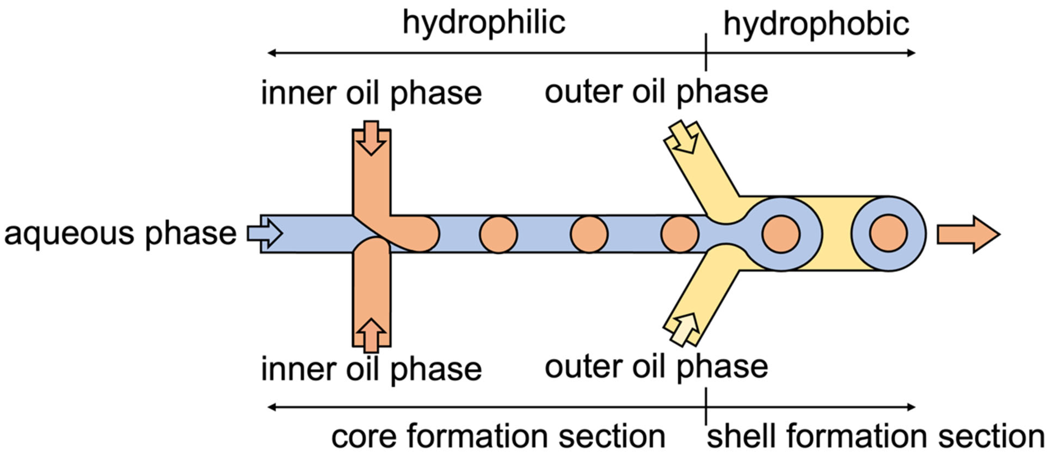
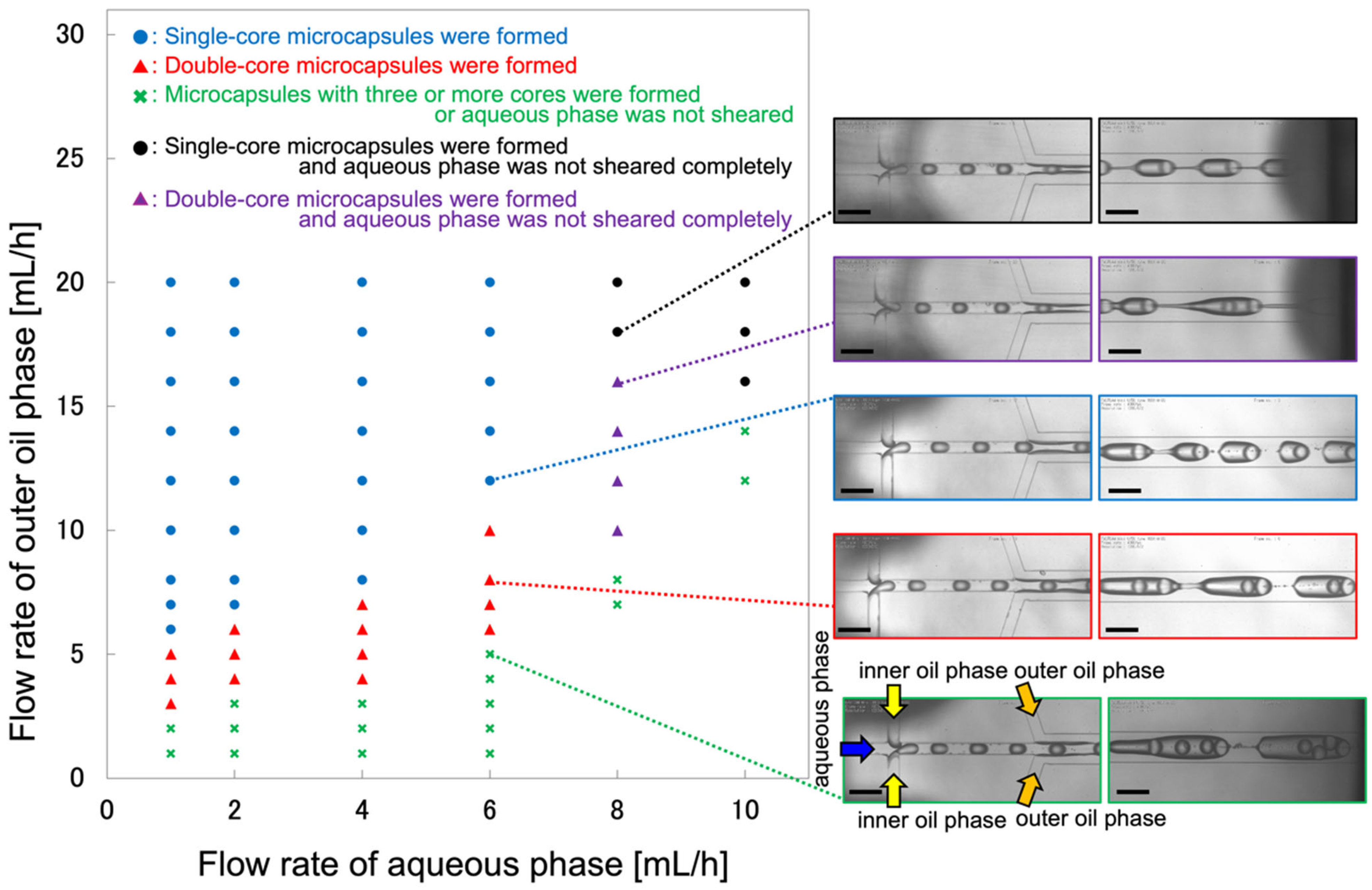
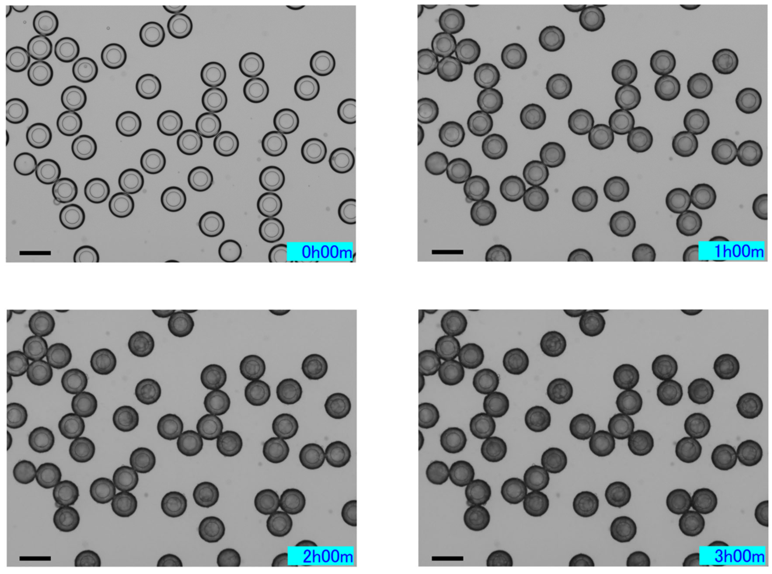
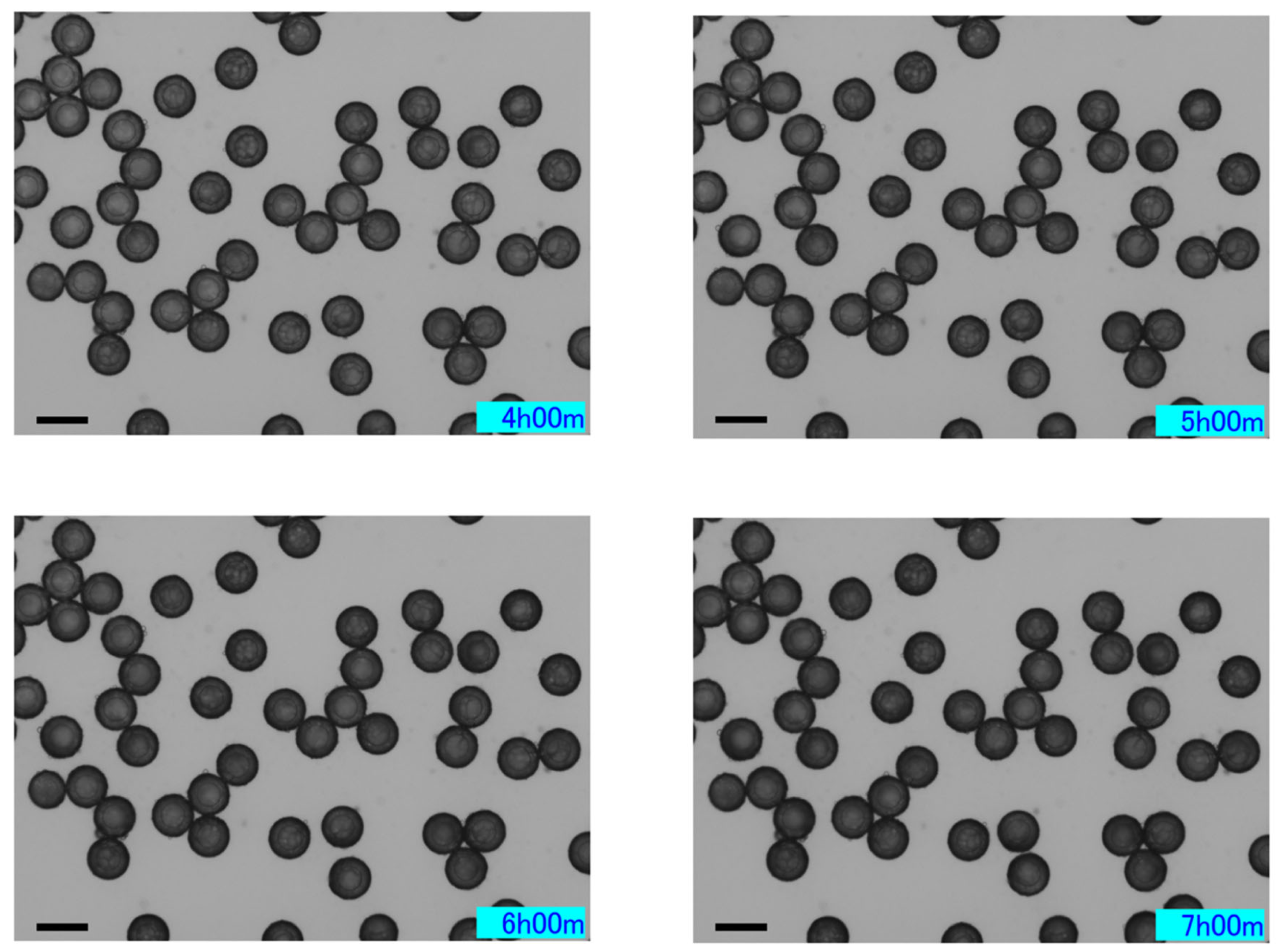
2.1. Microcapsule Formation Behavior

To produce O/W/O-type microcapsules, aqueous, inner oil, and outer oil phases were introduced into the glass microchannel (Figure 1). To produce O/W/O within a single microchannel, it is necessary to create upstream hydrophilic and downstream hydrophobic regions. For this reason, a microchannel with a partially modified wettability was used. The aqueous phase was a 0.5 wt% agarose/2.0 wt% polyvinyl alcohol (PVA) aqueous solution, the inner oil phase was a 1 vol% benzyl alcohol/mineral oil solution, and the outer oil phase was a 1 wt% SY-Glyster/mineral oil solution. Agarose (gelation point: 8–17 °C), which solidifies at a certain temperature or lower, was added to the aqueous phase; however, at this concentration and composition, it remained in the liquid state without gelation at room temperature (approximately 25 °C). This was confirmed experimentally, as the agarose solution could flow through the microchannel without causing any blockage or solidification, allowing stable droplet generation (Figure 2). The formation behavior of the microcapsules was investigated under different flow conditions (Figure 2). To simplify the control parameters, the inner oil phase flow rate was fixed at a constant value (2.0 mL/h), and the flow rates of the aqueous and outer oil phases were varied. Under the flow conditions indicated by ○, microcapsules containing a single oil droplet (single core) were formed. More specifically, at the cross-section upstream of the microchannel, oil droplets (formed from the inner oil phase) were generated alternately while facing each other. Under these conditions, where the flow rate of the outer oil phase was relatively high, single-core microcapsules were generated because the shear stress exerted by the outer oil phase on the aqueous phase increased with the flow rate of the outer oil phase. Under the flow rate conditions indicated by Δ, the shear interval of the aqueous phase was shortened and microcapsules containing two oil droplets were produced. Under the flow rate conditions indicated by X, microcapsules containing three or more oil droplets were produced, or microcapsules were not produced because the aqueous phase was not sheared.

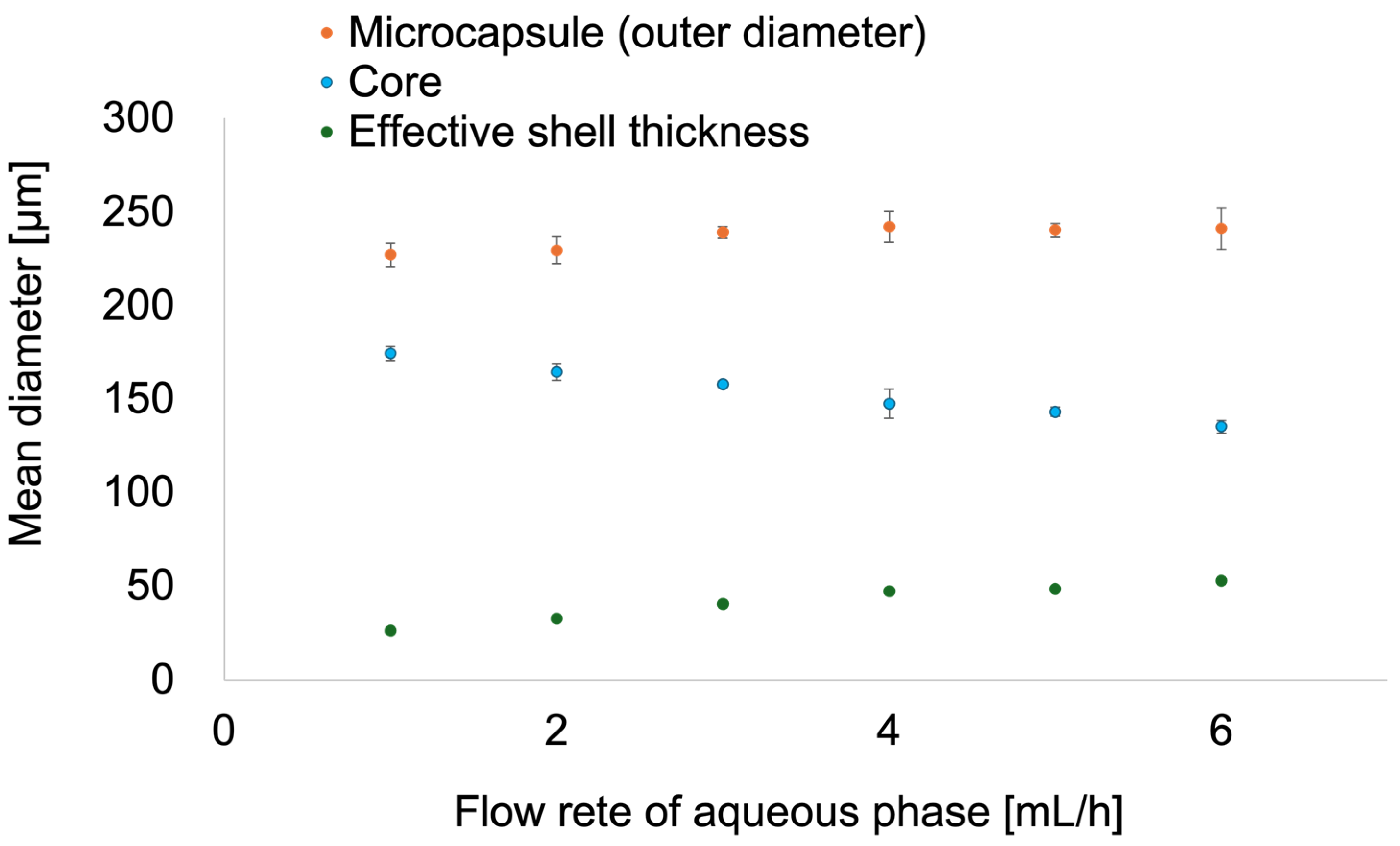
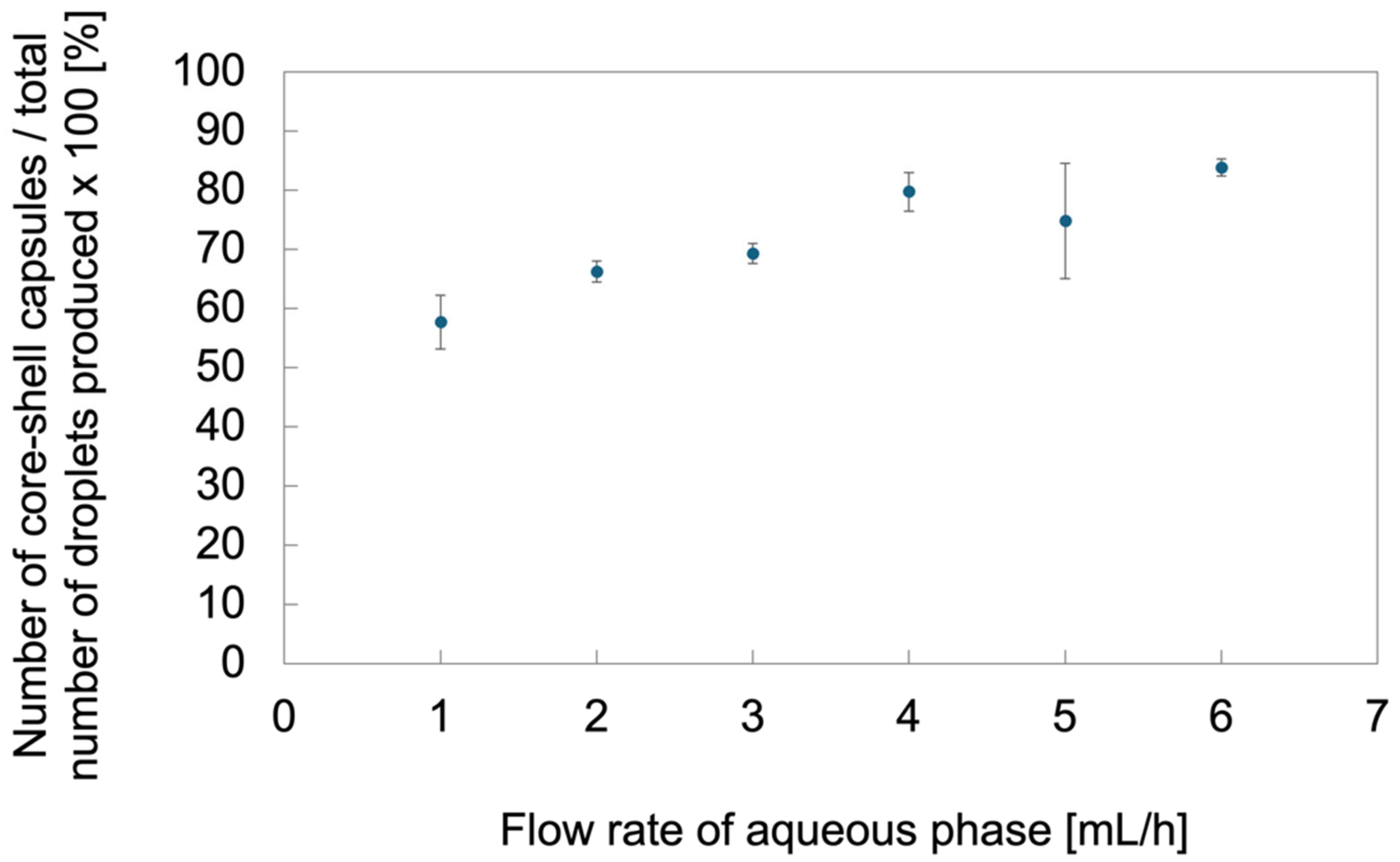
2.2. Effect of Flow Conditions on the Morphology and Dimensions of the Microcapsules

The relationship between the flow conditions and the microcapsule dimensions was investigated. This is because, in general, the dimensions of microcapsules generated using microchannels vary depending on the flow rate [17]. To simplify the control parameters, the inner oil phase flow rate, Qi, was fixed at a constant value (2 mL/h). In this study, to investigate whether the shell thickness (difficult to control in the interface polymerization and sol-gel methods) could be significantly varied, the flow rate was set such that the diameter (outer diameter) of the microcapsules remained approximately constant, whereas the core size (linked to the shell size) could be significantly varied. Specifically, the outer oil phase flow rate Qo was varied such that the ratio of the total flow rate of the inner oil phase and aqueous phase to the outer oil phase flow rate was constant according to the following equation:
Q o = 2 ( Q i + Q a )
The aqueous phase flow rate Qa was varied in the range of 1–6 mL/h, as shown in Figure 2. Under all the flow conditions described above, the formation of single-core microcapsules was confirmed in the microchannel. To examine the morphology and dimensions of the microcapsules, they were collected in a dish and observed under a microscope. The collected microcapsules were mostly single-core microcapsules, with some containing multiple cores formed by the coalescence of single-core microcapsules, and others consisting solely of the aqueous phase with the core removed.
First, we measured the microcapsule diameter (outer diameter) and the core diameter of the collected single-core microcapsules (Figure 3). Shell thickness was calculated as the average value of the microcapsule outer diameter minus the average value of the core diameter divided by 2. In reality, because of the density difference between the inner oil and aqueous phases, the core is not centered within the microcapsule (in this case, the core floats upward). Therefore, the shell thickness formed around the core was uneven, depending on the location (in this case, thinner at the top and thicker at the bottom). Here, we defined the shell thickness as “effective shell thickness”. The coefficient of variation for the single-core microcapsules was 1.3–4.6% for the microcapsule outer diameter and 1.0–5.3% for the core diameter, indicating a narrow distribution. The higher the aqueous phase flow rate, the smaller the core diameter. However, the outer diameter of the microcapsules remained almost the same. The decrease in the core diameter is suggested to be due to an increase in the shear force exerted by the aqueous phase on the inner oil phase as the aqueous phase flow rate increased, thereby promoting the shear of the inner oil phase. Additionally, the fact that the outer diameter of the microcapsules remained nearly constant suggests that increasing the aqueous phase flow rate made the aqueous phase more resistant to shear by the outer oil phase, thereby increasing shell volume (thickness). In summary, within the flow rate range set in this study, the outer diameter of the microcapsules remained nearly constant, whereas the core dimensions could be controlled in response to changes in the flow rate.
Next, we investigated the ratio of single-core microcapsules to the total number of collected microcapsules (that is, the single-core rate) (Figure 4). The single-core rate increased at higher aqueous phase flow rates. Correlating this with Figure 3, the single-core rate increases as the core size decreases. We discuss the reasons for this from the perspectives of interfacial tension balance and local interfacial conditions within the microcapsules. First, we considered the balance of the interfacial tension. The spreading coefficient (S) is an important parameter that determines the behavior of droplets in a three-phase system, as defined in Equation (2) [18,19]. Using this parameter, it is possible to predict whether a liquid phase will spread on another liquid phase and the resulting morphology (Figure 5 and Table 1)
S i = γ j k ( γ i j + γ i k )
where γij, γik, and γjk represent the interfacial tensions between phase i and phase j, phase i and phase k, and phase j and phase k, respectively. In this study, the expansion Fowkes formula [20] shown in Equation (3) was used to calculate the interfacial tensions from the measured surface tensions of the aqueous phase (W), oil phase 1 (O1), and oil phase 2 (O2) (Table 2), and the spreading coefficients were determined (Table 3).
γ i j = σ i + σ j 2 ( σ i   ×   σ j ) 1 2
where σi and σj represent the surface tensions of phases i and j, respectively. The sign of the spreading coefficient (Table 4) suggests a non-engulfing morphology. However, the single-core microcapsules generated and collected in this study exhibited a complete engulfing morphology. This may have been due to the addition of agarose, polysaccharides, and PVA to the shell. These additives may have reduced the fluidity of the core within the microcapsules, contributing to their stabilization.
Subsequently, we considered the local interfacial conditions inside the microcapsules. As the aqueous phase flow rate decreased, the effective shell thickness also decreased. Simultaneously, the single-core ratio decreased. At this point, in addition to single cores, microcapsules consisting only of coalesced microcapsules and the aqueous phase (derived from the shell) were obtained. The cores inside the microcapsules can move easily owing to the influence of surrounding vibrations. Therefore, the smaller the effective shell thickness, the closer the core and shell interfaces. This also indicates that the core and outer oil phase became closer to each other. When the two interfaces come into contact and the pressure increases, the van der Waals forces amplify the fluctuations at the interface, destabilizing it and resulting in coalescence of the two interfaces [21]. In this study, the proximity of the core and outer oil phase interfaces may have caused the release of the core when there were no microcapsules in the vicinity, and may have facilitated coalescence when microcapsules were present in the vicinity.

2.3. Characterization of Microcapsules

2.3.1. Characteristics in Response to Temperature Stimulation

The odor release from microcapsules subjected to temperature stimulation was investigated (Table 5). First, microcapsules produced at a constant flow rate were collected in a glass bottle and closed with a septum cap. Temperature stimulation was applied to the bottle and the headspace vapor was measured using an odor sensor. For comparison, samples at room temperature and those in which the aqueous, inner oil, and outer oil phases were simply mixed (with and without heating) were measured. Preliminary experiments confirmed that the higher the fragrance concentration, the higher the sensor intensity obtained with the liquid combination (mineral oil and benzaldehyde) used in this experiment. The odor intensity was faint when sample no. 1 was sniffed. This confirmed that the microcapsules had odor-releasing properties. Sensor measurements showed that the sensor intensity was lower for the microcapsule samples than for the samples in which the liquids were simply mixed. This suggests that the microcapsules suppressed the release of the fragrance. In addition, higher sensor intensities were obtained for samples subjected to temperature stimulation than at room temperature. This may be because temperature stimulation increased the fluidity of the agarose aqueous solution shell, making it easier for the core and outer oil phase to come into contact. This may contribute to the release of the core into the outer oil phase, resulting in an increase in fragrance concentration in the outer oil phase. Furthermore, statistical analysis revealed that the sensor intensity of the thermally stimulated microcapsule sample (No. 1) was significantly higher than that of its non-stimulated counterpart (No. 2) (p = 0.04, Student’s t-test). This indicates that thermal stimulation had a measurable effect on odor release from the microcapsules.

2.3.2. Characteristics over Time

The characteristics of the microcapsules were investigated over time. After collecting microcapsules generated at a constant flow rate in a dish containing an outer oil phase, microscopic images (phase-contrast observation) were acquired over time. Two types of outer oil phases were used: mineral oil with and without surfactants. Specifically, the mineral oil with surfactant was the same 1 wt% SY-Glyster/mineral oil solution as the outer oil phase (the PVA used in this study is water-soluble; therefore, it was not used as a surfactant for the oil phase). To maximize the coagulation inhibition effect, the surfactant concentration was set to almost the maximum solubility in the mineral oil. When surfactant-containing mineral oil was used as the outer oil phase poured into the dish, the microcapsules gradually turned black over time (Figure 6). This change was caused by the surfactant in the outer oil phase acting on the water in the shell to form reverse micelles (a phenomenon called spontaneous emulsification), which then covered the microcapsules [22]. These reverse micelles may affect the release of odor from the microcapsules. Coalescence was significant when mineral oil without a surfactant was used as the outer oil phase and poured into the dish. The microcapsules (immediately after formation) were collected from a dish containing the outer oil phase. To reduce coalescence by increasing the shell fluidity, they were subsequently cooled at 4 °C (below the gelling point of the agarose gel used) for 17 h. The samples were then observed under a microscope (Figure 7). The microcapsules did not exhibit black discoloration. This indicates that reverse micelles formed by surfactants hardly formed around the microcapsules. In this study, mineral oil containing surfactants was used as the outer oil phase during microcapsule formation; therefore, surfactants were also added to the mineral oil in the dish. However, the surfactants were diluted with mineral oil in the dish. This may have had little effect on the formation of reverse micelles. In addition, it has been reported that reverse micelles change in size depending on the temperature [23]. The interfacial phenomenon related to this characteristic of the reverse micelles may have been involved in the suppression of reverse micelle formation. In this study, the microcapsules were stable for several days. However, further investigations are required to fully evaluate their long-term storage potential.

3. Conclusions

By employing a method for generating O/W/O-type microcapsules within microchannels with partially controlled wettability, we successfully produced monodisperse fragrance-encapsulated microcapsules with a hydrophilic polymer shell, without requiring a prior dispersion process. The core size (linked to the effective shell thickness) could be controlled by adjusting the flow rate. Microcapsules with relatively thick effective shells exhibited more stable interfacial states. The generated microcapsules promoted odor release in response to temperature stimulation. In addition, the microcapsule surface was covered with reverse micelles over time. The formation of these reverse micelles could be suppressed by placing microcapsules in cooled oil without surfactants. These fragrance-encapsulated microcapsules, which can be produced without a preliminary fragrance dispersion process, are expected to contribute to a wide range of applications, including cosmetics and food.

4. Materials and Methods

4.1. Materials

Mineral oil and agarose (A5030, melting point: 50 °C (0.8%), gelation point: 8–17 °C) were obtained from Sigma-Aldrich (St. Louis, MO, USA), and SY-Glyster (surfactant, polyglycerol esters of fatty acids, CRS-75) was obtained from Sakamoto Yakuhin Kogyo Co., Ltd. (Osaka, Japan), PVA (surfactant, Goseonol, GL-03) was obtained from Mitsubishi Chemical Corporation (Tokyo, Japan), and octadecyltrichlorosilane was obtained from Tokyo Chemical Industry Co. Ltd. (Tokyo, Japan), benzaldehyde (fragrance), and toluene were obtained from FUJIFILM Wako Pure Chemical Corporation (Osaka, Japan). As previously described, the three phases used in this study were as follows: namely, the aqueous phase was a 0.5 wt% agarose/2.0 wt% PVA aqueous solution, the inner oil phase was a 1 vol% benzyl alcohol/mineral oil solution, and the outer oil phase was a 1 wt% SY-Glyster/mineral oil solution.

4.2. Microcapsule Formation

O/W/O-type microcapsules were formed by introducing the aqueous, inner oil, and outer oil phases into a glass microchannel (depth: 200 µm, Figure S2). The microchannel was fabricated by bonding three layers: a top plate with through-holes for the inlets and outlets, a middle plate with through-holes forming the channel structure, and a bottom flat plate. The device was manufactured by KADOMI Optical Industry Co., Ltd., Japan. To form O/W/O-type microcapsules within a single microchannel, the wettability of the microchannel was partially modified using a method described in previous studies [24]. In summary, the microchannel was rendered hydrophobic by filling it with 1 vol% octadecyltrichlorosilane/toluene solution for 10 min. Then, the microchannel was partially irradiated with ultraviolet light through a photomask using a vacuum ultraviolet irradiation device (Min-excimer, Ushio Inc., Tokyo, Japan) for 120 min, and only the irradiated areas were hydrophilized. A polytetrafluoroethylene tube (inner diameter: 0.8 mm, outer diameter: 1.58 mm) was used to connect the glass syringe and microchannel. The aqueous, inner oil, and outer oil phases were filled into each syringe and introduced into the microchannel using a syringe pump (Legato 210, KD Scientific, Holliston, MA, USA). Microscopic images of microcapsule formation in the microchannel were obtained using an optical microscope (BX50, Olympus Corporation, Tokyo, Japan) equipped with a high-speed camera (FASTCAM Mini UX50, PHOTRON LIMITED, Tokyo, Japan). Microcapsules were collected in a glass-bottom dish (D11130H, Matsunami Glass Ind., Ltd., Osaka, Japan) and microscopic images of the microcapsules were obtained using an inverted optical microscope (BZ-X710, KEYENCE CORPORATION, Osaka, Japan). The dimensions of the microcapsules were analyzed using image analysis software (ImageJ, version 2.14.0). The mean diameter and coefficient of variation (=standard deviation/mean diameter) were calculated from the diameter values obtained for each microcapsule. Surface tension was measured using a surface tension meter (CBVP-Z, Kyowa Interface Science Co., Ltd., Saitama, Japan).

4.3. Characterization Methods for Microcapsules

To evaluate the characteristics of the microcapsules in response to temperature stimulation, the following procedures were performed. Microcapsules were generated at a constant flow rate (aqueous phase, 6 mL/h; inner oil phase, 2 mL/h; outer oil phase, 16 mL/h) and collected in a 20 mL glass bottle with a septum-sealed lid for 20 min. Approximately 5 g of the outer oil phase was removed from the samples using a syringe. For comparison, a solution was prepared by mixing 2 g of aqueous phase, 0.6 g of inner oil phase, and 0.8 g of outer oil phase. The three liquids were placed in a glass bottle, sealed with a cap, and mixed vigorously by repeatedly inverting the bottle. The headspaces of both the samples were replaced with air. The glass bottles were then placed in an oven set to 70 °C (SONW-300SB, AS ONE CORPORATION, Osaka, Japan) or room temperature (22–25 °C) for 30 min, followed by 10 min at room temperature. The headspace vapor in the glass bottles was measured using an odor sensor (NeOse Advance, Aryballe Technologies SA, Grenoble, France). To evaluate the characteristics over time, microcapsules were collected at a constant flow rate (aqueous phase: 6 mL/h, inner oil phase: 2 mL/h, outer oil phase: 16 mL/h) into a glass bottom dish containing 2 mL of the outer oil phase (with and without surfactant) and observed under a microscope.

Supplementary Materials

The following supporting information can be downloaded at: https://www.mdpi.com/article/10.3390/app15105650/s1, Figure S1: Structure of a conventional core–shell microcapsule requiring predispersion; Figure S2: Dimensional drawing of the microchannel device.

Author Contributions

Conceptualization, H.H.; methodology, H.H., H.M. and Y.M.; formal analysis, H.H., H.M. and Y.M.; investigation, H.H., H.M. and Y.M.; data curation, H.H., H.M. and Y.M.; writing—original draft preparation, H.H. and H.M.; writing—review and editing, H.H., H.M., Y.M. and M.H.; visualization, H.H. and H.M.; supervision, M.H.; project administration, H.H. All authors have read and agreed to the published version of the manuscript.

Funding

This research received no external funding.

Institutional Review Board Statement

Not applicable.

Informed Consent Statement

Not applicable.

Data Availability Statement

The original contributions presented in this study are included in the article. Further inquiries can be directed to the corresponding author.

Acknowledgments

I would like to thank Satoru Suzuki for his valuable assistance with the experiments.

Conflicts of Interest

The authors declare no conflicts of interest.

References

  1. Liu, Y.X.; Liu, K.Y.; Zhao, M.N.; Wang, S.H.; Zhou, Z.X.; Shen, Y.Q.; Jiang, L.M. A pH-responsive fragrance release system based on pseudopeptide polymeric micelles. React. Funct. Polym. 2018, 132, 138–144. [Google Scholar] [CrossRef]
  2. Trachsel, A.; Paret, N.; Berthier, D.L.; Herrmann, A. Light-Induced Fragrance Release from 2-Oxoacetates: Impact of Compound Mixtures on the Efficiency of the Norrish Type II Photoreaction in Solution and in Encapsulation Systems. Chemphotochem 2022, 6, e202200045. [Google Scholar] [CrossRef]
  3. Nicastro, G.; Black, L.M.; Ravarino, P.; D’Agostino, S.; Faccio, D.; Tomasini, C.; Giuri, D. Controlled Hydrolysis of Odorants Schiff Bases in Low-Molecular-Weight Gels. Int. J. Mol. Sci. 2022, 23, 3105. [Google Scholar] [CrossRef]
  4. Kim, Y.M.; Lee, K.; Lee, Y.; Yang, K.; Choe, D.; Roh, Y.H. Thermoresponsive semi-interpenetrating gelatin-alginate networks for encapsulation and controlled release of scent molecules. Int. J. Biol. Macromol. 2022, 208, 1096–1105. [Google Scholar] [CrossRef]
  5. Theisinger, S.; Schoeller, K.; Osborn, B.; Sarkar, M.; Landfester, K. Encapsulation of a Fragrance via Miniemulsion Polymerization for Temperature-Controlled Release. Macromol. Chem. Phys. 2009, 210, 411–420. [Google Scholar] [CrossRef]
  6. Sansukcharearnpon, A.; Wanichwecharungruang, S.; Leepipatpaiboon, N.; Kerdcharoen, T.; Arayachukeat, S. High loading fragrance encapsulation based on a polymer-blend: Preparation and release behavior. Int. J. Pharm. 2010, 391, 267–273. [Google Scholar] [CrossRef]
  7. Sadovoy, A.V.; Lomova, M.V.; Antipina, M.N.; Braun, N.A.; Sukhorukov, G.B.; Kiryukhin, M.V. Layer-by-Layer Assembled Multilayer Shells for Encapsulation and Release of Fragrance. ACS Appl. Mater. Interfaces 2013, 5, 8948–8954. [Google Scholar] [CrossRef]
  8. Utada, A.S.; Lorenceau, E.; Link, D.R.; Kaplan, P.D.; Stone, H.A.; Weitz, D.A. Monodisperse double emulsions generated from a microcapillary device. Science 2005, 308, 537–541. [Google Scholar] [CrossRef]
  9. Zhang, H.; Tumarkin, E.; Peerani, R.; Nie, Z.; Sullan, R.M.A.; Walker, G.C.; Kumacheva, E. Microfluidic production of biopolymer microcapsules with controlled morphology. J. Am. Chem. Soc. 2006, 128, 12205–12210. [Google Scholar] [CrossRef]
  10. Zhao, Y.J.; Shum, H.C.; Adams, L.L.A.; Sun, B.J.; Holtze, C.; Gu, Z.Z.; Weitz, D.A. Enhanced Encapsulation of Actives in Self-Sealing Microcapsules by Precipitation in Capsule Shells. Langmuir ACS J. Surf. Colloids 2011, 27, 13988–13991. [Google Scholar] [CrossRef]
  11. Abbaspourrad, A.; Datta, S.S.; Weitz, D.A. Controlling Release From pH-Responsive Microcapsules. Langmuir ACS J. Surf. Colloids 2013, 29, 12697–12702. [Google Scholar] [CrossRef] [PubMed]
  12. Abbaspourrad, A.; Duncanson, W.J.; Lebedeva, N.; Kim, S.H.; Zhushma, A.P.; Datta, S.S.; Dayton, P.A.; Sheiko, S.S.; Rubinstein, M.; Weitz, D.A. Microfluidic Fabrication of Stable Gas-Filled Microcapsules for Acoustic Contrast Enhancement. Langmuir ACS J. Surf. Colloids 2013, 29, 12352–12357. [Google Scholar] [CrossRef]
  13. Abbaspourrad, A.; Carroll, N.J.; Kim, S.H.; Weitz, D.A. Polymer Microcapsules with Programmable Active Release. J. Am. Chem. Soc. 2013, 135, 7744–7750. [Google Scholar] [CrossRef]
  14. Grolman, J.M.; Inci, B.; Moore, J.S. pH-Dependent Switchable Permeability from Core Shell Microcapsules. ACS Macro Lett. 2015, 4, 441–445. [Google Scholar] [CrossRef]
  15. Zieringer, M.A.; Carroll, N.J.; Abbaspourrad, A.; Koehler, S.A.; Weitz, D.A. Microcapsules for Enhanced Cargo Retention and Diversity. Small 2015, 11, 2903–2909. [Google Scholar] [CrossRef]
  16. Lee, H.; Choi, C.H.; Abbaspourrad, A.; Wesner, C.; Caggioni, M.; Zhu, T.; Weitz, D.A. Encapsulation and Enhanced Retention of Fragrance in Polymer Microcapsules. ACS Appl. Mater. Interfaces 2016, 8, 4007–4013. [Google Scholar] [CrossRef]
  17. Ling, S.D.; Geng, Y.H.; Chen, A.; Du, Y.A.; Xu, J.H. Enhanced single-cell encapsulation in microfluidic devices: From droplet generation to single-cell analysis. Biomicrofluidics 2020, 14, 061508. [Google Scholar] [CrossRef]
  18. Torza, S.; Mason, S.G. Three-phase interactions in shear and electrical fields. J. Colloid Interface Sci. 1970, 33, 67–83. [Google Scholar] [CrossRef]
  19. Xu, J.-H.; Ge, X.-H.; Chen, R.; Luo, G.-S. Microfluidic preparation and structure evolution of double emulsions with two-phase cores. RSC Adv. 2014, 4, 1900–1906. [Google Scholar] [CrossRef]
  20. Fowkes, F.M. Attractive Forces at Interfaces. Ind. Eng. Chem. 1964, 56, 40–52. [Google Scholar] [CrossRef]
  21. Chesters, A.K. The Modeling of Coalescence Processes in Fluid Liquid Dispersions—A Review of Current Understanding. Chem. Eng. Res. Des. 1991, 69, 259–270. [Google Scholar]
  22. Fukuyama, M.; Mizuguchi, T.; Santivongskul, P.; Ono, Y.; Kasuya, M.; Inagawa, A.; Hibara, A. Kinetic description of water transport during spontaneous emulsification induced by Span 80. Nanoscale 2024, 16, 4056–4062. [Google Scholar] [CrossRef] [PubMed]
  23. Penttilä, P.A.; Vierros, S.; Utriainen, K.; Carl, N.; Rautkari, L.; Sammalkorpi, M.; Österberg, M. Phospholipid-Based Reverse Micelle Structures in Vegetable Oil Modified by Water Content, Free Fatty Acid, and Temperature. Langmuir ACS J. Surf. Colloids 2019, 35, 8373–8382. [Google Scholar] [CrossRef]
  24. Hirama, H.; Wada, S.; Shimamura, J.; Komazaki, Y.; Inoue, T.; Torii, T. Surface modification of a glass microchannel for the formation of multiple emulsion droplets. Microfluid. Nanofluid. 2017, 21, 91. [Google Scholar] [CrossRef]
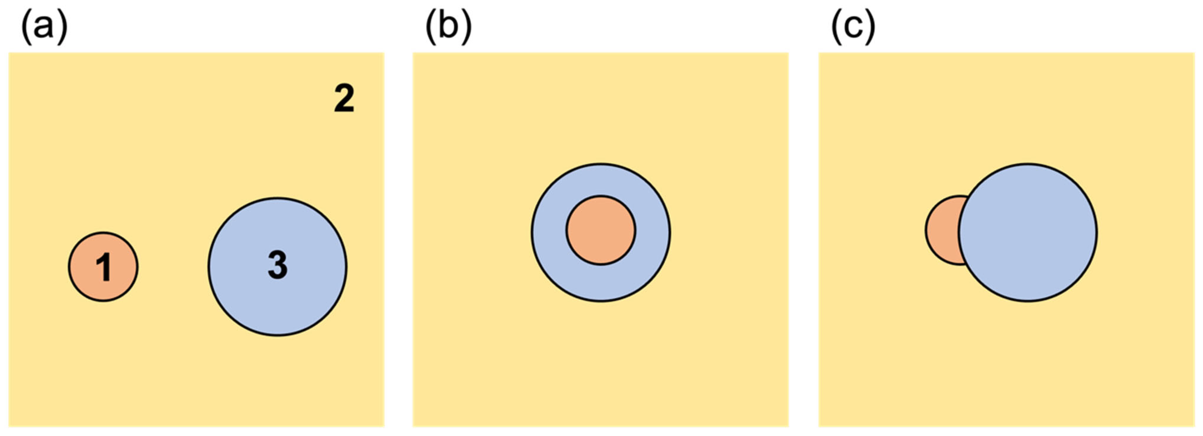
Figure 1. Schematic diagram of the formation of oil-in-water-in-oil (O/W/O) microcapsules in a microchannel.
Applsci 15 05650 g001
Figure 2. Microcapsule formation behavior under different flow conditions. The inset shows micrographs of the upstream and downstream microchannels. The flow rate of the inner oil phase was 2.0 mL/h. Scale bars are 300 µm.
Applsci 15 05650 g002
Figure 3. Relationship between aqueous phase flow rate and dimensions of single-core microcapsules. The effective shell thickness was calculated by subtracting the average core diameter from the average microcapsule outer diameter and dividing by 2. The flow rate of the inner oil phase was 2.0 mL/h. The outer oil phase flow rate was adjusted in the range of 6–16 mL/h in accordance with Equation (1) to correspond to the aqueous phase flow rate.
Applsci 15 05650 g003
Figure 4. Relationship between aqueous phase flow rate and single-core rate.
Applsci 15 05650 g004
Figure 5. Possible forms in three phases: (a) non-engulfing, (b) complete engulfing, and (c) partial engulfing. In this study, phases 1, 2, and 3 indicate oil phase 1, oil phase 2, and aqueous phase, respectively.
Applsci 15 05650 g005
Figure 6. Time-lapse microscopic images of microcapsules collected in the outer oil phase containing surfactants. Scale bars are 300 µm.
Applsci 15 05650 g006aApplsci 15 05650 g006b
Figure 7. Microscopic image of microcapsules collected and cooled in the outer oil phase without surfactants. The coalescence of the microcapsules that came into contact with each other during cooling was confirmed. Scale bar is 300 µm.
Applsci 15 05650 g007
Table 1. Droplet morphologies determined by sign combinations of spreading coefficients.
Droplet StateS1S2S3Description
Non-engulfing<0>0<0Phase 1 does not spread on Phase 2, and Phase 3 does not spread on Phase 2. The droplets exist independently.
Complete engulfing<0<0>0Phase 3 completely engulfs Phase 1, forming a stable O/W/O or W/O/W emulsion floating in Phase 2.
Partial engulfing<0<0<0Phase 3 partially engulfs Phase 1, with part of Phase 1 exposed to Phase 2.
Table 2. Surface tension. All the values were obtained at approximately 25 °C.
SymbolPhaseCompositionSurface Tension (Mean ± SD) (mN/m)
σWAqueous phase0.5 wt% agarose/2.0 wt% PVA in water39.6 ± 0.64
σO1Oil phase 11 vol% benzaldehyde in mineral oil29.2 ± 0.05
σO2Oil phase 2Mineral oil29.3 ± 0.15
Table 3. The interfacial tension was calculated using the expansion Fowkes formula.
SymbolInterfaceInterfacial Tension (mN/m)
γWO1Aqueous/oil phase 10.79
γWO2Aqueous/oil phase 20.77
γO1O2Oil phase 1/oil phase 28.5 × 10⁻⁵
Table 4. Spreading coefficient calculated from the interfacial tension.
SymbolSpreading Coefficient (mN/m)
SO1−0.016
SO20.016
SW−1.6
Table 5. The intensities of the sample vapors were measured using an odor sensor. * Significantly different from No. 2 (p = 0.04, Student’s t-test).
No.Liquid PropertiesThermal StimulationMeasured Sensor Intensity (Mean ± SD)
1MicrocapsuleWith7.1 ± 0.2 *
2MicrocapsuleWithout6.7 ± 0.4
3Simply stirred solutionWith9.3 ± 0.1
4Simply stirred solutionWithout8.8 ± 0.2
Disclaimer/Publisher’s Note: The statements, opinions and data contained in all publications are solely those of the individual author(s) and contributor(s) and not of MDPI and/or the editor(s). MDPI and/or the editor(s) disclaim responsibility for any injury to people or property resulting from any ideas, methods, instructions or products referred to in the content.

