Article

Increased Myo/Nog Cell Presence and Phagocytic Activity in Retinal Degeneration: Insights from a Mouse Model

by
Diana Crowley
,
Samantha Murad
,
Courtney Helm
,
Rachel Souza
,
Sarah Coughlan
,
Scott Serpico
,
Eric Sugarman
,
Kyle Margulies
,
Brian Heist
,
Kathryn D. Mitchell
,
Christopher K. Sutera
,
Mark Martin
,
Carlos Font
,
Mary Woodruff
,
E-Jine Tsai
,
Rushil Brahmbhatt
,
Paul Lecker
,
Grzegorz Gorski
,
John Benalcazar
,
Serena Young
,
Abey Martin
,
Lindsay Gugerty
,
Jacquelyn Gerhart
,
Mindy George-Weinstein
and
Arturo Bravo-Nuevo
*
add Show full author list remove Hide full author list
Department of Biomedical Sciences, Philadelphia College of Osteopathic Medicine, Philadelphia, PA 19131, USA
*
Author to whom correspondence should be addressed.
Appl. Sci. 2025, 15(10), 5486; https://doi.org/10.3390/app15105486
Submission received: 5 March 2025 / Revised: 30 April 2025 / Accepted: 8 May 2025 / Published: 14 May 2025
(This article belongs to the Section Applied Biosciences and Bioengineering)

Abstract

:
Myo/Nog cells play a pivotal role in ocular development and demonstrate a rapid response to stress and injury. This study investigates their behavior and distribution in a murine model of retinitis pigmentosa, specifically in C3H/HeJ mice, which exhibit photoreceptor degeneration due to a homozygous mutation in the Pde6brd1 gene. Retinal samples from C3H/HeJ and C57BL/6J mice were analyzed at postnatal weeks 2.5 to 6 using hematoxylin and eosin staining, immunofluorescence for brain-specific angiogenesis inhibitor 1 (BAI1) expressed in Myo/Nog cells, and TUNEL labeling for apoptotic cell detection. The results demonstrated a progressive thinning of the outer nuclear layer (ONL) in C3H mice, accompanied by a significant increase in Myo/Nog cell numbers. In normal retinas, Myo/Nog cells were primarily located in the inner nuclear and outer plexiform layers. However, in C3H/HeJ mice, they accumulated in the ONL near apoptotic photoreceptors and within the choroid. Notably, in these degenerative regions, Myo/Nog cells exhibited features of phagocytosis, suggesting a role in apoptotic cell clearance. Additionally, parallels between Myo/Nog cell responses in retinitis pigmentosa and models of oxygen-induced retinopathy, ocular hypertension, and light damage suggest that these cells may be leveraged for therapeutic purposes.

1. Introduction

Myo/Nog (M/N) cells were first identified in the chick embryo blastocyst, where they express MyoD, a muscle-specific transcription factor; Noggin, a bone morphogenetic protein (BMP) inhibitor; and brain-specific angiogenesis inhibitor 1 (BAI1) [1,2]. Their critical functions in early development have been demonstrated, with studies showing that the absence of Myo/Nog cells results in severe malformations, including the lack of skeletal muscle formation, central nervous system defects, and various ocular abnormalities such as anophthalmia, lens dysgenesis, and retinal overgrowth [3].
Beyond embryonic development, Myo/Nog cells persist in multiple tissues of adult organisms, including mice, rats, rabbits, and humans. Previous research has explored their role in ocular and systemic pathologies, demonstrating their capacity to respond to tissue injury. In the lens, these cells localize to wound sites, differentiate into contractile myofibroblasts, and contribute to posterior capsule opacification, a common complication following cataract surgery [4]. Their depletion in murine models of retinopathy of prematurity results in increased neuronal apoptosis, while their addition to the vitreous cavity following light-induced retinal damage enhances cell survival and preserves visual function [5]. Additionally, neuroprotective properties of Myo/Nog cells have been documented in models of ocular hypertension and localized brain injuries [6,7].
Although Myo/Nog cells respond to injury in the eye, the mechanisms through which they mediate neuroprotection are unknown. Most prior studies have focused on acute and artificially induced models of ocular pathology, limiting our understanding of Myo/Nog cell behavior in chronic retinal conditions. The role and anatomical locations of the Myo/Nog cells in a naturally occurring model of ocular pathology have not been looked at before. This study differs from prior research that focused on acute injury models by utilizing a congenital retinitis pigmentosa model, allowing us to examine Myo/Nog cell behavior during natural disease progression. For this, we used the C3H/HeJ mouse strain, which is homozygous for the cGMP phosphodiesterase 6B (PDE6) mutation, also known as the rd1 mutation. PDE6 is in the outer segments of photoreceptors and is an essential part of the visual signaling cascade [8]. In mice with the PDE6 mutation, an accumulation of intracellular cGMP leads to the deleterious influx of Ca2+ into the cell, which consequently leads to rod cell death through a mechanism likely mediated by apoptosis-inducing factor (AIF) in the mitochondria [9,10,11]. Rod cell death begins at postnatal day 7 (P7) and progresses quickly for 3–4 weeks [12]. Mice retain some pattern recognition ability up to postnatal day 40 (P40). Like humans with RP, the mutation eventually affects cones and leads to complete blindness by 6 weeks of age [13]. Other widely used models of RP include the rd10 mouse, which exhibits slower degeneration, and the Rho/ mouse, which lacks rhodopsin and undergoes rapid rod degeneration. Compared to these models, the C3H/HeJ strain offers a consistent, faster, and well-characterized timeline of degeneration useful for studying early to late-stage retinal pathology within a month after birth.
This study aims to quantify Myo/Nog cell distribution in response to retinal degeneration and assess their phagocytic activity in the ONL based on BAI1’s known function as a mediator of phagocytosis [14] and our previous study in which Myo/Nog cells internalized tattoo ink, dead cells in lens cultures, and microbeads injected into the anterior chamber of the eye [15].

2. Materials and Methods

2.1. Animal Model and Housing Conditions

C57BL/6J (C57) and C3H/HeJ (C3H) mice were acquired from Jackson Laboratories (Bar Harbor, ME, USA). Mice were housed under standard conditions with a 12 h light/dark cycle, an ambient temperature of 22 ± 1 °C, and humidity control (50 ± 10%). Food and water were provided ad libitum. An equal distribution of male and female mice was used to control for sex-dependent variability in retinal degeneration. All procedures adhered to guidelines approved by the Institutional Animal Care and Use Committee (IACUC) at the Philadelphia College of Osteopathic Medicine (A17-007).

2.2. Tissue Collection and Processing

Mice were euthanized via CO2 inhalation followed by cervical dislocation, in accordance with AVMA guidelines. Eyes were enucleated immediately, punctured at the cornea, and fixed in 4% paraformaldehyde (pH 7.4) for three hours. Following fixation, tissues were rinsed three times with phosphate-buffered saline (PBS) (15 min per rinse) before cryoprotection in 30% sucrose overnight at 4 °C. Eyes were embedded in Tissue-Plus OCT compound (ThermoFisher Scientific, Waltham, MA, USA), frozen, and sectioned at 20 μm thickness using a Leica Biosystems cryostat. Sections were stored at −80 °C until further analysis.

2.3. Immunohistochemistry and Imaging

Tissue sections were stained with hematoxylin and eosin (H&E) for morphological assessment. The ONL and INL thicknesses were measured at eight standardized locations along the retina relative to the optic nerve using NIS Elements software (Basic Research version) (Nikon, Belmont, CA, USA). For immunofluorescence, retinal sections were labeled with anti-BAI1 monoclonal antibodies (mAbs) to detect Myo/Nog cells. Sections were permeabilized with 0.1% Triton X-100, blocked with 10% goat serum, and incubated with primary antibodies overnight at 4 °C. Fluorescein-conjugated secondary antibodies (Jackson ImmunoResearch, West Grove, PA, USA) were used for visualization. Apoptotic cells were identified using the In Situ Cell Death Detection Kit (TUNEL assay, Roche, Germany). Confocal microscopy (Olympus FLUOVIEW FV3000, Evident Scientific, Waltham, MA, USA) was used for imaging, and cell quantification was performed on 10 sections per eye, and 5–10 eyes per group were analyzed.

2.4. Statistical Analyses

Statistical analyses were performed using GraphPad Prism 10. All datasets were tested for normality using the Shapiro–Wilk test. Two-way ANOVA followed by Tukey’s post hoc test was used to compare ONL/INL ratios and Myo/Nog cell counts between groups. Significance was set at p ≤ 0.05. Data are presented as mean ± SEM.

3. Results

3.1. Photoreceptor Degeneration in the C3H Mice

C3H mice exhibited a progressive thinning of the ONL from 2.5 to 6 weeks, with a significant reduction in the ONL/INL ratio compared to C57 mice (p < 0.05). Myo/Nog cells were observed in significantly higher numbers in the C3H retina compared to the C57 retina at all time points (p < 0.05). In normal retinas, Myo/Nog cells were predominantly located in the inner nuclear and outer plexiform layers. However, in C3H mice, they were primarily localized to the ONL and choroid, increasing in number over time.
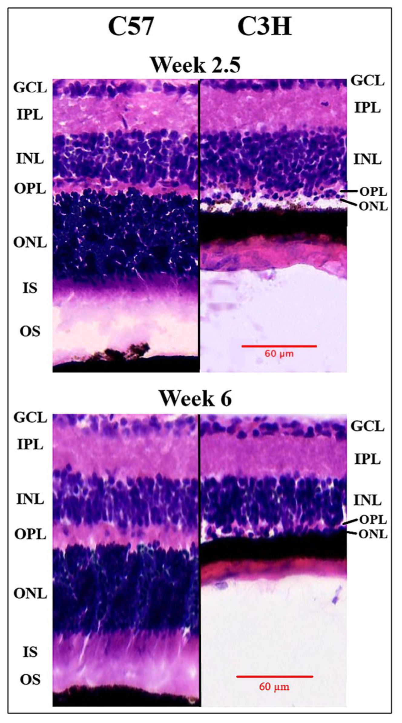
As shown in Figure 1 and Figure 2, ONL thinning was most pronounced between weeks 3 and 4 in C3H mice, with a significantly greater loss of photoreceptor cells occurring in the central retina compared to the periphery. The ONL/INL ratio was consistently higher in C57 mice at all four measured retinal locations (p < 0.05), confirming that retinal degeneration in C3H mice progresses in a uniform but slightly regionally dependent manner.

3.2. Distribution of Myo/Nog Cells Shift in Number and Location with Stress

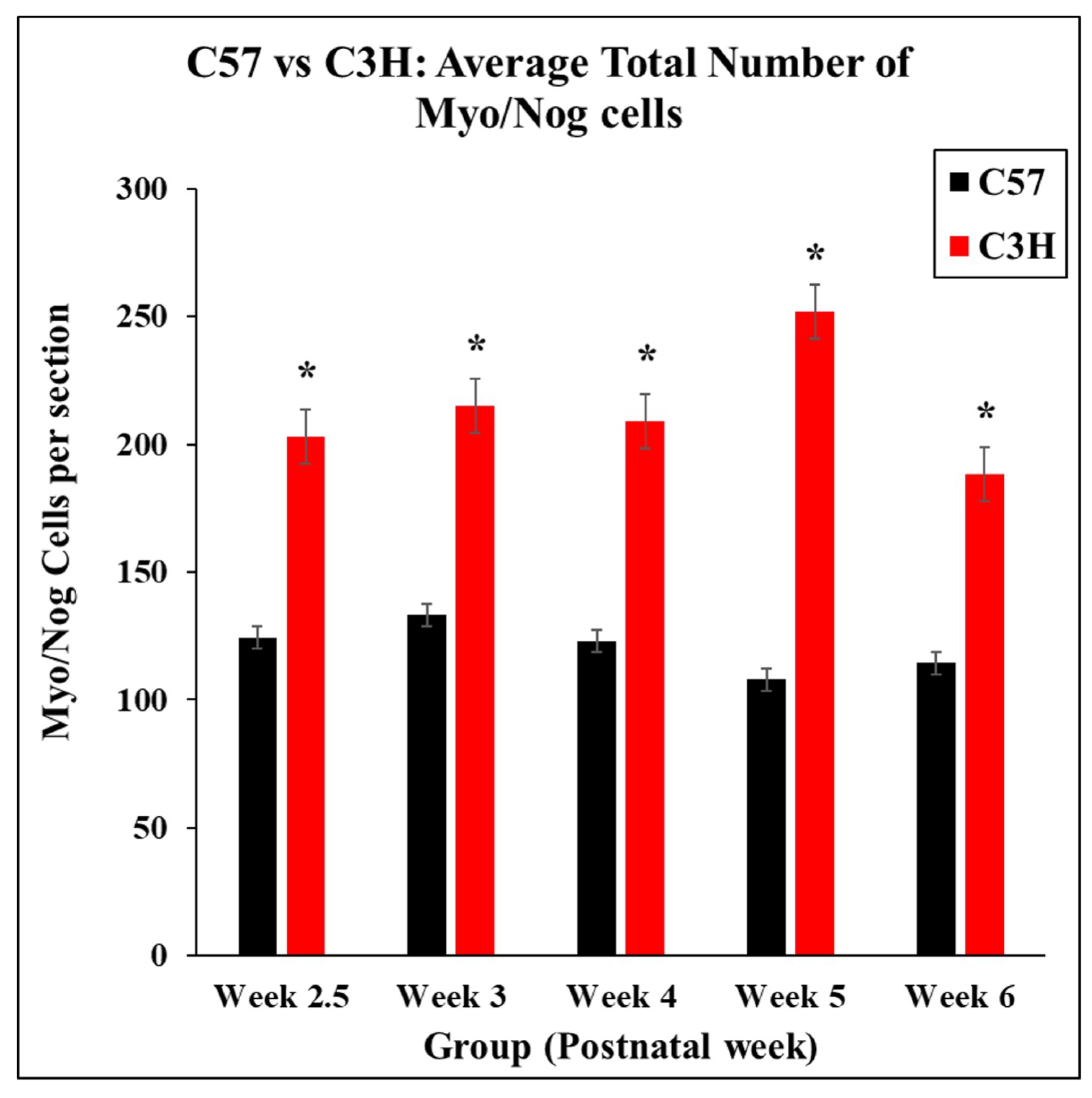
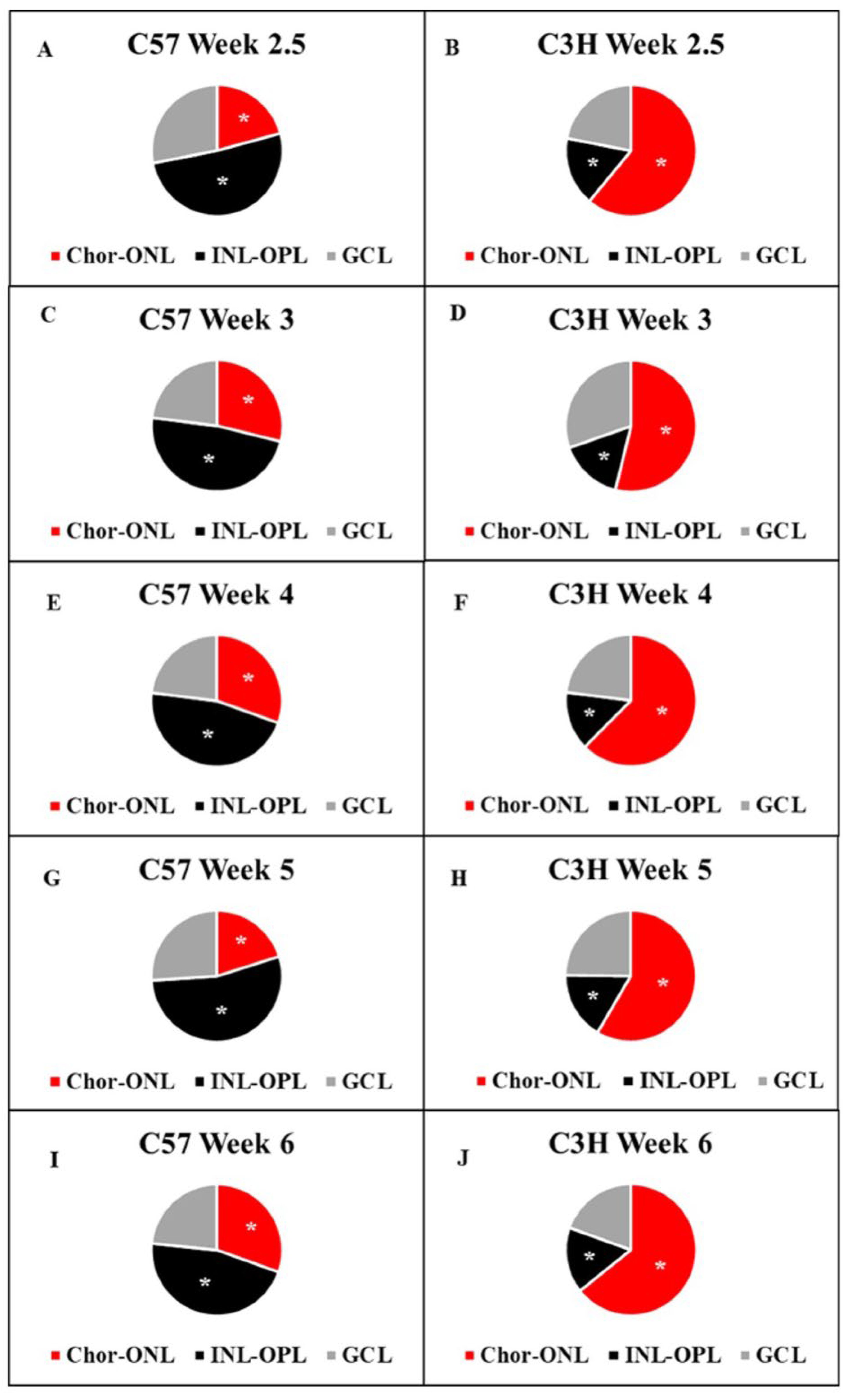
The spatial distribution of Myo/Nog cells also changed dynamically, with increasing numbers detected in the ONL and choroid over time, particularly in areas with the highest levels of TUNEL+ apoptotic photoreceptors (Figure 3, Figure 4, Figure 5 and Figure 6).
The quantification of Myo/Nog cells revealed a statistically significant increase in their numbers at week 3 compared to week 2.5 (p < 0.05), indicating a rapid response to early-stage photoreceptor degeneration (Figure 4). By week 5, Myo/Nog cells were highly concentrated in the ONL, with the highest densities in regions adjacent to apoptotic photoreceptors. This distribution suggests that Myo/Nog cells migrate toward sites of photoreceptor loss and may contribute to apoptotic cell clearance (Figure 4, Figure 5 and Figure 6).
Additionally, comparisons with C57 mice showed that Myo/Nog cells in healthy retinas remained relatively stable in their numbers and localization, reinforcing the notion that their proliferation and redistribution in C3H mice are driven by retinal degeneration (Figure 3, Figure 4, Figure 5 and Figure 6).

3.3. Localization of Nuclear Remnants Inside Myo/Nog Cells Capable of Phagocytosis of Degenerating Photoreceptors

As the resident immune cells of the retina, microglia are highly responsive to injury or stress. In retinal degeneration, they migrate to areas of cell death, including the ONL, as well as phagocytose apoptotic cells and debris. Microglia are considered professional phagocytes and are essential for maintaining retinal homeostasis and preventing secondary inflammation. Myo/Nog cells were previously shown to be phagocytic in the skin and anterior segment of the eye [15]. Though typically less numerous than microglia, Myo/Nog cells may also play a supportive role in clearing debris if they migrate to the ONL during stress. Sections of C3H mice were double-labeled with the BAI1mAb and TUNEL reagents to examine whether Myo/Nog cells might be involved in clearing degenerating photoreceptors.
The imaging of the choroid and outer nuclear layer (ONL) of the retina in C3H mice revealed a notable overlap in Myo/Nog cell labeling (G8+) with TUNEL-positive cells (Figure 7), which are markers of apoptotic photoreceptors. This colocalization confirmed that Myo/Nog cells actively participate in phagocytosing apoptotic photoreceptors. Unlike microglia, which are typically regarded as the primary phagocytes in retinal degeneration, Myo/Nog cells exhibited non-professional phagocytic behavior in response to ongoing retinal stress and cell death in the ONL.
The BAI+ labeling in conjunction with TUNEL+ signals highlights the capacity of Myo/Nog cells to respond dynamically to photoreceptor degeneration. This response includes their migration to affected areas within the ONL and choroid and subsequent engulfment of apoptotic photoreceptor nuclei. These findings suggest that Myo/Nog cells may supplement the function of microglia in retinal degeneration, providing additional clearance of apoptotic cells that may otherwise accumulate and exacerbate retinal stress.
This phagocytic activity observed in Myo/Nog cells challenges the traditional understanding that microglia are solely responsible for debris clearance in retinal degeneration. While microglia remain the primary immune cells in the retina, Myo/Nog cells appear to play a supportive phagocytic role, particularly in environments of high cell death, as seen in progressive retinal degenerative conditions like retinitis pigmentosa. These results open new avenues for considering Myo/Nog cells in therapeutic strategies aimed at enhancing retinal debris clearance and potentially slowing the progression of retinal degeneration by reducing inflammation and secondary cell damage.

4. Discussion

This study investigated the role of Myo/Nog cells in the context of retinal degeneration, emphasizing their phagocytic activity and potential neuroprotective contributions in the C3H/HeJ mouse model of retinitis pigmentosa (RP). The findings align with and expand upon existing research, underscoring the multifaceted roles of Myo/Nog cells in response to tissue stress and degeneration.
The increased presence of Myo/Nog cells in the ONL and choroid of C3H/HeJ mice observed in this study complements earlier reports of their migration to injury sites in both ocular and non-ocular tissues. Prior work has established that Myo/Nog cells rapidly respond to acute stress, such as light-induced damage [16] and glaucomatous injury [6]. This study extends these observations to a chronic model of degeneration, showing that Myo/Nog cells dynamically engage in apoptotic debris clearance, a process critical for maintaining retinal homeostasis. In previous studies, we have shown that Myo/Nog cells, under certain conditions, work as non-professional phagocytes [15]. Non-professional phagocytes are cells whose main role is not immune defense, but which can still phagocytose under certain conditions [14,17,18,19]. This role is consistent with the capacity of Myo/Nog cells to express BAI1, a mediator of apoptotic cell phagocytosis [14].
In addition to Myo/Nog cells functioning as non-professional phagocytes, these cells also express BAI1, which is known to be a mediator in phagocytosis [14,20]. It is believed that BAI1’s role in phagocytosis is through its interaction with the lipid phosphatidylserine (PtdSer), which signals the phagocytosis of apoptotic cells [14]. After BAI1 directly binds to PtdSer, BAI1 interacts with the ELMO/Dock180/Rac proteins, leading to actin cytoskeleton rearrangement and thus allowing for the uptake of apoptotic cells [14,21]. The phagocytic behavior of the BAI1-expressing Myo/Nog cells is likely triggered by this mechanism through phosphatidylserine (PtdSer), which accumulates on the outer leaflet of apoptotic photoreceptor membranes. Oxidative stress and damage-associated molecular patterns (DAMPs) such as HMGB1 may further augment this activity, as observed in other phagocytic contexts [22,23,24]. While our study focused on BAI1 as a marker of Myo/Nog cells, additional phagocytic markers, such as MERTK, TIM-4, or Gas6, have not yet been characterized in this cell type. Future investigations should assess whether Myo/Nog cells express these or other phagocytic regulators in the context of retinal degeneration.
In the retina, microglia are traditionally considered the primary immune cells responsible for debris clearance [25,26]. However, our findings suggest that Myo/Nog cells provide complementary support to microglia in the stressed ONL of degenerating retinas. This dual engagement of phagocytic cells may enhance the efficiency of apoptotic cell clearance and reduce the risk of chronic inflammation, which is a known contributor to secondary retinal damage [27]. The combined effect of Myo/Nog cells acting as non-professional phagocytes and BAI1 being a mediator in phagocytosis aligns well with their proposed function in neuroprotection, as the phagocytosis of this debris may protect the retina from further damage.
Recent research has emphasized the complex interplay between microglia and other support cells in shaping inflammatory responses during retinal degeneration. The transcriptomic profiling of degenerating retinas has identified distinct shifts in phagocytic gene expression across multiple cell types, including Müller glia and astrocytes, which may work in tandem with or independently of microglia [25,26]. In this context, our findings add Myo/Nog cells to the growing list of retinal cell types capable of engaging in apoptotic clearance. Moreover, recent studies have proposed targeting phagocytosis-related receptors like BAI1 as a means to modulate neuroinflammatory outcomes in degenerative eye diseases [14]. These insights collectively suggest a broader and more cooperative network of phagocytic players in the retina than previously appreciated.
The process of photoreceptor death in the rd1 model has been previously linked to apoptosis-inducing factor (AIF) translocation from the mitochondria to the nucleus, independent of caspase activation [11]. Recent studies have identified oxidative stress, particularly elevated hydrogen peroxide (H2O2), as a key upstream signal that promotes AIF-mediated cell death [11]. It is proposed that reactive oxygen species activate mitochondrial proteases, such as calpain 2, which cleave AIF and facilitate its nuclear translocation [27]. While this study did not directly assess oxidative stress markers, it is likely that H2O2 levels are elevated in the degenerating ONL [28], providing a potential explanation for the observed apoptosis and recruitment of phagocytic cells, including Myo/Nog cells.
Interestingly, the spatial progression of photoreceptor degeneration in C3H mice revealed a more rapid decline at the retinal periphery compared to central regions, despite the relatively uniform rod and cone distribution in murine retinas [12]. This pattern differs from the peripheral rod-dominant degeneration observed in human RP [29]. While these differences underscore species-specific variations in retinal architecture, they also highlight the importance of studying regional degeneration dynamics to better understand disease progression and therapeutic targeting. Studies in the murine model of retinopathy of prematurity, ocular hypertension, acute brain injury, and retinal light damage indicate that Myo/Nog cells mitigate tissue damage [5,6,7,16]. Applying such strategies to C3H/HeJ mice could reveal whether their removal accelerates photoreceptor death or has an effect on inflammation. These experiments would provide direct evidence for their protective role in RP pathology.
The therapeutic potential of Myo/Nog cells is particularly noteworthy. Previous studies have shown that the addition of brain-derived Myo/Nog cells mitigates neuronal cell death in models of ocular hypertension and retinal light damage, improving functional outcomes [5,16]. In focal brain injury, supplementation with these cells reduced neuronal cell death [7]. Additionally, in a glaucoma model, Myo/Nog cell addition mitigated ganglion cell loss without altering intraocular pressure [6]. These studies collectively indicate that Myo/Nog cells are not merely passive responders but actively modulate neurodegenerative processes. In the context of retinitis pigmentosa, our current findings of Myo/Nog cell presence in degenerating retinal regions and their phagocytic activity suggest a similar modulatory role. In this study, the observation of endogenous Myo/Nog cells actively phagocytosing apoptotic photoreceptors suggests that augmenting their activity could further enhance retinal resilience. Leveraging Myo/Nog cell functions may offer a novel therapeutic strategy to slow the progression of RP. The genetic overexpression of BAI1 or the delivery of BAI1 agonists could enhance apoptotic debris clearance and neuroprotection. The transplantation of Myo/Nog cells, shown to preserve visual function in light-damage and brain injury models [7,16], may be tested in RP models. Myo/Nog supplementation has the potential to enhance clearance as well as the delivery of neuroprotective molecules.
The genetic alteration of BAI1 or targeting BAI1-mediated pathways and promoting Myo/Nog cell proliferation and migration to stressed regions are promising directions for future research. The transcriptomic profiling of Myo/Nog cells isolated from early versus late stages of degeneration could clarify whether their functional state evolves in response to retinal cues. Such data could identify signatures associated with neuroprotection, inflammation, or fibrosis. Myo/Nog cells exhibit functional plasticity that is shaped by local signals. In lens injury models, Myo/Nog cells differentiate into contractile myofibroblasts that cause posterior capsule opacification [4]. Similarly, Myo/Nog cells express muscle proteins in epiretinal membranes removed from patients and in a mouse model of proliferative vitreoretinopathy [30,31]. In the rd1 model, the cytokine milieu may not support Myo/Nog muscle differentiation and, instead, favor their neuroprotective roles. Understanding the signaling switches that govern these divergent outcomes is essential for therapeutic targeting.
Despite the promising insights, this study has limitations. The use of a single animal model restricts the generalizability of findings to other forms of RP or retinal diseases. Moreover, the lack of functional assessments precludes conclusions about the impact of Myo/Nog cell activity on visual outcomes. Future studies will investigate these cells in additional models of retinal degeneration, explore their interactions with microglia and other retinal cells, and evaluate long-term functional effects of modulating their activity. A limitation of our study is that Figure 7 shows only representative immunofluorescent images suggestive of phagocytic activity, without quantitative data. While these results are consistent with our previous studies demonstrating Myo/Nog cell engulfment of human lens epithelial cell corpses, beads injected into the mouse eye to induce ocular hypertension, and tattoo ink in human skin [6,15], future studies employing 3D confocal reconstruction, single-cell imaging, or electron microscopy would enhance the resolution of their phagocytic activity in the RD1 model.
In conclusion, this study positions Myo/Nog cells as integral players in the retinal response to chronic degeneration. Their ability to clear apoptotic debris and provide neuroprotection highlights their therapeutic promise. Building on these findings, further exploration of their mechanisms and interactions could pave the way for innovative treatments aimed at preserving vision in retinal degenerative diseases.

5. Conclusions

In summary, this study demonstrates that Myo/Nog cells proliferate and are dynamically recruited to degenerating regions of the retina, where they contribute to apoptotic cell clearance. Additional studies will reveal whether the supplementation of the RP retina with exogenous Myo/Nog cells is neuroprotective, as they are in other retinopathies, and slow the progression of vision loss in this murine model of RP.

Author Contributions

Conceptualization, A.B.-N.; Methodology, D.C., S.M., C.H., R.S., S.C., S.S., E.S., K.M., R.B., C.K.S., M.M., C.F., M.W., E.-J.T., P.L., J.B., S.Y., A.M., L.G., J.G., M.G.-W. and A.B.-N.; Validation, S.M., C.H., R.S., R.B., M.W., J.G., M.G.-W. and A.B.-N.; Formal analysis, C.H., S.C., B.H., M.M., C.F., E.-J.T., G.G. and P.L.; Investigation, D.C., S.M., C.H., R.S., S.C., S.S., E.S., K.M., B.H., R.B., C.K.S., M.M., C.F., M.W., E.-J.T., G.G., P.L., J.B., S.Y., L.G., J.G. and A.B.-N.; Resources, C.H., M.W. and M.G.-W.; Data curation, D.C., S.M., R.S., S.C., B.H., K.D.M., R.B., C.K.S., M.M., M.W., E.-J.T., P.L., J.G., M.G.-W. and A.B.-N.; Writing—original draft, S.M., R.S., S.C., B.H., K.D.M., C.K.S., E.-J.T., P.L., M.G.-W. and A.B.-N.; Writing—review & editing, D.C., S.M., S.S., K.M., A.M., J.G., M.G.-W. and A.B.-N.; Visualization, K.M. and R.B.; Supervision, D.C. and A.B.-N.; Project administration, R.S., K.D.M., R.B., E.-J.T. and M.G.-W.; Funding acquisition, M.G.-W. and A.B.-N. All authors have read and agreed to the published version of the manuscript.

Funding

Funding for these studies was provided by an anonymous donation to ABN and MGW, and an award by the Division of Research at the Philadelphia College of Osteopathic Medicine to ABN.

Institutional Review Board Statement

This study was approved by the Institutional Animal Care and Use Committee of the Philadelphia College of Osteopathic Medicine (A17-007).

Data Availability Statement

The raw data supporting the conclusions of this article will be made available by the authors on request.

Conflicts of Interest

The authors declare no conflicts of interest. The funders had no role in the design of the study; in the collection, analyses, or interpretation of data; in the writing of the manuscript; or in the decision to publish the results.

Abbreviations

The following abbreviations are used in this manuscript:
Electroretinogram (ERG), outer nuclear layer (ONL), inner nuclear layer (INL), ganglion cell layer (GCL), retinitis pigmentosa (RP), immunofluorescence (IF), phosphate-buffered saline (PBS), postnatal day (P), phosphodiesterase 6B (PDE6), apoptosis-inducing factor (AIF), monoclonal antibody (mAb), C57BL/6J (C57), C3H/HeJ (C3H), Myo/Nog (M/N).

References

  1. Gerhart, J.; Elder, J.; Neely, C.; Schure, J.; Kvist, T.; Knudsen, K.; George-Weinstein, M. MyoD-positive epiblast cells regulate skeletal muscle differentiation in the embryo. J. Cell Biol. 2006, 175, 283–292. [Google Scholar] [CrossRef]
  2. Gerhart, J.; Baytion, M.; DeLuca, S.; Getts, R.; Lopez, C.; Niewenhuis, R.; Nilsen, T.; Olex, S.; Weintraub, H.; George-Weinstein, M. DNA dendrimers localize MyoD mRNA in presomitic tissues of the chick embryo. J. Cell Biol. 2000, 149, 825–834. [Google Scholar] [CrossRef]
  3. Gerhart, J.; Pfautz, J.; Neely, C.; Elder, J.; DuPrey, K.; Menko, A.S.; Knudsen, K.; George-Weinstein, M. Noggin producing, MyoD-positive cells are crucial for eye development. Dev. Biol. 2009, 336, 30–41. [Google Scholar] [CrossRef]
  4. Gerhart, J.; Werner, L.; Mamalis, N.; Infanti, J.; Withers, C.; Abdalla, F.; Gerhart, C.; Bravo-Nuevo, A.; Gerhart, O.; Getts, L.; et al. Depletion of Myo/Nog Cells in the Lens Mitigates Posterior Capsule Opacification in Rabbits. Investig. Ophthalmol. Vis. Sci. 2019, 60, 1813–1823. [Google Scholar] [CrossRef]
  5. Bravo-Nuevo, A.; Brandli, A.A.; Gerhart, J.; Nichols, J.; Pitts, M.; Sutera, C.K.; Assali, S.; Scheinfeld, V.; Prendergast, G.C.; Stone, J.; et al. Neuroprotective effect of Myo/Nog cells in the stressed retina. Exp. Eye Res. 2016, 146, 22–25. [Google Scholar] [CrossRef]
  6. Lecker, P.; Johal, K.; McGrath, A.; Spikes, J.; Bernstein, J.; MacPherson, V.; Brahmbhatt, R.; Fadl, N.; Weyback-Liogier, E.; Adams, S.; et al. Myo/Nog Cells Increase in Response to Elevated Intraocular Pressure and Mitigate Ganglion Cell Death in a Mouse Model of Glaucoma. Appl. Sci. 2023, 13, 12423. [Google Scholar] [CrossRef]
  7. Joseph-Pauline, S.; Morrison, N.; Braccia, M.; Payne, A.; Gugerty, L.; Mostoller, J.; Lecker, P.; Tsai, E.J.; Kim, J.; Martin, M.; et al. Acute Response and Neuroprotective Role of Myo/Nog Cells Assessed in a Rat Model of Focal Brain Injury. Front. Neurosci. 2021, 15, 780707. [Google Scholar] [CrossRef]
  8. Chang, B. Animal Models of Retinitis Pigmentosa (RP). In Animal Models of Ophthalmic Diseases; Chan, C.-C., Ed.; Springer International Publishing: Cham, Switzerland, 2016; pp. 101–116. [Google Scholar]
  9. Paquet-Durand, F.; Silva, J.; Talukdar, T.; Johnson, L.E.; Azadi, S.; van Veen, T.; Ueffing, M.; Hauck, S.M.; Ekstrom, P.A. Excessive activation of poly(ADP-ribose) polymerase contributes to inherited photoreceptor degeneration in the retinal degeneration 1 mouse. J. Neurosci. 2007, 27, 10311–10319. [Google Scholar] [CrossRef]
  10. Paquet-Durand, F.; Johnson, L.; Ekstrom, P. Calpain activity in retinal degeneration. J. Neurosci. Res. 2007, 85, 693–702. [Google Scholar] [CrossRef] [PubMed]
  11. Sanges, D.; Comitato, A.; Tammaro, R.; Marigo, V. Apoptosis in retinal degeneration involves cross-talk between apoptosis-inducing factor (AIF) and caspase-12 and is blocked by calpain inhibitors. Proc. Natl. Acad. Sci. USA 2006, 103, 17366–17371. [Google Scholar] [CrossRef] [PubMed]
  12. Portera-Cailliau, C.; Sung, C.H.; Nathans, J.; Adler, R. Apoptotic photoreceptor cell death in mouse models of retinitis pigmentosa. Proc. Natl. Acad. Sci. USA 1994, 91, 974–978. [Google Scholar] [CrossRef] [PubMed]
  13. Nambu, H.; Yuge, K.; Shikata, N.; Tsubura, A.; Matsuzawa, A. Fas-independent apoptosis of photoreceptor cells in C3H mice. Exp. Anim. 1996, 45, 309–315. [Google Scholar] [CrossRef]
  14. Arandjelovic, S.; Ravichandran, K.S. Phagocytosis of apoptotic cells in homeostasis. Nat. Immunol. 2015, 16, 907–917. [Google Scholar] [CrossRef]
  15. Gerhart, J.; Gugerty, L.; Lecker, P.; Abdalla, F.; Martin, M.; Gerhart, O.; Gerhart, C.; Johal, K.; Bernstein, J.; Spikes, J.; et al. Myo/Nog cells are nonprofessional phagocytes. PLoS ONE 2020, 15, e0235898. [Google Scholar] [CrossRef]
  16. Brandli, A.; Gerhart, J.; Sutera, C.K.; Purushothuman, S.; George-Weinstein, M.; Stone, J.; Bravo-Nuevo, A. Role of Myo/Nog Cells in Neuroprotection: Evidence from the Light Damaged Retina. PLoS ONE 2017, 12, e0169744. [Google Scholar] [CrossRef] [PubMed]
  17. Stuart, L.M.; Ezekowitz, R.A. Phagocytosis: Elegant complexity. Immunity 2005, 22, 539–550. [Google Scholar] [CrossRef] [PubMed]
  18. Krysko, D.V.; Vandenabeele, P. Phagocytosis of Dying Cells: From Molecular Mechanisms to Human Diseases; Springer: Dordrecht, The Netherlands, 2009. [Google Scholar]
  19. Penberthy, K.K.; Ravichandran, K.S. Apoptotic cell recognition receptors and scavenger receptors. Immunol. Rev. 2016, 269, 44–59. [Google Scholar] [CrossRef]
  20. Gerhart, J.; Bowers, J.; Gugerty, L.; Gerhart, C.; Martin, M.; Abdalla, F.; Bravo-Nuevo, A.; Sullivan, J.T.; Rimkunas, R.; Albertus, A.; et al. Brain-specific angiogenesis inhibitor 1 is expressed in the Myo/Nog cell lineage. PLoS ONE 2020, 15, e0234792. [Google Scholar] [CrossRef]
  21. Park, D.; Tosello-Trampont, A.C.; Elliott, M.R.; Lu, M.; Haney, L.B.; Ma, Z.; Klibanov, A.L.; Mandell, J.W.; Ravichandran, K.S. BAI1 is an engulfment receptor for apoptotic cells upstream of the ELMO/Dock180/Rac module. Nature 2007, 450, 430–434. [Google Scholar] [CrossRef]
  22. Patel, V.; Dial, K.; Wu, J.; Gauthier, A.G.; Wu, W.; Lin, M.; Espey, M.G.; Thomas, D.D.; Ashby, C.R., Jr.; Mantell, L.L. Dietary Antioxidants Significantly Attenuate Hyperoxia-Induced Acute Inflammatory Lung Injury by Enhancing Macrophage Function via Reducing the Accumulation of Airway HMGB1. Int. J. Mol. Sci. 2020, 21, 977. [Google Scholar] [CrossRef]
  23. Gauthier, A.G.; Lin, M.; Zefi, S.; Kulkarni, A.; Thakur, G.A.; Ashby, C.R., Jr.; Mantell, L.L. GAT107-mediated α7 nicotinic acetylcholine receptor signaling attenuates inflammatory lung injury and mortality in a mouse model of ventilator-associated pneumonia by alleviating macrophage mitochondrial oxidative stress via reducing MnSOD-S-glutathionylation. Redox Biol. 2023, 60, 102614. [Google Scholar] [CrossRef]
  24. Yimam, M.; Horm, T.; O’Neal, A.; Jiao, P.; Hong, M.; Brownell, L.; Jia, Q.; Lin, M.; Gauthier, A.; Wu, J.; et al. A Standardized Botanical Composition Mitigated Acute Inflammatory Lung Injury and Reduced Mortality through Extracellular HMGB1 Reduction. Molecules 2023, 28, 6560. [Google Scholar] [CrossRef] [PubMed]
  25. Rashid, K.; Akhtar-Schaefer, I.; Langmann, T. Microglia in Retinal Degeneration. Front. Immunol. 2019, 10, 1975. [Google Scholar] [CrossRef] [PubMed]
  26. Xiao, R.; Huang, X.; Gao, S.; Duan, J.; Zhang, Y.; Zhang, M. Microglia in retinal diseases: From pathogenesis towards therapeutic strategies. Biochem. Pharmacol. 2024, 230, 116550. [Google Scholar] [CrossRef]
  27. Newton, F.; Megaw, R. Mechanisms of Photoreceptor Death in Retinitis Pigmentosa. Genes 2020, 11, 1120. [Google Scholar] [CrossRef]
  28. Dong, S.; Zhen, F.; Xu, H.; Li, Q.; Wang, J. Leukemia inhibitory factor protects photoreceptor cone cells against oxidative damage through activating JAK/STAT3 signaling. Ann. Transl. Med. 2021, 9, 152. [Google Scholar] [CrossRef]
  29. Hartong, D.T.; Berson, E.L.; Dryja, T.P. Retinitis pigmentosa. Lancet 2006, 368, 1795–1809. [Google Scholar] [CrossRef]
  30. Gerhart, J.; George-Weinstein, M. Myo/Nog Cells: The Jekylls and Hydes of the Lens. Cells 2023, 12, 1725. [Google Scholar] [CrossRef]
  31. Crispin, M.; Gerhart, J.; Heffer, A.; Martin, M.; Abdalla, F.; Bravo-Nuevo, A.; Philp, N.J.; Kuriyan, A.E.; George-Weinstein, M. Myo/Nog Cells Give Rise to Myofibroblasts During Epiretinal Membrane Formation in a Mouse Model of Proliferative Vitreoretinopathy. Investig. Ophthalmol. Vis. Sci. 2023, 64, 1. [Google Scholar] [CrossRef]
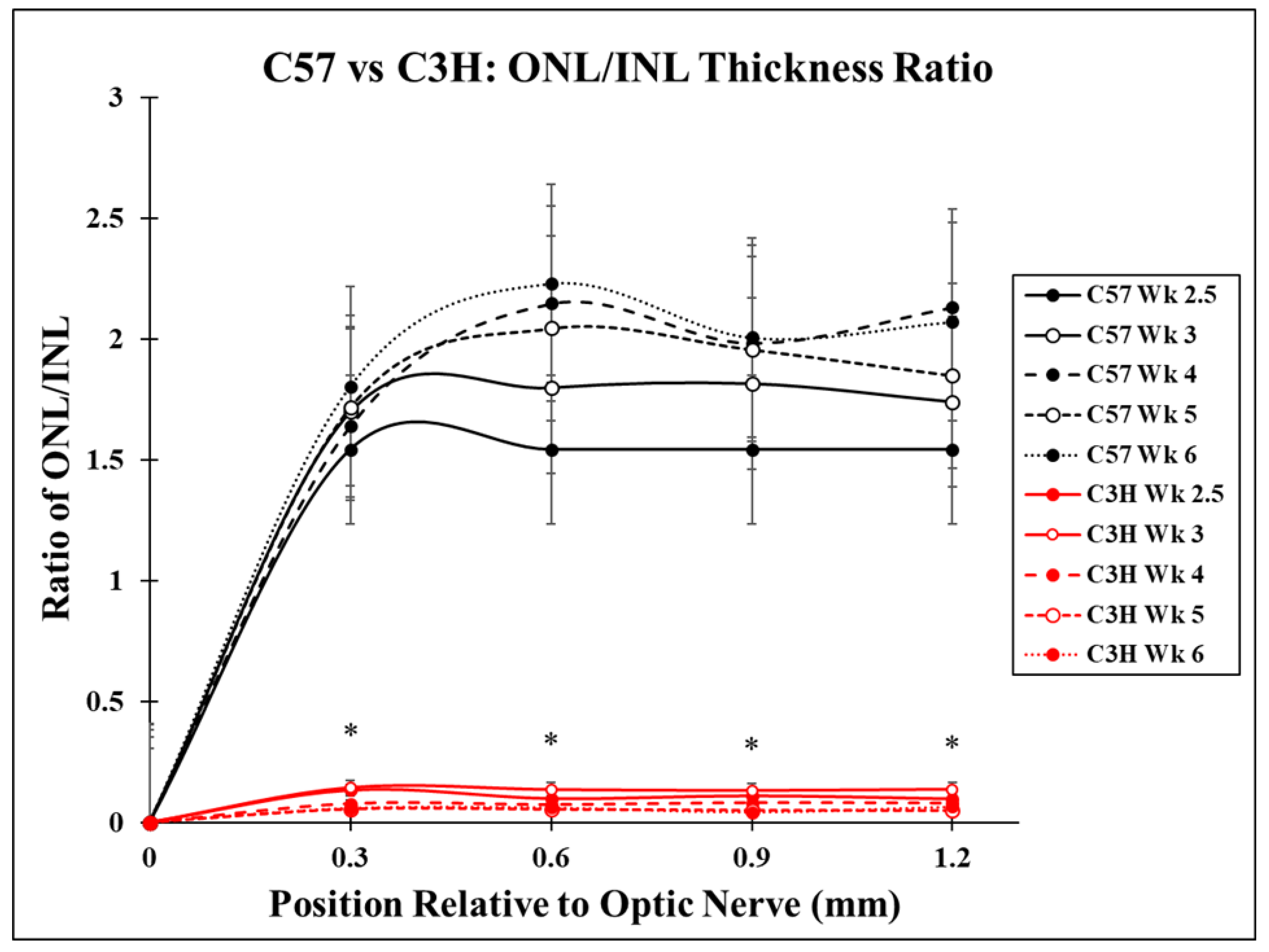
Figure 1. Difference in ONL/INL ratios between C57 and C3H mice from postnatal week 2.5 to postnatal week 6. The ONL/INL ratio was calculated by measuring the depth of each layer in fields surrounding the optic nerve and dividing the ONL by the INL. This ratio was significantly larger for C57 mice than the C3H mice for each time point measured along the retina. The difference between the C57 and C3H ONL/INL ratio increased as time progressed from week 2.5 to week 6. The C3H mice showed a significant decrease in ONL/INL ratios between weeks 2.5 and 3, as well as between weeks 3 and 4 (p < 0.05). A significant difference was found for ONL/INL ratios of all C3H time point groups compared to their C57 counterparts at each retinal location (* indicates p ≤ 0.05).
Applsci 15 05486 g001
Figure 2. Comparison of C57 and C3H retinal layers at week 2.5 and week 6. Eye sections from the mid-periphery of each animal’s retina, stained with H&E, show the retinal layers, as indicated by the text above. Significantly reduced OPL and ONL can be seen in the C3H sections (right) when compared to their C57 counterparts (left).
Applsci 15 05486 g002
Figure 3. Number of Myo/Nog cells in the retinas of C57 and C3H mice. Total Myo/Nog cells in all retinal layers were counted and averaged for each time point. Myo/Nog cells were significantly more numerous at all time points in C3H mice compared to C57 mice (* indicates p ≤ 0.05).
Applsci 15 05486 g003
Figure 4. Distribution of Myo/Nog cells throughout the retinal layers at different time points of C57 and C3H mice. (A,C,E,G,I) represent C57 mice in which the majority of Myo/Nog cells are in the INL-OPL layers of the retina. (B,D,F,H,J) represent C3H mice with the majority of Myo/Nog cells in the ONL and choroid. There was no statistical difference between the distribution of Myo/Nog cells in the GCL between the C3H and C57 mice. The distributions of Myo/Nog cells found in the choroid–ONL and INL-OPL layers of the retina were statistically different in C3H mice compared to C57 mice (* indicates p ≤ 0.05).
Applsci 15 05486 g004
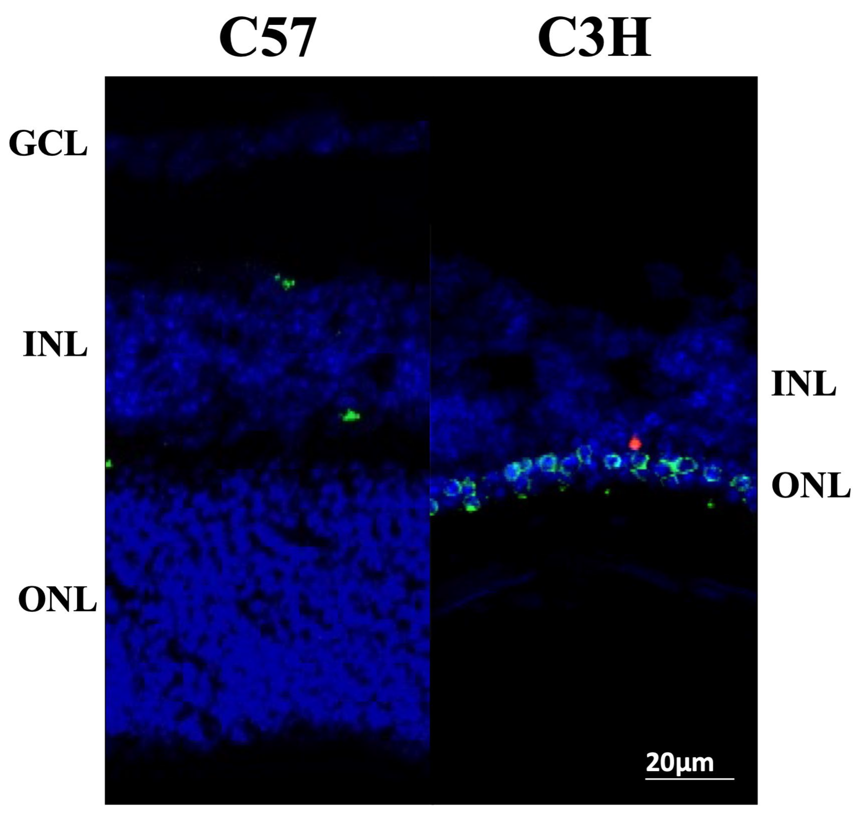
Figure 5. Distribution of Myo/Nog cells throughout the retinal layers in 3-week-old C57 and C3H mice. Tissue sections of a C57 and C3H retina labeled with TUNEL reagents (red), anti-BAI1 mAb (green), and DAPI for nuclei (blue). This figure shows a shift in the distribution of Myo/Nog cells from the INL in C57 mice to the ONL in C3H mice.
Applsci 15 05486 g005
Figure 6. Number of Myo/Nog cells in different layers of the retina in C57 and C3H mice. The number of Myo/Nog cells was counted in five sections per group and combined for all eight subsequent regions moving from the optic nerve toward the periphery. Significantly more Myo/Nog cells were seen in all layers of the C3H mice compared to the corresponding layers of the C57 mice (* indicates p ≤ 0.05).
Applsci 15 05486 g006
Figure 7. Myo/Nog cells phagocytose apoptotic photoreceptors in C3H retinas. Tissue section of a C3H retina labeled with TUNEL reagents (red) (B,C,E,F), anti-BAI1 mAb (green) (A,C,D,F), and DAPI for nuclei (blue) (AF). Arrows indicate BAI1+ Myo/Nog cells in the ONL phagocytosing apoptotic cellular debris.
Applsci 15 05486 g007
Disclaimer/Publisher’s Note: The statements, opinions and data contained in all publications are solely those of the individual author(s) and contributor(s) and not of MDPI and/or the editor(s). MDPI and/or the editor(s) disclaim responsibility for any injury to people or property resulting from any ideas, methods, instructions or products referred to in the content.