
The Concept of Using 2D Self-Assembly of Magnetic Nanoparticles for Bioassays

 , , , , and
Abstract
1. Introduction
2. Materials and Methods
2.1. Synthesis of Magnetic Fe3O4 Nanoparticles
2.2. APTES Functionalized Nanoparticles
2.3. Preparation of Magnetic Chain Structures
2.4. Preparation of Solution Containing Fluorescein
2.5. Measurement Characterization
- Atomic-force microscopy (AFM) measurements provided by Flex Axiom-Nanosurf to visualize deposits from drying magnetic colloids;
- Optical microscopy provided by Keyence VHX-970F digital microscope;
- Zeta potential for determining stability of magnetic colloids, and dynamic light scattering (DLS) method, where Zetasizer Ultra Malvern was used;
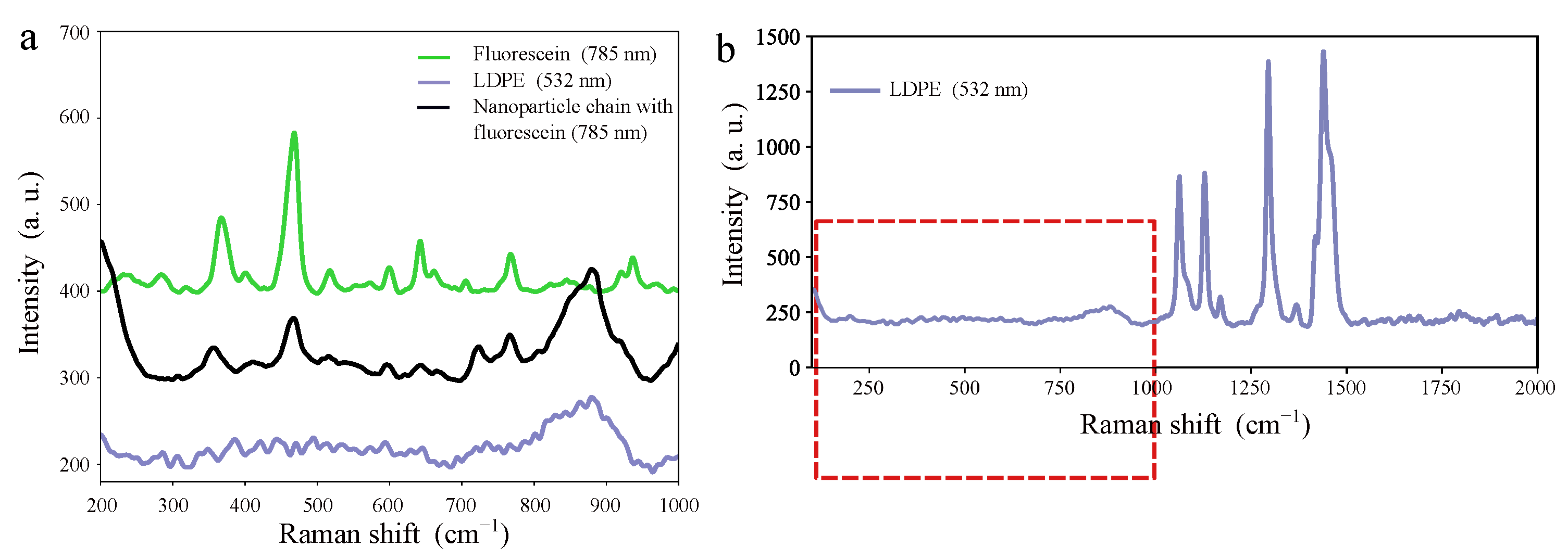
- Raman spectroscopy with the use of the Renishaw InVia Quantor spectrometer to determine the kind of magnetic iron oxides in the magnetic colloids and confirm the presence of fluorescein deposited on a 2D array of ordered chains of magnetic nanoparticles;
- Transmission electron microscopy (TEM) with the energy-dispersive X-ray spectrometer (EDX), to confirm the size of the coated magnetic nanoparticles and their chemical composition.
3. Results
- (i)
- preparation of a 2D array of magnetic-nanoparticle chains;
- (ii)
- preparing a solution containing fluorescein and placing a droplet of this solution on the substrate with chains of magnetic nanoparticles;
- (iii)
- taking measurements to confirm the presence of fluorescein on the chains of magnetic nanoparticles.
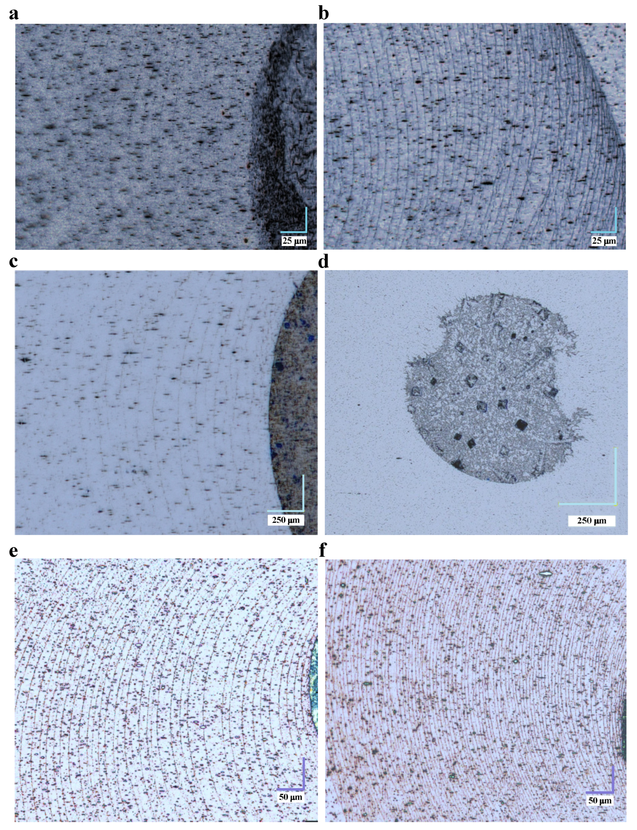
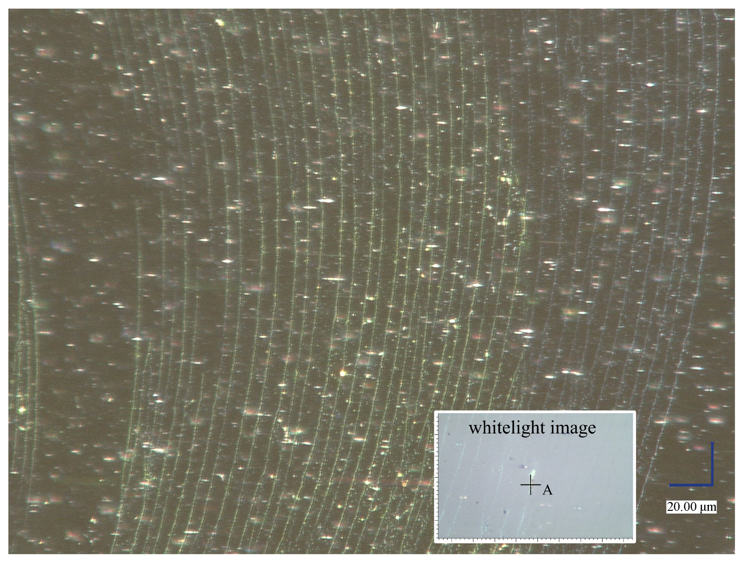
3.1. Preparation of 2D Array of Magnetic-Nanoparticle Chains
3.2. Deposition of Fluorescein on Magnetic Chains
4. Discussion
5. Conclusions
Author Contributions
Funding
Institutional Review Board Statement
Informed Consent Statement
Data Availability Statement
Conflicts of Interest
Abbreviations
| AFM | atomic-force microscopy |
| TEM | transmission electron microscopy |
| EDX | energy-dispersive X-ray spectrometer |
| APTES | 3-aminopropyltriethoxysilane |
| LDPE | low-density polyethylene |
| SPR | surface-plasmon resonance |
| rpm | rotations per minute |
| PZC | point of zero charge |
References
- Hussain, S.; Youngs, I.J.; Ford, I.J. The electromagnetic properties of nanoparticle colloids at radio and microwave frequencies. J. Phys. D Appl. Phys. 2007, 40, 5331. [Google Scholar] [CrossRef]
- Materón, E.M.; Miyazaki, C.M.; Carr, O.; Joshi, N.; Picciani, P.H.S.; Dalmaschio, C.J.; Davis; Shimizu, F.F.M. Magnetic nanoparticles in biomedical applications: A review. Appl. Surf. Sci. Adv. 2021, 6, 100163. [Google Scholar] [CrossRef]
- Giustini, A.J.; Petryk, A.A.; Cassim, S.M.; Tate, J.A.; Baker, I.; Hoopes, P.J. Magnetic nanoparticle hyperthermia in cancer treatment. Nano Life 2010, 1, 17–32. [Google Scholar] [CrossRef]
- Liu, X.; Zhang, Y.; Wang, Y.; Zhu, W.; Li, G.; Ma, X.; Zhang, Y.; Chen, S.; Tiwari, S.; Shi, K.; et al. Comprehensive understanding of magnetic hyperthermia for improving antitumor therapeutic efficacy. Theranostics 2020, 10, 3793–3815. [Google Scholar] [CrossRef] [PubMed]
- Zhang, H.; Liu, X.L.; Fan, H.M. Advances in magnetic nanoparticle-based magnetic resonance imaging contrast agents. Nano Res. 2023, 16, 12531–12542. [Google Scholar] [CrossRef]
- Stueber, D.D.; Villanova, J.; Aponte, I.; Xiao, Z.; Colvin, V.L. Magnetic nanoparticles in biology and medicine: Past, present, and future trends. Pharmaceutics 2021, 13, 943. [Google Scholar] [CrossRef]
- Reddy, L.H.; Arias, J.L.; Nicolas, J.; Couvreur, P. Magnetic Nanoparticles: Design and Characterization, Toxicity and Biocompatibility, Pharmaceutical and Biomedical Applications. Chem. Rev. 2012, 112, 5818–5878. [Google Scholar] [CrossRef] [PubMed]
- Rui, M.; Ma, C.; Hao, Y.; Guo, J.; Rui, Y.; Tang, X.; Zhao, Q.; Fan, X.; Zhang, Z.; Hou, T.; et al. Nanoparticles as a Potential iron fertilizer for peanut (Arachis hypogaea). Front. Plant Sci. 2016, 7, 815. [Google Scholar]
- Zahn, M. Magnetic fluid and nanoparticle applications to nanotechnology. J. Nanoparticle Res. 2001, 3, 73–78. [Google Scholar] [CrossRef]
- Corzo, D.; Tostado-Blázquez, G.; Baran, D. Flexible Electronics: Status, Challenges and Opportunities. Front. Electron. 2020, 1, 594003. [Google Scholar] [CrossRef]
- Gilchrist, R.K.; Medal, R.; Shorey, W.D.; Hanselman, R.C.; Parrott, J.C.; Taylor, C.B. Selective inductive heating of lymph nodes. Ann. Surg. 1957, 146, 596–606. [Google Scholar] [CrossRef]
- Wolak, W.; Kolomeisky, A.B.; Dudek, M.R.; Marć, M.; Najder-Kozdrowska, L. Enhancing silica surface deprotonation by using magnetic nanoparticles as heating agents. J. Phys. D Appl. Phys. 2019, 52, 465001. [Google Scholar] [CrossRef]
- Mleczko, J.; Defort, A.; Kozioł, J.J.; Nguyen, T.T.; Mirończyk, A.; Zapotoczny, B.; Nowak-Jary, J.; Gronczewska, E.; Marć, M.; Dudek, M.R. Limitation of tuning the antibody-antigen reaction by changing the value of pH and its consequence for hyperthermia. J. Biochem. 2016, 159, 421–427. [Google Scholar] [CrossRef]
- Carreón, Y.J.P.; Gómez-López, M.L.; Díaz-Hernández, O.; Vazquez-Vergara, P.; Moctezuma, R.E.; Saniger, J.M.; González-Gutiérrez, J. Patterns in dried droplets to detect unfolded BSA. Sensors 2022, 22, 1156. [Google Scholar] [CrossRef]
- Deegan, R.D.; Bakajin, O.; Dupont, T.F.; Huber, G.; Nagel, S.R.; Witten, T.A. Capillary flow as the cause of ring stains from dried liquid drops. Nature 1977, 389, 827–829. [Google Scholar] [CrossRef]
- Krisman, A. The fern reaction of cervical mucus. Canad. Med. Ass. J. 1964, 91, 805–807. [Google Scholar]
- Priya, B.S.; Pushpaja, M.; Siva Kumar, A.V.; Maruthy, K.N. Does the salivary fern pattern determine fertile period in reproductive female? Clin. Epidemiol. Glob. Health 2020, 8, 698–701. [Google Scholar] [CrossRef]
- Masmali, A.M.; Purslow, C.; Murphy, P.J. The tear ferning test: A simple clinical technique to evaluate the ocular tear film. Clin. Exp. Optom. 2014, 97, 399–406. [Google Scholar] [CrossRef]
- Leow, W.W.; Hwang, W. Epitaxially guided assembly of collagen layers on mica surfaces. Langmuir 2011, 27, 10907–10913. [Google Scholar] [CrossRef] [PubMed]
- Röthel, C.; Radziown, M.; Resel, R.; Zimmer, A.; Simbrunner, C.; Werzer, O. Complex behavior of caffeine crystallites on muscovite mica surfaces. Cryst. Growth Des. 2015, 15, 4563–4570. [Google Scholar] [CrossRef]
- Kralj, S.; Makovec, D. Magnetic assembly of superparamagnetic iron oxide nanoparticle clusters into nanochains and nanobundles. ACS Nano 2015, 9, 9700–9707. [Google Scholar] [CrossRef] [PubMed]
- Zhao, S.; Hao, N.; Zhang, J.X.J.; Hoopes, P.J.; Shubitidze, F.; Chen, Z. Fabrication of monodisperse magnetic nanorods for improving hyperthermia efficacy. J. Nanobiotechnol. 2021, 19, 63. [Google Scholar] [CrossRef] [PubMed]
- Potrč, T.; Kralj, S.; Nemec, S.; Kocbek, P.; Kreft, M.E. The shape anisotropy of magnetic nanoparticles: An approach to cell-type selective and enhanced internalization. Nanoscale 2023, 15, 8611. [Google Scholar] [CrossRef] [PubMed]
- Fu, Q.; Feng, H.; Liu, L.; Li, Z.; Li, J.; Hu, J.; Hu, C.; Yan, X.; Yang, H.; Song, J. Spatiotemporally Controlled Formation and Rotation of Magnetic Nanochains In Vivo for Precise Mechanotherapy of Tumors. Angew. Chem. Int. Ed. 2022, 61, e202213319. [Google Scholar] [CrossRef] [PubMed]
- Xia, L.; Zhao, X.; Ma, X.; Hu, Y.; Zhang, Y.; Li, S.; Wang, J.; Zhao, Y.; Chai, R. Controllable growth of spiral ganglion neurons by magnetic colloidal nanochains. Nano Today 2022, 44, 101507. [Google Scholar] [CrossRef]
- Amstad, E.; Textora, M.; Reimhult, E. Stabilization and functionalization of iron oxide nanoparticles for biomedical applications. Nanoscale 2011, 3, 2819–2843. [Google Scholar] [CrossRef]
- Marć, M.; Wolak, W.; Drzewiński, A.; Dudek, M.R. Coffee-ring formation through the use of the multi-ring mechanism guided by the self-assembly of magnetic nanoparticles. Sci. Rep. 2022, 12, 20131. [Google Scholar] [CrossRef]
- Moffat, J.R.; Sefiane, K.; Shanahan, M.E.R. Effect of TiO2 nanoparticles on contact line stick-slip behavior of volatile drops. J. Phys. Chem. B 2009, 113, 8860–8866. [Google Scholar] [CrossRef]
- Askounis, A.; Orejon, D.; Koutsos, V.; Sefiane, K.; Shanahan, M.E.R. Nanoparticle deposits near the contact line of pinned volatile droplets: Size and shape revealed by atomic force microscopy. Soft Matter 2011, 7, 4152. [Google Scholar] [CrossRef]
- Kim, D.-O.; Pack, M.; Rokoni, A.; Kaneelil, P.; Sun, Y. The EECT of particle wettability on the stick-slip motion of the contact line. Soft Matter 2018, 14, 9599. [Google Scholar] [CrossRef]
- An, M.; Cui, B.; Duan, X. Preparation and applications of linear low-density polyethylene. J. Phys. Conf. Ser. 2022, 2229, 012009. [Google Scholar] [CrossRef]
- Larson, I.; Attard, P. Surface charge of silver iodide and several metal oxides, are all surfaces nernstian? J. Colloid. Interface Sci. 2000, 227, 152–163. [Google Scholar] [CrossRef] [PubMed]


Disclaimer/Publisher’s Note: The statements, opinions and data contained in all publications are solely those of the individual author(s) and contributor(s) and not of MDPI and/or the editor(s). MDPI and/or the editor(s) disclaim responsibility for any injury to people or property resulting from any ideas, methods, instructions or products referred to in the content. |
© 2024 by the authors. Licensee MDPI, Basel, Switzerland. This article is an open access article distributed under the terms and conditions of the Creative Commons Attribution (CC BY) license (https://creativecommons.org/licenses/by/4.0/).
Share and Cite
Marć, M.; Wolak, W.; Drzewiński, A.; Mudry, S.; Shtablavyi, I.; Dudek, M.R. The Concept of Using 2D Self-Assembly of Magnetic Nanoparticles for Bioassays. Appl. Sci. 2024, 14, 1906. https://doi.org/10.3390/app14051906
Marć M, Wolak W, Drzewiński A, Mudry S, Shtablavyi I, Dudek MR. The Concept of Using 2D Self-Assembly of Magnetic Nanoparticles for Bioassays. Applied Sciences. 2024; 14(5):1906. https://doi.org/10.3390/app14051906
Chicago/Turabian StyleMarć, Maciej, Wiktor Wolak, Andrzej Drzewiński, Stepan Mudry, Ihor Shtablavyi, and Mirosław R. Dudek. 2024. "The Concept of Using 2D Self-Assembly of Magnetic Nanoparticles for Bioassays" Applied Sciences 14, no. 5: 1906. https://doi.org/10.3390/app14051906
APA StyleMarć, M., Wolak, W., Drzewiński, A., Mudry, S., Shtablavyi, I., & Dudek, M. R. (2024). The Concept of Using 2D Self-Assembly of Magnetic Nanoparticles for Bioassays. Applied Sciences, 14(5), 1906. https://doi.org/10.3390/app14051906