Article

Waferless Orthognathic Surgery with Customized Osteosynthesis and Surgical Guides: A Prospective Study

by
Marc Espino-Segura-Illa
1,2,
Octavi Camps-Font
3,4,*,
Ada Ferrer-Fuertes
5,
Fran Cuesta-González
5,
Ignacio Zubillaga-Rodríguez
6 and
Ramón Sieira-Gil
1,4,5,*
1
Facial Plastic and Maxillofacial Reconstructive Surgery, SIES Maxillofacial, 08011 Barcelona, Spain
2
Department of Oral and Maxillofacial Surgery, Bellvitge University Hospital, University of Barcelona, 08907 Barcelona, Spain
3
Faculty of Medicine and Health Sciences, University of Barcelona, 08907 Barcelona, Spain
4
Bellvitge Biomedical Research Institute (IDIBELL) Research Institute, Feixa Llarga s/n, 08907 L’Hospitalet de Llobregat, Spain
5
Department of Oral and Maxillofacial Surgery, Hospital Clínic and Barnaclínic, Hospital Clínic de Barcelona, University of Barcelona, Villarroel 170, 08036 Barcelona, Spain
6
Department of Oral and Maxillofacial Surgery, Doce de Octubre University Hospital, Complutense University, 28041 Madrid, Spain
*
Authors to whom correspondence should be addressed.
Appl. Sci. 2024, 14(5), 1893; https://doi.org/10.3390/app14051893
Submission received: 11 January 2024 / Revised: 23 February 2024 / Accepted: 23 February 2024 / Published: 25 February 2024

Abstract

:
The purpose of this paper was to describe and to evaluate the accuracy of a protocol that involves CAD/CAM-generated cutting guides and customized titanium plates for waferless orthognathic surgery. Twenty-one patients consecutively treated between January 2021 and January 2023 were included. The preoperative virtual surgical plan (VSP) was compared with the final position determined from the postoperative CT scan and STL files. An alignment algorithm was employed to adjust the skull position in areas unaffected by the surgery. Absolute and signed deviations were calculated across all three dimensions for each maxilla, mandible and chin landmark. The accuracy analysis revealed an overall deviation of 0.93 mm (95% confidence interval [95%CI]: 0.86 to 0.99), which was < 2 mm for all assessed landmarks (p < 0.05; one-sample t-test). The mandibular landmarks showed greater deviation than the maxillary ones (p < 0.001; independent-samples t-test). Considering the deviations along the three axes, statistically significant differences were identified (p < 0.001; one-way analysis of variance). The reported protocol provides evidence on the benefit of guided orthognathic surgery when performed using a defined VSP protocol, improving accuracy in the maxilla, mandible and chin position, considered both globally and as isolated variables.

1. Introduction

Computer-assisted planning and orthognathic surgery (OS) for the treatment of dentofacial deformities has been extensively documented over the last decade [1,2]. The transformative influence of three-dimensional (3D) imaging, coupled with the rapid evolution of computer-aided design (CAD) and computer-aided manufacturing (CAM) technologies, has marked a significant paradigm shift in the field [3]. Despite these advances, challenges remain, particularly with regard to achieving optimal 3D maxillary positioning using CAD/CAM occlusal surgical splints, as highlighted by multiple studies [4,5,6].
The era of digital treatment planning, augmented by patient-specific guides, has ushered in a new era characterized by improved feasibility, precision, surgical efficiency, and enhanced clinical outcomes in complex orthognathic procedures [1,7,8,9,10]. Recognizing the limitations of traditional two-dimensional (2D) treatment planning [11], recent innovations have introduced diverse methodologies for seamlessly translating 3D virtual treatment plans into the intraoperative sphere [2,8,12,13,14]. However, the absence of a universally accepted standard for implementing various designs of CAD/CAM customized surgical cutting guides and fixation plates has led to inherent variations in surgical protocols and outcomes [15,16,17].
The present study describes and evaluates a new protocol involving CAD/CAM-generated cutting guides and customized titanium plates (CMTPs) for waferless positioning of the maxilla in OS. Notably, this approach enables independent maxillary positioning, eliminating the necessity for an intermediate splint. The study hypothesis is that waferless OS using customized surgical guides (CMSGs) and CMTPs for osteosynthesis is an accurate procedure with potential transformative implications for the field. Through this exploration, this study aims to offer valuable insights into refining surgical techniques and advancing the overall efficacy of orthognathic interventions.

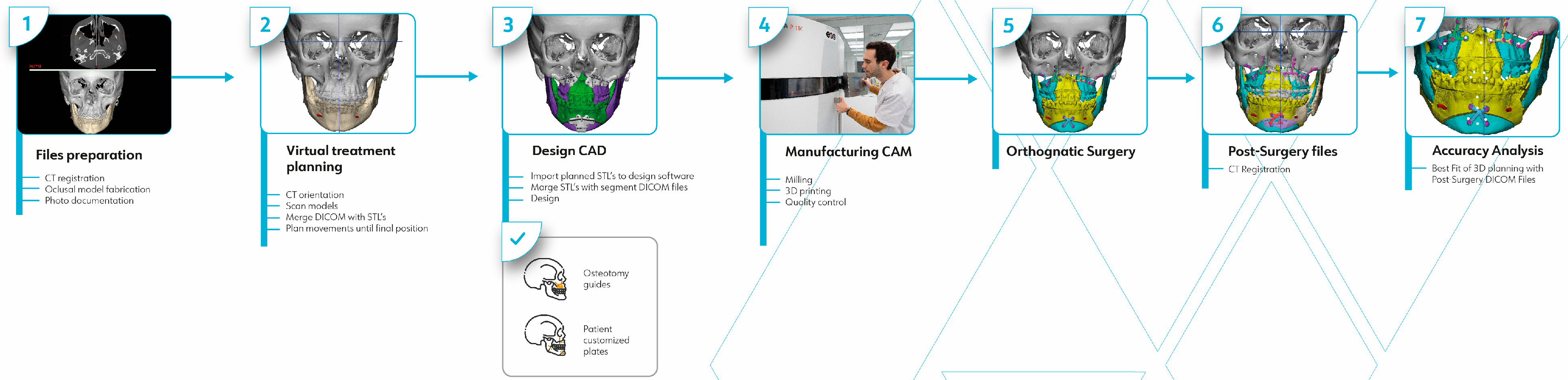
2. Materials and Methods

A prospective analysis was made of 21 patients consecutively subjected to orthognathic surgery at the Barnaclínic and Hospital Clínic (Barcelona, Spain), based on a waferless, guided and customized protocol between January 2021 and January 2023. The study design followed the Strengthening the Reporting of Observational Studies in Epidemiology (STROBE) guidelines for observational studies [18], and the study protocol was approved by the Institutional Review Board of the Hospital Clínic (Ref. REG.HCB/2022/0625).
Patients with temporomandibular joint (TMJ) disorders, cleft-lip palate, craniofacial syndromes, or systemic or coagulation disorders were excluded, in the same way as subjects under 18 years of age.
All surgeries were performed by experienced surgeons (R.S.-G. and M.E.-S.-I.) using a maxilla first protocol consisting of a guided Le Fort or segmented Le Fort osteotomy, followed by a non-guided sagittal split osteotomy and a guided chin osteotomy, depending on the surgical indication. Demographic characteristics, the type of dentofacial deformity and surgical procedure, complications, and the duration of hospital stay were recorded.

2.1. Patient Orthognathic Planning Protocol

The preoperative evaluation, virtual surgical planning (VSP), surgery and postoperative follow-up were standardized before conducting this study. Facial images of the patient in the neutral head position (NHP) were calibrated for future analysis, drawing a 20 mm line on the forehead and cheek of the patient.
A preoperative computed tomography (CT) or cone-beam computed tomography (CBCT) scan was performed with 0.5 mm to 1 mm slices for 3D imaging reconstruction. The Digital Imaging and Communications in Medicine (DICOM) files were converted into Standard Tessellation Language (STL) files for the VSP.

2.2. Two-Dimensional Virtual Surgical Planning

A lateral teleradiograph obtained from the CT scan was required for the initial 2D cephalometric diagnosis in NHP. Dolphin® software version 12.0 (Chatsworth, CA, USA) and the Arnett protocol [19] were used for the 2D analysis and the initial treatment decision plan. The distance from the cutaneous glabella (Gb) to the vertical line at the subnasale (Vsn) was used as the true vertical line (TVL) for referencing head orientation in NHP. This 2D surgical plan was performed following functional and aesthetic criteria of the main authors. Once the 2D movements were decided, the VSP was continued in the 3D setting.

2.3. Three-Dimensional Virtual Surgical Planning
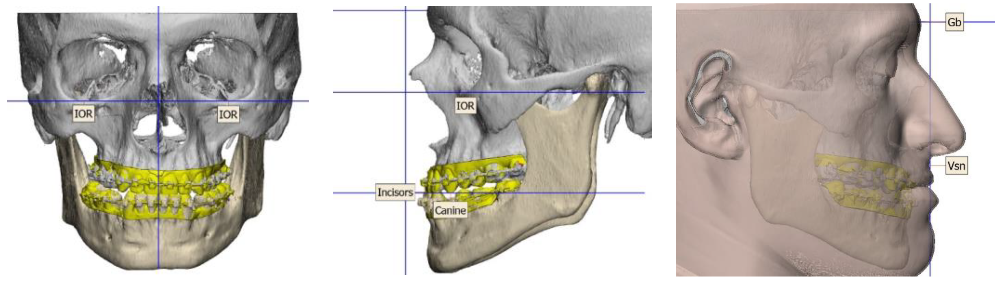
A 3D intraoral scanner (TRIOS, 3shape, Copenhagen, Denmark) was used to obtain an STL file of the teeth and preoperative occlusion. The STL files were used for 3D casts printing and for superimposition of the skull 3D STL file with the preoperative dental occlusion STL. Model surgery of the 3D printed casts was performed either virtually or manually, obtaining an STL file of the final dental occlusion. The three-dimensional VSP was performed using craniomaxillofacial (CMF) software Materialise Mimics Medical version 26.0 (Materialise®, Leuven, Belgium), with the contribution of the biomedical engineers from Avinent® (Avinent Implant System, Santpedor, Spain). The 3D study protocol and postoperative analysis are detailed in Figure 1.
For 3D skull positioning in a frontal view, the midline plane was traced, following clinical evaluation of the patient. A horizontal plane passing over the infraorbital rim (IOR), perpendicular to the midline, was the reference in the horizontal plane. In a lateral view, skull positioning was decided following clinical NHP, using the distance from the cutaneous Gb to Vsn as the clinical reference (Figure 2).
Once the skull was positioned in 3D, movement of the facial skeleton could be performed. Measurements were made of the vertical dimension in a lateral view, from a plane passing through the inferior orbital rim (IOR) to the canine and the central incisors. The maxilla was then advanced to its final position, as planned in 2D. Final occlusion was achieved following the scanned final occlusion, moving the mandible to its final position simulating bilateral sagittal split osteotomies (SSO) when needed. At this point the maxillomandibular complex, canting, yaw and roll were revised. The maxillomandibular complex was moved to improve the occlusal plane following clinical evaluation (pitch). The vertical dimension was revised, comparing vertical dimension to the preoperative measurements. This was a clinical criterion, based on the facial and smile evaluation made by the main authors. A genioplasty was finally designed to improve facial aesthetics, lip competence and chin symmetry when required.
After completing the VSP, CMSGs and patient-specific implants for osteosynthesis together with screw positions and length were decided by the main authors R.S.-G. and M.E.-S.-I. and designed for the maxilla and the chin by the bioengineers from Avinent® (Avinent Implant System, Santpedor, Spain). CMSGs were 3D printed in polyamide (PA2200) using an EOS-Formiga P110 printer (EOS GmbH, Krailing, Germany) and CMTP were milled from titanium (ti6A14V) using an 5-axis milling machine DMG Sauer HSC 20 Linear (DMG Mori Altingesellschaft, Bielefeld, Germany).
A CAD/CAM palatal splint was designed and printed in Bio Splint P HI (Dental Direkt GmbH, Spenge, Germany) using a Roland DWX-52D 5-axis dental milling machine (Roland DG Corporation, Hamamatsu-shi, Japan) for maxillary expansion stability in the Le Fort segmented maxilla cases. Decisions were validated by the main authors (SGR and ESIM), with fabrication by Avinent® (Avinent Implant System, Santpedor, Spain).
The routine CT scan performed preoperatively (T0) was compared with a new CT scan performed 1–3 months after surgery (T1).

2.4. Data Sampling

Surgical accuracy was evaluated by the same trained examiner (MR) who compared the preoperative VSP with the final position determined from the postoperative CT scan and STL files. A 3-matic Medical® algorithm (Materialise®, Leuven, Belgium), was used to adjust the skull position in areas unaffected by the surgery. Deviation calculations across all three dimensions between the planned and postoperative positions were performed using 3-matic Medical® 17.0 software (Materialise®, Leuven, Belgium). The following landmarks were specified [16]:
-
Occlusal: tips of the mesiobuccal cuspids of the first molars, tips of the upper canines, upper incisor point, and lower incisor point.
-
Skeletal: A point, B point, and pogonion point.
-
Titanium plates: anterior and posterior upper plates, inferior chin plate, and midpoint chin plate.
To test intra-examiner reliability, an assessment of 18 randomly selected landmarks (54 measurements) was repeated after four weeks. The intraclass correlation coefficient (ICC) was 0.94 (95% confidence interval [95%CI]: 0.90–0.96; p < 0.001) for absolute agreement.

2.5. Statistical Analysis
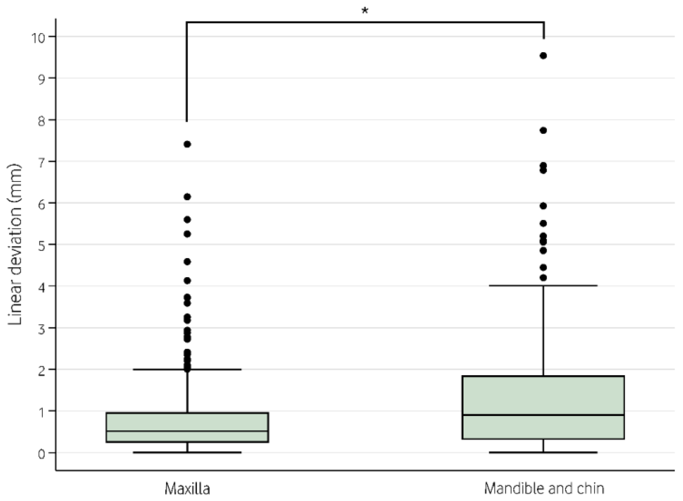
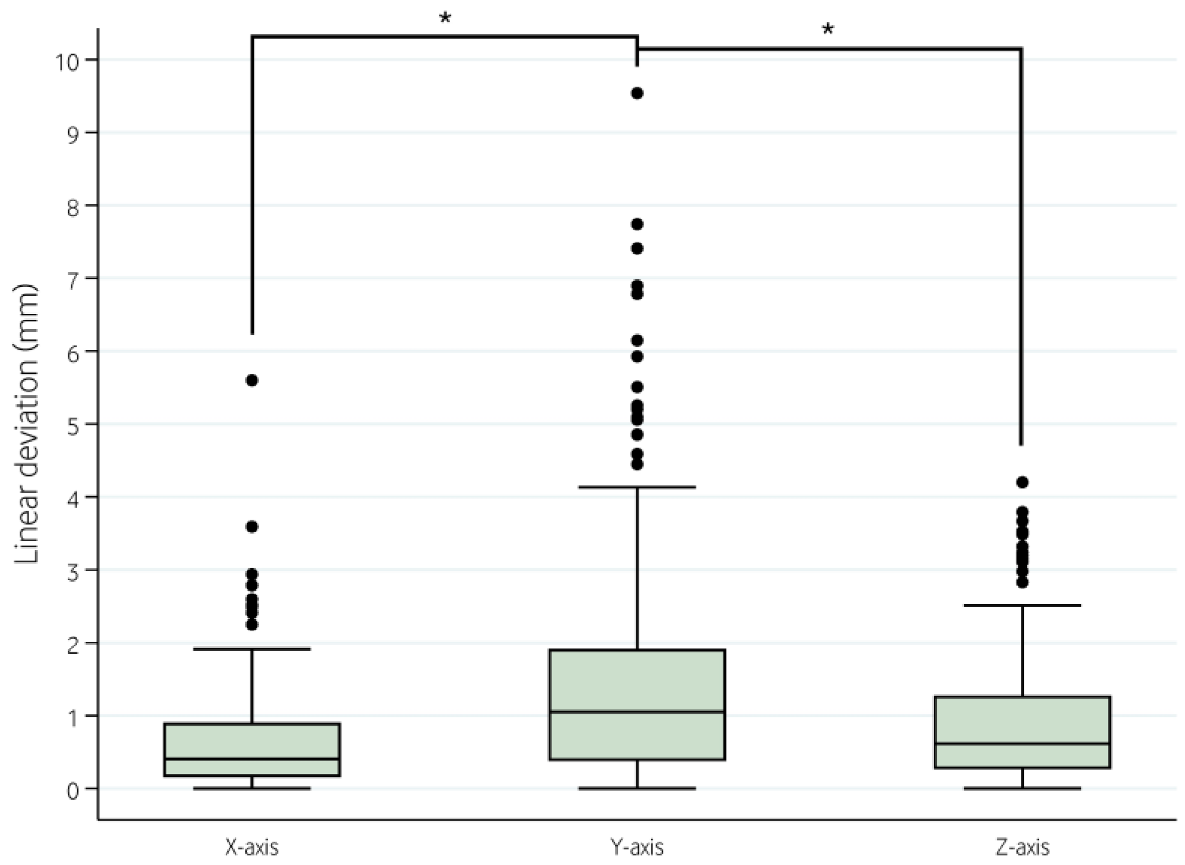
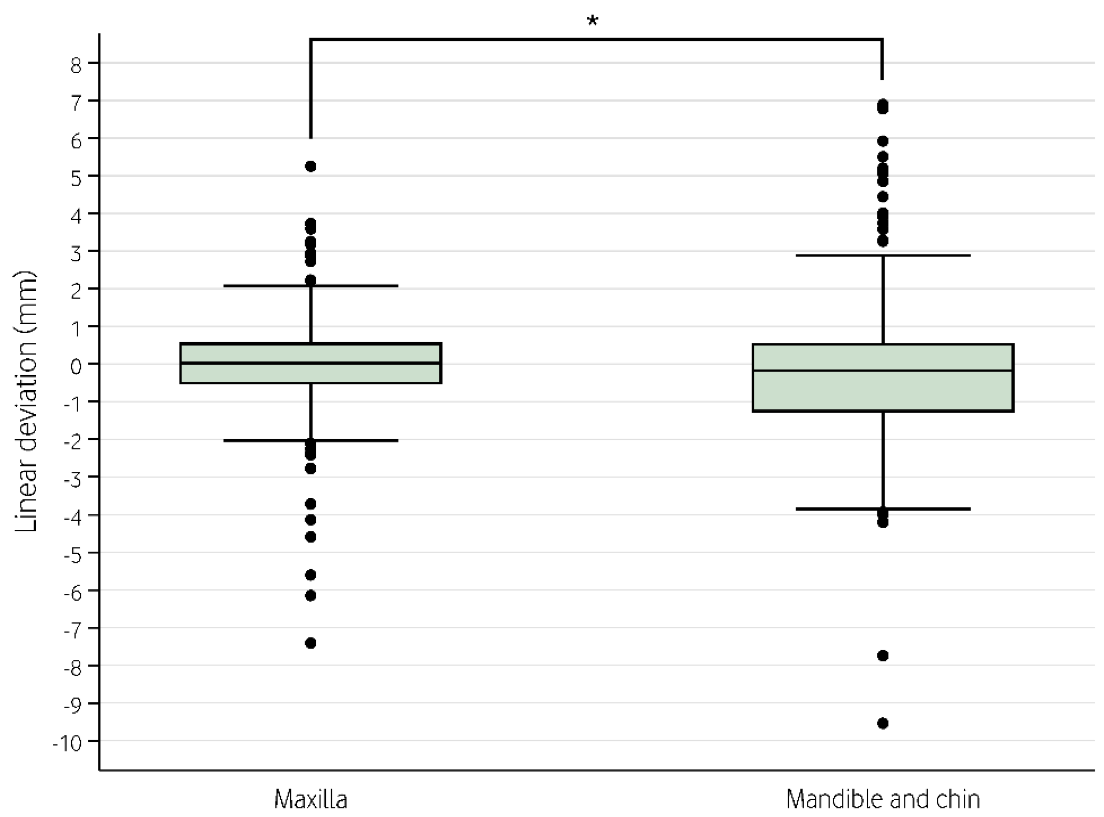
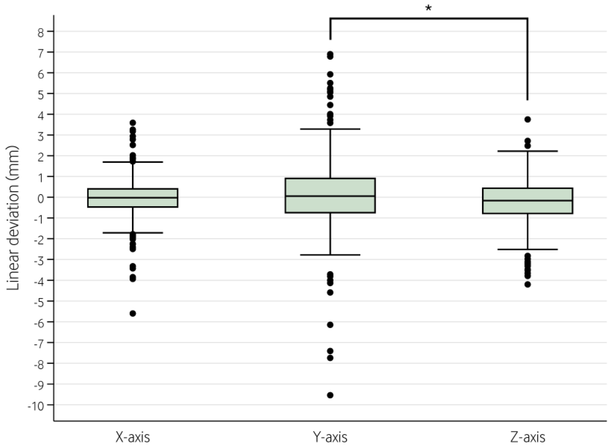
The sample size was determined based on a preliminary sample of data, involving a retrospective analysis of 4 patients who underwent surgery before January 2021. The primary outcome variable, absolute accuracy, guided this calculation, aiming to detect a minimum expected effect size of 1 mm. A standard deviation of 1.5 mm and a 10% exclusion rate were expected. Under these assumptions, a total of 21 patients were required (two-sided one-sample mean z-test, α = 0.05 and 1 − β = 0.80).
Categorical outcomes were presented as absolute and relative frequencies. The normality of scale variables was explored using the Shapiro–Wilk test and through visual analysis of the P–P plots and box plots. Where normal data distribution was rejected, the interquartile range (IQR) and median were calculated. Where the distribution was compatible with normality, the mean and standard deviation (SD) were used. Differences between groups of scale variables were explored using parametric (Student t-test for independent-samples) or non-parametric tests (Mann–Whitney U-test).
A one-sample t-test was used to examine whether the mean absolute and signed deviations were significantly below 2 mm.
To analyze the deviation in each plane in space, one-way analyses of variance (ANOVAs) were conducted. Assumption fulfillment was verified through tests of normality and homogeneity of variances (i.e., Shapiro–Wilk and Levene tests, respectively). Pairwise comparisons between groups were made using Bonferroni’s tests.
Statistical analysis was carried out using the Stata 14.2 package (StataCorp®, College Station, TX, USA). The level of significance was set at p < 0.05, using the Bonferroni correction for multiple comparisons.

3. Results

Twenty-one patients (13 females and 8 males) were enrolled in this study. The mean age was 30.45 years (SD = 7.18; range: 21.13–46.16). Fifteen cases were performed as bimaxillary segmented procedures using segmented LeFort I osteotomy in the maxilla and a bilateral sagittal split osteotomy (BSSO) in the mandible. No major complications were recorded. Days of discharge were one day for all patients except for a male patient who received bimaxillary surgery for facial asymmetry and discharged after two days. The main demographic and surgical variables of the sample are shown in Table 1.

3.1. Absolute Values

All measurements were made without registering any deviation from the protocol. Accuracy analysis revealed an overall deviation of 0.93 mm (95%CI: 0.86–0.99).
Maxillary landmarks were associated with a mean deviation of 0.72 mm (SD = 0.75), whereas in the mandible the mean deviation was 1.30 mm (SD = 1.34). This difference was statistically significant (mean difference [MD]: 0.58 mm; 95%CI: 0.45–0.70; p < 0.001) (Figure 3). Nevertheless, the magnitude of change (deviation versus 2 mm reference value) was < 2 mm for all assessed landmarks (p < 0.05; one-sample t-test).
Considering the deviations along the three axes, the mean deviation was 0.70 mm (SD = 0.74) on the X-axis, 1.20 mm (SD = 1.36) on the Y-axis, and 0.87 mm (SD = 0.81) on the Z-axis (F [2, 1053] = 22.49, p < 0.001). Specifically, statistically significant differences were found between the X and Y axis (MD: −0.50 mm; 95%CI: −0.68 to −0.32; p < 0.001) as well as between the Z and Y axis (MD: −0.33 mm; 95%CI: −0.51 to −0.16; p < 0.001) (Figure 4).
Table 2 shows the deviations observed for each assessed landmark according to its location (maxilla, mandible and chin) and axis (X, Y or Z).

3.2. Signed Values

The accuracy analysis revealed an overall deviation of −0.08 mm (95%CI: −0.16–0.01). Mandibular landmarks exhibited greater deviation compared to those associated with the maxilla (MD: 0.19 mm; 95%CI: 0.01–0.36; p = 0.037) (Figure 5). As in the previous analysis, the magnitude of change (deviation versus 2 mm reference value) remained <2 mm for all assessed landmarks (p < 0.001; one-sample t-test).
Examining the deviations along the three axes, the mean deviation was −0.08 mm (SD = 1.02) on the X-axis, 0.14 mm (SD = 1.82) on the Y-axis, and −0.27 mm (SD = 1.16) on the Z-axis (F [2, 1053] = 7.82, p < 0.001). Notably, statistically significant differences were identified between the Z and Y axis (MD: −0.41 mm; 95%CI: −0.65 to −0.17; p < 0.001) (Figure 6).
Table 2 provides an overview of the deviations observed for each assessed landmark, categorized by its location (maxilla or mandible) and axis (X, Y, or Z).

4. Discussion

Securing accurate and predictable results with validated treatment protocols should be the goal in OS. In this regard, CAD/CAM technology and digital protocol workflows contribute to improve the surgical treatment plan and reduce the preoperative steps—leading to increased accuracy and efficiency in affording improved patient outcomes.
The incorporation of 3D printing to OS has paved the way for the development and production of customized surgical guides and customized titanium plates. Such guides not only enhance surgical accuracy but also contribute to reduce surgery times and possible postoperative complications [13,14,16,20,21]. CAD/CAM technology also allows us to design and fabricate CMTPs that seamlessly fit the anatomy of the patient, facilitating optimal outcomes and reducing the complications associated with ill-fitting titanium plates.
Traditionally, occlusal wafers (also known as splints) were used to guide repositioning of the jaws during OS. These wafers were created based on dental impressions and plaster models, which could prove time consuming and sometimes led to inaccuracies due to the manual fabrication process involved [22]. Additionally, patients were required to wear the wafers for an extended period of time, which could cause some discomfort.
Novel approaches in orthognathic guided osteotomies seek to eliminate the wafer entirely, using CMSGs and CMTPs to reposition the maxilla independently from the mandible.
Several authors have assessed the accuracy of splint-guided orthognathic surgery, reporting that the greatest deviations between virtual planning and the final outcome were found in the vertical dimension [23,24]. In 2020, Kraeima et al. conducted a randomized controlled trial in which 31 patients underwent conventional model surgery (control group) and 27 underwent orthognathic surgery without a surgical wafer. The results showed the control group to have a mean deviation of 1.74 mm versus 1.05 mm in the waferless group [25].
With the use of CMSGs and CMTPs there is no need for an occlusal splint to achieve the planned bone movements, as described in the present study. Our protocol converts a bimaxillary technique into a procedure where the maxilla is positioned and fixed independently from the mandible as in a monomaxillary procedure, and the mandibular osteotomy is then performed.
An additional advantage of using CMSGs in combination with CMTPs is preoperative planning of the screw position with regard to bone thickness and dental roots. This is especially important when segmenting the maxilla, choosing the best plate and screw position, and avoiding the risk of damaging the dental roots.
The main disadvantage is a current turnaround time of approximately 2–4 weeks from approval of the virtual plan to delivery of the procedure kit to the hospital. Therefore, planning of the entire procedure has to be made weeks in advance and must be adapted into the clinical process. In our protocol, the minimum time was 5 working days to print and send the guides and plates to the hospital. This is because the manufacturing plant of the osteosynthesis company is located less than 50 km from our hospital. In any case, this timeframe for planning and fabrication in elective OS should be a minor problem. All cases included in our study were completed without unexpected incidents.
Due to the lack of published randomized controlled trials, the current evidence is not strong enough to recommend the use of CMSGs and CMTPs for OS. In this regard, more well designed randomized controlled trials are needed to confirm significantly superior efficacy of waferless surgery.
The main objective of our study was to assess the accuracy of waferless OS using CMSGs and CMTPs after virtually planned surgery. Previous studies have reported that deviations of <2.0 mm in the maxilla are clinically insignificant [12,26,27]. In contrast, inaccuracies of >1.0 mm in the anterior maxilla could result in undesirable positioning of the maxillary midline, which could lead to a undesired aesthetic consequences for the patient [13,14,28]. On the other hand, discrepancies of 2 mm traditionally have been considered acceptable, as they may be resolved through orthodontic treatment [20].
Our results demonstrated a mean overall (all 18 landmarks and three axes) deviation of <1 mm which, to our knowledge, is consistent with the results of other published articles [13,14,16,21,23,24,25]. We performed a segmented analysis of maxillary, mandibular and chin landmarks, evidencing greater accuracy in the maxilla than in the mandible or chin. These discrepancies could be explained by different situations such as positioning of the mandible during the CT scan, the timing of the CT scan after OS, or the orthodontic treatment carried out at the time of the CT scan.
In our opinion, the most important issue when performing guided OS is to secure precise positioning of the maxilla. No surgical guides were used in the present study for the mandible or SSO. We consider that there are many reasons for not using guides in the mandible. Nevertheless, no studies have been found reporting significant improvements with the use of CMSGs and CMTPs for the mandible. Positioning of the mandible may be affected by factors such as the condyle position, bone interferences, occlusal interferences and the type of osteosynthesis. All these variables need to be managed by the surgeon during surgery. In this regard, leaving all such variables in the hands of a surgical guide and moreover in a defined plating system, could affect the final results. Maxillofacial surgeons need to take all these aspects into account when performing mandibular OS. Condyle positioning and mandibular osteosynthesis form part of the art of OS, and should be managed by experienced surgeons.
The present study was designed evaluating variables in the maxilla, mandible and chin. In this regard, CMSGs and CMTPs were only used in the maxilla and chin. We expected less accuracy in the mandibular and chin landmarks, due to possible variations in mandibular position when performing the CT scan. Even considering this possible bias, the mandibular and chin landmarks yielded significant results when a deviation of >2 mm was taken to be the maximum acceptable deviation, as described in the literature [12,26,27]. In any case, we found positioning of the maxilla to be accurate to <1 mm (0.72 mm, SD = 0.75). This is the real accuracy that can be achieved when positioning the maxilla, following the virtual guided treatment plan in the hands of a surgical team experienced with customized procedures in maxillary OS.
A potential additional problem in waferless mandibular positioning is the position of the condyle during the preoperative CT scan, which is the basis for computer-assisted planning of mandibular bone segment positioning. In computer-assisted planning of waferless mandibular positioning, the condyles must be in the centric position during scanning. From our perspective, this is difficult to achieve in many cases, even when wearing a relaxation splint for several weeks before the preoperative CT scan is made.
In our opinion, no better protocol is available when performing OS, though each surgical team should assess its own protocol to understand and improve the clinical results obtained.
OS has a great impact on individual’s face features which can positively impact their mental, emotional, and psychosocial health over time [29,30]. This is something paramount to consider when evaluating the importance of promoting and achieving the most accurate and predictable results possible.
CAD/CAM surgery of the maxilla can be performed not only as a maxilla first protocol. Considering the level of accuracy achieved in our study, a mandible first with an intermediate splint protocol could also be used. The decision to choose either a maxilla first or a mandible protocol should be made in advance to surgery, when performing the virtual surgical planning. The patient and the surgeon may benefit from one or the other depending on factors such as final occlusal stability, segmenting of the maxilla, the bone characteristics, or the type and degree of the surgical movements. However, future research is needed about prosthetic materials such as plates and screws in order to test their reliability also in particular techniques such waferless OS [31,32].
The present study has several inherent limitations that deserve consideration within a scientific context. Firstly, the absence of a control group in the study design precludes direct comparisons with alternative treatment modalities or traditional approaches. This fact, in turn, has the potential to undermine the external validity of the proposed waferless OS approach. Furthermore, due to the observational nature of our study, the absence of randomization or blinding procedures may influence the internal validity of the findings and compromises the ability to draw robust causal inferences. Finally, the duration and follow-up period of this study may not adequately capture long-term outcomes and potential complications linked to the suggested surgical approach. Therefore, additional research with extended follow-up periods is essential to further understand the durability and sustainability of the proposed procedure.

5. Conclusions

The reported protocol contributes evidence on the benefit of guided orthognathic surgery when performed using a defined VSP protocol, improving accuracy in the maxilla, mandible and chin position, considered both globally and as isolated variables.

Author Contributions

Conceptualization, R.S.-G. and M.E.-S.-I.; methodology, R.S.-G.; software, R.S.-G.; validation, R.S.-G., M.E.-S.-I. and O.C.-F.; formal analysis, O.C.-F.; investigation, M.E.-S.-I. and R.S.-G.; resources, R.S.-G., M.E.-S.-I., A.F.-F. and F.C.-G.; data curation, O.C.-F.; writing—original draft preparation, R.S.-G. and M.E.-S.-I.; writing—review and editing, R.S.-G., M.E.-S.-I. and O.C.-F.; visualization, I.Z.-R.; supervision, R.S.-G.; project administration, M.E.-S.-I. All authors have read and agreed to the published version of the manuscript.

Funding

This research received no external funding.

Institutional Review Board Statement

This study was conducted in accordance with the Declaration of Helsinki, and approved by the Institutional Review Board of the Hospital Clínic (protocol code REG.HCB/2022/0625).

Informed Consent Statement

An informed consent was obtained from all subjects involved in this study.

Data Availability Statement

The data presented in this study are available on request from the corresponding author. The data are not publicly available due to privacy.

Acknowledgments

The authors wish to thank Maria Reyes (MR) for support in data collection during the file overlay process, and Avinent Implant System S.L.U. for technical support.

Conflicts of Interest

The authors declare no conflicts of interest.

References

  1. Swennen, G.R.J.; Mollemans, W.; Schutyser, F. Three-dimensional treatment planning of orthognathic surgery in the era of virtual imaging. J. Oral Maxillofac. Surg. 2009, 67, 2080–2092. [Google Scholar] [CrossRef] [PubMed]
  2. Bai, S.; Shang, H.; Liu, Y.; Zhao, J.; Zhao, Y. Computer-aided design and computer-aided manufacturing locating guides accompanied with prebent titanium plates in orthognathic surgery. J. Oral Maxillofac. Surg. 2012, 70, 2419–2426. [Google Scholar] [CrossRef] [PubMed]
  3. Lee, Y.C.; Kim, S.G. Redefining precision and efficiency in orthognathic surgery through virtual surgical planning and 3D printing: A narrative review. Maxillofac. Plast. Reconstr. Surg. 2023, 45, 42. [Google Scholar] [CrossRef] [PubMed]
  4. Sun, Y.; Luebbers, H.T.; Agbaje, J.O.; Schepers, S.; Vrielinck, L.; Lambrichts, I.; Politis, C. Accuracy of upper jaw positioning with intermediate splint fabrication after virtual planning in bimaxillary orthognathic surgery. J. Craniofac. Surg. 2013, 24, 1871–1876. [Google Scholar] [CrossRef] [PubMed]
  5. Song, K.; Baek, S.H. Comparison of the accuracy of the three-dimensional virtual method and the conventional manual method for model surgery and intermediate wafer fabrication. Oral Surg. Oral Med. Oral Pathol. Oral Radiol. Endod. 2009, 107, 13–21. [Google Scholar] [CrossRef] [PubMed]
  6. Lee, R.; Goonewardene, M.S.; Mian, A.; Allan, B.; Brock, D.; Trevenen, M. Accuracy of orthognathic surgery using 3D computer-assisted surgical simulation. Aust. Orthod. 2018, 34, 17–26. [Google Scholar] [CrossRef]
  7. Xia, J.J.; Gateno, J.; Teichgraeber, J.F.; Christensen, A.M.; Lasky, R.E.; Lemoine, J.J. Accuracy of the Computer-Aided Surgical Simulation (CASS) System in the Treatment of Patients With Complex Craniomaxillofacial Deformity: A Pilot Study. J. Oral Maxillofac. Surg. 2007, 65, 248–254. [Google Scholar] [CrossRef] [PubMed]
  8. Metzger, M.C.; Hohlweg-Majert, B.; Schwarz, U.; Teschner, M.; Hammer, B.; Schmelzeisen, R. Manufacturing splints for orthognathic surgery using a three-dimensional printer. Oral Surg. Oral Med. Oral Pathol. Oral Radiol. Endod. 2008, 105, e1–e7. [Google Scholar] [CrossRef]
  9. Xia, J.J.; Gateno, J.; Teichgraeber, J.F. Three-dimensional computer-aided surgical simulation for maxillofacial surgery. Atlas Oral Maxillofac. Surg. Clin. N. Am. 2005, 13, 25–39. [Google Scholar] [CrossRef]
  10. Tucker, S.; Cevidanes, L.H.S.; Styner, M.; Kim, H.; Reyes, M.; Proffit, W. Comparison of actual surgical outcomes and 3-dimensional surgical simulations. J. Oral Maxillofac. Surg. 2010, 68, 2412–2421. [Google Scholar] [CrossRef]
  11. Sharifi, A.; Jones, R.; Ayoub, A.; Moos, K.; Walker, F.; Khambay, B. How accurate is model planning for orthognathic surgery? Int. J. Oral Maxillofac. Surg. 2008, 37, 1089–1093. [Google Scholar] [CrossRef] [PubMed]
  12. Schouman, T.; Rouch, P.; Imholz, B.; Fasel, J.; Courvoisier, D.; Scolozzi, P. Accuracy evaluation of CAD/CAM generated splints in orthognathic surgery: A cadaveric study. Head Face Med. 2015, 11, 24. [Google Scholar] [CrossRef] [PubMed]
  13. Li, B.; Shen, S.; Jiang, W.; Li, J.; Jiang, T.; Xia, J.J. A new approach of splint-less orthognathic surgery using a personalized orthognathic surgical guide system: A preliminary study. Int. J. Oral Maxillofac. Surg. 2017, 46, 1298–1305. [Google Scholar] [CrossRef] [PubMed]
  14. Mazzoni, S.; Bianchi, A.; Schiariti, G.; Badiali, G.; Marchetti, C. Computer-aided design and computer-aided manufacturing cutting guides and customized titanium plates are useful in upper maxilla waferless repositioning. J. Oral Maxillofac. Surg. 2015, 73, 701–707. [Google Scholar] [CrossRef] [PubMed]
  15. Shakoori, P.; Yang, R.; Nah, H.D.; Scott, M.; Swanson, J.W.; Taylor, J.A. Computer-aided Surgical Planning and Osteosynthesis Plates for Bimaxillary Orthognathic Surgery: A Study of 14 Consecutive Patients. Plast Reconstr. Surg. Glob. Open 2022, 10, e4609. [Google Scholar] [CrossRef] [PubMed]
  16. Heufelder, M.; Wilde, F.; Pietzka, S.; Mascha, F.; Winter, K.; Schramm, A. Clinical accuracy of waferless maxillary positioning using customized surgical guides and patient specific osteosynthesis in bimaxillary orthognathic surgery. J. Craniomaxillofac. Surg. 2017, 45, 1578–1585. [Google Scholar] [CrossRef]
  17. Wong, A.; Goonewardene, M.S.; Allan, B.P.; Mian, A.S.; Rea, A. Accuracy of maxillary repositioning surgery using CAD/CAM customized surgical guides and fixation plates. Int. J. Oral Maxillofac. Surg. 2021, 50, 494–500. [Google Scholar] [CrossRef]
  18. Vandenbroucke, J.P.; von Elm, E.; Altman, D.G.; Gøtzsche, P.C.; Mulrow, C.D.; Pocock, S.J.; Poole, C.; Schlesselman, J.J.; Egger, M. STROBE Initiative. Strengthening the Reporting of Observational Studies in Epidemiology (STROBE): Explanation and elaboration. PLoS Med. 2007, 4, 1628–1655. [Google Scholar] [CrossRef]
  19. Arnett, G.W.; Jelic, J.S.; Kim, J.; Cummings, D.R.; Beress, A.; Worley, C.M. Soft tissue cephalometric analysis: Diagnosis and treatment planning of dentofacial deformity. Am. J. Orthod. Dentofacial. Orthop. 1999, 116, 239–253. [Google Scholar] [CrossRef]
  20. Williams, A.; Walker, K.; Hughes, D.; Goodson, A.M.C.; Mustafa, S.F. Accuracy and cost effectiveness of a waferless osteotomy approach, using patient specific guides and plates in orthognathic surgery: A systematic review. Br. J. Oral Maxillofac. Surg. 2022, 60, 537–546. [Google Scholar] [CrossRef]
  21. Suojanen, J.; Leikola, J.; Stoor, P. The use of patient-specific implants in orthognathic surgery: A series of 32 maxillary osteotomy patients. J. Craniomaxillofac. Surg. 2016, 44, 1913–1916. [Google Scholar] [CrossRef]
  22. Zinser, M.J.; Sailer, H.F.; Ritter, L.; Braumann, B.; Maegele, M.; Zöller, J.E. A paradigm shift in orthognathic surgery? A comparison of navigation, computer-aided designed/computer-aided manufactured splints, and “classic” intermaxillary splints to surgical transfer of virtual orthognathic planning. J. Oral Maxillofac. Surg. 2013, 71, 2151. [Google Scholar] [CrossRef]
  23. Karanxha, L.; Rossi, D.; Hamanaka, R.; Giannì, A.B.; Baj, A.; Moon, W. Accuracy of splint vs splintless technique for virtually planned orthognathic surgery: A voxel-based three-dimensional analysis. J. Craniomaxillofac. Surg. 2021, 49, 1–8. [Google Scholar] [CrossRef]
  24. Sánchez-Jáuregui, E.; Baranda-Manterola, E.; Ranz-Colio, Á.; Bueno de Vicente, Á.; Acero-Sanz, J. Custom made cutting guides and osteosynthesis plates versus CAD/CAM occlusal splints in positioning and fixation of the maxilla in orthognathic surgery: A prospective randomized study. J. Craniomaxillofac. Surg. 2022, 50, 609–614. [Google Scholar] [CrossRef]
  25. Kraeima, J.; Schepers, R.H.; Spijkervet, F.K.L.; Maal, T.J.J.; Baan, F.; Witjes, M.J.H. Splintless surgery using patient-specific osteosynthesis in Le Fort I osteotomies: A randomized controlled multi-centre trial. Int. J. Oral Maxillofac. Surg. 2020, 49, 454–460. [Google Scholar] [CrossRef] [PubMed]
  26. Marchetti, C.; Bianchi, A.; Bassi, M.; Gori, R.; Lamberti, C.; Sarti, A. Mathematical modeling and numerical simulation in maxillofacial virtual surgery. J. Craniofac. Surg. 2007, 18, 826–832. [Google Scholar] [CrossRef] [PubMed]
  27. Ong, T.K.; Banks, R.J.; Hildreth, A.J. Surgical accuracy in Le Fort I maxillary osteotomies. Br. J. Oral Maxillofac. Surg. 2001, 39, 96–102. [Google Scholar] [CrossRef] [PubMed]
  28. Figueiredo, C.E.; Paranhos, L.R.; da Silva, R.P.; Herval, M.; Blumenberg, C.; Zanetta-Barbosa, D. Accuracy of orthognathic surgery with customized titanium plates—Systematic review. J. Stomatol. Oral Maxillofac. Surg. 2021, 122, 88–97. [Google Scholar] [CrossRef] [PubMed]
  29. Brucoli, M.; Baena, R.R.Y.; Boffano, P.; Benech, A. Psychological profiles in patients undergoing orthognathic surgery or rhinoplasty: A preoperative and preliminary comparison. Oral Maxillofac. Surg. 2019, 23, 179–186. [Google Scholar] [CrossRef] [PubMed]
  30. Brucoli, M.; Zeppegno, P.; Benech, R.; Boffano, P.; Benech, A. Psychodynamic Features Associated With Orthognathic Surgery: A Comparison Between Conventional Orthognathic Treatment and “Surgery-First” Approach. J. Oral Maxillofac. Surg. 2019, 77, 157–163. [Google Scholar] [CrossRef] [PubMed]
  31. Gupta, A.; Dutta, A.; Dutta, K.; Mukherjee, K. Biomechanical influence of plate configurations on mandible subcondylar fracture fixation: A finite element study. Med. Biol. Eng. Comput. 2023, 61, 2581–2591. [Google Scholar] [CrossRef] [PubMed]
  32. Sfondrini, M.F.; Gandini, P.; Alcozer, R.; Vallittu, P.K.; Scribante, A. Failure load and stress analysis of orthodontic miniscrews with different transmucosal collar diameter. J. Mech. Behav. Biomed. Mater. 2018, 87, 132–137. [Google Scholar] [CrossRef] [PubMed]
Figure 1. Standardized protocol for treatment planning and postoperative analysis.
Applsci 14 01893 g001
Figure 2. Three-dimensional skull orientation reference lines for positioning in NHP.
Applsci 14 01893 g002
Figure 3. Box plots illustrating absolute deviations between the preoperative plan and the final result, stratified by location. * Statistically significant (p < 0.001).
Applsci 14 01893 g003
Figure 4. Box plots depicting absolute linear deviations between the preoperative plan and the final result, stratified by axis (X/Y/Z). * Statistically significant (Bonferroni test, p < 0.001).
Applsci 14 01893 g004
Figure 5. Box plots illustrating signed deviations between the preoperative plan and the final result, stratified by location. * Statistically significant (p < 0.001).
Applsci 14 01893 g005
Figure 6. Box plots depicting signed linear deviations between the preoperative plan and the final result, categorized by axis (X/Y/Z). * Statistically significant (Bonferroni test, p < 0.001).
Applsci 14 01893 g006
Table 1. Description of the treated patients.
CaseGenderAge (years)TODTOSComplications
1Female21.13AsymmetryBS + GNone
2Female32.12AsymmetryBS + GNone
3Female46.16Class IIIMSNone
4Female22.66Class II subdivision 1BSNone
5Female30.65AsymmetryBS + GNone
6Female38.34AsymmetryBS + GNone
7Male37.67Class II subdivision 2B + GNone
8Male37.13Class IIIBS + GNone
9Male39.76Class I retrusiveB + GNone
10Female21.31Class II subdivision 1BSNone
11Male28.19AsymmetryBSNone
12Female24.84Class II subdivision 1BS + GNone
13Female29.69Class II subdivision 1BS + GNone
14Female38.33Class II subdivision 1BRevision surgery
15Female24.33Class II subdivision 2BSNone
16Male40.82AsymmetryBSNone
17Male29.08Class IIIMSNone
18Female31.44AsymmetryBS + GNone
19Female22.47AsymmetryBS + GNone
20Male29.90AsymmetryB + GRevision surgery
21Male34.39Class II subdivision 1BS + GRevision surgery
TOD: type of dentofacial deformity; TOS: type of orthognathic surgery; BS: bimaxillary segmented; B: bimaxillary; G: genioplasty; MS: monomaxillary segmented.
Table 2. Accuracy values, expressed as mean and SD, stratified by arch and axis.
AbsoluteSigned
XYZXYZ
MeanSDMeanSDMeanSDMeanSDMeanSDMeanSD
MaxillaFirst right molar0.630.551.141.250.770.61−0.130.830.691.560.110.99
First left molar0.630.650.730.490.660.520.220.890.190.87−0.050.85
Right canine1.341.351.221.370.470.35−0.071.92−0.491.780.110.58
Left canine0.660.481.221.730.590.370.280.78−0.342.11−0.030.71
Right central incisor (upper)0.600.460.920.880.600.46−0.090.760.001.29−0.200.74
Left central incisor (upper)0.500.411.040.950.700.420.080.65−0.041.43−0.220.80
A0.580.760.940.840.580.740.120.950.910.87−0.510.79
Anterior right miniplate0.400.360.660.470.560.49−0.210.510.180.800.310.68
Anterior left miniplate0.470.390.860.580.680.51−0.190.58−0.291.01−0.170.85
Posterior right miniplate0.420.220.780.660.640.440.050.48−0.540.87−0.020.78
Posterior left miniplate0.460.350.790.600.710.520.090.58−0.360.940.400.80
MandibleRight central incisor (lower)0.880.701.321.031.170.96−0.131.140.051.70−0.421.48
Left central incisor (lower)0.750.801.281.121.150.92−0.011.11−0.041.72−0.431.44
Pogonion0.970.812.132.661.211.01−0.111.270.883.33−0.951.27
B0.380.631.821.681.161.28−0.270.690.352.49−0.631.62
Midpoint chin miniplate1.081.041.681.851.730.93−0.521.430.732.43−1.131.65
Right inferior chin miniplate1.241.021.922.171.451.24−0.761.440.832.82−0.921.70
Left inferior chin miniplate1.220.961.941.761.671.03−0.251.570.302.66−1.021.72
Disclaimer/Publisher’s Note: The statements, opinions and data contained in all publications are solely those of the individual author(s) and contributor(s) and not of MDPI and/or the editor(s). MDPI and/or the editor(s) disclaim responsibility for any injury to people or property resulting from any ideas, methods, instructions or products referred to in the content.

