Review

Exploring the Potential of Nanoparticles in the Treatment of Breast Cancer: Current Applications and Future Directions

by
Puja Patel
1,
Vadanasundari Vedarethinam
2,
Maame A. Korsah
3,
Michael K. Danquah
4 and
Jaison Jeevanandam
5,*
1
Department of Animal Health and Management, Alagappa University, Karaikudi 630003, Tamil Nadu, India
2
Department of Biomedical Engineering, Stony Brook University, New York, NY 17790, USA
3
College of Arts and Sciences, University of Tennessee, Chattanooga, TN 37403, USA
4
Chemical and Biomolecular Engineering, University of Tennessee, Knoxville, TN 37996, USA
5
CQM—Centro de Quimica da Madeira, Universidade da Madeira, Campus da Penteada, 9020-105 Funchal, Portugal
*
Author to whom correspondence should be addressed.
Appl. Sci. 2024, 14(5), 1809; https://doi.org/10.3390/app14051809
Submission received: 15 January 2024 / Revised: 13 February 2024 / Accepted: 19 February 2024 / Published: 22 February 2024

Abstract

:

Featured Application

This review explores an innovative approach to combating resistance to breast cancer through the application of nanotechnology.

Abstract

Breast cancer (BC) ranks among the most diagnosed solid tumors worldwide. For decades, significant research efforts have been dedicated to finding selective treatments for these solid tumors. Currently, the primary treatment method for BC involves surgery, with the subsequent utilization of radiotherapy and chemotherapy. However, these subsequent treatments often fall short of effectively treating BC due to their side effects and harm to healthy tissues. Today, a range of nanoparticles are being developed to target BC cells without affecting the surrounding healthy tissues. This in-depth review, based on studies, seeks to shed light on these specially designed nanoparticles and their potential in BC treatment. Typically, therapeutic drugs or naturally occurring bioactive compounds are incorporated into precisely crafted nanoparticles. This enhances their solubility, longevity in the bloodstream, and distribution in the body while also minimizing side effects and immune reactions. Nanoparticles have been designed to address the shortcomings of standalone therapeutics and traverse various biological obstacles spanning the systemic, microenvironmental, and cellular that differ among patients and diseases. We prioritize breakthroughs in nanoparticle design to surpass diverse delivery obstacles and believe that smart nanoparticle engineering not only enhances effectiveness for general delivery but also allows customized solutions for specific needs, ultimately leading to better outcomes for patients.

1. Introduction

Despite significant strides in technology in medical science, cancer continues to present challenges with limited treatment options. The Global Cancer Observatory (GCO) estimates that the annual mortality rate from cancer will reach around 30 million individuals by the year 2030 [1]. The development of cancer is commonly attributed to gene mutations. Globally, breast cancer (BC) ranks as the most frequently diagnosed neoplasm in women. In 2020, breast cancer held the highest incidence rate of 11.7% and mortality rate of 6.9% among all cancers affecting females worldwide [2]. Due to the closure of medical institutions brought on by the COVID-19 epidemic, there has been a hindrance to the detection and treatment of cancer. Moreover, given the lag of 2 to 3 years in population-based cancer incidence and mortality statistics, it will take some time to precisely determine the full degree of its influence on the community. It is anticipated that the prolonged delays in diagnosis and treatment may lead to increased rates of severe disease and mortality [3]. The treatment approaches for breast cancer range from localized strategic treatments, like surgery and radiation, to systemic therapies, which encompass chemotherapy, immunotherapy, hormonal therapy, and endocrine therapy [4]. Disability and mortality rates are significantly influenced by cancer metastasis and recurrence, yet the precise underlying mechanisms remain unclear. Apart from the significant mortality associated with cancer, an enormous economic burden exists on both the families of cancer patients and society. Hence, prioritizing efforts toward cancer prevention, diagnosis, and treatment is of paramount importance.
In cases of early-stage breast cancer, the effective form of treatment recommended is surgery. The objectives of breast cancer surgery include the pathological staging of both the tumor and axillary lymph nodes to offer crucial prognostic insights and full excision of the primary tumor with negative margins to limit the chance of local recurrences [4]. In instances of locally advanced breast cancer, neoadjuvant chemotherapy is administered to shrink the tumor in preparation for definitive surgery, potentially allowing for a less invasive procedure [5]. The effectiveness of neoadjuvant therapy in patients with triple-negative, which includes human epidermal growth factor receptor 2 (HER2), progesterone receptors, and estrogen receptors, helps to determine the best adjuvant therapy [6]. In breast cancer research, nanotechnology has proven to be effective for detection, diagnosis, and treatment. Numerous nanoformulations, including liposomes, nanofibers, nanocapsules, and nanoparticles, have been developed and investigated to inhibit breast cancer cell proliferation, prevent recurrence, and address postchemotherapy metastasis [7].
Research in nanotechnology for cancer therapy extends beyond conventional medication delivery, delving into the development of innovative medicines made possible by the distinctive characteristics of nanomaterials [5]. In addition, nanotechnology has successfully addressed the critical issue of nontargeted and nonspecific damage to bodily tissues resulting from conventional therapeutic measures. It offers distinctive benefits by improving the efficacy of medications and radiation therapy while minimizing the potential side effects [8]. It is noteworthy that a total of 573 agents were approved by the United States Food and Drug Administration (USFDA) from January 2020 to October 2022 for several oncological applications, such as targeted drugs (48%), biological therapies (43%), and cytotoxic treatments (9%) [9]. One such advantage is the use of multifunctional nanocarriers, which capitalize on the distinction between tumor and normal tissues to preferentially deliver therapeutic pharmaceuticals, improving medication permeability and retention. Moreover, nanotechnology leverages the specific physical and chemical characteristics of the tumor microenvironment, such as a lower environment, weak acidity, overexpressed proteins and enzymes, hypoxia, and anomalous temperature gradients. The drug delivery rate of loaded medications from nanocarriers can be accurately controlled due to these unique features. Nanoparticles possess a size that is substantial enough to accommodate numerous tiny molecules and yet remain comparatively small in relation to cells. The relatively large surface area of nanoparticles can also be functionalized by ligands such as DNA or RNA strands, aptamers, peptides, and antibodies [5]. These ligands can serve as therapeutic measures or be utilized to regulate the in vivo behavior of nanoparticles [5]. These features enable multimodal therapy, theragnostic action, and the administration of several medications. Similar to the application of laser ablation, nanoparticles can utilize their physical properties of energy absorption and reradiation when applied to diseased tissues. Hence, the aim of this review is to explore various innovative approaches to combat breast cancer through the application of nanotechnology. It covers an overview of the classification of breast cancer, traditional breast cancer treatments, the significant properties of conventional breast cancer drugs, and nanomedicine as a potential alternative drug delivery candidate for breast cancer therapy.

2. Classification of Breast Cancer by Histological, Molecular, and Morphological Characteristics

Breast cancer (BC), the predominant cancer among women, poses a significant health concern for the female population [2]. The development of BC involves a complex process influenced by factors such as age, genetics, hormones, and the environment [2,10,11,12]. Chronic inflammation, which is linked to both cancer development and progression, is one of the most recent factors to be linked to an elevated risk of BC. A multidimensional framework that takes into account histological classification, clinical traits, and cutting-edge molecular analysis is used to categorize human breast carcinomas. Figure 1 illustrates a visual classification of breast cancer based on distribution, histology, and machine learning methods.

2.1. Histopathological Classification of Breast Cancer

Histologically, tumors are broadly classified as either invasive carcinomas or in situ carcinomas, depending on whether malignant cells have spread from breast lobules or ducts into the surrounding stroma at the time of diagnosis [12].
Ductal carcinoma in situ (DCIS) is the most prevalent form of preinvasive breast cancer, with about 10% to 30% of cases progressing to invasive cancer, as there are insufficient prognostic bioindicators for predicting the development of invasive or metastatic disease [10]. In the last 2 decades, there has been a notable rise in the incidence of ductal carcinoma in situ, constituting 20–25% of newly identified cases of breast cancer [11]. The widespread adoption of mammographic screening in numerous countries is a major factor contributing to this upsurge. The rising trend is evident across various age groups and all genders, underscoring the need for additional considerations. As the incidence of DCIS is increasing in developed nations alongside a general rise in life expectancy, a growing number of elderly women will face a DCIS diagnosis, prompting questions about the feasibility of reducing local–regional therapy. Common risk factors for male breast cancer, including DCIS, involve aging, hyperestrogenism, and a positive family history [12]. While the precise lifetime risk of transitioning from DCIS to invasive breast cancer remains uncertain, it is believed to be less than 50% [12]. This study systematically examines authors focused on two distinct subgroups: male patients and older women with concomitant conditions [12].
Invasive ductal carcinoma (IDC), which accounts for 60–75% of cases, and invasive lobular carcinoma (ILC), which makes up 10–15% of tumors [16], are the two most frequent types of invasive carcinomas. This category of carcinomas is characterized by heterogeneity and is further classified based on cell shape. An uncommon phenomenon, invasive carcinoma with neuroendocrine characteristics makes for 2–5% of all invasive carcinomas of the breast. With varying proportions of neuroendocrine markers, it shares several histological characteristics with other neuroendocrine tumors of the bronchopulmonary system, pancreas, and digestive tract. Immunohistochemistry markers, which are usually not employed to detect breast cancers, are utilized to validate the diagnosis of neuroendocrine tumors in the breast. As a result, the precise prevalence is yet unknown. Studies suggest that invasive carcinomas, including solid papillary carcinoma, invasive ductal carcinoma, and mucinous carcinoma, are frequently associated with neuroendocrine tumors, observed in up to 30% of breast cancer cases [17]. There are several subtypes of invasive ductal carcinoma, each with distinct characteristics.
  • Medullary carcinoma is characterized by its slow growth, featuring soft and fleshy tumors that bear a resemblance to the medulla of the brain. This subtype accounts for less than 1% of all breast cancers [18];
  • Tubular carcinoma is a rare histological subtype of invasive ductal carcinoma, constituting 1% to 5% of all invasive breast cancers. These tumors exhibit slow growth and are characterized by cancer cells with tube-like structures [19]. TC is recognized as a well-differentiated invasive carcinoma and is further classified into two categories: pure TC and mixed TC. Pure TC refers to tumors with a tubular content of more than 90% and a low nuclear grade, with few-to-no mitoses [20]. On the other hand, mixed TC has a tubular composition of less than 75% [20];
  • Mucinous carcinoma is characterized by low-grade tumors composed of cancer cells that are situated within the mucin substance found in mucus. This subtype accounts for fewer than 2% of all breast cancers [21];
  • Papillary carcinomas are tiny cancerous cells that have finger-like appendages. Less than 1% of all breast cancers are papillary carcinomas, making them extremely uncommon [22];
  • Cribriform carcinoma is characterized as an unusual subtype with a Swiss cheese-like pattern of holes. This type of cancer accounts for fewer than 1% of breast cancer cases [23];
  • Adenoid cystic carcinoma: In contrast to typical ductal cancer cells, adenoid cystic carcinoma resembles cancerous cells found in the salivary gland. This subtype constitutes less than 1% of all breast cancers [24];
  • Metaplastic carcinoma: This takes place when ductal cells transform into new cell types. Less than 1% of all breast cancers are metaplastic carcinomas, which are typically more aggressive cancers [25].

2.2. Molecular Classification of Breast Cancer

The conventional classification of breast cancer divides the tumors into various categories with varying prognoses and behaviors using morphology [26]. It has limits despite offering high-quality data for a low cost; therefore, there has been a hope that the new molecular approaches may aid in the improvement of the classification algorithms. Although much has been learned in recent years, molecular taxonomy is still evolving and is likely to alter over the next few years. It remains to be seen whether molecular categorization is as helpful for specific subtypes of breast cancer as it has been for ductal carcinoma, which is not a distinct kind. With the possibility of stratifying this neoplasm into multiple entities that call for particular treatments and monitoring measures, as well as a better knowledge of the pathophysiological pattern and clinical prognosis, molecular subtyping altered the perspective on breast cancer. Breast cancer can be categorized into different molecular subtypes based on the status of the progesterone receptor (PR), estrogen receptor (ER), and HER2. These subtypes include HER2+ (ER−/PR−/HER2+) luminal B (ER+/PR+/HER2+), luminal type A (ER+/PR+/HER2−), and basal-like (ER−/PR−/HER2−) [27,28].
Luminal A, the most prevalent subtype of breast cancer, accounts for 60–70% of all cases and is characterized by positive statuses for estrogen and progesterone receptors but a negative HER2 status [29]. As a result, endocrine therapies are considered the primary treatment approach for luminal A breast cancer. For instance, by preventing the aromatization of androgens to estrogens, aromatase inhibitors prevent the synthesis of estrogen. On the other hand, selective estrogen receptor degraders (SERDs), such as elacestrant and fulvestrant, hinder the translocation of estrogen receptors to the nucleus and destroy them [30], while selective estrogen receptor modulators (SERMs), such as tamoxifen, prevent the binding of estrogen and estrogen receptors [30]. The prognosis of luminal A breast cancer is more varied than that of other breast cancer subtypes, and some patients with this subtype of breast cancer have intrinsic or acquired resistance to these endocrine therapies.
About 20% to 30% of cases of invasive breast cancer are caused by the luminal B molecular subtype, with a lower expression of estrogen receptors (ERs) [31]. The more biologically and clinically diverse luminal B subtype necessitates a thorough characterization to determine the best course of treatment for every patient [32]. Patients with luminal B (HER2-negative) BC were specifically categorized for survival results and recurrence scores using progesterone receptor (PR) and proliferative regulator (Ki-67) experiments [33]. Triple-negative breast cancers (TNBCs) make up 15% to 20% of all breast cancer cases [34]. TNBC is more prevalent in premenopausal women under the age of 40 and is extremely aggressive, developing faster, having a higher risk of metastasizing, and having a worse clinical result than hormone receptor-positive and HER2-enriched breast tumors [35]. The age-related increases in cancer risk are as follows: 1.5% at age 40, 3% at age 50, and at least 4% at age 70 [36], which consist of ER, PR, and HER2 [37].

3. Traditional Methods for Breast Cancer Treatment

Most patients undergo a surgical procedure with the choice of process influenced by factors such as the patient’s preferences, the identified type of breast cancer, tumor size and location, and other considerations to remove a breast tumor. A commonly selected option is breast-conserving surgery, also known as a lumpectomy, which involves removing the tumor and some surrounding healthy tissue. This procedure may also include the removal of nearby lymph nodes to check for cancer spread. Alternatively, mastectomy, the removal of the entire breast along with lymph nodes, is another viable option. There are three types of mastectomy: total (simple) mastectomy, modified radical mastectomy, and radical mastectomy, each with its own set of risks and benefits. The latter procedure, radical mastectomy, is rarely performed unless breast cancer has progressed to the chest muscles [38].
Following a mastectomy, patients may opt for breast reconstruction, utilizing nonbreast tissue or implants to create a new breast. Simultaneously, a lymph node biopsy or lymph node dissection is conducted to determine whether cancer cells are present in the nodes, which indicates potential disease spread. Radiation therapy targets tumors and kills cancer cells by using energy from radiation beams, radioactive isotopes, or charged particles. After a lumpectomy, external radiation is usually always necessary to prevent the cancer from coming back. In cases where the tumor is large or has spread to the lymph nodes, bones, or the brain (common after a mastectomy), external radiation is occasionally employed. Some breast cancer patients may also be administered radiation internally (brachytherapy) [39]. Chemotherapy kills cancer cells by administering medications or medication mixtures intravenously or orally. Patients with breast cancer might receive chemotherapy either before or after surgery. Chemotherapy may be the main treatment when the cancer is in an advanced stage.
However, the quality of life a patient experiences during and after treatment can be significantly impacted by the treatment options. This may include the body’s physical alterations, emotional anguish, and the cost of treatment. Hair loss, nausea, exhaustion, and an increased risk of infection are just a few of the serious side effects that are frequently associated with conventional therapies like chemotherapy and radiation therapy [39]. Even after a successful course of treatment, the cancer may still come back [39]. This is especially true for breast cancer kinds that are aggressive. Traditional therapies like hormone therapy are less effective against some forms of breast cancer, like triple-negative breast cancer (TNBC). In some circumstances, especially for elderly patients or those with other serious pre-existing medical issues, the risks associated with the treatment option may outweigh its benefits. Additionally, factors such as cost, geographic location, and variations in healthcare systems contribute to the limited accessibility of treatments to all patients. Nonetheless, the field of breast cancer treatment is continuously advancing, placing a greater emphasis on personalized medicine and targeted therapies designed to overcome current limitations [39].

4. Nanotechnology and Cancer

Recent advancements in nanotechnology provide promising strategies to circumvent the nonspecific harm to healthy tissues resulting from conventional cancer therapies, such as chemotherapy, radiotherapy, and immunotherapy [39]. Nanotechnology’s distinctive benefits center on augmenting the effectiveness of drug and radiation therapy while minimizing adverse reactions. Multifunctional nanocarriers, for instance, can exploit the discrepancies between tumor and normal tissues to selectively improve drug delivery and retention [39]. Moreover, the tumor microenvironment (TME) exhibits several physical and chemical characteristics, such as abnormal temperature gradients, reductive conditions, mild acidity, hypoxia, and overexpressed proteins and enzymes. Exploiting these features can enable the controlled release of drugs from nanocarriers, furthering the potential for targeted, effective cancer treatments. With the rapid advancement of nanotechnology over the past few decades, countless nanomaterials have been developed. However, only a limited number of nanoparticulate-based systems are applicable in the biomedical field, and even fewer meet the stringent requirements set by the US FDA (United States Food and Drug Administration) for clinical applications. The use of engineered nanoparticles (NPs) for targeted cancer therapy, including breast cancer, is a growing field of research [39]. The goal is to improve the efficacy of treatment, minimize side effects, and elevate the overall quality of life for patients. These NPs can be customized to bind specifically to cancer cells, thus ensuring that therapeutic drugs are delivered to the targeted location [39].

4.1. Nanoparticles in Cancer Therapy

Current cancer treatments are constrained to surgical methods, radiation therapy, and chemotherapy, each of which poses the risk of damaging healthy tissues or not eliminating the cancer. Nanotechnology, however, presents a solution to these issues by enabling the precise and targeted delivery of chemotherapy drugs to cancer cells and tumors, contributing to the surgical removal of tumors and enhancing the efficacy of radiation and other established treatment approaches [39]. This progress promises a lower patient risk and increases the likelihood of survival. Further advancements in nanotechnology-based cancer treatment have expanded beyond merely improving drug delivery to creating new treatment options that are only possible through the unique properties of nanomaterials [39]. Nanoparticles’ sizable surface area can be tailored with ligands, which can be antibodies, small molecules, peptides, DNA or RNA strands, and aptamers [39]. These ligands can serve both therapeutic purposes and guide the nanoparticle’s behavior within the body. The versatility of these properties allows for combination drug delivery, multimodality treatment, and the combination of therapy and diagnosis, referred to as “theranostic” action. Nanoparticles used in cancer therapy include liposomes, polymeric nanoparticles, gold nanoparticles, magnetic nanoparticles, and dendrimers [39]. Each type has unique properties that make it suitable for certain applications. For instance, liposomes are often used as drug carriers due to their biocompatibility and ability to encapsulate both hydrophilic and hydrophobic drugs [39].

4.2. Targeting Strategies

Nanoparticles hold the transformative potential to innovate the diagnosis and treatment of various diseases, including enabling the accurate targeted delivery of drugs to cell types [39]. Progress in the engineering of nanoparticles and a deeper comprehension of the significance of characteristics like size, shape, and surface properties for biological interactions are paving the way for new prospects in the development of therapeutic nanoparticles [39]. NPs can be tailored to target cancer cells in several ways. Passive targeting exploits the Enhanced Permeability and Retention (EPR) effect, which is due to the irregular vasculature and inadequate lymphatic drainage in tumors, facilitating the accumulation of nanoparticles in tumor tissues [40,41]. Active targeting involves modifying the surface of NPs with molecules that can bind specifically to receptors overexpressed on cancer cells. The first obstacles faced are largely determined by the preferred method of administration. The extent to which each of these administration methods can be successfully used is significantly impacted by the characteristics of the nanoparticle [42].
Managing the particle size, surface characteristics, and release profiles of bioactive compounds is the major objective when building nanoformulations to ensure the site-targeted accumulation and activity of pharmaceuticals at the therapeutically ideal rate and dose regimen. Nanoformulations, which are typically 10–200 nm in diameter, are made by either encapsulating pharmaceuticals inside the nanocarriers or complexing medications with the nanocarriers by covalent bonding to enhance the characteristics of the bioactive chemicals. To increase the bioavailability, solubility, extended blood circulation, and targeted delivery of medications while reducing their negative effects, these NFs have been used in drug delivery studies. Nanoformulations may interact with biological systems when ingested, injected, skin-penetrated, or inhaled into the body.
Loading drugs is a crucial approach in drug delivery. An effective nanodelivery system should possess substantial drug-loading capabilities, enabling a reduction in the required amount of matrix materials for delivery. Two primary methods are employed for drug loading. The first is the incorporation method, where the drug is integrated during the nanoparticle formulation process. The second is the adsorption/absorption method, where the drug is absorbed after nanoparticle creation by immersing the nanocarrier in a concentrated drug solution. The effectiveness of drug loading and entrapment is contingent upon the solubility of the drug in the excipient matrix material, which is influenced by various factors, such as molecular weight, matrix composition, interactions between the drug and matrix, and the existence of end functional groups in either the drug or matrix [43]. Loading macromolecules, pharmaceuticals, or proteins into nanoparticles at or near their isoelectric point maximizes loading efficiency, as indicated by studies [42]. Ionic contact between the drug and matrix materials enhances drug loading for small molecules. When developing a nanoparticle delivery system, careful consideration of both drug release and polymer biodegradation is essential. Various factors collectively influence the rate at which the drug exits the body, including the drug’s solubility, desorption of surface-bound or adsorbed drugs, drug diffusion through the nanoparticle matrix, and degradation or erosion of the nanoparticle matrix. The delivery mechanism is controlled by considering the solubility, diffusion, and biodegradation of the particle-matrix.
In the context of nanotechnology, matrix diffusion or erosion is the mechanism through which drug release occurs when the medication is evenly dispersed. If the drug’s diffusion is more rapid than the matrix’s erosion, then a diffusion process is mostly in charge of the release mechanism. The initial rapid release is primarily attributed to the weak binding or adsorption of the medication to the relatively extensive surface of nanoparticles. The incorporation approach significantly impacts the release profile, with sustained release characteristics and a relatively small burst effect observed when employing the inclusion approach for drug loading.

4.3. Targeted Drug Delivery

The field of nanotechnology for tumors has the potential to transform both cancer diagnostics and treatment [44]. Advances in protein engineering and material science have recently resulted in the creation of novel nanoscale targeting methods, providing renewed optimism for individuals dealing with breast cancer (BC) [45]. Moreover, nanoparticle therapy plays a role in reducing negative impacts on healthy tissues and organs [46]. Acknowledging its paradigm-shifting potential, the National Cancer Institute recognizes nanotechnology as an excellent strategy for advancing the diagnosis and therapy of breast cancer [42]. With positive clinical outcomes, numerous therapeutic nanoparticles have garnered approval and are extensively utilized in adjuvant therapy for breast cancer [47]. Drug delivery systems based on nanoparticles present a variety of designs determined by the shape, size, and composition of the biomaterials carrying the drugs [48]. These configurations enhance drug solubility, stability, circulatory half-life, biodistribution, and release rate while simultaneously reducing toxicity, immunogenicity, and side effects. Moreover, to precisely target breast cancer (BC) cells, targeting ligands can be incorporated onto the surface of nanoparticles (NPs), binding to the surface receptors of BC cells [49]. The modifiability of NPs is crucial for their effectiveness against cancer, overcoming drug resistance, and inhibiting metastasis [50]. NPs typically possess active or passive targeting capabilities, with sizes ranging from 1 to 100 nm, and their properties are dictated by the organic or inorganic coatings enveloping them. These properties not only decrease systemic toxicity in healthy tissues but also enhance drug concentration within the tumor [51]. Numerous studies have explored the advantages of NPs in drug delivery systems (DDSs) for BC therapy, emphasizing characteristics such as water dispersion, biocompatibility, biodegradability, stability, half-life in portal circulation, renal clearance, accumulation, and absorption [42]. Drug delivery systems (DDSs) are essential for comprehending the cellular and tissue-level responses of living systems to nanoparticles (NPs). Liposomes, exemplifying bilayered phospholipids, offer the capability to encapsulate both hydrophilic and hydrophobic medications [42,52]. Customized liposomes demonstrate effective storage of medications until disruption, offering support for the sustained administration of drug formulations. Furthermore, they accumulate in cancer cells, enhancing drug selectivity and consequently reducing toxicity [53].
The recent literature has delved into the progress of nanoparticle delivery systems designed for precise drug administration. Targeted delivery can be achieved through two approaches: active and passive. In active targeting, the medicinal substance or carrier system is bound to a ligand specific to a tissue or cell [54]. On the other hand, passive targeting involves incorporating the medicinal substance into a macromolecule or nanoparticle that naturally reaches the target organ [42]. Tumors can be passively targeted by nanoparticles containing the medicine or pharmaceuticals coupled to macromolecules via the Enhanced Permeability and Retention effect [42,55]. Another method includes the use of catheters to directly infuse nanoparticles into the target organ or tissues [56]. For instance, localized administration of drug-laden nanoparticles to regions of vascular restenosis can facilitate prolonged drug release at specific areas of the arterial wall [42]. Refer to Figure 2 for a pictorial depiction of nanomaterials and carrier types employed as controlled therapeutic release systems.

5. Therapeutic Properties of Nanoparticles in Breast Cancer Treatment

The challenges posed by conventional therapies, causing indiscriminate damage to body tissues, have been effectively addressed by the rapid advancements in nanotechnology [58]. This technology provides distinct advantages by enhancing the efficacy of radiation therapy and medication while mitigating side effects [59]. Multifunctional nanocarriers leverage the differences between tumor and normal tissue, facilitating the selective transport of therapeutic medications and increasing drug permeability and retention. Moreover, the unique physical and chemical characteristics of the tumor microenvironment (TME), including hypoxia, weak acidity, lower pH, unusual temperature gradient, and overexpressed proteins and enzymes, can be harnessed to control the release rate of pharmaceuticals from nanocarriers [60].
Nanomaterials are broadly divided into two main subcategories: nanostructured and nanocrystalline. Within the classification of nanostructured materials, lipid-based nanoparticles, along with both nonpolymer and polymer-based nanoparticles, fall under this category [42]. Polymer-based nanoparticles encompass dendrimers, micelles, nanogels, protein nanoparticles, and drug conjugates. Nonpolymeric nanoparticles include silica-based nanoparticles, metallic nanoparticles, quantum dots, carbon nanotubes, and nanodiamonds [61]. Lipid-based nanoparticles are categorized into two types: liposomes and solid lipid nanoparticles. The nanoparticles clinically approved for therapeutic purposes predominantly consist of polymer- or lipid-based components [62]. In addition to nanostructured particles based on polymers, nonpolymers, or lipids, specific therapeutic applications also involve the use of nanocrystalline particles formed through the crystallization of medicinal substances [42].
In the selection of therapeutic nanoparticles, the critical factors to consider include uptake, distribution, release, interaction with cells and molecules, modulation of the immune system, prolonged existence, and overall efficiency. The mechanisms mentioned in this section are rooted in the inherent characteristics of nanoparticles [63]. Refer to Figure 3 for a visual representation of systemic drug delivery mechanisms utilizing novel nanomaterials for breast cancer treatment.

5.1. pH-Responsive NPs

The acidic microenvironment induced by intense anaerobic glycolysis is a key characteristic of malignant tumors and a significant factor contributing to breast cancer (BC) incidence, metastasis, and therapy resistance [64]. In recent years, this acidic microenvironment has emerged as a novel target for both tumor detection and treatment. This recognition holds significant importance in the progress of pH-responsive nanomedicine and nanodiagnostic approaches [65]. A recent study focused on the intracellular delivery of Dox and pH-responsive drug release observed 76% of drug release under acidic conditions using bimetallic Prussian blue analogs with cobalt iron and polyethylene glycol methacrylate (PEGMA) as an intermediate, AS1411 aptamer (CuFePBA@PEGMA@AS1411), and CoFePBA@PEGMA@AS1411 [66]. The bimetallic Prussian blue analogs and cobalt–iron acted as a carrier of doxorubicin. The biocompatibility of these bimetallic materials was found to be pH-dependent, showcasing excellent survival at a responsive pH [66]. ZnO nanoparticles with pH-responsive properties were developed by Kundu et al. These nanoparticles were conjugated with phenylboronic acid (PBA), enhancing the absorption of drug molecules in tumor tissue through interaction with sialic acid. In acidic environments, ZnO undergoes degradation, leading to a higher release of curcumin in tumor cells compared to normal cells. This innovative approach presents a targeted treatment for breast cancer, with the goal of minimizing systemic toxicity. The study sheds light on how PBA functionalization precisely targets tumor cells. The anticancer efficacy of curcumin-loaded, pH-sensitive nanohybrids arises from the unique oxidative stress-triggering capabilities of both curcumin and Zn+2 ions [67]. Liu et al. also developed pH-responsive nanohybrids, where they designed a dual pH-responsive nanoparticle system to specifically target BC by merging immunotherapy and chemotherapy treatments [68]. While the combination of these treatments shows promise as a potential breakthrough in cancer therapy, a significant challenge lies in simultaneously and accurately targeting both cancerous and immune cells. This inventive system employs poly(L-histidine) and hyaluronic acid, coencapsulating an immune regulator (R848) and chemotherapy drug (doxorubicin) using distinct encapsulation techniques. Exploiting the acidic pH levels within the tumor environment and specific intracellular structures, the nanoparticle system releases R848 externally while precisely guiding doxorubicin to breast cancer cells. This strategy opens up possibilities for a synergistic and enhanced therapeutic impact against breast cancer [68].
A recent study introduced a formulation involving folic acid-conjugated polyacrylic acid-coated mesoporous silica nanoparticles for pH-sensitive targeted delivery of chrysin to breast cancer cells [69]. These nanoparticles loaded with chrysin were observed to induce apoptosis in MCF-7 cells by causing oxidative stress and mitochondrial dysfunction, leading to G1 arrest. In mouse tumor tests, the intravenous injection of chrysin-loaded nanoparticles enhanced the tumor-fighting effects of chrysin through specific accumulation at the tumor site [69]. This heightened chrysin cytotoxicity resulted in significant tumor reduction, restoration of typical tissue structure, and stable body weight. The research highlights chrysin’s anticancer potential and its improved efficacy when delivered through folic acid-conjugated mesoporous silica nanoparticles, indicating a promising avenue for future biomedical studies [69].
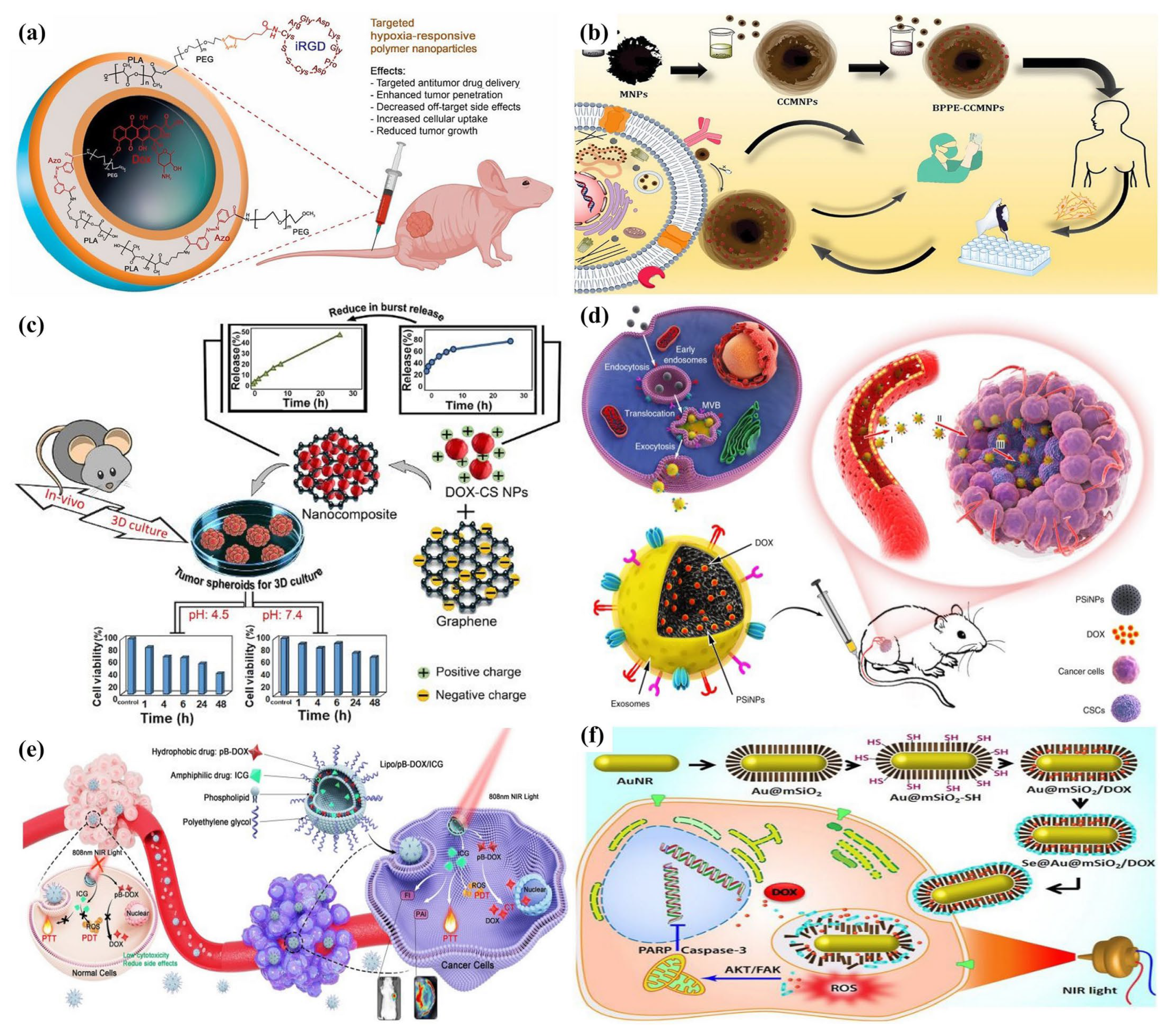
Figure 3. Systemic drug delivery using novel nanomaterials for breast cancer treatment. (a) Hypoxia-responsive polymersomes encapsulating Dox and conjugated with iRGD serve as NRP-1 receptor-targeted delivery vehicles for introducing Dox into solid TNBC tumors [70], reproduced with permission from Mamnoon et al., (2021), ©American Chemical Society; (b) black pomegranate peel extract used as a novel drug for BC treatment using chitosan-coated magnetic nanoparticles [71], reproduced with permission from Taherian et al., (2021), Creative Commons Attribution 4.0; (c) pH-sensitive BSA-stabilized graphene/chitosan nanocomposites conjugated with breast cancer drugs to control BC cells at acidic pH [72], reproduced with permission from Gooneh-Farahani et al., (2021), Creative Commons Attributions 4.0; (d) porous silicon nanoparticles that mimic biocompatible exosomes excreted by tumor cells serve as drug carriers for bulk cancer cells and cancer-stem-cell-targeted cancer chemotherapy [73], reproduced with permission from Yong et al., (2019), Creative Commons Attribution 4.0; (e) combining photodynamic treatment and chemotherapy with a reactive oxygen species-responsive drug delivery nanosystem [74], reproduced with permission from Yi et al., (2021), Creative Commons Attributions 4.0; (f) Se@Au@mSiO2 nanocomposite for inhibition of Src/FAK/AKT pathway of metastatic breast cancer [75], reproduced with permission from Ramasamy et al., (2018), Creative Commons Attributions 4.0.
Applsci 14 01809 g003

5.2. Temperature-Sensitive Nanoparticles

Tumor tissues typically exhibit higher temperatures than normal tissues, allowing for controlled drug delivery at the tumor site through external heating. This characteristic has been harnessed in the development of intelligent drug delivery systems. Several preclinical studies have demonstrated that the efficacy of both radiotherapy and chemotherapy can be enhanced when combined with hyperthermic therapy [76]. Cen et al. evaluated the photothermal efficacy of a palladium–ruthenium nanohybrid with a polypyridyl complex (PdRu-RCE@PCM NPs) against breast cancer cell lines [76]. The photothermal properties of PdRu facilitate the melting of the heat-sensitive PCM material under near-infrared light, leading to the release of PdRu and RCE in a laboratory setting. Additionally, PdRu not only damages tumor cells through photothermal therapy (PTT) but also catalyzes H2O2 to produce O2, thereby enhancing photodynamic therapy (PDT). The enzyme-like activity further elevates reactive oxygen species (ROS) levels within tumors [76]. Significantly, due to the effective accumulation of RCE, PdRu-RCE@PCM nanoparticles not only serve as photosensitizers but also function as superior fluorescent imaging agents [76]. Studies indicate that these nanoparticles, when combined with PTT and PDT, offer promising therapeutic outcomes in suppressing both primary and metastatic tumors while maintaining favorable biocompatibility in the body [76]. This provides valuable insights into the treatment of breast cancer, particularly the metastatic type. Pd is known for its robust catalytic activity, and Ru, with its ability to produce oxidation species at low potentials, enhances catalytic activity through both bifunctional and electronic mechanisms. The alloy of Pd and Ru is considered a crucial element often used as a cocatalyst [76]. Pd and Ru have demonstrated effectiveness as photothermal conversion agents, making them suitable for photothermal therapy in tumor treatments [76]. Bao et al. have developed a gold-promoting satellite with a copper chalcogenide nanocrystal (Cu2-xS) [76]. Triple-negative breast cancer (TNBC) exhibits a high expression of programmed cell death-ligand 1 (PD-L1), which serves as an active targeting site. The authors leveraged this by attaching the extracellular domain of PD-1 to gold-promoting satellites on the Cu2-xSe surface. This resulted in the PD-1-modified GPS–CS@PD-1 nanocomplex, which demonstrated robust binding to PD-L1-expressing TNBC cells [76]. When this complex was introduced to TNBC tumor-bearing mice, photoacoustic and photothermal imaging revealed that the nanocomplex’s accumulation in tumor areas peaked. This was evidenced by a prominent photoacoustic imaging (PAI) signal contrast and a peak temperature of 53.4 °C [76]. This elevated temperature induced hyperthermia, leading to the necrosis of cancer cells [77]. A targeted nanocarrier system enhances drug delivery to the epidermal growth factor receptor (EGFR)-expressing breast cancer (BC) cells, thereby amplifying the effects of photothermal therapy. Dorjsuren et al. developed liposome-mediated magnetic nanoparticles conjugated with cetuximab and doxorubicin to specifically target EGFR [78]. These liposomes, sensitive to heat, encapsulate iron oxide nanoparticles within heat-sensitive liposomes that can be activated by near-infrared light for controlled drug release, showcasing effective photothermal therapy. This delivery mechanism presents a potential advancement in breast cancer treatment techniques [78]. Luo et al. designed a thermo-responsive hydrogel by conjugating it with triptolide. This injectable hydrogel significantly enhances survival rates and minimizes side effects compared to unbound triptolide [79]. In another study, heat treatment at around 43 °C combined with lauric acid encapsulated in a biocompatible silica shell induced oxidative stress, variations in caspase levels, and morphometric changes [80]. These nanoparticles were particularly effective in disrupting breast cancer cell lines (MCF-7) through thermal activation at 43 °C, which is lauric acid’s melting point [80]. Consequently, the MCF-7 cell lines were doubly affected, first due to the elevated temperature and second, the therapeutic impact of lauric acid, functioning as a chemotherapy agent, inducing oxidative stress, apoptosis, and morphometric changes [80]. Chemo-photothermal therapy is emerging as a future direction in cancer treatment. In their study, Pakravan et al. fabricated hollow gold nanostars (HGNSs) and gold nanocages (GNCs) [80]. They then attached doxorubicin (Dox) to the GNSs@Pol structures, demonstrating the effectiveness of combined therapy in MCF-7 BC cells using HGNSs@Dox-Pol as well as GNCs@Dox-Pol for photothermal therapy [80].

5.3. Enzyme-Responsive Nanoparticles

Engineered nanoparticles responsive to enzymes are designed to selectively interact with particular enzymes present in tumor tissues [81]. This leads to the precise delivery of anticancer drugs, minimizing systemic side effects and improving treatment outcomes. Additionally, these nanoparticles effectively engage with internal enzymes under mild conditions, such as near-neutral pH levels, water-based environments, and low temperatures [82]. Typically, tumor cells exhibit higher concentrations of gelatinase, a proteolytic enzyme, compared to normal tissues [83]. In this regard, a chitosan/gelatin hybrid nanogel incorporating gold nanoparticles (CS/AuNPs@Gel-Dox nanogel) was designed as an enzyme-responsive nanoparticle platform for cancer therapy [84]. The zeta potential and the size of the CS/AuNPs were influenced by cross-linking. Upon exposure to gelatinase, the nanogel’s structure undergoes enzyme-specific degradation, enabling targeted drug delivery from the CS/AuNPs@Gel-Dox nanogel [84]. The nanogel’s compatibility with cells and its efficacy as a cancer drug carrier were validated through cytotoxicity tests. Flow cytometry results indicated that the CS/AuNPs@Gel-Dox nanogel was successfully absorbed by cells. Therefore, this tailor-made, enzyme-responsive nanogel holds promise for targeted treatment of various solid tumors [84]. Similar research was carried out by Xiao et al. with disulfiram and doxorubicin conjugated with polymeric nanoparticles [85]. Another interesting study was recently conducted with peptide conjugate crafted to combine mild photothermal therapy with immunotherapy in a unified nanosystem for treating breast cancer. The peptide–photosensitizer conjugate nanospheres have a distinctive property: they release the PD-L1 (programmed cell death-ligand 1) antagonist peptide in response to MMP (matrix metalloproteinase) and undergo a coassembled morphology change. Both of these characteristics were studied both in vitro and in vivo [86]. With laser irradiation, these PPC nanospheres demonstrate impressive antitumor effects, efficiently curbing the growth of localized tumors, distant tumors, and in vivo lung metastases [86]. The PPC (peptide–photosensitizer conjugate) is modular in design, allowing for adjustments in its responsiveness, the delivery speed of immune checkpoint antagonists, and the photosensitizer’s aggregated state for different uses. Sun et al., in their study, noted the localized enzyme response and carrier transformation through changes in fluorescence intensity [86]. A drug delivery system (POL-MSN) was developed that responds to both pH and esterase. This system was created by enclosing Dox-loaded MSNs with poly (β-amino ester) and boronate esters. Given the acidic pH and elevated esterase levels in cancer cells, POL-MSN is designed to smartly discharge its drug payload at the tumor location, targeting and eliminating the cancer cells. This system has demonstrated potent cytotoxic effects against MDA-MB-231 human breast cancer cells [87]. In addition, microbots that are fabricated using nanomaterials are gaining attention among researchers for the treatment of specific cancer types via targeted and controlled drug delivery [88,89,90]. Table 1 provides a concise overview of nanotherapeutics for breast cancer investigated in prior studies, detailing the delivery techniques employed and their respective findings.

6. Properties of Breast Cancer Drugs

Current cancer treatments encompass surgery, radiation, and chemotherapy, each posing threats to healthy tissues or potentially leaving behind cancerous cells. However, nanotechnology promises a direct and selective approach to target cancer cells, aid in tumor surgeries, and amplify the effects of current treatments [39]. The potential outcomes include enhanced patient safety and better chances of recovery. Going beyond the improved drug delivery system, nanotechnology plays a significant role in cancer treatment by enabling innovative approaches tailored to the unique attributes of nanomaterials [39]. Despite being smaller than cells, nanoparticles can house various types of small molecule compounds. Their ample surface area can be customized with several ligands, like peptides, DNA or RNA strands, and even antibodies. These ligands serve dual purposes: they can both treat and guide the nanoparticles in the body. This multifunctionality facilitates combined treatment methods, simultaneous diagnosis, and therapy-termed theragnostics.
Commercially available therapeutic drugs for BC can be classified as hydrophilic or hydrophobic, depending on their solubility in water. Additionally, depending on their electrostatic characteristics, they can be classified as either highly charged or neutral. When creating NPs to serve as carriers for particular drug classes, understanding the drug’s behaviors and properties is crucial to ensure optimal encapsulation and the desired release traits. NPs that are functionalized or tailored are particularly promising in drug delivery systems due to their unique sizes, adaptable surface properties, and controlled drug release capabilities. Hydrophilic drugs are crucial in treating BC subtypes and encompass both macromolecules and various small molecular compounds. Most therapeutic BC drugs used in clinics are hydrophobic, which poses ongoing delivery challenges to their intended targets [87]. Given their water-insolubility, hydrophobic drugs struggle to traverse through bodily fluids and find it challenging to penetrate cell membranes and reach intracellular targets. Furthermore, administering these drugs intravenously can result in clinical complications, such as tissue damage and embolisms.
Existing breast cancer treatments encounter several challenges, including a lack of targeted toxicity, leading to reduced treatment effectiveness and compromised medical diagnosis [91]. These treatments can also cause harm to healthy tissues, requiring reduced doses of anticancer drugs to lessen this toxicity. In solid tumors, there is often inadequate distribution and penetration, and the diverse blood vessels in tumor areas can lead to excessive drug leakage [91]. In comparison to tumor sites, normal organs often receive 10–20 times more drug deposits. Additionally, many chemotherapy drugs cannot spread beyond 40–50 mm from the blood vessels, potentially causing multidrug resistance (MDR) and treatment failure. When tumor cells develop MDR after exposure to one anticancer drug, they may become resistant to multiple drugs due to the heightened expression of drug-removal proteins [92].
The combined innovation of nanoparticle designs with new pharmaceutical components broadens the spectrum of active ingredients beyond those conventionally considered safe or effective. There is also an exploration into immune-boosting components and coatings, acting as supplementary measures to conventional radio- and chemotherapy or as independent treatments. One exciting approach involves crafting nanoparticles as synthetic antigen-presenting cells and in vivo reservoirs of immune-enhancing factors, harnessing the power of nanotech for sustained anticancer activities [92].
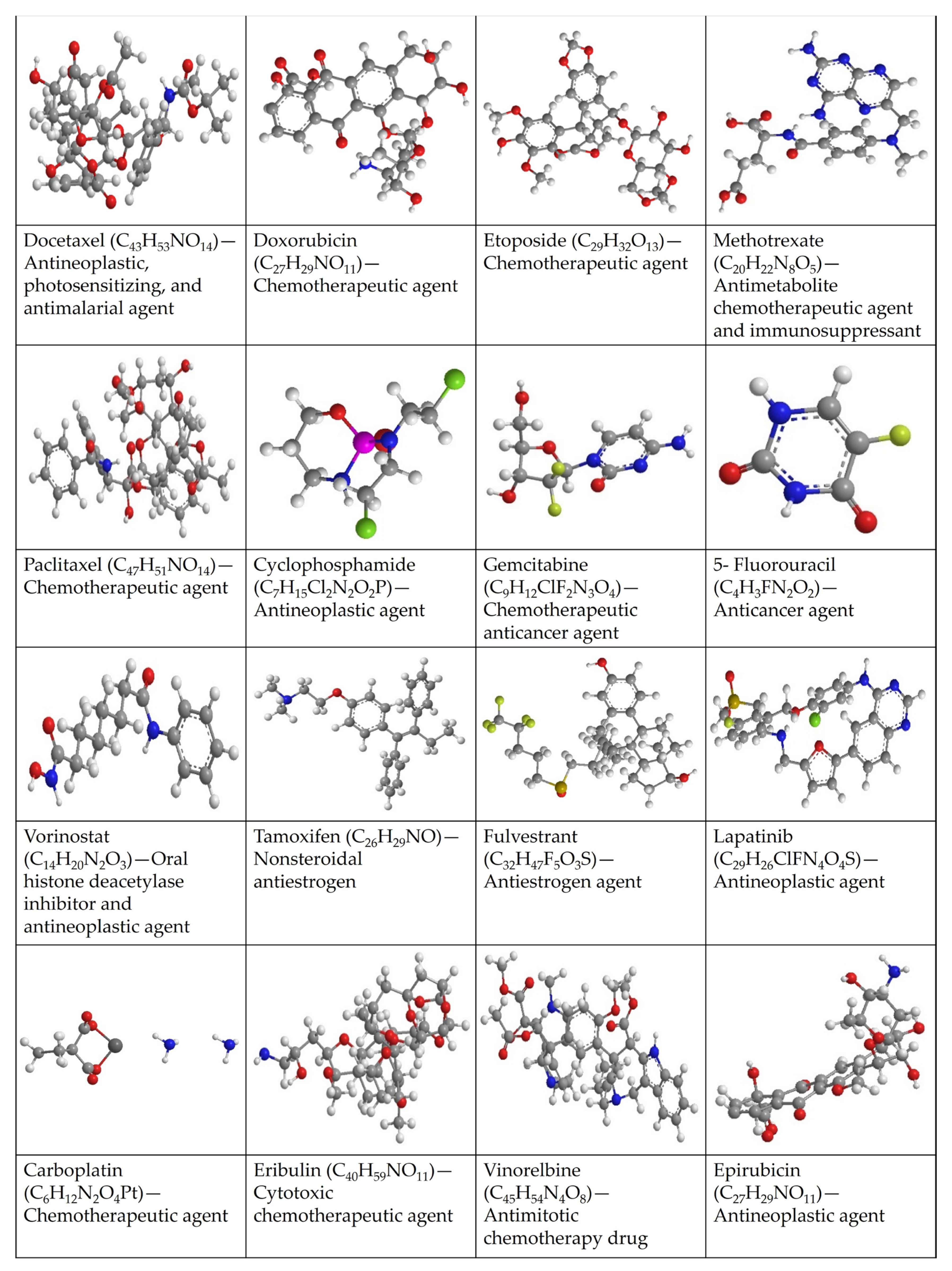
Technological advancements in cancer treatment have significantly improved the standard therapeutic strategies for BC, leading to a reduced mortality rate and aiding many patients in their cancer recovery. However, challenges remain in treating BC using the current therapeutic approaches. Chemotherapy is widely recognized as the primary treatment for BC [93]. However, traditional chemotherapy approaches have notable limitations. One major concern is the nonspecific distribution of chemotherapeutic agents to tumors. These agents harm the body’s normal cells along with their rapid-dividing cancerous targets. This lack of specificity often leads to unavoidable side effects [93]. The subsequent side effects include hair loss, vomiting, nausea, diarrhea, mouth ulcers, fatigue, heightened vulnerability to infectious diseases, myelosuppression, and issues like leucopenia, anemia, and increased propensity for bruising or bleeding [94]. Moreover, specific drugs have unique side effects, such as cardiotoxicity from anthracyclines and ototoxicity and nephrotoxicity from cisplatin. Secondly, drug resistance poses another challenge for traditional chemotherapeutic agents, diminishing their effectiveness in treating cancer cells [94]. This resistance can be categorized into innate and acquired types, depending on when they appear. Chemoresistance has intricate underlying causes, such as heightened drug expulsion, tumor diversity, improved repair of DNA damage, epigenetic changes, inhibition of apoptosis, modifications in drug targets, deactivation of the anticancer drugs, alterations in drug metabolism, and shifts in the TME [94]. Given the aforementioned challenges with conventional treatments, key obstacles in BC treatment involve tackling multidrug resistance and recurrence and mitigating or sidestepping treatment-induced side effects. Consequently, there is a pressing need for innovative novel therapeutic approaches to effectively manage BC and meet the critical medical demands of BC patients. Figure 4 enlists the FDA-approved drugs currently available in the market for the treatment of breast cancer.

7. Nanomedicine to Enhance the Therapeutic Effectiveness of BC

In recent years, nanomedicine has shown significant potential in addressing the limitations of traditional BC treatments. Nanotechnology focuses on the fabrication of nanomaterials/nanoparticles with sizes between 1 and 100 nm in at least one dimension. For instance, nanoparticles (NPs) can be engineered for selective medications and are utilized as drug delivery systems, commonly known as nanomedicines. These NPs offer substantial advantages by enhancing the biological distribution of drugs, directing active molecules precisely to affected tissues, and safeguarding healthy tissues against unnecessary drug exposure [94,95]. The distinctive characteristics of NPs encompass their minuscule size, extensive surface-to-volume ratio, modifiable physicochemical properties, capability to carry substantial drug quantities, prolonged circulation time, elevated absorption and retention, efficient tumor targeting, extended drug release, biocompatibility, improved bioavailability, and capability to counteract multidrug resistance [94,95]. Additionally, the minute size of nanoparticles enables them to penetrate biological barriers, offering a treatment avenue for BC patients with brain metastases [94,95]. Moreover, these nanoparticle-based medicines possess all the significant properties of a conventional breast cancer drug along with enhanced abilities to overcome some of the major limitations discussed earlier.
Drug delivery systems based on nanocarriers have been identified to possess enhanced effectiveness for targeting multiple cancer sites. The predominant methods of nanocarrier-based drug delivery utilize both organic and inorganic particles. Common organic particles employed in drug delivery include micelles, liposomes, polymers, dendrimers, and nanogels, known for their adaptable surface structures that aid in efficient drug loading and cellular uptake. Further, nanoparticles as carriers possess surfaces that can be tailored to direct drugs specifically to tumor blood vessels. Recently, the utilization of nanodevices to encapsulate chemotherapeutic drugs has proven beneficial in reducing side effects and enhancing the drugs’ bioavailability, especially for breast cancer [94,95]. Hence, nanocarrier platforms offer the most effective method for targeting drug-resistant cells in breast tumors.

7.1. Nanoparticle-Mediated Drug Delivery

Nanomedicine offers numerous advantages over traditional cancer treatments, including better protection against in vivo biochemical degradation, fewer adverse effects as a result of enhanced biocompatibility and precision targeting, and a higher dose of chemotherapy reaching the cancerous tissue. The field of delivery systems has seen significant advancements aimed at transporting therapeutic agents or naturally derived active compounds to targeted locations for treating various ailments. Despite recent successes with several drug delivery systems, there are persisting challenges that necessitate attention. Advanced technology is crucial to ensure the effective delivery of drugs to targeted sites [96]. Consequently, the current research is dedicated to advancing nano-based drug delivery systems as the next-generation approach. NPs possess active or passive targeting capabilities and are enveloped in layers of various organic or inorganic materials that define their characteristics. These attributes can enhance drug concentration within tumors while minimizing toxicity in healthy tissues. Numerous studies underscore the benefits of utilizing NPs in drug delivery systems (DDSs) for BC treatment, including factors like water dispersion, biocompatibility, biodegradability, stability, circulation half-life, renal processing, accumulation, and cellular uptake [94,95]. Thus, DDSs play a crucial role in probing the response of nanoparticles toward living systems at cellular and tissue levels. For instance, tailored liposomes can securely hold drugs until triggered; thus, they can facilitate prolonged drug release. Additionally, they tend to accumulate in cancer cells, enhancing drug selectivity and consequently reducing toxicity. Nanomedicine holds significant promise in targeting and eradicating BC stem cells, which could play a crucial role in BC initiation, recurrence, and resistance to chemo/radiotherapy. Several nanoparticle-based chemotherapy delivery systems are either FDA-approved or undergoing clinical trials for cancer treatment [47,97].

7.1.1. Liposomal Nanocarriers

Recent studies by Luo et al. delved into treatments for TNBC using the drug cabazitaxel [98]. Women who experience brain metastasis from TNBC face severe therapeutic obstacles, mainly due to the tumor’s heterogeneity. To address this, the authors employed a platelet–membrane hybrid liposome delivery system, known as pVAP-PL, to target orthotopic breast cancer [99]. They found that the pVAP-PL/CNC drug delivery system was effective in preventing the premature release of cabazitaxel and in extending the in vivo circulation time of CNC [99]. Further, Hu et al. also developed a liposome responsive to matrix metalloproteinase-2 (MMP-2), which dual-targets and co-delivers AUNP-12 and NLG919 to the intended targets, enhancing the synergy in remodeling the immunosuppressive microenvironment [98]. They encapsulated the indoleamine 2,3-dioxygenase 1 (IDO-1) inhibitor, NLG919, into the liposome, resulting in a tumor cascade-responsive liposome drug delivery system named NLG919@Lip-pep1 [98]. This system targets both the T cells and tumor cells associated with the PD-1 signaling pathway, particularly at the tumor’s invasive margins. Furthermore, Hu et al. successfully introduced a novel MMP-2-responsive cascade target liposome delivery system [98]. Their findings underscore the potential of an MMP-2-responsive cascade-targeted immunotherapy approach for metastatic BC [98]. This method synergistically modulates the tumor’s immunosuppressive environment, prompting a robust immune response against metastatic breast tumors [98]. Moreover, tumor-homing peptide–capecitabine liposomes (THP-CAP-LPs) were notably absorbed by cells, and the destructive impact of capecitabine was enhanced with THP-CAP-LPs by decreasing antiapoptotic proteins and increasing proapoptotic ones, as confirmed by Western blot analysis [100]. Thus, THP-CAP-LPs for delivering CAP could be a viable strategy to boost antitumor effects while minimizing unintended consequences [100].

7.1.2. Solid Lipid Nanoparticles

Recently, solid lipid nanoparticles (SLNs) based on glyceryl monostearate were employed to encapsulate methotrexate for breast cancer treatment [101]. The methotrexate NPs hindered the cell cycle’s advancement into the S phase. Compared to standard methotrexate, the methotrexate NPs demonstrated enhanced cellular absorption. Moreover, these nanoparticles curtailed the movement and growth of cancer cells and stimulated cell death both in vitro and in vivo. Cytotoxicity assessments showed promising tumor suppression and biocompatibility with minimal side effects [101]. Similar results were obtained by Abd-Ellatef et al. when SLNs were loaded with curcumin to become biocompatible nanocarriers for reducing P-glycoprotein expression, enhancing the effectiveness of doxorubicin against resistant TNBC tumors and using smaller, harmless doses of CURC. Previous P-glycoprotein inhibitors have been ineffective due to their lack of specificity, high toxicity, and interference with the natural functions of P-glycoprotein in healthy tissues. SLNs have proven to be both biocompatible and safe. This finding is crucial since doxorubicin-based chemotherapy is a primary treatment for TNBC. Unfortunately, this breast cancer subtype is less responsive to doxorubicin because of the prevalent presence of P-glycoprotein [102]. Furthermore, psoralen-loaded polymeric lipid nanoparticles demonstrated enhanced antimetastatic and antitumor effects of paclitaxel in both in vitro and in vivo studies against TNBC [103].

7.1.3. Other Polymeric Nanoparticles

Doxorubicin and cisplatin loaded with polylactic-co-glycolic acid (PLGA)-based nanosystems are currently being employed for breast cancer treatment, demonstrating controlled delivery in both in vitro and in vivo studies [104]. Likewise, methotrexate combined with chitosan biopolymer nanoparticles has also been utilized for breast cancer treatment. The serum biomarkers were evaluated with controlled and treated biofluids to confirm the controlled release and biodistribution cytotoxic effect [105]. Hence, a novel approach was used to synthesize MSN-COOH and encapsulate Dox within its pores to address the nontargeted adverse effects of doxorubicin. Subsequently, Chang et al. modified the surface of MSN-COOH with polyethylenimine (PEI) and amino acids (AAs) using amide bonds [106]. This system not only ensured Dox retention until it reached tumor sites but also facilitated efficient Dox release, leveraging the inherent pH sensitivity of tumor cells. Furthermore, AA-enhanced nanoparticles improved uptake in tumor cells and their spread within breast tumors by specifically binding to sigma receptors. This system achieved a coordinated antitumor effect by combining a pH-driven controlled release with targeted delivery, both in lab tests and live subjects, to minimize harm to healthy tissues [106]. Similarly, paclitaxel-loaded PLGA polymeric nanoparticles were used to treat TNBC. The drug-loaded nanoparticles were employed to assess adenosine receptor expression (a TNBC marker) in both controlled and treated breast cancer cell lines, revealing higher levels of apoptosis [107]. Another research group, Guo et al. developed a double-network hydrogel polymer infused with paclitaxel nanoparticles, targeting sustained local drug delivery for the treatment of BC. This formulation demonstrated enhanced stability, tolerability, and effectiveness [108]. Folic acid–PEGylated nanoliposomes were engineered to codeliver both water-soluble (cisplatin/CIS) and water-insoluble (epirubicin/EPI) chemotherapy drugs for breast cancer treatment. These FA-PEGylated noisome nanocarriers, optimized for the codelivery of CIS and EPI, showed increased stability over a two-month period and sustained delivery at a physiological pH. Cellular tests revealed anticancer effects on SKBR3 and 4T1 cancer cells, with reduced toxicity to healthy cells. Additionally, the FA-PEGylated noisome CIS and EPI (FPNCE) and epirubicin-loaded noisome groups were more effective in preventing the migration and division of cancer cells compared to free drugs [109].

7.1.4. Carbon-Based Nanoparticles

Recently, a novel study on a dual-drug delivery system was carried out using potassium-containing graphene oxide as a fluorescent nanocarrier, where the cell viability assay showed that only 18% of the breast cancer cell lines survived. The authors suggested that potassium-containing graphene oxide could provide a viable nanocarrier for dual-drug delivery [110]. An alkylating chemotherapy medication for a variety of malignancies is carboplatin. Using chitosan-coated magnetite graphene oxide, carboplatin was loaded for breast cancer treatment. The results from the research verified the drug-loading capacity as opposed to other nanomaterials. The drug releasing/biodistribution within the BC cell lines were also examined as good nanocarriers for drug delivery, particularly for carboplatin delivery [111]. The encapsulation and carboplatin capacities were maximized by PEGylated multiwall carbon nanotubes (MWCNTs). The release of carboplatin from PEGylated MWCNTs, particularly at pH 6.8, indicated pH-dependent drug activity, making them a promising carrier for chemotherapy drugs facing high resistance, significant side effects, or limited oral bioavailability [112].

7.1.5. Other Novel Nanoparticles

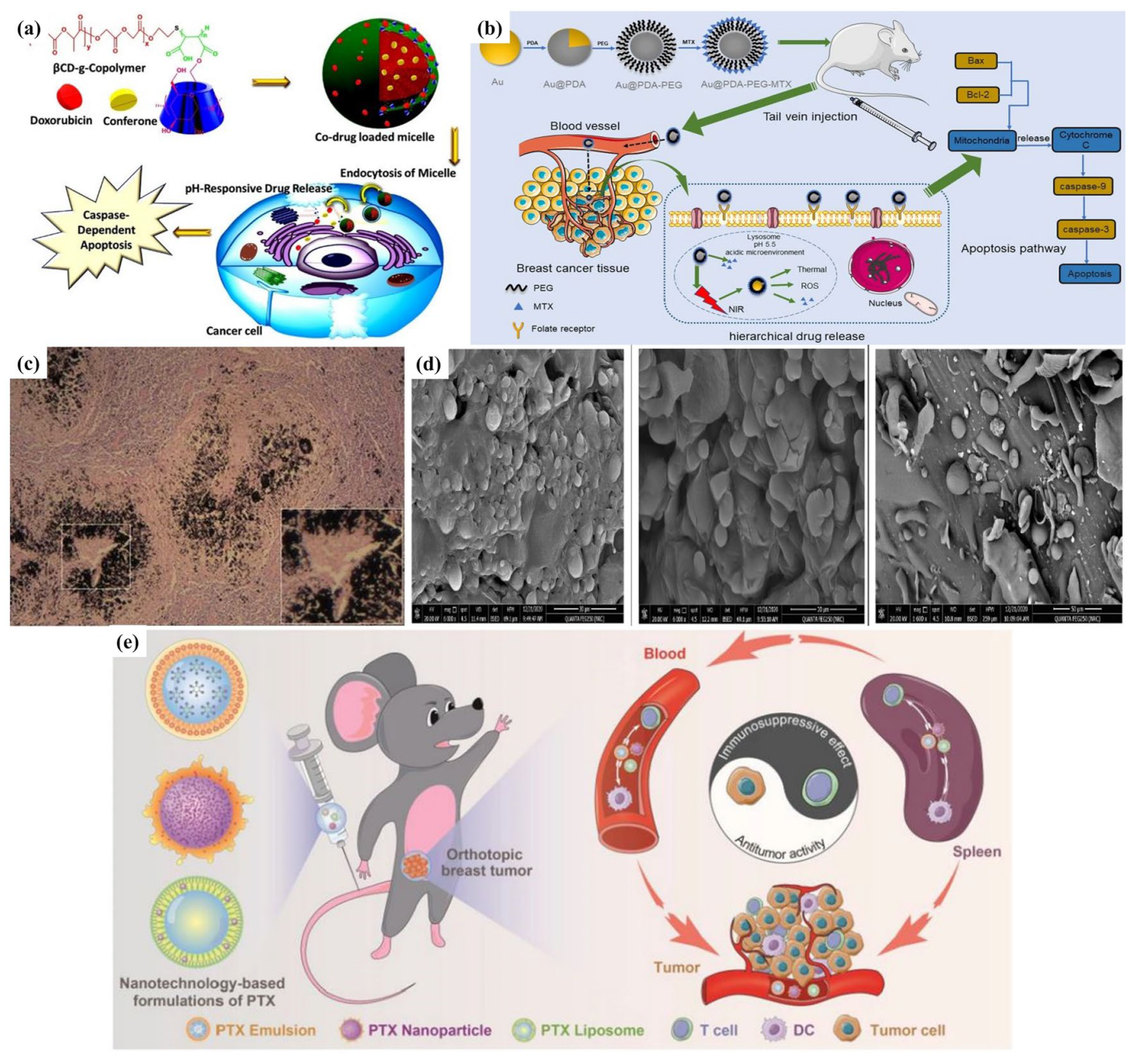
In recent times, certain nanoparticles have been used as therapeutic carriers that can encapsulate drugs in their core for both single and combined treatments. Letrozole and cyclophosphamide loaded with noisome nanoparticles were used to check the synergic effect. Folic acid was used to coat the nanoliposomes to enhance the targeting capabilities of the carriers, allowing them to bind with folate receptors that are more prevalent in breast cancer cells. Subsequently, biological tests were conducted in vitro, evaluating cell viability, the expression of apoptotic genes, and apoptosis ratios using breast cancer cell lines [113]. Another study was conducted by Tohidi et al. using MIL-100 (Fe) to treat breast cancer, and the observed results concluded that drug release was controlled by NPs, with histopathological experiments providing additional evidence for the usage of NPs in drug delivery systems [114]. Although nanoparticle-based systemic drug delivery technologies can potentially provide early treatment for breast cancer, there remain limited options available for patients with metastatic breast cancer. Figure 5 illustrates an insightful exploration of the applications of nanomaterials in drug delivery systems for breast cancer treatment.

7.2. Nanotechnology to Enhance Immunotherapy for BC Treatment

Immunotherapy offers a hopeful avenue in cancer treatment, featuring various methods such as checkpoint inhibition and cellular therapies. While some patients have seen remarkable results, only a limited number benefit long-term, especially for specific cancer types. A deeper comprehension of the interrelations between the immune system of the host and tumors is necessary to broaden the advantages of immunotherapy. The exploration of nanotechnologies for immunotherapy delivery involves utilizing nanoparticles to carry immune-boosting or immune-altering molecules, combined with chemo- or radiotherapy or as supplements to other immunotherapies. Separate nanoparticle vaccines are in development to enhance the T-cell response to eliminate tumors. This can be achieved by codelivering antigens and adjuvants, incorporating various antigens for stimulating multiple dendritic cell targets, and maintaining a steady release of antigens for extended immune activation [120]. Nanotechnology’s role in immunotherapy also encompasses the placement of immune depots within or close to tumors for on-site vaccination and the creation of synthetic antigen-presenting cells [121].
Numerous attempts have been made to enhance antitumor immunity through immune modulation. Nanomaterials have gained substantial attention for their capacity to tackle current problems in cancer immunotherapy. In a notable approach, the Toll-like receptor 7/8 agonist, which is a small immunomodulatory molecule, was chemically bonded to a biodegradable polymeric nanoparticle for use in treating metastatic breast cancer alongside PD-1 checkpoint blockade immunotherapy [122]. The nanoparticle treatments demonstrated a notable absence of toxicity typically associated with the direct use of resiquimod. This suggests that embedding resiquimod into nanoparticles reduced the toxicity concerns, making these nanoparticles potential candidates for clinical application [122]. A combined immunotherapy system utilizing the recombinant Nap protein from Helicobacter pylori was developed for breast tumor therapy [123]. Chitosan nanoparticles, which are pseudo-spherical and positively charged, were engineered to transport HP-Nap. In vitro experiments were conducted on mice (4T1) and human (MCF7) BC cell lines. In vivo, testing occurred on mice with 4T1 tumors. Both TUNEL assays and real-time PCR tests were executed on tumor-bearing mice postnanoparticle therapy [124]. In vivo, tests on female BALB/c mice showcased reduced tumor size after receiving the HP-Nap-loaded nanoparticle treatment. The TUNEL assay also indicated cell death in retrieved mouse BC cells [123]. A decline in the expression of VEGF and MMP9 genes in 4T1 cells was confirmed by real-time PCR. This evidence implies the nanocomplex might reduce mouse tumor growth by modifying cytokine production and amplifying the immune system’s tumor-killing actions [123].
A vaccine based on the λ phage targeting aspartate β-hydroxylase (ASPH) paired with a checkpoint inhibitor effectively activates antigen-specific CD8+T and B cell-driven cellular and humoral immunity in both hepatocellular carcinoma (HCC) and TNBC [124]. By targeting ASPH, an oncofetal protein and optimal tumor-associated antigen that bypasses immunological tolerance, this innovative vaccination strategy has broad applicability across tumors expressing ASPH. It presents distinct chances for crafting precision treatments [124]. When Bai et al. combined an ASPH-based λ phage vaccine blueprint with checkpoint inhibitors, the result was an intensified synergistic antitumor immune reaction in preliminary mouse models of HCC and TNBC [124]. Both cancer types have historically lacked effective treatments and often result in notably adverse outcomes [124].
A recent study employed a new nanoparticle-based approach to enhance targeted therapy for tumors, integrating chemotherapeutics like doxorubicin (Dox) and melittin (Mel) with an immune checkpoint inhibitor, PD-L1 DsiRNA [125]. The designed nanoparticle was crafted by pairing Mel with PD-L1 DsiRNA and then incorporating Dox. The resulting particles (DoxMel/PD-L1 DsiRNA) were coated with hyaluronic acid (HA) to boost stability and distribution [125]. Additionally, HA offers tumor-targeting capabilities by binding to the CD44 receptor on cancer cells. The experiments confirmed that the HA-coated DoxMel/PD-L1 DsiRNA displayed enhanced specificity in breast cancer cells. Bahreyni et al. noticed a marked drop in PD-L1 expression and a combined impact of Dox and Mel in eradicating cancer cells and inducing immune-mediated apoptosis [125]. This led to a notable reduction in tumor progression in Balb/c mice with 4T1 breast tumors, a heightened survival rate, and an increased presence of immune cells, including cytotoxic T cells, in the tumor surroundings [125].

7.3. Nanotechnology-Augmented Gene Therapy for BC Treatment

The significance of nanomaterial delivery is evident, especially for novel therapeutics like nucleic acids, which degrade easily in systemic circulation. This encompasses DNA and RNA genetic treatments, including small interfering RNAs (siRNAs) and microRNAs (miRNAs). SiRNAs, known for gene silencing, display notably longer half-lives when transported via nanoparticles either encapsulated or attached to their surface. Often, these treatments target elusive cancer proteins that other drugs cannot address. Furthermore, the stability of genetic treatments, when conveyed through nanocarriers and combined with controlled release, tends to extend their impact. A recent study explored the combination of a chemotherapy drug with siRNAs for treating TNBC [126]. In an effort to increase the effectiveness of therapy for advanced breast cancer, a versatile carrier system that utilizes gold nanoparticles (AuNPs) was designed to deliver siRNAs that target the antiapoptotic Bcl-2 gene and the well-known chemotherapeutic medication doxorubicin concurrently [126]. The Bcl-2-siRNAs were affixed to 13 nm AuNPs, with Dox instinctively inserted into the siRNA molecules, eliminating the necessity for intricate chemical alterations or coatings. The results of the study confirmed that both siRNA and Dox could be successfully integrated into the AuNPs to produce a multifunctional carrier. This Dox-infused system displayed toxicity toward TNBC cells and ensured efficient drug uptake [126]. Furthermore, codelivering Bcl-2 siRNA and Dox using the AuNPs resulted in increased cell death and reduced cancer cell growth [126]. This combined approach also led to decreased BC cell colony development and mobility. Ultimately, suppressing Bcl-2 expression amplified Dox’s therapeutic impact on TNBC [126]. Chaudhari et al., in a recent study, explored a technique to administer delicate therapeutics such as miRNA, and a parallel technique can be employed for the delivery of siRNAs as well as other treatments [127]. The crafted nanocomplex, composed of AuNPs and NH2-PEG-SH, results in PEGylated AuNPs. Beyond its capacity to bind electrostatically to miRNAs, this nanocomplex offers the significant benefit of easy conjugation with therapeutic agents [127]. Even at nanomolar concentrations, the loaded nanocomplex can induce cancer cell death. Administering MiR-206 through this gold nanocomplex halted cell growth, triggered G0–G1 cell arrest, and altered the mitochondrial membrane potential [127]. Another study conducted by Han et al. supports the findings that miRNAs have the potential to target various immune checkpoints, sparking interest in investigating their combination with immune checkpoint inhibitors (ICIs) for BC treatment [128].
LINC01094 impacts cell cycle progression and breast cancer cell growth and reduces apoptosis by influencing the miR-340-5p/E2F3 molecular pathway. The objective of a study conducted by Wu et al. was to comprehend the impact of long intergenic nonprotein coding RNA 1094 (LINC01094) on breast cancer cell growth, cell cycle dynamics, cell death, and the underlying mechanism involved [129]. By manipulating the expression levels of both LINC01094 and miR-340-5p in breast cancer cell lines, the authors analyzed their impacts on cell progression, cell cycle, and cell death using techniques like the cell counting kit-8 (CCK-8), 5-bromo-2′-deoxyuridine, and flow cytometry. The qRT-PCR findings indicated that LINC01094 expression was significantly elevated in BC tissues compared to neighboring noncancerous tissues. Additionally, a decline in patient survival time has been associated with higher expression levels of LINC01094 [129].
Nanomaterial-based cancer treatments offer benefits over conventional free drugs, especially in targeted delivery. Such treatments lead to reduced toxicity, lesser degradation, longer half-life, and improved efficiency compared to free drugs. Innovations in nanomaterial-targeted drug delivery include both active and passive targeting. Active targeting uses either antibodies or nanoparticles attached to small molecules, while passive targeting leverages enhanced permeability effects [42]. Active targeting, with its enhanced tumor localization abilities, offers a promising alternative to passive targeting due to its improved efficiency and retention. In comparison to conventional chemical therapies, drugs based on nanomaterials are more specific and have better bioavailability, lower cytotoxicity, superior loading capacity, and an extended half-life. The field of nanoscience and technology has seen significant advancements, leading to the emergence of several nanomaterials for cancer therapy.

8. Conclusions and Future Perspectives

From our review, we have found significant progress in understanding cancer biology, leading to the realization of the diverse nature of breast cancer. Biomarkers are pivotal for distinguishing cancer types and identifying cellular anomalies driving cancer growth. However, despite such progress, chemotherapy continues to be the primary treatment for breast cancer due to the insensitivity of metastatic cells to treatments. Nanomaterial-based therapeutics have been developed and explored to alleviate and reduce side effects associated with chemotherapy. Traditional drug delivery systems have shown limitations in targeted therapy, often with reduced efficacy. In contrast, nanoparticles offer benefits like surface modification, targeted delivery, and heightened efficacy. Utilizing nanotechnology for precise drug delivery increases therapeutic success and facilitates the integration of molecular biomarkers for specific recognition in breast cancers and their subtypes. Additionally, the bioaccumulation of nanoparticles after drug release, site-specific nanoparticle interactions with drug molecules, possible physicochemical reactions during passage through distinct organs before reaching the targeted drugs, and the self-transformation and/or self-digestion of certain nanoparticles must be evaluated in the future to enhance efficacy in the treatment and diagnosis of breast cancer.
However, despite the promising potential of nanoparticles in breast cancer therapy, few nanotherapeutics reach clinical use due to intricate designs, manufacturing challenges, regulatory hurdles, costs, and testing constraints. As nanotherapeutics become more central to diagnosis and treatment, the establishment of updated policies is crucial to overcome these barriers. While the use of nanoparticles for targeted cancer therapy holds great promise, there are several challenges to be addressed. These include the potential toxicity of nanoparticles, the difficulty in controlling drug release, and the immune system’s potential clearance of nanoparticles. However, with ongoing research and development, it is anticipated that nanoparticle-based cancer therapies will become an important part of oncology in the future. In conclusion, engineered nanoparticles offer a promising and innovative approach for targeted breast cancer therapy, with the potential to substantially enhance patient outcomes. Nevertheless, additional studies and clinical trials are imperative to comprehensively grasp their capabilities and limitations.

Author Contributions

Conceptualization, J.J.; writing—original draft preparation, P.P.; writing—review and editing, V.V., M.A.K. and M.K.D. All authors have read and agreed to the published version of the manuscript.

Funding

This research received no external funding.

Acknowledgments

All the authors acknowledge their respective department and university/institution for their support.

Conflicts of Interest

The authors declare no conflicts of interest.

References

  1. Chhikara, B.S.; Parang, K. Global Cancer Statistics 2022: The trends projection analysis. Chem. Biol. Lett. 2023, 10, 451. [Google Scholar]
  2. Sung, H.; Ferlay, J.; Siegel, R.L.; Laversanne, M.; Soerjomataram, I.; Jemal, A.; Bray, F. Global cancer statistics 2020: GLOBOCAN estimates of incidence and mortality worldwide for 36 cancers in 185 countries. CA—A Cancer J. Clin. 2021, 71, 209–249. [Google Scholar] [CrossRef]
  3. Siegel, R.L.; Miller, K.D.; Wagle, N.S.; Jemal, A. Cancer statistics, 2023. CA—A Cancer J. Clin. 2023, 73, 17–48. [Google Scholar] [CrossRef]
  4. Ben-Dror, J.; Shalamov, M.; Sonnenblick, A. The History of Early Breast Cancer Treatment. Genes 2022, 13, 960. [Google Scholar] [CrossRef]
  5. Yusuf, A.; Almotairy, A.R.Z.; Henidi, H.; Alshehri, O.Y.; Aldughaim, M.S. Nanoparticles as Drug Delivery Systems: A Review of the Implication of Nanoparticles’ Physicochemical Properties on Responses in Biological Systems. Polymers 2023, 15, 1596. [Google Scholar] [CrossRef] [PubMed]
  6. PDQ Adult Treatment Editorial Board. Breast Cancer Treatment (PDQ®). 2023. Available online: https://www.ncbi.nlm.nih.gov/books/NBK65969/ (accessed on 6 December 2023).
  7. Dissanayake, R.; Towner, R.; Ahmed, M. Metastatic Breast Cancer: Review of Emerging Nanotherapeutics. Cancers 2023, 15, 2906. [Google Scholar] [CrossRef] [PubMed]
  8. Patra, J.K.; Das, G.; Fraceto, L.F.; Campos, E.V.R.; Rodriguez-Torres, M.D.P.; Acosta-Torres, L.S.; Diaz-Torres, L.A.; Grillo, R.; Swamy, M.K.; Sharma, S.; et al. Nano based drug delivery systems: Recent developments and future prospects. J. Nanobiotechnol. 2018, 16, 71. [Google Scholar] [CrossRef]
  9. Michaeli, D.T.; Michaeli, T. Overall survival, progression-free survival, and tumor response benefit supporting initial US food and drug administration approval and indication extension of new cancer drugs, 2003–2021. J. Clin. Oncol. 2022, 40, 4095–4106. [Google Scholar] [CrossRef] [PubMed]
  10. Goransson, S.; Chen, S.; Olofsson, H.; Larsson, O.; Stromblad, S. An extracellular matrix stiffness-induced breast cancer cell transcriptome resembles the transition from ductal carcinoma in situ (DCIS) to invasive ductal carcinoma (IDC). Biochem. Biophys. Res. Commun. 2023, 654, 73–79. [Google Scholar] [CrossRef] [PubMed]
  11. Krawczyk, N.; Fehm, T.; Banys-Paluchowski, M. DCIS in Male and Aged Women with Comorbidities. Chirurgia 2021, 116, S120–S127. [Google Scholar] [CrossRef] [PubMed]
  12. Rodin, D.; Sutradhar, R.; Nofech-Mozes, S.; Gu, S.M.; Faught, N.; Hahn, E.; Fong, C.; Trebinjac, S.; Paszat, L.; Rakovitch, E. Long-term outcomes of women with large DCIS lesions treated with breast-conserving therapy. Breast Cancer Res. Treat. 2022, 192, 223–233. [Google Scholar] [CrossRef] [PubMed]
  13. Fatima, N.; Liu, L.; Hong, S.; Ahmed, H. Prediction of Breast Cancer, Comparative Review of Machine Learning Techniques, and Their Analysis. IEEE Access 2020, 8, 150360–150376. [Google Scholar] [CrossRef]
  14. Girithar, H.N.; Pires, A.S.; Ahn, S.B.; Guillemin, G.J.; Gluch, L.; Heng, B. Involvement of the kynurenine pathway in breast cancer: Updates on clinical research and trials. Br. J. Cancer 2023, 129, 185–203. [Google Scholar] [CrossRef]
  15. Rechsteiner, A.; Dietrich, D.; Varga, Z. Prognostic relevance of mixed histological subtypes in invasive breast carcinoma: A retrospective analysis. J. Cancer Res. Clin. Oncol. 2023, 149, 4967–4978. [Google Scholar] [CrossRef] [PubMed]
  16. Reed, A.E.M.; Kalinowski, L.; Simpson, P.T.; Lakhani, S.R. Invasive lobular carcinoma of the breast: The increasing importance of this special subtype. Breast Cancer Res. 2021, 23, 6. [Google Scholar] [CrossRef]
  17. Takada, K.; Kashiwagi, S.; Asano, Y.; Goto, W.; Morisaki, T.; Takahashi, K.; Fujita, H.; Takashima, T.; Tomita, S.; Hirakawa, K.; et al. Factors predictive of invasive ductal carcinoma in cases preoperatively diagnosed as ductal carcinoma in situ. BMC Cancer 2020, 20, 513. [Google Scholar] [CrossRef]
  18. Aihara, T.; Kumamaru, H.; Ishitobi, M.; Miyashita, M.; Miyata, H.; Tamura, K.; Yoshida, M.; Ogo, E.; Nagahashi, M.; Asaga, S.; et al. Prognosis and effectiveness of chemotherapy for medullary breast carcinoma. Breast Cancer Res. Treat. 2022, 196, 635–645. [Google Scholar] [CrossRef]
  19. Nakagawa, T.; Oda, G.; Mori, H.; Uemura, N.; Onishi, I.; Sagawa, N.; Fujioka, T.; Mori, M.; Kubota, K.; Ishikawa, T.; et al. Prognosis of Subcutaneous Mastectomy for Special Types of Breast Cancer. Med.-Lith. 2022, 58, 112. [Google Scholar] [CrossRef]
  20. Min, Y.; Bae, S.Y.; Lee, H.C.; Lee, J.H.; Kim, M.; Kim, J.; Lee, S.K.; Kil, W.H.; Kim, S.W.; Lee, J.E.; et al. Tubular Carcinoma of the Breast: Clinicopathologic Features and Survival Outcome Compared with Ductal Carcinoma In Situ. J. Breast Cancer 2013, 16, 404–409. [Google Scholar] [CrossRef]
  21. Tsoukalas, N.; Kiakou, M.; Tolia, M.; Kostakis, I.D.; Galanopoulos, M.; Nakos, G.; Tryfonopoulos, D.; Kyrgias, G.; Koumakis, G. Mucinous breast carcinoma with tall columnar cells. Ann. R. Coll. Surg. Engl. 2018, 100, E132–E135. [Google Scholar] [CrossRef]
  22. Arif, F.Z.; Breese, R.O.; Friend, K. Tall Cell Variant of Invasive Papillary Breast Carcinoma. Am. Surg. 2023, 89, 3875–3876. [Google Scholar] [CrossRef]
  23. Xia, L.Q.; Chen, X. Invasive cribriform carcinoma of the breast: A case report. Asian J. Surg. 2023, 46, 1130–1131. [Google Scholar] [CrossRef]
  24. Samar, M.R.; Khan, W.; Mooghal, M.; Anjum, S.; Mohammad, A.T.V.; Vohra, L.M. Breast adenoid cystic carcinoma: An uncommon neoplasm- Case report. Int. J. Surg. Case Rep. 2023, 107, 108333. [Google Scholar] [CrossRef]
  25. Tower, A.; Hughes, J.; Moore, L.; Srivastava, K. Mixed metaplastic carcinoma of the breast: A case report. J. Surg. Case Rep. 2023, 2023, rjad144. [Google Scholar] [CrossRef]
  26. Goetz, M.P.; Gradishar, W.J.; Anderson, B.O.; Abraham, J.; Aft, R.; Allison, K.H.; Blair, S.L.; Burstein, H.J.; Dang, C.; Elias, A.D. NCCN guidelines insights: Breast cancer, Version 3.2018: Featured updates to the NCCN guidelines. J. Natl. Compr. Cancer Netw. 2019, 17, 118–126. [Google Scholar] [CrossRef] [PubMed]
  27. Bulut, N.; Ulas, A.; Altundag, K.; Altundag, K.; Bulut, N.; Ulas, A.; Bulut, N. Histopathological Characteristics, Clinical Course, and 10-year Follow-up Results of Breast Cancer Subtypes with Regard to ER (+)(−)/PR (+)(−) Receptor Status. Available online: https://www.researchgate.net/profile/Kadri-Altundag/publication/311949386_Histopathological_characteristics_clinical_course_and_10-_year_follow-up_results_of_breast_cancer_subtypes_with_regard_to_ER_-PR_-_receptor_status_Short_title_Clinic_course_in_subtypes_of_breast_cance/links/5864253408aebf17d3974378/Histopathological-characteristics-clinical-course-and-10-year-follow-up-results-of-breast-cancer-subtypes-with-regard-to-ER-PR-receptor-status-Short-title-Clinic-course-in-subtypes-of-breast.pdf (accessed on 30 December 2016).
  28. Subramaniam, S.; Bhoo-Pathy, N.; Taib, N.; Tan, G.; See, M.; Jamaris, S.; Ho, G.; Looi, L.; Yip, C. Breast cancer outcomes as defined by the estrogen receptor, progesterone receptor, and human growth factor receptor-2 in a multi-ethnic asian country. World J. Surg. 2015, 39, 2450–2458. [Google Scholar] [CrossRef]
  29. Harbeck, N.; Penault-Llorca, F.; Cortes, J.; Gnant, M.; Houssami, N.; Poortmans, P.; Ruddy, K.; Tsang, J.; Cardoso, F. Breast cancer. Nat. Rev. Dis. Primers 2019, 5, 66. [Google Scholar] [CrossRef]
  30. Hernando, C.; Ortega-Morillo, B.; Tapia, M.; Moragon, S.; Martinez, M.T.; Eroles, P.; Garrido-Cano, I.; Adam-Artigues, A.; Lluch, A.; Bermejo, B.; et al. Oral Selective Estrogen Receptor Degraders (SERDs) as a Novel Breast Cancer Therapy: Present and Future from a Clinical Perspective. Int. J. Mol. Sci. 2021, 22, 7812. [Google Scholar] [CrossRef]
  31. Yu, N.Y.; Iftimi, A.; Yau, C.; Tobin, N.P.; van ‘t Veer, L.; Hoadley, K.A.; Benz, C.C.; Nordenskjold, B.; Fornander, T.; Stal, O.; et al. Assessment of Long-term Distant Recurrence-Free Survival Associated With Tamoxifen Therapy in Postmenopausal Patients With Luminal A or Luminal B Breast Cancer. JAMA Oncol. 2019, 5, 1304–1309. [Google Scholar] [CrossRef]
  32. Goncalves, A.; Finetti, P.; Birnbaum, D.; Bertucci, F. The CINSARC signature predicts the clinical outcome in patients with Luminal B breast cancer. NPJ Breast Cancer 2021, 7, 48. [Google Scholar] [CrossRef]
  33. Yang, Z.J.; Liu, Y.X.; Huang, Y.; Chen, Z.J.; Zhang, H.Z.; Yu, Y.; Wang, X.; Cao, X.C. The regrouping of Luminal B (HER2 negative), a better discriminator of outcome and recurrence score. Cancer Med. 2022, 12, 2493–2504. [Google Scholar] [CrossRef]
  34. Tsang, J.Y.S.; Tse, G.M. Molecular Classification of Breast Cancer. Adv. Anat. Pathol. 2020, 27, 27–35. [Google Scholar] [CrossRef]
  35. Shen, M.Y.; Pan, H.W.; Chen, Y.X.; Xu, Y.H.; Yang, W.X.; Wu, Z.J. A review of current progress in triple-negative breast cancer therapy. Open Med. 2020, 15, 1143–1149. [Google Scholar] [CrossRef]
  36. Lukasiewicz, S.; Czeczelewski, M.; Forma, A.; Baj, J.; Sitarz, R.; Stanislawek, A. Breast Cancer-Epidemiology, Risk Factors, Classification, Prognostic Markers, and Current Treatment Strategies-An Updated Review. Cancers 2021, 13, 4287. [Google Scholar] [CrossRef] [PubMed]
  37. Sakach, E.; O’Regan, R.; Meisel, J.; Li, X.X. Molecular Classification of Triple Negative Breast Cancer and the Emergence of Targeted Therapies. Clin. Breast Cancer 2021, 21, 509–520. [Google Scholar] [CrossRef]
  38. Chu, C.R.K.; Bedrosian, I. Prophylactic Mastectomy and Breast Reconstruction in Patients at High Risk for Breast Cancer. Curr. Breast Cancer Rep. 2020, 12, 13–20. [Google Scholar] [CrossRef]
  39. Pandit, S.; Sapkota, S.; Suwal, S.; Adhikari, A.; Karki, P.; Jha, A.K. Interstitial brachytherapy for internal mammary node in breast cancer: A case report. J. Contemp. Brachyther. 2023, 15, 229–233. [Google Scholar] [CrossRef]
  40. Subhan, M.A.; Yalamarty, S.S.K.; Filipczak, N.; Parveen, F.; Torchilin, V.P. Recent advances in tumor targeting via EPR effect for cancer treatment. J. Pers. Med. 2021, 11, 571. [Google Scholar] [CrossRef] [PubMed]
  41. Patel, J.K.; Patel, A.P. Passive targeting of nanoparticles to cancer. Surf. Modif. Nanopart. Target. Drug Deliv. 2019, 2019, 125–143. [Google Scholar]
  42. Sung, J.C.; Pulliam, B.L.; Edwards, D.A. Nanoparticles for drug delivery to the lungs. Trends Biotechnol. 2007, 25, 563–570. [Google Scholar] [CrossRef]
  43. Felice, B.; Prabhakaran, M.P.; Rodríguez, A.P.; Ramakrishna, S. Drug delivery vehicles on a nano-engineering perspective. Mater. Sci. Eng. C 2014, 41, 178–195. [Google Scholar] [CrossRef] [PubMed]
  44. Chaturvedi, V.K.; Singh, A.; Singh, V.K.; Singh, M.P. Cancer nanotechnology: A new revolution for cancer diagnosis and therapy. Curr. Drug Metab. 2019, 20, 416–429. [Google Scholar] [CrossRef] [PubMed]
  45. Vaez, A.; Abbasi, M.; Shabani, L.; Azizipour, E.; Shafiee, M.; Zare, M.A.; Rahbar, O.; Azari, A.; Amani, A.M.; Golchin, A. A Bright Horizon of Intelligent Targeted-cancer Therapy: Nanoparticles Against Breast Cancer Stem Cells. Curr. Stem Cell Res. Ther. 2023, 18, 787–799. [Google Scholar] [CrossRef] [PubMed]
  46. Wu, T.; Tang, M. Review of the effects of manufactured nanoparticles on mammalian target organs. J. Appl. Toxicol. 2018, 38, 25–40. [Google Scholar] [CrossRef]
  47. Liyanage, P.Y.; Hettiarachchi, S.D.; Zhou, Y.; Ouhtit, A.; Seven, E.S.; Oztan, C.Y.; Celik, E.; Leblanc, R.M. Nanoparticle-mediated targeted drug delivery for breast cancer treatment. Biochim. Biophys. Acta (BBA)-Rev. Cancer 2019, 1871, 419–433. [Google Scholar] [CrossRef]
  48. Adepu, S.; Ramakrishna, S. Controlled drug delivery systems: Current status and future directions. Molecules 2021, 26, 5905. [Google Scholar] [CrossRef]
  49. Ganesan, K.; Wang, Y.; Gao, F.; Liu, Q.; Zhang, C.; Li, P.; Zhang, J.; Chen, J. Targeting engineered nanoparticles for breast cancer therapy. Pharmaceutics 2021, 13, 1829. [Google Scholar] [CrossRef]
  50. Ulldemolins, A.; Seras-Franzoso, J.; Andrade, F.; Rafael, D.; Abasolo, I.; Gener, P.; Schwartz, S., Jr. Perspectives of nano-carrier drug delivery systems to overcome cancer drug resistance in the clinics. Cancer Drug Resist. 2021, 4, 44. [Google Scholar] [CrossRef]
  51. De Almeida, M.S.; Susnik, E.; Drasler, B.; Taladriz-Blanco, P.; Petri-Fink, A.; Rothen-Rutishauser, B. Understanding nanoparticle endocytosis to improve targeting strategies in nanomedicine. Chem. Soc. Rev. 2021, 50, 5397–5434. [Google Scholar] [CrossRef]
  52. Grumezescu, A.; Holban, A.M. Materials for Biomedical Engineering: Organic Micro and Nanostructures; Elsevier: Amsterdam, The Netherlands, 2019. [Google Scholar]
  53. Nikolova, M.P.; Kumar, E.M.; Chavali, M.S. Updates on responsive drug delivery based on liposome vehicles for cancer treatment. Pharmaceutics 2022, 14, 2195. [Google Scholar] [CrossRef] [PubMed]
  54. Hirsjarvi, S.; Passirani, C.; Benoit, J.-P. Passive and active tumour targeting with nanocarriers. Curr. Drug Discov. Technol. 2011, 8, 188–196. [Google Scholar] [CrossRef]
  55. Ejigah, V.; Owoseni, O.; Bataille-Backer, P.; Ogundipe, O.D.; Fisusi, F.A.; Adesina, S.K. Approaches to improve macromolecule and nanoparticle accumulation in the tumor microenvironment by the enhanced permeability and retention effect. Polymers 2022, 14, 2601. [Google Scholar] [CrossRef]
  56. Roe, D.; Karandikar, B.; Bonn-Savage, N.; Gibbins, B.; Roullet, J.-B. Antimicrobial surface functionalization of plastic catheters by silver nanoparticles. J. Antimicrob. Chemother. 2008, 61, 869–876. [Google Scholar] [CrossRef]
  57. Senapati, S.; Mahanta, A.K.; Kumar, S.; Maiti, P. Controlled drug delivery vehicles for cancer treatment and their performance. Signal Transduct. Target. Ther. 2018, 3, 7. [Google Scholar] [CrossRef]
  58. Mirza, Z.; Karim, S. Nanoparticles-Based Drug Delivery and Gene Therapy for Breast Cancer: Recent Advancements and Future Challenges. Semin. Cancer Biol. 2021, 69, 226–237. [Google Scholar]
  59. Chehelgerdi, M.; Chehelgerdi, M.; Allela, O.Q.B.; Pecho, R.D.C.; Jayasankar, N.; Rao, D.P.; Thamaraikani, T.; Vasanthan, M.; Viktor, P.; Lakshmaiya, N. Progressing nanotechnology to improve targeted cancer treatment: Overcoming hurdles in its clinical implementation. Mol. Cancer 2023, 22, 169. [Google Scholar] [CrossRef]
  60. Yadav, P.; Jain, J.; Sherje, A.P. Recent advances in nanocarriers-based drug delivery for cancer therapeutics: A review. React. Funct. Polym. 2021, 165, 104970. [Google Scholar] [CrossRef]
  61. Vordos, N.; Gkika, D.A.; Pradakis, N.; Mitropoulos, A.C.; Kyzas, G.Z. Therapeutic and Diagnostic Potential of Nanomaterials for Enhanced Biomedical Applications. In Advanced and Innovative Approaches of Environmental Biotechnology in Industrial Wastewater Treatment; Springer: Berlin/Heidelberg, Germany, 2023; pp. 277–300. [Google Scholar]
  62. Amoabediny, G.; Haghiralsadat, F.; Naderinezhad, S.; Helder, M.N.; Akhoundi Kharanaghi, E.; Mohammadnejad Arough, J.; Zandieh-Doulabi, B. Overview of preparation methods of polymeric and lipid-based (niosome, solid lipid, liposome) nanoparticles: A comprehensive review. Int. J. Polym. Mater. Polym. Biomater. 2018, 67, 383–400. [Google Scholar] [CrossRef]
  63. Boraschi, D.; Italiani, P.; Palomba, R.; Decuzzi, P.; Duschl, A.; Fadeel, B.; Moghimi, S.M. Nanoparticles and Innate Immunity: New Perspectives on Host Defence. Semin. Immunol. 2017, 34, 33–51. [Google Scholar]
  64. Parks, S.K.; Mueller-Klieser, W.; Pouysségur, J. Lactate and acidity in the cancer microenvironment. Annu. Rev. Cancer Biol. 2020, 4, 141–158. [Google Scholar] [CrossRef]
  65. Li, J.P.; Wang, Y.S.; Xu, C.Q.; Yu, Q.W.; Wang, X.H.; Xie, H.B.; Tian, L.F.; Qiu, Y.; Guo, R.; Lu, Z.Z.; et al. Rapid pH-responsive self-disintegrating nanoassemblies balance tumor accumulation and penetration for enhanced anti-breast cancer therapy. Acta Biomater. 2021, 134, 546–558. [Google Scholar] [CrossRef]
  66. Chen, Q.; Huang, X.Y.; Zhang, G.Y.; Li, J.N.; Liu, Y.; Yan, X. Novel targeted pH-responsive drug delivery systems based on PEGMA-modified bimetallic Prussian blue analogs for breast cancer chemotherapy. RSC Adv. 2023, 13, 1684–1700. [Google Scholar] [CrossRef]
  67. Kundu, M.; Sadhukhan, P.; Ghosh, N.; Chatterjee, S.; Manna, P.; Das, J.; Sil, P.C. pH-responsive and targeted delivery of curcumin via phenylboronic acid-functionalized ZnO nanoparticles for breast cancer therapy. J. Adv. Res. 2019, 18, 161–172. [Google Scholar] [CrossRef]
  68. Liu, Y.Y.; Qiao, L.N.; Zhang, S.P.; Wan, G.Y.; Chen, B.W.; Zhou, P.; Zhang, N.; Wang, Y.S. Dual pH-responsive multifunctional nanoparticles for targeted treatment of breast cancer by combining immunotherapy and chemotherapy. Acta Biomater. 2018, 66, 310–324. [Google Scholar] [CrossRef]
  69. Ghosh, N.; Kundu, M.; Ghosh, S.; Das, A.K.; De, S.; Das, J.; Sil, P.C. pH-responsive and targeted delivery of chrysin via folic acid-functionalized mesoporous silica nanocarrier for breast cancer therapy. Int. J. Pharm. 2023, 631, 122555. [Google Scholar] [CrossRef]
  70. Mamnoon, B.; Loganathan, J.; Confeld, M.I.; De Fonseka, N.; Feng, L.; Froberg, J.; Choi, Y.; Tuvin, D.M.; Sathish, V.; Mallik, S. Targeted Polymeric Nanoparticles for Drug Delivery to Hypoxic, Triple-Negative Breast Tumors. Acs Appl. Bio Mater. 2021, 4, 1450–1460. [Google Scholar] [CrossRef] [PubMed]
  71. Taherian, A.; Esfandiari, N.; Rouhani, S. Breast cancer drug delivery by novel drug-loaded chitosan-coated magnetic nanoparticles. Cancer Nanotechnol. 2021, 12, 15. [Google Scholar] [CrossRef]
  72. Gooneh-Farahani, S.; Naghib, S.M.; Naimi-Jamal, M.R.; Seyfoori, A. A pH-sensitive nanocarrier based on BSA-stabilized graphene-chitosan nanocomposite for sustained and prolonged release of anticancer agents. Sci. Rep. 2021, 11, 17404. [Google Scholar] [CrossRef] [PubMed]
  73. Yong, T.Y.; Zhang, X.Q.; Bie, N.N.; Zhang, H.B.; Zhang, X.T.; Li, F.Y.; Hakeem, A.; Hu, J.; Gan, L.; Santos, H.A.; et al. Tumor exosome-based nanoparticles are efficient drug carriers for chemotherapy. Nat. Commun. 2019, 10, 3838. [Google Scholar] [CrossRef] [PubMed]
  74. Yi, H.X.; Lu, W.X.; Liu, F.; Zhang, G.Q.; Xie, F.F.; Liu, W.J.; Wang, L.; Zhou, W.H.; Cheng, Z.N. ROS-responsive liposomes with NIR light-triggered doxorubicin release for combinatorial therapy of breast cancer. J. Nanobiotechnol. 2021, 19, 134. [Google Scholar] [CrossRef] [PubMed]
  75. Ramasamy, T.; Ruttala, H.B.; Sundaramoorthy, P.; Poudel, B.K.; Youn, Y.S.; Ku, S.K.; Choi, H.G.; Yong, C.S.; Kim, J.O. Multimodal selenium nanoshell-capped Au@mSiO(2) nanoplatform for NIR-responsive chemo-photothermal therapy against metastatic breast cancer. NPG Asia Mater. 2018, 10, 197–216. [Google Scholar] [CrossRef]
  76. Cen, J.Q.; Huang, Y.Q.; Liu, J.; Liu, Y.A. Thermo-responsive palladium-ruthenium nanozyme synergistic photodynamic therapy for metastatic breast cancer management. J. Mater. Chem. B 2022, 10, 10027–10041. [Google Scholar] [CrossRef] [PubMed]
  77. Bao, J.; Wang, Y.; Li, C.; Yang, C.; Xu, H.; Liang, Q.; Zhou, Y.; Zhang, L.; He, Y.; Tong, H.; et al. Gold-promoting-satellite to boost photothermal conversion efficiency of Cu2-xSe for triple-negative breast cancer targeting therapy. Mater. Today Nano 2022, 18, 100211. [Google Scholar] [CrossRef]
  78. Dorjsuren, B.; Chaurasiya, B.; Ye, Z.X.; Liu, Y.Y.; Li, W.; Wang, C.Y.; Shi, D.; Evans, C.E.; Webster, T.J.; Shen, Y. Cetuximab-Coated Thermo-Sensitive Liposomes Loaded with Magnetic Nanoparticles and Doxorubicin for Targeted EGFR-Expressing Breast Cancer Combined Therapy. Int. J. Nanomed. 2020, 15, 8201–8215. [Google Scholar] [CrossRef]
  79. Luo, Y.Y.; Li, J.J.; Hu, Y.C.; Gao, F.; Leung, G.P.H.; Geng, F.N.; Fu, C.M.; Zhang, J.M. Injectable thermo-responsive nano-hydrogel loading triptolide for the anti-breast cancer enhancement via localized treatment based on “two strikes” effects. Acta Pharm. Sin. B 2020, 10, 2227–2245. [Google Scholar] [CrossRef]
  80. De Matteis, V.; Cascione, M.; De Giorgi, M.L.; Leporatti, S.; Rinaldi, R. Encapsulation of Thermo-Sensitive Lauric Acid in Silica Shell: A Green Derivate for Chemo-Thermal Therapy in Breast Cancer Cell. Molecules 2019, 24, 2034. [Google Scholar] [CrossRef]
  81. Mu, J.; Lin, J.; Huang, P.; Chen, X. Development of endogenous enzyme-responsive nanomaterials for theranostics. Chem. Soc. Rev. 2018, 47, 5554–5573. [Google Scholar] [CrossRef]
  82. Khizar, S.; Zine, N.; Errachid, A.; Elaissari, A. Introduction to Stimuli-Responsive Materials and Their Biomedical Applications. In Stimuli-Responsive Materials for Biomedical Applications; ACS Publications: Washington, DC, USA, 2023; pp. 1–30. [Google Scholar]
  83. Moll, U.M.; Youngleib, G.L.; Rosinski, K.B.; Quigley, J.P. Tumor promoter-stimulated M r 92,000 gelatinase secreted by normal and malignant human cells: Isolation and characterization of the enzyme from HT1080 tumor cells. Cancer Res. 1990, 50, 6162–6170. [Google Scholar]
  84. Aslzad, S.; Heydari, P.; Abdolahinia, E.D.; Amiryaghoubi, N.; Safary, A.; Fathi, M.; Erfan-Niya, H. Chitosan/gelatin hybrid nanogel containing doxorubicin as enzyme-responsive drug delivery system for breast cancer treatment. Colloid Polym. Sci. 2023, 301, 273–281. [Google Scholar] [CrossRef]
  85. Xiao, P.F.; Tao, X.G.; Wang, H.X.; Liu, H.B.; Feng, Y.P.; Zhu, Y.Q.; Jiang, Z.Z.; Yin, T.; Zhang, Y.; He, H.B.; et al. Enzyme/pH dual stimuli-responsive nanoplatform co-deliver disulfiram and doxorubicin for effective treatment of breast cancer lung metastasis. Expert Opin. Drug Deliv. 2023, 20, 1015–1031. [Google Scholar] [CrossRef]
  86. Sun, Y.A.; Lyu, B.; Yang, C.; He, B.; Zhang, H.; Wang, X.Q.; Zhang, Q.; Dai, W.B. An enzyme-responsive and transformable PD-L1 blocking peptide-photosensitizer conjugate enables efficient photothermal immunotherapy for breast cancer. Bioact. Mater. 2023, 22, 47–59. [Google Scholar] [CrossRef]
  87. Fernando, I.R.; Ferris, D.P.; Frasconi, M.; Malin, D.; Strekalova, E.; Yilmaz, M.D.; Ambrogio, M.W.; Algaradah, M.M.; Hong, M.P.; Chen, X.Q.; et al. Esterase- and pH-responsive poly(beta-amino ester)-capped mesoporous silica nanoparticles for drug delivery. Nanoscale 2015, 7, 7178–7183. [Google Scholar] [CrossRef]
  88. Gong, D.; Sun, L.; Li, X.; Zhang, W.; Zhang, D.; Cai, J. Micro/Nanofabrication, Assembly, and Actuation Based on Microorganisms: Recent Advances and Perspectives. Small Struct. 2023, 4, 2200356. [Google Scholar] [CrossRef]
  89. Gong, D.; Celi, N.; Zhang, D.; Cai, J. Magnetic biohybrid microrobot multimers based on chlorella cells for enhanced targeted drug delivery. ACS Appl. Mater. Interfaces 2022, 14, 6320–6330. [Google Scholar] [CrossRef]
  90. Zhu, Y.X.; Jia, H.R.; Jiang, Y.W.; Guo, Y.; Duan, Q.Y.; Xu, K.F.; Shan, B.H.; Liu, X.; Chen, X.; Wu, F.G. A Red Blood Cell-Derived Bionic Microrobot Capable of Hierarchically Adapting to Five Critical Stages in Systemic Drug Delivery; Exploration, 2023; Wiley Online Library: Hoboken, NJ, USA, 2023; p. 20230105. [Google Scholar]
  91. Zhong, L.; Li, Y.S.; Xiong, L.; Wang, W.J.; Wu, M.; Yuan, T.; Yang, W.; Tian, C.Y.; Miao, Z.; Wang, T.Q.; et al. Small molecules in targeted cancer therapy: Advances, challenges, and future perspectives. Signal Transduct. Target. Ther. 2021, 6, 201. [Google Scholar] [CrossRef]
  92. Duan, C.Y.; Yu, M.J.; Xu, J.Y.; Li, B.Y.; Zhao, Y.; Kankala, R.K. Overcoming Cancer Multi-drug Resistance (MDR): Reasons, mechanisms, nanotherapeutic solutions, and challenges. Biomed. Pharmacother. 2023, 162, 114643. [Google Scholar] [CrossRef]
  93. Omidi, Y.; Mobasher, M.; Castejon, A.M.; Mahmoudi, M. Recent advances in nanoscale targeted therapy of HER2-positive breast cancer. J. Drug Target. 2022, 30, 687–708. [Google Scholar] [CrossRef]
  94. Wang, X.; Zhang, H.Y.; Chen, X.Z. Drug resistance and combating drug resistance in cancer. Cancer Drug Resist. 2019, 2, 141–160. [Google Scholar] [CrossRef]
  95. Tagde, P.; Najda, A.; Nagpal, K.; Kulkarni, G.T.; Shah, M.D.S.; Ullah, O.; Balant, S.; Rahman, M.H. Nanomedicine-Based Delivery Strategies for Breast Cancer Treatment and Management. Int. J. Mol. Sci. 2022, 23, 2856. [Google Scholar] [CrossRef]
  96. Behl, A.; Chhillar, A.K. Nano-Based Drug Delivery of Anticancer Chemotherapeutic Drugs Targeting Breast Cancer. Recent Pat. Anti-Cancer Drug Discov. 2023, 18, 325–342. [Google Scholar] [CrossRef]
  97. Bonizzi, A.; Truffi, M.; Sevieri, M.; Allevi, R.; Sitia, L.; Ottria, R.; Sorrentino, L.; Sottani, C.; Negri, S.; Grignani, E.; et al. Everolimus Nanoformulation in Biological Nanoparticles Increases Drug Responsiveness in Resistant and Low-Responsive Breast Cancer Cell Lines. Pharmaceutics 2019, 11, 384. [Google Scholar] [CrossRef]
  98. Hu, C.; Song, Y.J.; Zhang, Y.W.; He, S.Q.; Liu, X.Y.; Yang, X.T.; Gong, T.; Huang, Y.; Gao, H.L. Sequential delivery of PD-1/PD-L1 blockade peptide and IDO inhibitor for immunosuppressive microenvironment remodeling via an MMP-2 responsive dual-targeting liposome. Acta Pharm. Sin. B 2023, 13, 2176–2187. [Google Scholar] [CrossRef]
  99. Luo, Z.M.; Wu, S.Y.; Zhou, J.F.; Xu, W.X.; Xu, Q.Z.; Lu, L.W.; Xie, C.; Liu, Y.; Lu, W.Y. All-stage targeted therapy for the brain metastasis from triple-negative breast cancer. Acta Pharm. Sin. B 2023, 13, 359–371. [Google Scholar] [CrossRef]
  100. Singh, M.K.; Pindiprolu, S.; Sanapalli, B.K.R.; Yele, V.; Ganesh, G.N.K. Tumor homing peptide modified liposomes of capecitabine for improved apoptotic activity and HER2 targeted therapy in breast cancer: In vitro studies. RSC Adv. 2019, 9, 24987–24994. [Google Scholar] [CrossRef]
  101. Surve, C.; Banerjee, A.; Anupriya, S.; Chakraborty, R.; Kumar, D.; Butti, R.; Gorain, M.; Parida, S.; Kundu, G.C.; Shidhaye, S.; et al. Antiproliferative and apoptotic potential of methotrexate lipid nanoparticle in murine breast cancer model. Nanomedicine 2022, 17, 753–764. [Google Scholar] [CrossRef]
  102. Abd-Ellatef, G.E.F.; Gazzano, E.; Chirio, D.; Hamed, A.R.; Belisario, D.C.; Zuddas, C.; Peira, E.; Rolando, B.; Kopecka, J.; Marie, M.A.S.; et al. Curcumin-Loaded Solid Lipid Nanoparticles Bypass P-Glycoprotein Mediated Doxorubicin Resistance in Triple Negative Breast Cancer Cells. Pharmaceutics 2020, 12, 96. [Google Scholar] [CrossRef]
  103. Liu, F.J.; Li, L.H.; Lan, M.; Zou, T.T.; Kong, Z.D.; Cai, T.G.; Wu, X.Y.; Cai, Y. Psoralen-loaded polymeric lipid nanoparticles combined with paclitaxel for the treatment of triple-negative breast cancer. Nanomedicine 2021, 16, 2411–2430. [Google Scholar] [CrossRef]
  104. Kefayat, A.; Vaezifar, S. Biodegradable PLGA implants containing doxorubicin-loaded chitosan nanoparticles for treatment of breast tumor-bearing mice. Int. J. Biol. Macromol. 2019, 136, 48–56. [Google Scholar] [CrossRef]
  105. Verma, R.; Rani, V.; Kumar, M. In-vivo anticancer efficacy of self-targeted methotrexate-loaded polymeric nanoparticles in solid tumor-bearing rat. Int. Immunopharmacol. 2023, 119, 110147. [Google Scholar] [CrossRef]
  106. Chang, J.; Mo, L.F.; Song, J.F.; Wang, X.C.; Liu, H.H.; Meng, C.C.; Wu, Y.J. A pH-responsive mesoporous silica nanoparticle-based drug delivery system for targeted breast cancer therapy. J. Mater. Chem. B 2022, 10, 3375–3385. [Google Scholar] [CrossRef]
  107. Chaudhari, D.; Kuche, K.; Yadav, V.; Ghadi, R.; Date, T.; Bhargavi, N.; Jain, S. Exploring paclitaxel-loaded adenosine-conjugated PEGylated PLGA nanoparticles for targeting triple-negative breast cancer. Drug Deliv. Transl. Res. 2023, 13, 1074–1087. [Google Scholar] [CrossRef]
  108. Wang, M.C.; Chen, J.; Li, W.J.; Zang, F.; Liu, X.X.; Qin, S. Paclitaxel-nanoparticles-loaded double network hydrogel for local treatment of breast cancer after surgical resection. Mater. Sci. Eng. C-Mater. Biol. Appl. 2020, 114, 111046. [Google Scholar] [CrossRef]
  109. Moammeri, A.; Abbaspour, K.; Zafarian, A.; Jamshidifar, E.; Motasadizadeh, H.; Moghaddam, F.D.; Salehi, Z.; Makvandi, P.; Dinarvand, R. pH-Responsive, Adorned Nanoniosomes for Codelivery of Cisplatin and Epirubicin: Synergistic Treatment of Breast Cancer. ACS Appl. Bio Mater. 2022, 5, 675–690. [Google Scholar] [CrossRef]
  110. Tiwari, H.; Karki, N.; Tewari, C.; Pandey, N.; Rana, A.; Rana, S.; Pal, M.; Sahoo, N.G. Dual Drug Loaded Potassium-contained Graphene Oxide as a Nanocarrier in Cocktailed Drug Delivery for the Treatment of Human Breast Cancer. Curr. Drug Deliv. 2023, 20, 943–950. [Google Scholar] [CrossRef]
  111. Ramadan, I.; Nassar, M.Y.; Gomaa, A. In-vitro Investigation of the Anticancer Efficacy of Carboplatin-Loaded Chitosan Nanocomposites Against Breast and Liver Cancer Cell Lines. J. Polym. Environ. 2023, 31, 1102–1115. [Google Scholar] [CrossRef]
  112. Sharma, S.; Naskar, S.; Kuotsu, K. Metronomic chemotherapy of carboplatin-loaded PEGylated MWCNTs: Synthesis, characterization and in vitro toxicity in human breast cancer. Carbon Lett. 2020, 30, 435–447. [Google Scholar] [CrossRef]
  113. Sahrayi, H.; Hosseini, E.; Karimifard, S.; Khayam, N.; Meybodi, S.M.; Amiri, S.; Bourbour, M.; Far, B.F.; Akbarzadeh, I.; Bhia, M.; et al. Co-Delivery of Letrozole and Cyclophosphamide via Folic Acid-Decorated Nanoniosomes for Breast Cancer Therapy: Synergic Effect, Augmentation of Cytotoxicity, and Apoptosis Gene Expression. Pharmaceuticals 2022, 15, 6. [Google Scholar] [CrossRef]
  114. Tohidi, S.; Aghaie-Khafri, M. Cyclophosphamide Loading and Controlled Release in MIL-100(Fe) as an Anti-breast Cancer Carrier: In vivo In vitro Study. Curr. Drug Deliv. 2023, 21, 283–294. [Google Scholar] [CrossRef]
  115. Rahmani, A.; Rahimi, F.; Iranshahi, M.; Kahroba, H.; Zarebkohan, A.; Talebi, M.; Salehi, R.; Mousavi, H.Z. Co-delivery of doxorubicin and conferone by novel pH-responsive beta-cyclodextrin grafted micelles triggers apoptosis of metastatic human breast cancer cells. Sci. Rep. 2021, 11, 21425. [Google Scholar] [CrossRef]
  116. Li, W.; Cao, Z.W.; Yu, L.C.Y.; Huang, Q.C.; Zhu, D.J.; Lu, C.; Lu, A.P.; Liu, Y.Y. Hierarchical drug release designed Au @PDA-PEG-MTX NPs for targeted delivery to breast cancer with combined photothermal-chemotherapy. J. Nanobiotechnol. 2021, 19, 143. [Google Scholar] [CrossRef]
  117. Du, J.Z.; Zhang, Y.S.; Ming, J.; Liu, J.; Zhong, L.; Liang, Q.K.; Fan, L.J.; Jiang, J. Evaluation of the tracing effect of carbon nanoparticle and carbon nanoparticle-epirubicin suspension in axillary lymph node dissection for breast cancer treatment. World J. Surg. Oncol. 2016, 14, 164. [Google Scholar] [CrossRef]
  118. Samy, M.; Abdallah, H.M.; Awad, H.M.; Ayoub, M.M.H. Preparation, Characterization and In vitro Biological activity of 5-Fluorouracil Loaded onto poly (D, L-lactic-co-glycolic acid) Nanoparticles. Polym. Bull. 2023, 80, 6197–6219. [Google Scholar] [CrossRef]
  119. Ye, J.; Li, R.J.; Yang, Y.F.; Dong, W.J.; Wang, Y.J.; Wang, H.L.; Sun, T.; Li, L.; Shen, Q.Q.; Qin, C.Y.; et al. Comparative colloidal stability, antitumor efficacy, and immunosuppressive effect of commercial paclitaxel nanoformulations. J. Nanobiotechnol. 2021, 19, 199. [Google Scholar] [CrossRef]
  120. Emens, L.A.; Adams, S.; Cimino-Mathews, A.; Disis, M.L.; Gatti-Mays, M.E.; Ho, A.Y.; Kalinsky, K.; McArthur, H.L.; Mittendorf, E.A.; Nanda, R. Society for Immunotherapy of Cancer (SITC) clinical practice guideline on immunotherapy for the treatment of breast cancer. J. Immunother. Cancer 2021, 9, e002597. [Google Scholar] [CrossRef]
  121. Gao, S.; Yang, X.; Xu, J.; Qiu, N.; Zhai, G. Nanotechnology for boosting cancer immunotherapy and remodeling tumor microenvironment: The horizons in cancer treatment. ACS Nano 2021, 15, 12567–12603. [Google Scholar] [CrossRef]
  122. Kakwere, H.; Zhang, H.; Ingham, E.S.; Nura-Raie, M.; Tumbale, S.K.; Allen, R.; Tam, S.M.; Wu, B.; Liu, C.; Kheirolomoom, A.; et al. Systemic Immunotherapy with Micellar Resiquimod-Polymer Conjugates Triggers a Robust Antitumor Response in a Breast Cancer Model. Adv. Healthc. Mater. 2021, 10, 2100008. [Google Scholar] [CrossRef]
  123. Mobarez, A.M.; Soleimani, N.; Esmaeili, S.A.; Farhangi, B. Nanoparticle-based immunotherapy of breast cancer using recombinant Helicobacter pylori proteins. Eur. J. Pharm. Biopharm. 2020, 155, 69–76. [Google Scholar] [CrossRef]
  124. Bai, X.W.; Zhou, Y.M.; Yokota, Y.; Matsumoto, Y.; Zhai, B.; Maarouf, N.; Hayashi, H.; Carlson, R.; Zhang, S.H.; Sousa, A.; et al. Adaptive antitumor immune response stimulated by bio-nanoparticle based vaccine and checkpoint blockade. J. Exp. Clin. Cancer Res. 2022, 41, 132. [Google Scholar] [CrossRef]
  125. Bahreyni, A.; Mohamud, Y.; Zhang, J.C.; Luo, H.L. Engineering a facile and versatile nanoplatform to facilitate the delivery of multiple agents for targeted breast cancer chemo-immunotherapy. Biomed. Pharmacother. 2023, 163, 114789. [Google Scholar] [CrossRef]
  126. Tunc, C.U.; Aydin, O. Co-delivery of Bcl-2 siRNA and doxorubicin through gold nanoparticle-based delivery system for a combined cancer therapy approach. J. Drug Deliv. Sci. Technol. 2022, 74, 103603. [Google Scholar] [CrossRef]
  127. Chaudhari, R.; Nasra, S.; Meghani, N.; Kumar, A. MiR-206 conjugated gold nanoparticle based targeted therapy in breast cancer cells. Sci. Rep. 2022, 12, 4713. [Google Scholar] [CrossRef]
  128. Han, R.; Zhao, J.; Lu, L.G. MicroRNA-34a expression affects breast cancer invasion in vitro and patient survival via downregulation of E2F1 and E2F3 expression. Oncol. Rep. 2020, 43, 2062–2072. [Google Scholar] [CrossRef] [PubMed]
  129. Wu, X.; Kong, C.; Wu, Y.L. Long intergenic non-protein coding RNA 1094 (LINC01094) promotes the progression of breast cancer (BC) by regulating the microRNA-340-5p (miR-340-5p)/E2F transcription factor 3 (E2F3) axis. Bioengineered 2021, 12, 9046–9057. [Google Scholar] [CrossRef] [PubMed]
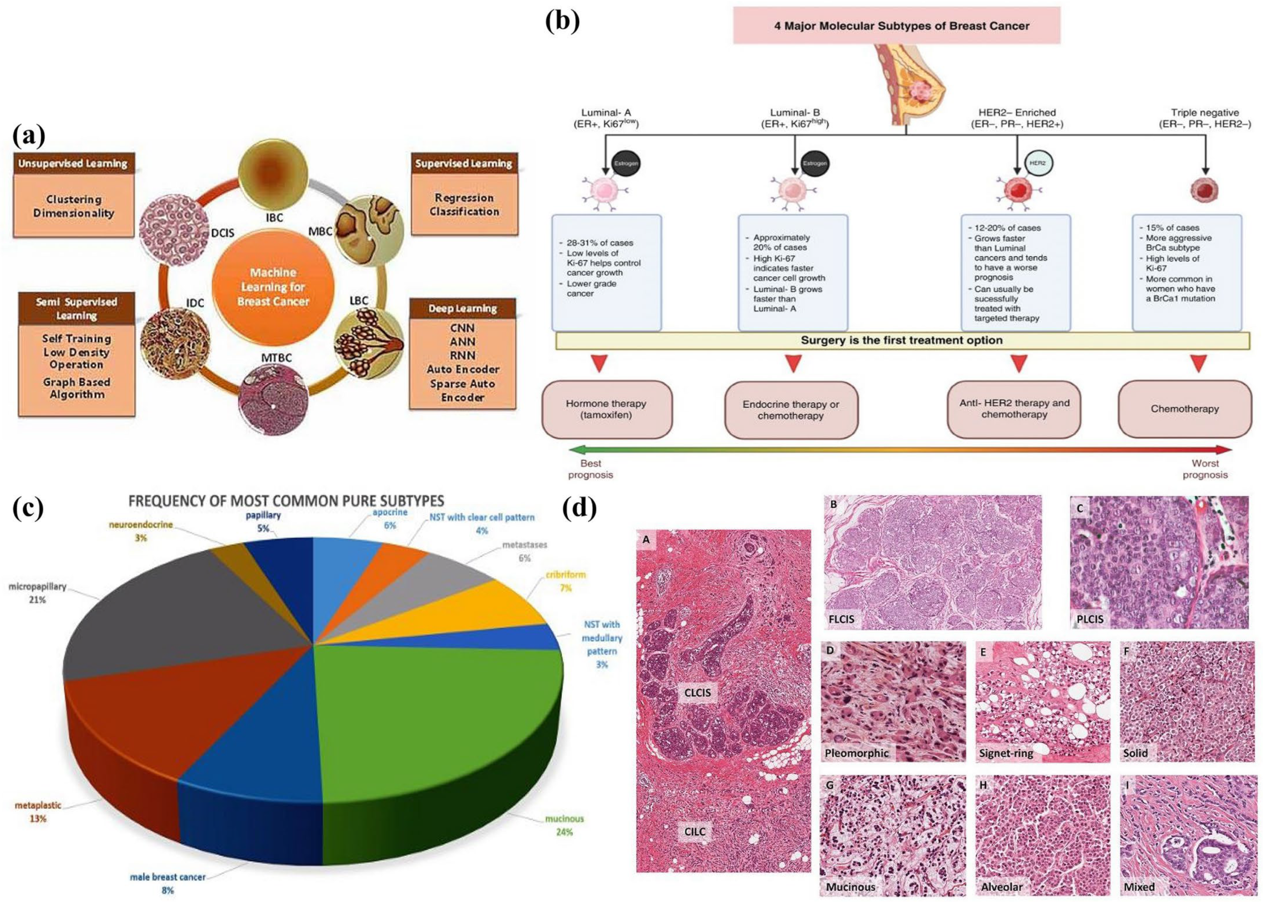
Figure 1. Classifications of breast cancer by distribution, histology, and machine learning methods: (a) techniques in machine learning for predicting various forms of breast cancer [13], reproduced with permission from Fatima et al., (2020), Creative Common Attribution 4.0; (b) classification of breast cancer subtypes [14], reproduced with permission from Girithar et al. (2023), Creative Common Attribution 4.0; (c) histological subtypes and distribution rates [15], reproduced with permission from Rechsteiner et al., (2023), Creative Common Attribution 4.0; (d) classification of breast cancer types by immune staining of tumor tissue [16], reproduced with permission from McCart Reed et al., (2020), Creative Common Attribution 4.0.
Applsci 14 01809 g001
Figure 2. Common nanomaterials and carrier types utilized as controlled release systems for therapeutic applications. Row 1 (left to right): zero-dimensional (0-D) material, where drugs can be loaded on the surface; one-dimensional (1-D) material (example: carbon nanotube), where drugs are loaded into the tube’s interior; and two-dimensional (2-D) material, where drugs can be loaded in-between the layers. Row 2 (left to right): mesoporous materials, where drugs are loaded onto the porous; liposomes, where drugs can be loaded on the interior lipid bilayer; and micelle, where drugs can be tagged with the polar/nonpolar layer. Row 3 (left to right): dendrimer, where drugs can be loaded into the void spaces; polymeric nanoparticles, where drugs can be functionalized in the terminal layers; and hydrogel, where drugs can be interlinked in the polymer chains [57], reproduced with permission from Senapati et al., (2018), © Springer Nature (Creative Commons Attribution 4.0).
Applsci 14 01809 g002
Figure 4. FDA-approved drugs for the treatment of breast cancer currently available in the market.
Applsci 14 01809 g004
Figure 5. Applications of nanomaterials in drug delivery systems. (a) Doxorubicin-, curcumin-, and perfluorooctyl bromide-loaded PLGA NPs for treatment of BC [115], reproduced with permission from Ramasamy et al., (2018), Creative Commons Attributions 4.0; (b) methotrexate-modified Au @PDA-PEG NPs for improved photothermal therapy and chemotherapy for BC [116], reproduced with permission from Li et al., (2021), Creative Commons Attributions 4.0; (c) epirubicin-encapsulated carbon nanoparticles for tracing and as a local chemotherapeutic on breast cancer with axillary metastasis [117], reproduced with permission from Du et al., (2016), Creative Commons Attributions 4.0; (d) 5-fluorouracil-incorporated PLG nanoparticles for slower drug release toward various cancer cell lines, including BC cell lines [118], reproduced with permission from Samy et al., (2023), Creative Commons Attributions 4.0; (e) paclitaxel liposomal nanoformulations for TNBC treatment [119], reproduced with permission from Ye et al., (2021), Creative Commons Attributions 4.0.
Applsci 14 01809 g005
Table 1. A table of nanotherapeutics for breast cancer explored in previous studies, the techniques employed based on the nature of nanomaterials, and their findings.
Nanomaterials/NanoformulationsNature of Nanomaterial/Delivery MechanismResultsReferences
Bimetallic Prussian blue analogs and cobalt–iron loaded with DoxpH-dependent mechanismGood biocompatibility of PBA-DDSs[66]
Curcumin-loaded ZnO nanoparticles conjugated with phenylboronic acid (PBA)pH-dependentSuccessful reduction in tumor growth in mice with Ehrlich ascites carcinoma (EAC) tumors[66]
HA-Dox/PHIS/R848 nanoparticles: R848, was encapsulated with poly(L-histidine) (PHIS) to form PHIS/R848 nanocores, and doxorubicin (Dox) was conjugated to hyaluronic acid (HA)Dual pH-dependent multifunctional nanoparticle combining chemotherapy and immunotherapyImpressive tumor-targeting ability and effective inhibition tumor growth by regulating immunity and directly eliminating cancer cells[66]
Folic acid-conjugated polyacrylic acid-coated mesoporous silica nanoparticles loaded with chrysinpH-sensitiveIn vivo regression of tumors, restoration of normal tissue structure, and the preservation of a healthy body weight[66]
Phenylboronic acid (PBA)-conjugated zinc oxide nanoparticles (PBA-ZnO), loaded with quercetinpH-sensitiveReduction in tumor growth[77]
BSA-stabilized graphene/chitosan nanocomposites conjugated with breast cancer drugspH-sensitiveReduced the burst release observed compared to that for pure chitosan nanoparticles[72]
Palladium–ruthenium nanohybrid with polypyridyl complex (PdRu-RCE@PCM NPs)Temperature-sensitive (photothermal targeting therapy)Inhibition of primary tumor growth and tumor metastasis[77]
Programmed death-1 (PD-1)-modified gold-promoting satellite copper selenide nanocrystalsPhotothermal targeting therapyApoptosis of cancer cells[77]
Cetuximab- and doxorubicin-conjugated liposome-mediated magnetic nanoparticlesCombined treatment of photothermal therapy and targeted chemotherapy in thermo-sensitive nanocarriersReduction in viability of breast cancer cells[77]
(Triptolide) TPL@nanogelInjectable thermo-responsive hydrogelReduced systemic toxicity and increased antitumor efficacy[77]
Lauric acid encapsulated in a biocompatible silica shellCombined effect of temperature and lauric acid activityDual activity in anticancer treatments due to the two combined mechanisms[77]
Hollow gold nanostars (HGNSs) and gold nanocages (GNCs) with doxorubicin (Dox) attached Temperature/pH-dependent mechanismA high cell mortality and apoptotic effects were observed[80]
Chitosan/gelatin hybrid nanogel incorporating gold nanoparticles (CS/AuNPs@Gel-Dox nanogel)Enzyme responsiveSuccessful absorption by cells. Cell cytotoxicity revealed that the drug carrier was efficient[84]
Disulfiram and doxorubicin conjugated with polymeric nanoparticlesEnzyme/pH dual responsiveIncreased cytotoxicity against 4T1 cells[84]
Peptide conjugate crafted to combined mild photothermal therapy with immunotherapy in a unified nanosystempH and enzyme responsiveEffective inhibition of tumor growth while preventing the formation of lung metastases[86]
(POL-MSN) Dox-loaded MSNs with poly (β-amino ester) and boronate esterspH and enzyme responsiveReduced cancer cell viability[86]
Disclaimer/Publisher’s Note: The statements, opinions and data contained in all publications are solely those of the individual author(s) and contributor(s) and not of MDPI and/or the editor(s). MDPI and/or the editor(s) disclaim responsibility for any injury to people or property resulting from any ideas, methods, instructions or products referred to in the content.