
Anatomical Plausibility in Deformable Image Registration Using Bayesian Optimization for Brain MRI Analysis

 , , and
Abstract
1. Introduction
2. Materials and Methods
2.1. Datasets
2.2. Deformable Registration of Point Clouds
| Algorithm 1 Estimation of transformation parameters |
| Require: Initial correspondences between and Require: Geodesic surface partitions 1: while Convergence criterion not met do 2: for each patch i do 3: for each neighboring ring j do 4: Compute local transformation by minimizing local energy 5: Update positions of vertices in 6: end for 7: Compute intermediate transformation by minimizing intermediate energy 8: Update positions of vertices in 9: end for 10: Compute global transformation by minimizing global energy 11: Update positions of all vertices in 12: end while |
Energy Model
Local Energy
Intermediate Energy
Global Energy
2.3. Anatomical Plausibility
2.3.1. Regularization Terms
L2 Norm Regularization
Total Variation (TV) Regularization
Bending Energy Regularization
Smoothness Constraints
2.3.2. Similarity Metric
2.4. Conformal Bayesian Optimization with Gaussian Process Priors
- Number of clusters (c): This parameter determines the granularity of local deformations. A higher number of clusters allows for finer transformations; however, excessively high values may lead to overfitting or increase computational complexity. The range ensures adaptability without compromising efficiency.
- Regularization weight (): This controls the trade-off between alignment precision and deformation smoothness. Higher values promote smoother transformations, which are crucial for maintaining anatomical plausibility, particularly in sensitive brain structures.
- The number of neighboring rings (r): This parameter defines the local neighborhood around each point and influences how adjacent points contribute to the transformation. Smaller values encourage localized deformations, while higher values capture broader contextual relationships.
- Influence factor (): This modulates the importance of local energy contributions from each neighboring ring. This ensures that the algorithm appropriately balances the impact of different regions on the final deformation, which is essential for aligning structures with varying geometric properties.
- Threshold for global energy correction (d): This parameter determines when global transformations, such as translation and rotation, are applied. A lower threshold triggers more frequent global adjustments, ensuring accurate alignment across large-scale transformations.
3. Results
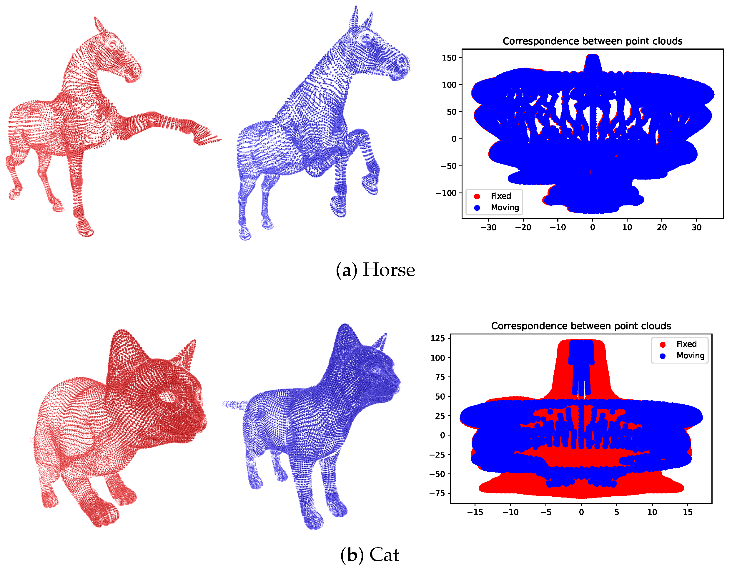
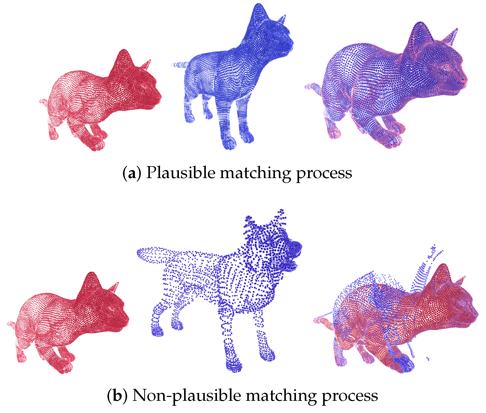
3.1. Tosca Database
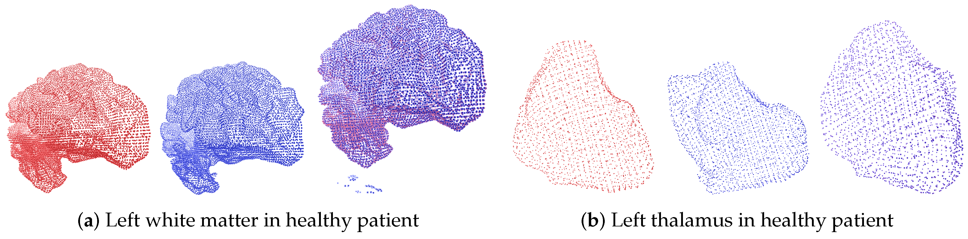
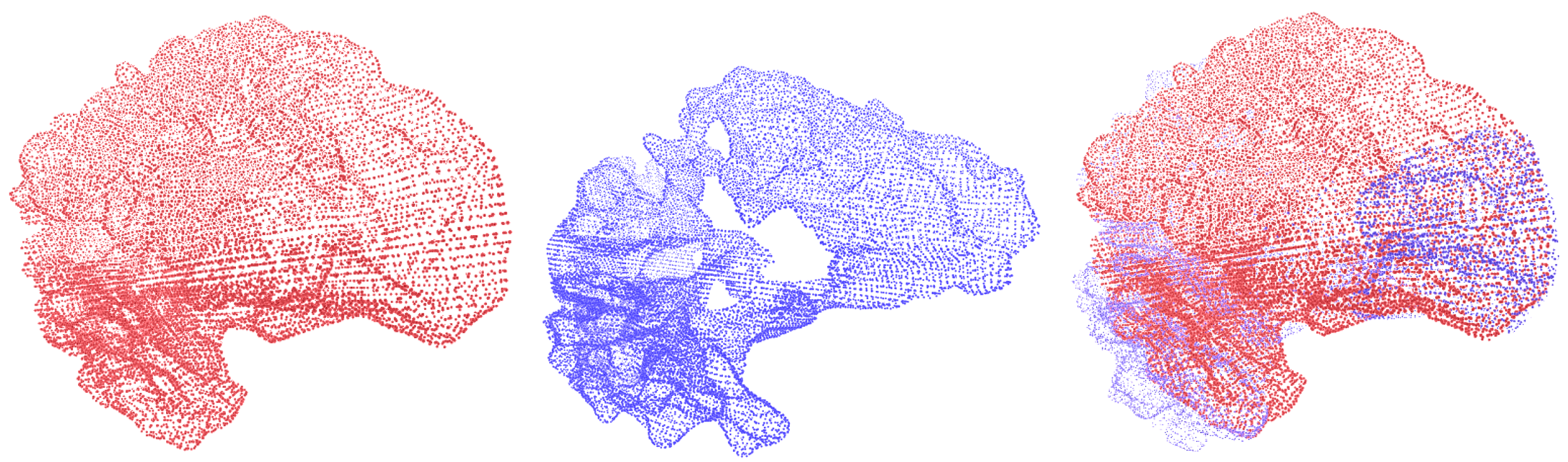
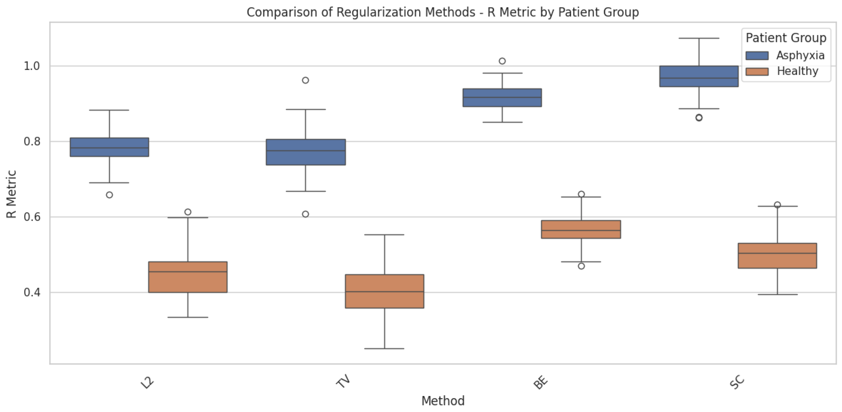
3.2. Brain Asphyxia Database
4. Conclusions
Author Contributions
Funding
Institutional Review Board Statement
Informed Consent Statement
Data Availability Statement
Acknowledgments
Conflicts of Interest
Abbreviations
| HIE | Hypoxic-ischemic encephalopathy |
| MRI | Magnetic Resonance Imaging |
| CBO | Conformal Bayesian Optimization |
| FFD | Free-form deformation |
References
- Darzi, F.; Bocklitz, T. A Review of Medical Image Registration for Different Modalities. Bioengineering 2024, 11, 786. [Google Scholar] [CrossRef] [PubMed]
- Khawaled, S.; Freiman, M. Npbdreg: Uncertainty assessment in diffeomorphic brain mri registration using a non-parametric bayesian deep-learning based approach. Comput. Med. Imaging Graph. 2022, 99, 102087. [Google Scholar] [CrossRef] [PubMed]
- Krebs, J. Robust Medical Image Registration and Motion Modeling Based on Machine Learning. Ph.D. Thesis, Université Côte d’Azur, Côte d’Azur, France, 2020. [Google Scholar]
- Zitová, B.; Flusser, J. Image Registration Methods: A Survey. Image Vis. Comput. 2003, 21, 977–1000. [Google Scholar] [CrossRef]
- Maintz, J.; Viergever, M. A Survey of Medical Image Registration. Med. Image Anal. 1998, 2, 1–36. [Google Scholar] [CrossRef]
- Oliveira, F.P.M.; Tavares, J. Medical image registration: A review. Comput. Methods Biomech. Biomed. Eng. 2014, 17, 73–93. [Google Scholar] [CrossRef]
- McKenzie, E.M.; Santhanam, A.; Ruan, D.; O’Connor, D.; Cao, M.; Sheng, K. Multimodality image registration in the head-and-neck using a deep learning-derived synthetic CT as a bridge. Med. Phys. 2020, 47, 1094–1104. [Google Scholar] [CrossRef]
- Rueckert, D.; Sonoda, L.I.; Hayes, C.; Hill, D.L.; Leach, M.O.; Hawkes, D.J. Nonrigid Registration Using Free-Form Deformations: Application to Breast MR Images. IEEE Trans. Med. Imaging 1999, 18, 712–721. [Google Scholar] [CrossRef]
- de Vos, B.D.; Berendsen, F.F.; Viergever, M.A.; Sokooti, H.; Staring, M.; Išgum, I. A Deep Learning Framework for Unsupervised Affine and Deformable Image Registration. Med. Image Anal. 2019, 52, 128–143. [Google Scholar] [CrossRef] [PubMed]
- Ashburner, J. A Fast Diffeomorphic Image Registration Algorithm. NeuroImage 2007, 38, 95–113. [Google Scholar] [CrossRef]
- Beg, M.F.; Miller, M.I.; Trouvé, A.; Younes, L. Computing Large Deformation Metric Mappings via Geodesic Flows of Diffeomorphisms. Int. J. Comput. Vis. 2005, 61, 139–157. [Google Scholar] [CrossRef]
- Xiong, F.; Kong, Y.; Hu, M.; Zhang, Z.; Shen, C.; Kuang, L.; Han, X. A multi-scale covariance matrix descriptor and an accurate transformation estimation for robust point cloud registration. Res. Sq. 2024. preprint. [Google Scholar] [CrossRef]
- Balakrishnan, G.; Zhao, A.; Sabuncu, M.; Guttag, J.; Dalca, A.V. VoxelMorph: A Learning Framework for Deformable Medical Image Registration. IEEE Trans. Med. Imaging 2019, 38, 1788–1800. [Google Scholar] [CrossRef] [PubMed]
- Krebs, J.; Delingette, H.; Mailhé, B.; Ayache, N.; Mansi, T. Learning a Probabilistic Model for Diffeomorphic Registration. IEEE Trans. Med. Imaging 2019, 38, 2165–2176. [Google Scholar] [CrossRef] [PubMed]
- Mansilla, L.; Milone, D.H.; Ferrante, E. Learning Deformable Registration of Medical Images with Anatomical Constraints. Neural Netw. 2020, 124, 269–279. [Google Scholar] [CrossRef] [PubMed]
- Fong, E.; Holmes, C.C. Conformal Bayesian Computation. In Advances in Neural Information Processing Systems; Ranzato, M., Beygelzimer, A., Dauphin, Y., Liang, P., Vaughan, J.W., Eds.; Curran Associates, Inc.: Newry, UK, 2021; Volume 34, pp. 18268–18279. [Google Scholar]
- Stanton, S.; Maddox, W.J.; Wilson, A.G. Bayesian Optimization with Conformal Coverage Guarantees. arXiv 2022, arXiv:2210.12496. [Google Scholar]
- Bronstein, A.M.; Bronstein, M.M.; Kimmel, R. Efficient Computation of Isometry-Invariant Distances Between Surfaces. SIAM J. Sci. Comput. 2006, 28, 1812–1836. [Google Scholar] [CrossRef]
- Bronstein, A.M.; Bronstein, M.M.; Kimmel, R. Calculus of Nonrigid Surfaces for Geometry and Texture Manipulation. IEEE Trans. Vis. Comput. Graph. 2007, 13, 902–913. [Google Scholar] [CrossRef] [PubMed]
- Fischl, B.R. FreeSurfer. NeuroImage 2012, 62, 774–781. [Google Scholar] [CrossRef]
- Satheesan, A.P.; Chinnappa, A.R.; Goudar, G.; Raghoji, C.R. Correlation between early magnetic resonance imaging brain abnormalities in term infants with perinatal asphyxia and neuro developmental outcome at one year. Int. J. Contemp. Pediatr. 2020, 7, 1957–1961. [Google Scholar] [CrossRef]
- Miller, S.P.; Ramaswamy, V.; Michelson, D.J.; Barkovich, A.J.; Holshouser, B.A.; Wycliffe, N.; Glidden, D.V.; Deming, D.D.; Partridge, J.C.; Wu, Y.W.; et al. Patterns of brain injury in term neonatal encephalopathy. J. Pediatr. 2005, 146, 453–460. [Google Scholar] [CrossRef]
- Gutiérrez-Becker, B.; Wachinger, C. Deep Multi-structural Shape Analysis: Application to Neuroanatomy. In Proceedings of the Medical Image Computing and Computer Assisted Intervention—MICCAI 2018, Granada, Spain, 16–20 September 2018; Frangi, A.F., Schnabel, J.A., Davatzikos, C., Alberola-López, C., Fichtinger, G., Eds.; Springer: Cham, Switzerland, 2018; pp. 523–531. [Google Scholar]
- Hu, Y.; Modat, M.; Gibson, E.; Li, W.; Ghavami, N.; Bonmati, E.; Wang, G.; Bandula, S.; Moore, C.; Emberton, M.; et al. DeepReg: A deep learning toolkit for medical image registration. J. Open Source Softw. 2020, 5, 2121. [Google Scholar]


| Hyperparameter | Symbol | Range |
|---|---|---|
| Number of clusters | c | |
| Regularization weight | ||
| Number of neighboring rings | r | |
| Influence factors for local energy | ||
| Threshold for global energy correction | d |
Disclaimer/Publisher’s Note: The statements, opinions and data contained in all publications are solely those of the individual author(s) and contributor(s) and not of MDPI and/or the editor(s). MDPI and/or the editor(s) disclaim responsibility for any injury to people or property resulting from any ideas, methods, instructions or products referred to in the content. |
© 2024 by the authors. Licensee MDPI, Basel, Switzerland. This article is an open access article distributed under the terms and conditions of the Creative Commons Attribution (CC BY) license (https://creativecommons.org/licenses/by/4.0/).
Share and Cite
Castaño-Aguirre, M.; García, H.F.; Cárdenas-Peña, D.; Porras-Hurtado, G.L.; Orozco-Gutiérrez, Á.Á. Anatomical Plausibility in Deformable Image Registration Using Bayesian Optimization for Brain MRI Analysis. Appl. Sci. 2024, 14, 10890. https://doi.org/10.3390/app142310890
Castaño-Aguirre M, García HF, Cárdenas-Peña D, Porras-Hurtado GL, Orozco-Gutiérrez ÁÁ. Anatomical Plausibility in Deformable Image Registration Using Bayesian Optimization for Brain MRI Analysis. Applied Sciences. 2024; 14(23):10890. https://doi.org/10.3390/app142310890
Chicago/Turabian StyleCastaño-Aguirre, Mauricio, Hernán Felipe García, David Cárdenas-Peña, Gloria Liliana Porras-Hurtado, and Álvaro Ángel Orozco-Gutiérrez. 2024. "Anatomical Plausibility in Deformable Image Registration Using Bayesian Optimization for Brain MRI Analysis" Applied Sciences 14, no. 23: 10890. https://doi.org/10.3390/app142310890
APA StyleCastaño-Aguirre, M., García, H. F., Cárdenas-Peña, D., Porras-Hurtado, G. L., & Orozco-Gutiérrez, Á. Á. (2024). Anatomical Plausibility in Deformable Image Registration Using Bayesian Optimization for Brain MRI Analysis. Applied Sciences, 14(23), 10890. https://doi.org/10.3390/app142310890