Article

The Effect of Silver Nanoparticles on Bond Strength of Calcium Silicate-Based Sealer: An In Vitro Study

1
Division of Endodontics, Department of Restorative Dental Science, College of Dentistry, King Saud University, Riyadh 13313, Saudi Arabia
2
Division of Dental Materials, Department of Restorative Dental Science, College of Dentistry, King Saud University, Riyadh 13313, Saudi Arabia
*
Author to whom correspondence should be addressed.
Appl. Sci. 2024, 14(21), 9817; https://doi.org/10.3390/app14219817
Submission received: 26 September 2024 / Revised: 15 October 2024 / Accepted: 23 October 2024 / Published: 27 October 2024

Abstract

:
The aim of this study was to evaluate the bond strength of the calcium silicate-based sealer (CSS) modified with the silver nanoparticles (AgNPs) using the single-cone technique (SC) and the continuous wave condensation (CWC) technique, measured by a universal testing machine. The AgNPs and the modified sealers were characterized by scanning electron microscopy and transmission electron microscopy. One hundred single-rooted extracted human permanent teeth with a single root canal were cleaned and shaped with a Protaper Next system. The teeth were randomly divided into four groups (n = 25) as follows: Group 1, canals were obturated using the SC technique with TotalFill® BC Sealer. Group 2, canals were obturated using the SC technique with TotalFill® BC Sealer mixed with AgNPs. Group 3, canals were obturated using the CWC technique with TotalFill® HiFlow BC Sealer. Group 4, canals were obturated using the CWC technique with TotalFill® HiFlow BC Sealer mixed with AgNPs. After two weeks, 1 mm-thick dentin slices were cut and exposed to a push-out bond strength test using a universal testing machine. Specimens were examined under a digital microscope to determine the mode of failure. Statistical analysis was performed using ANOVA and Tukey multiple comparison tests (p < 0.05). The nanoparticle characterization revealed a spherical morphology with no obvious aggregations. The results showed that group 4 had the highest dislodgement resistance compared to all groups (p < 0.05). Group 4 was significantly higher in push-out bond strength value than group 1 (p < 0.001) and group 3 (p < 0.003), but not significantly higher than group 2. Cohesive failure was the most prevalent failure mode among all groups. It can be concluded that the incorporation of silver nanoparticles into the calcium silicate-based sealer significantly increased the bond strength. The warm obturation approach demonstrated significantly higher resistance to dislodgment as compared to the single-cone technique.

1. Introduction

Endodontic sealer creates a long-lasting and durable connection between the root canal filling material and dentinal walls [1]. This remarkable barrier prevents root canal infections and also ceases the growth of microorganisms within the root canal system, which gives the host a chance to heal and grow new periapical tissues [2]. The di- and tricalcium silicate-based sealer proved to be biocompatible and bioactive with the surrounding tissues, provide an antibacterial effect, and form a hermetic seal [3]. When the calcium silicate-based sealer (CSS) comes into contact with the phosphate from the tissue fluids, it initiates a hydration reaction that releases calcium hydroxide and forms a hard mineral layer at the dentin wall. The CSS is bound to the dentin because of the interaction between this mineral layer and the micromechanical infiltration within the dentinal tubules [4,5]. To measure the strength of the bond, the push-out bond strength (POBS) test, also known as the dislodgment resistance of the core filling material from the canal walls, was used widely in several studies [6,7].
TotalFill® BC Sealer (FKG, La Chaux-de-Fonds, Switzerland), also goes by the brand names EndoSequence® BC Sealer (BUSA, Savannah, GA, USA) and iRoot® SP (Innovative BioCeramix, Vancouver, BC, Canada); it is hydrophilic and has a low contact angle, which lets it fill the dentinal tubules and form tag-like structures. It can also expand by about 0.20 percent when it gets wet, creating a gapless bond between the sealer and the dentin [8,9]. A study reported that the iRoot® SP and AH Plus sealers had significantly higher bond strengths when compared to MTA Fillapex [10]. Others reported that the TotalFill® BC Sealer showed the highest POBS compared to BioRoot RCS and Endo CPM-Sealer® [11]. When the TotalFill® BC sealer was compared to the AH Plus sealer, it showed a higher bond strength. Moreover, the obturation methods had a significant influence on bond strength; the warm vertical compaction reduced the bond strength of the AH Plus sealer compared to cold lateral compaction, but this is not significant with the TotalFill® BC sealer [12]. However, other studies found that the bond strength of the TotalFill® BC Sealer increased when used in warm vertical compaction compared to cold obturation techniques [13].
Recently, nanoscale substances have been utilized to enhance the properties of various dental materials [14]. Researchers successfully synthesized and integrated silver nanoparticles (AgNPs) into resin-based materials, enhancing their antimicrobial effects while preserving the material’s physical, mechanical, and optical properties [15,16]. The AgNPs exhibited broad-spectrum antibacterial effects against various cariogenic and endodontic bacteria, as their nanoscale size allowed the material to penetrate the bacterial cell walls and alter the bacterial membrane structure [17].
Adding silver nanoparticles to a calcium silicate-based sealer was studied recently, and the results indicated that these enhancements favorably influenced the sealer’s physicochemical properties. The sealer preserved an alkaline pH, enhancing antibacterial effectiveness through the release of calcium and hydroxyl ions. The alkaline environment is crucial for enhancing osteogenic potential, ensuring biocompatibility, and providing antibacterial characteristics. Furthermore, this integration has demonstrated the ability to preserve contraction within 1% of the original dimension [18]. Moreover, nanoparticles (NPs) incorporated into sealers can facilitate penetration into dentinal tubules and allow for prolonged release of antimicrobial components [19,20]. Complying with the requirements established by ISO and ANSI/ADA, this outcome further underscores the advantages of incorporating silver nanoparticles into a calcium silicate-based sealer since dimensional stability is crucial for the efficacy of root canal treatments to mitigate microleakage.
Nonetheless, there are limited studies in the literature conducted to evaluate the bond strength of the calcium silicate-based sealer incorporated with silver nanoparticles. Thus, the purpose of this study was to evaluate the bond strength of the calcium silicate-based sealer modified with silver nanoparticles using the single-cone technique and the warm obturation technique, which were measured with a universal testing machine.

2. Material and Methods

2.1. Characterization of AgNPs

The silver nanoparticles, measuring 20 nm (Zhengzhou Dongyao Nano Materials Co., Ltd., Zhengzhou, China), were procured and analyzed using scanning electron microscopy (SEM) (JEOL, JSM-7610F Schottky Field Emission, Hitachi, Ibaraki, Japan) and transmission electron microscopy (TEM) (JEOL, JEM, 1400 Plus, Hitachi, Ibaraki, Japan) to characterize the size, shape, size distribution, and degree of aggregation of the particles prior to and following their incorporation into the sealer.

2.2. Specimen Preparation

This research was approved by the Institutional Review Board (E-23-8135) and College of Dentistry Research Center (FR 0707), King Saud University, Riyadh, Saudi Arabia. Sample size calculation was performed using G*Power 3.1.9.4 software at an alpha error probability of 0.05 with an effect size of 0.4 and power of 0.9. The power analysis showed that a total of 96 specimens (24 per group) were required. One hundred single-rooted extracted human permanent teeth with a single root canal were used in this study. Roots exhibiting anatomical variations, curvature, or prior endodontic therapy were excluded. The crowns were sectioned off to achieve a standardized root length of 16 mm. Root canals were prepared with Protaper Next system (Dentsply, Sirona, Charlotte, NC, USA) up to instrument X3, which corresponds to tip ISO size 30 and 7% taper. Canals were irrigated with 5 mL 5.25% sodium hypochlorite followed by 1 mL 17% ethylenediaminetetraacetic acid.

2.3. Experimental Design

The teeth were randomly divided into four experimental groups (n = 25) as follows:
Group 1, canals were obturated using SC technique with TotalFill® BC Sealer.
Group 2, canals were obturated using SC technique with TotalFill® BC Sealer mixed with AgNPs.
Group 3, canals were obturated using CWC technique with TotalFill® HiFlow BC Sealer.
Group 4, canals were obturated using CWC technique with TotalFill® HiFlow BC Sealer mixed with AgNPs.
To prepare the modified sealer (AgNPs incorporated with calcium silicate-based sealer), 0.06 mg of nanoparticles was measured for every 1 g of sealer using a digital analytical balance, resulting in a nanoparticle concentration of 0.06% in the modified sealer in line with previous work [21]. Manual mixing was conducted with a metal spatula on a glass plate, then for 1 min using a vortex mixer. This was succeeded by ultrasonication using a probe sonicator (Sigma-Aldrich, St Louis, MO, USA) for 1 h to improve the dispersion of NPs. This was conducted at 25 °C and in the absence of light. Table 1 lists the experimental groups used in the study.
In the SC technique, the BC sealer was injected by the intracanal tip, then the size-matched ISO size 30/0.06 taper TotalFill® BC Point (TotalFill® BC point, FKG, La Chaux-de-Fonds, Switzerland) was coated with the TotalFill® BC sealer followed by its slow insertion to the full working length. After that, the cone was cut off with activated system B pluggers (SybronEndo, Orange, CA, USA) just below the orifice, and a quick-setting temporary filling Cavit G (3 M ESPE, Seefeld, Germany) was placed. In the CWC technique, we used TotalFill® HiFlow BC Sealer (FKG, La Chaux-de-Fonds, Switzerland) because, according to the manufacturer, it exhibits lower viscosity and increased radiopacity when used with the heated obturation procedure than TotalFill® BC Sealer. In this technique, the placement of the BC sealer and the BC point were the same as in the SC technique. However, the BC point was cut 3 mm from the working length using activated system B pluggers, then the root canal was backfilled with plasticized GP using TotalFill® BC pellets (TotalFill® BC Pellets, FKG, La Chaux-de-Fonds, Switzerland), and the orifice was sealed with Cavit G. All treatment procedures were performed by a single operator. Specimens were stored at 37 °C in 100% humidity for 2 weeks to enable a complete set of the sealers. Afterwards, roots were horizontally sectioned using a 0.3 mm-thick, water-cooled slow-speed diamond blade (IsoMet 4000; Buehler, Lake Bluff, IL, USA) to obtain 1 mm-thick dentin slices from the middle third of each root.

2.4. Push-Out Bond Strength Assessment

Each specimen was positioned on a metal platform, ensuring that the apical surface was placed on top. Then, vertical load was applied using an Instron® 5965 universal testing machine (INSTRON Corp., Norwood, MA, USA) at a controlled speed of 1 mm per minute using a 0.6 mm diameter tip. A graphical representation was generated utilizing the BlueHill software 3.22.1373 (INSTRON Corp., Norwood, MA, USA). The bond failure was automatically detected upon observing a rapid decrease in load recorded in Newtons (N).
The push-out bond strength (MPa) of each specimen was then calculated by dividing the bond failure load (N) by the lateral surface area (LSA) of the bonded interface, which corresponds to the lateral surface of the root canal. The LSA was calculated using the following formula:
LSA = π (R + r)[(h2 + (R − r))2]0.5
where R represents the radius of the coronal root canal, r represents the radius of the apical root canal, and h represents the slice thickness, which denotes the distance between the two circles measured along the lateral face. All measurements were recorded in millimeters.

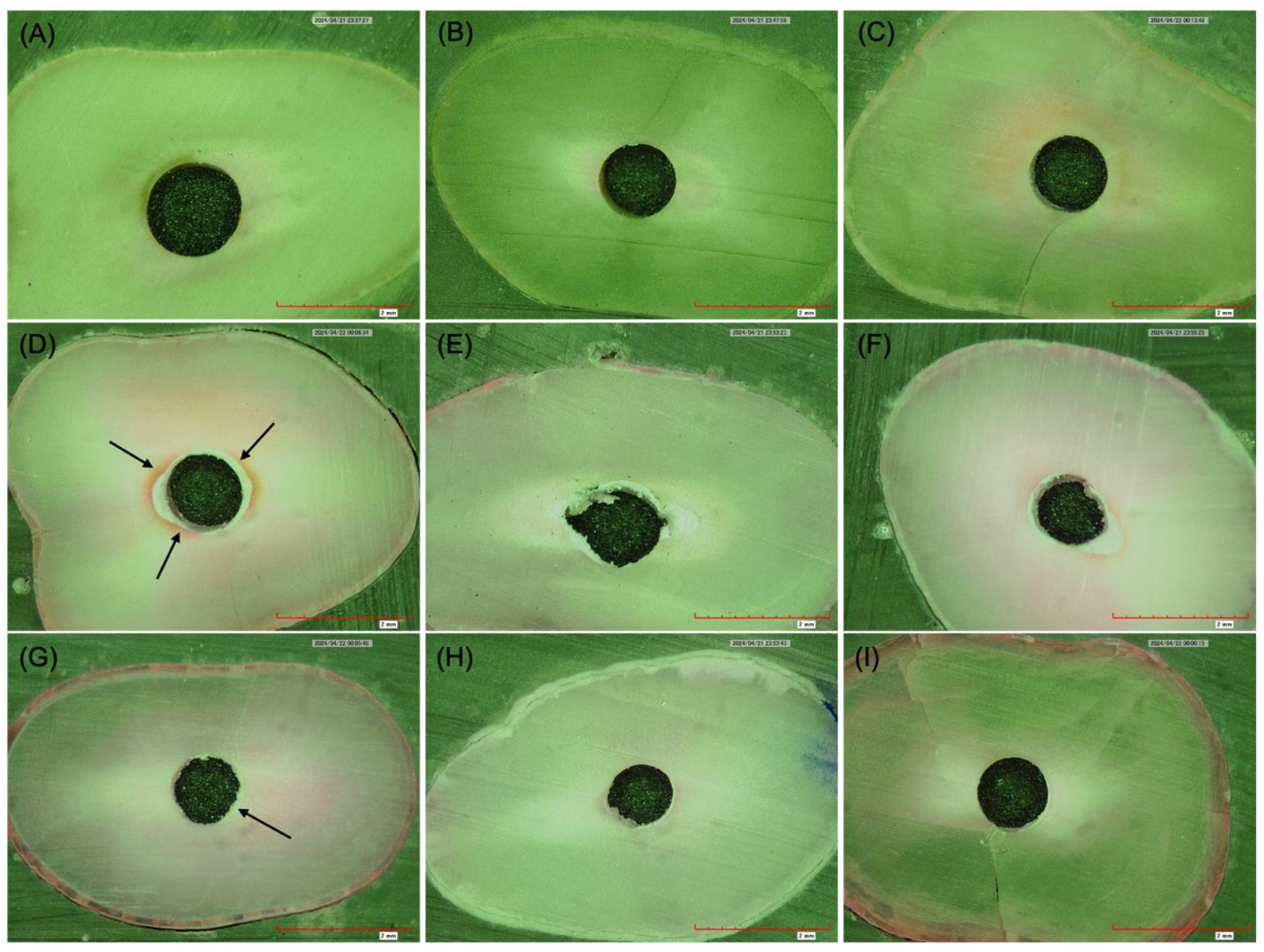
2.5. Failure Modes Assessment

Each specimen was visualized with a digital microscope (KH-7700, Hirox, Tokyo, Japan) under 50× magnification, and photographs were obtained after dislodgement of the root canal filling material. The photographs were separately evaluated by two blinded operators, and the mode of failure was recorded. Failure modes were classified into one of the following three categories according to Stelzer R et al. [22]: Class I: adhesive failure, characterized by less than 25% of the dentin surface being covered by sealer; Class II: cohesive failure, indicated when more than 75% of the dentin surface was covered by sealer; Class III: mixed failure, when the dentin surface covered by sealer was between 25% and 75%.

2.6. Statistical Analysis

Statistical analysis of POBS values was performed using one-way ANOVA and the Tukey multiple comparison test (p < 0.05), as data were distributed normally according to the Shapiro–Wilk test (p < 0.05). A chi-square test was performed to compare modes of failure between the groups.

3. Results

3.1. Characterization of AgNPs

SEM images of the AgNPs demonstrated a relatively spherical structure with single spread of the NPs (Figure 1A,B). The TEM images of the AgNPs confirmed their spherical morphology and uniform distribution, with an average size ranging from 5 to 22 nm (Figure 1C,D). After the incorporation of the AgNPs with calcium silicate-based sealer, TEM images demonstrated that NPs maintained their spherical shape within the sealer. Furthermore, the NPs precipitated within the structure of the sealer, exhibiting no signs of aggregation (Figure 1E,F).

3.2. Push-Out Bond Strength Assessment

Throughout the experiment, no premature failure occurred. The mean push-out bond strengths are summarized in Table 2. The one-way ANOVA tests revealed a significant difference among the groups (p < 0.001). Group 4 showed the highest dislodgement resistance compared to all the groups (p < 0.05). The Tukey post hoc tests showed that group 4 was significantly higher in push-out bond strength value than group 1 (p < 0.001) and group 3 (p < 0.003), but not significantly higher than group 2 (Table 2).

3.3. Failure Modes Assessment

The results of the mode of failure analysis are shown in Table 3. Chi-square test showed there was a significant association between the groups and mode of failure (p = 0.003). Cohesive failure was the most prevalent failure mode among all groups. Examples of modes of failure are shown in Figure 2.

4. Discussion

The impact of the silver nanoparticles on the bond strength and failure mode during the dislodgment of the calcium silicate-based sealer was investigated in this study. Nanoparticle characterization can be carried out using microscopy-based techniques and other extensive methods, which provide information about the size, shape, size distribution, degree of aggregation, and other surface chemistry [23]. In this current study, AgNPs exhibited a spherical morphology with an even distribution in both the SEM and TEM techniques. Additionally, the dimensions of the NPs are between 5 and 22 nanometers. The images also showed a singular dispersion of the sphere-shaped nanoparticles, without any significant aggregation within the calcium silicate-based sealer structure.
This study’s findings demonstrated a significant increase in dislodging resistance when the BC sealer was incorporated with AgNPs. The inclusion of the nano-sized particles, which had a high surface area and wetting capacity that led to enhanced contact with the surface and increased bond strength of the sealer, may be responsible for the remarkable results. Additionally, the dispersion of the nanoparticles acting as a reinforcing filler would improve the mechanical properties of the sealer, improving its tensile and shear strength [24,25].
This study’s findings indicate that the heated obturation approach, when combined with the inclusion of AgNPs, resulted in the highest mean values, which were statistically significant. This discovery aligns with many studies that have documented enhanced resistance to the dislodgment of bioceramic sealer when employed in warm obturation procedures [12,26]. The heat-induced increase in gutta-percha flow into deep depressions, auxiliary canals, and fins unfilled by sealer cement is associated with increased retention of root canal filling material to the canal walls. However, these findings contradict other research that indicated a reduction or no differences in the binding strengths of different bioceramic sealers when using warm obturation methods; different experimental protocols and parameters may explain the variability in the results [27,28]. Nonetheless, when the sealer was not combined with AgNPs, this study’s findings demonstrated comparable bond strength in both the warm obturation approach and the single-cone technique.
In this present work, 20 nm-sized particles were used to ensure infiltration through the dentinal tubules, which have an average diameter of 2.5 µm. It is proposed that particle size significantly influences the degree of dentin infiltration; smaller diameter particles show a greater infiltrative capacity [29]. The utilization of nanosized particles is reliant on both dose and time. In this investigation, a concentration of 0.06% of AgNPs was employed since it was shown to have the maximum antibacterial activity when mixed with calcium hydroxide and used as an intracanal medicament, and the efficacy improved considerably over time [30]. The ratio of the pin to root canal diameter is a significant component in the experimental design. In this current investigation, we employed a pin diameter of 0.6 mm in order to avoid any effect from the test machine, as per a previously published study, which states that the pin diameter should not be less than 0.6 mm or greater than 0.85 mm [31].
Among the clinically available root canal sealers, calcium silicate-based sealers are currently in widespread use. Thus, the resin-based root canal sealers were not included in the experimental groups due to their differing material compositions and properties. Additionally, earlier studies showed that resin-based sealers had weaker bond strengths compared to calcium silicate-based sealers [12,32,33]. In terms of obturation techniques, many practitioners may find it difficult to utilize the single-cone technique and prefer to use a thermoplasticized approach, despite the manufacturer’s guidelines. Thus, in this current investigation, both approaches were employed.
Adhesive root canal sealers are generally recognized for their high bond strength and tend to fail cohesively. Cohesive failure refers to the breaking or fracturing of the sealer within its own structure, without detaching from the dentin surface. This study’s results indicated that the predominant mode of failure seen was cohesive failure, aligning with earlier studies that proved the adhesive properties of the TotalFill® BC sealer with dentin [11,34]. The inclusion of AgNPs in this study may have contributed to the adhesive capabilities. However, it is not feasible to make direct comparisons with other studies, as this is the first study, to our knowledge, that examines the impact of adding AgNPs to a bioceramic sealer in terms of dislodgment resistance.
Under the constraints of this in vitro study, silver nanoparticles revealed an unexpected effect on the binding capacity of the BC sealer to dentin. It broadens our understanding of the integration of nano-sized particles into root canal sealers; yet, more testing in both laboratory and clinical settings is necessary to assess this incorporation.

5. Conclusions

This current study concludes that the incorporation of silver nanoparticles into the calcium silicate-based sealer significantly increased the bond strength. The warm obturation approach demonstrated significantly higher resistance to dislodgment as compared to the single-cone technique. In addition, the cohesive mode of failure was the most common failure type.

Author Contributions

Conceptualization, S.B.; methodology, S.B., S.A., D.A.; software, S.B.; validation, S.B., S.A.; formal analysis, S.B.; investigation, S.B., D.A.; resources, S.B.; data curation, S.B.; writing—original draft preparation, S.B., S.A.; writing—review and editing, S.B. All authors have read and agreed to the published version of the manuscript.

Funding

This research received no external funding.

Institutional Review Board Statement

This study was approved by the Institutional Review Board (E-23-8135) and College of Dentistry Research Center (FR 0707), King Saud University, Riyadh, Saudi Arabia (15 October 2023).

Informed Consent Statement

Not applicable.

Data Availability Statement

The raw data supporting the conclusions of this article will be made available by the authors upon request.

Acknowledgments

The authors acknowledge the staff and the use of the facilities of the physical research laboratory at College of Dentistry, King Saud University.

Conflicts of Interest

The authors declare no conflicts of interest.

References

  1. Madison, S.; Wilcox, L.R. An Evaluation of Coronal Microleakage in Endodontically Treated Teeth. Part III. In Vivo Study. J. Endod. 1988, 14, 455–458. [Google Scholar] [CrossRef] [PubMed]
  2. Wu, M.-K.; Fan, B.; Wesselink, P.R. Diminished Leakage along Root Canals Filled with Gutta-percha without Sealer over Time: A Laboratory Study. Int. Endod. J. 2000, 33, 121–125. [Google Scholar] [CrossRef] [PubMed]
  3. Prüllage, R.-K.; Urban, K.; Schäfer, E.; Dammaschke, T. Material Properties of a Tricalcium Silicate–Containing, a Mineral Trioxide Aggregate–Containing, and an Epoxy Resin–Based Root Canal Sealer. J. Endod. 2016, 42, 1784–1788. [Google Scholar] [CrossRef]
  4. Atmeh, A.R.; Chong, E.Z.; Richard, G.; Festy, F.; Watson, T.F. Dentin-Cement Interfacial Interaction: Calcium Silicates and Polyalkenoates. J. Dent. Res. 2012, 91, 454–459. [Google Scholar] [CrossRef]
  5. Camps, J.; Jeanneau, C.; El Ayachi, I.; Laurent, P.; About, I. Bioactivity of a Calcium Silicate–Based Endodontic Cement (BioRoot RCS): Interactions with Human Periodontal Ligament Cells In Vitro. J. Endod. 2015, 41, 1469–1473. [Google Scholar] [CrossRef]
  6. Pane, E.S.; Palamara, J.E.A.; Messer, H.H. Critical Evaluation of the Push-out Test for Root Canal Filling Materials. J. Endod. 2013, 39, 669–673. [Google Scholar] [CrossRef]
  7. Gesi, A.; Raffaelli, O.; Goracci, C.; Pashley, D.H.; Tay, F.R.; Ferrari, M. Interfacial Strength of Resilon and Gutta-Percha to Intraradicular Dentin. J. Endod. 2005, 31, 809–813. [Google Scholar] [CrossRef]
  8. Chisnoiu, R.; Moldovan, M.; Chisnoiu, A.; Hrab, D.; Rotaru, D.; Păstrav, O.; Delean, A. Comparative Apical Sealing Evaluation of Two Bioceramic Endodontic Sealers. Med. Pharm. Rep. 2019, 92, S55–S60. [Google Scholar] [CrossRef] [PubMed]
  9. Chen, I.; Salhab, I.; Setzer, F.C.; Kim, S.; Nah, H.-D. A New Calcium Silicate–Based Bioceramic Material Promotes Human Osteo- and Odontogenic Stem Cell Proliferation and Survival via the Extracellular Signal-Regulated Kinase Signaling Pathway. J. Endod. 2016, 42, 480–486. [Google Scholar] [CrossRef]
  10. Sagsen, B.; Ustün, Y.; Demirbuga, S.; Pala, K. Push-out Bond Strength of Two New Calcium Silicate-based Endodontic Sealers to Root Canal Dentine. Int. Endod. J. 2011, 44, 1088–1091. [Google Scholar] [CrossRef]
  11. Donnermeyer, D.; Dornseifer, P.; Schäfer, E.; Dammaschke, T. The Push-out Bond Strength of Calcium Silicate-Based Endodontic Sealers. Head Face Med. 2018, 14, 13. [Google Scholar] [CrossRef] [PubMed]
  12. Al-Hiyasat, A.S.; Alfirjani, S.A. The Effect of Obturation Techniques on the Push-out Bond Strength of a Premixed Bioceramic Root Canal Sealer. J. Dent. 2019, 89, 103169. [Google Scholar] [CrossRef] [PubMed]
  13. Mounes, B.B.; Alhashimi, R. The Push out Bond Strength of Bioceramic Seal-Er (Total Fill) after Warm and Cold Obturation Tech-Niques An in Vitro Comparative Study. J. Baghdad Coll. Dent. 2022, 34, 7–16. [Google Scholar] [CrossRef]
  14. Bolenwar, A.; Reche, A.; Dhamdhere, N.; Rathi, S. Applications of Silver Nanoparticles in Dentistry. Cureus 2023, 15, e44090. [Google Scholar] [CrossRef]
  15. Wang, J.; Jiang, W.; Liang, J.; Ran, S. Influence of Silver Nanoparticles on the Resin-Dentin Bond Strength and Antibacterial Activity of a Self-Etch Adhesive System. J. Prosthet. Dent. 2022, 128, 1363.e1–1363.e10. [Google Scholar] [CrossRef]
  16. Jowkar, Z.; Shafiei, F.; Asadmanesh, E.; Koohpeima, F. Influence of Silver Nanoparticles on Resin-Dentin Bond Strength Durability in a Self-Etch and an Etch-and-Rinse Adhesive System. Restor. Dent. Endod. 2019, 44, e13. [Google Scholar] [CrossRef]
  17. Yin, I.X.; Zhang, J.; Zhao, I.S.; Mei, M.L.; Li, Q.; Chu, C.H. The Antibacterial Mechanism of Silver Nanoparticles and Its Application in Dentistry. Int. J. Nanomed. 2020, 15, 2555–2562. [Google Scholar] [CrossRef]
  18. Sheethal Dsouza, T.; Shetty, A.; Dsouza, N. Evaluation of pH, Calcium Ion Release, and Dimensional Stability of an Experimental Silver Nanoparticle-Incorporated Calcium Silicate-Based Cement. Bioinorg. Chem. Appl. 2021, 2021, 3919543. [Google Scholar] [CrossRef] [PubMed]
  19. Zand, V.; Mokhtari, H.; Hasani, A.; Jabbari, G. Comparison of the Penetration Depth of Conventional and Nano-Particle Calcium Hydroxide into Dentinal Tubules. Iran. Endod. J. 2017, 12, 366–370. [Google Scholar] [CrossRef]
  20. Hashmi, A.; Zhang, X.; Kishen, A. Impact of Dentin Substrate Modification with Chitosan-Hydroxyapatite Precursor Nanocomplexes on Sealer Penetration and Tensile Strength. J. Endod. 2019, 45, 935–942. [Google Scholar] [CrossRef]
  21. Balto, H.; Bukhary, S.; Al-Omran, O.; BaHammam, A.; Al-Mutairi, B. Combined Effect of a Mixture of Silver Nanoparticles and Calcium Hydroxide against Enterococcus Faecalis Biofilm. J. Endod. 2020, 46, 1689–1694. [Google Scholar] [CrossRef] [PubMed]
  22. Stelzer, R.; Schaller, H.-G.; Gernhardt, C.R. Push-out Bond Strength of RealSeal SE and AH Plus after Using Different Irrigation Solutions. J. Endod. 2014, 40, 1654–1657. [Google Scholar] [CrossRef]
  23. Mourdikoudis, S.; Pallares, R.M.; Thanh, N.T.K. Characterization techniques for nanoparticles: Comparison and complementarity upon studying nanoparticle properties. Nanoscale 2018, 10, 12871–12934. [Google Scholar] [CrossRef]
  24. Nie, B.; Huo, S.; Qu, X.; Guo, J.; Liu, X.; Hong, Q.; Wang, Y.; Yang, J.; Yue, B. Bone Infection Site Targeting Nanoparticle-Antibiotics Delivery Vehicle to Enhance Treatment Efficacy of Orthopedic Implant Related Infection. Bioact. Mater. 2022, 16, 134–148. [Google Scholar] [CrossRef]
  25. Mallineni, S.K.; Sakhamuri, S.; Kotha, S.L.; AlAsmari, A.R.G.M.; AlJefri, G.H.; Almotawah, F.N.; Mallineni, S.; Sajja, R. Silver Nanoparticles in Dental Applications: A Descriptive Review. Bioengineering 2023, 10, 327. [Google Scholar] [CrossRef]
  26. AbdelWahed, A.; Roshdy, N.; Elbanna, A. Evaluation of Push-out Bond Strength of CeraSeal Bioceramic Sealer with Different Obturation Techniques. Egypt. Dent. J. 2021, 67, 2655–2662. [Google Scholar] [CrossRef]
  27. DeLong, C.; He, J.; Woodmansey, K.F. The Effect of Obturation Technique on the Push-out Bond Strength of Calcium Silicate Sealers. J. Endod. 2015, 41, 385–388. [Google Scholar] [CrossRef]
  28. Alberdi (Koki), J.; Martin, G.; Risso, L.; Kaplan, A. Effect of Heat Generated by Endodontic Obturation Techniques on Bond Strength of Bioceramic Sealers to Dentine. J. Endod. 2023, 49, 1565–1569. [Google Scholar] [CrossRef]
  29. Besinis, A.; Van Noort, R.; Martin, N. Infiltration of Demineralized Dentin with Silica and Hydroxyapatite Nanoparticles. Dent. Mater. 2012, 28, 1012–1023. [Google Scholar] [CrossRef]
  30. AlGazlan, A.S.; Auda, S.H.; Balto, H.; Alsalleeh, F. Antibiofilm Efficacy of Silver Nanoparticles Alone or Mixed with Calcium Hydroxide as Intracanal Medicaments: An Ex-Vivo Analysis. J. Endod. 2022, 48, 1294–1300. [Google Scholar] [CrossRef]
  31. Chen, W.-P.; Chen, Y.-Y.; Huang, S.-H.; Lin, C.-P. Limitations of Push-out Test in Bond Strength Measurement. J. Endod. 2013, 39, 283–287. [Google Scholar] [CrossRef] [PubMed]
  32. Dewi, A.; Upara, C.; Sastraruji, T.; Louwakul, P. Effect of a Heat-based Root Canal Obturation Technique on Push-out Bond Strength of the Classical Bioceramic and New HiFlow Sealer. Aust. Endod. J. 2022, 48, 116–120. [Google Scholar] [CrossRef] [PubMed]
  33. Cimpean, S.I.; Burtea, A.L.C.; Chiorean, R.S.; Dudescu, M.C.; Antoniac, A.; Robu, A.; Campian, R.S.; Timis, L.I. Evaluation of Bond Strength of Four Different Root Canal Sealers. Materials 2022, 15, 4966. [Google Scholar] [CrossRef]
  34. Shokouhinejad, N.; Gorjestani, H.; Nasseh, A.A.; Hoseini, A.; Mohammadi, M.; Shamshiri, A.R. Push-out Bond Strength of Gutta-percha with a New Bioceramic Sealer in the Presence or Absence of Smear Layer. Aust. Endod. J. 2013, 39, 102–106. [Google Scholar] [CrossRef] [PubMed]
Figure 1. (A,B) SEM images of AgNPs illustrating the spherical shape and size range ( × 130,000); (C,D) TEM images of AgNPs illustrate the distribution of spherical and semi-identical particles, as well as the absence of particle aggregations ( × 300,000); (E,F) TEM images of AgNPs mixed with calcium silicate-based sealer demonstrate the spherical shape of AgNPs, with no obvious aggregation ( × 50,000–100,000).
Applsci 14 09817 g001
Figure 2. Images obtained by digital microscopy at 50× magnification for analysis of the mode of failure after push-out test. (AC) Adhesive mode of failure; no sealer presents on the canal wall. (DF) Cohesive mode of failure; sealer is located circumferentially on the canal wall. (GI) Mixed mode of failure; sealer is present partially on the canal wall.
Applsci 14 09817 g002
Table 1. Summary of the experimental groups.
GroupsSealer TypeFilling Technique
Group 1TotalFill® BC SealerSingle-cone technique
Group 2TotalFill® BC Sealer + AgNPsSingle-cone technique
Group 3TotalFill® HiFlow BC SealerContinuous wave condensation technique
Group 4TotalFill® HiFlow BC Sealer + AgNPsContinuous wave condensation technique
AgNPs = silver nanoparticles.
Table 2. Comparing the groups by the average of Push-out bond strength at Maximum Load.
NMeanStd. Deviationp-Value95% Confidence Interval for MeanMultiple Comparison Test (Tukey)MCT *
Lower BoundUpper BoundGroup 1Group 2Group 3Group 4
Group 1259.3221.6290.0018.6499.9941 a
Group 22510.5831.7629.85711.3100.0901 ab
Group 3259.9152.2478.98810.8430.5930.6811 a
Group 42511.4061.83110.65012.1620.0010.4140.0311b
* MCT: multiple comparison test where b significantly different from a, but not from ab.
Table 3. The association between the groups and mode of failures Crosstabulation.
Mode of FailureTotalCh-sq p-Value
AdhesiveCohesiveMixed
Group 1Count8125250.003
% within Group32.0%48.0%20.0%100.0%
Group 2Count913325
% within Group36.0%52.0%12.0%100.0%
Group 3Count515525
% within Group20.0%60.0%20.0%100.0%
Group 4Count316625
% within Group12.0%64.0%24.0%100.0%
Disclaimer/Publisher’s Note: The statements, opinions and data contained in all publications are solely those of the individual author(s) and contributor(s) and not of MDPI and/or the editor(s). MDPI and/or the editor(s) disclaim responsibility for any injury to people or property resulting from any ideas, methods, instructions or products referred to in the content.

