Article

In Silico Investigation of Taurodispacamide A and Strepoxazine A from Agelas oroides S. as Potential Inhibitors of Neuroblastoma Targets Reveals Promising Anticancer Activity

1
Fundamental Sciences Laboratory, Amar Telidji University, Laghouat 03000, Algeria
2
Biology Department, Faculty of Sciences, Amar Telidji University, Laghouat 03000, Algeria
3
Laboratoire de Sciences Appliquées et Didactiques, Ecole Normale Supérieure de Laghouat, Laghouat 03000, Algeria
4
Unit of Scientific Research, Applied College, Qassim University, Buraydah 52571, Saudi Arabia
*
Authors to whom correspondence should be addressed.
Appl. Sci. 2024, 14(20), 9306; https://doi.org/10.3390/app14209306
Submission received: 18 July 2024 / Revised: 21 September 2024 / Accepted: 10 October 2024 / Published: 12 October 2024

Abstract

:
This study investigated the potential of five pyrrole-imidazole alkaloids from the marine sponge Agelas sp. to inhibit key targets in neuroblastoma, the most common pediatric malignant solid tumor. Molecular docking analysis using GOLD software (v4.1.2) revealed that Strepoxazine A (Mol3) and Taurodispacamide A (Mol5) exhibited the strongest inhibition of focal adhesion kinase 1 (FAK), caspase-3 (ca3), phosphatidylinositol 4,5-bisphosphate 3-kinase catalytic subunit gamma isoform (PI3K), telomerase reverse transcriptase (TERT), osm-9-like TRP channel 1 (TRPV1), and RAC-alpha serine/threonine-protein kinase (AKT1). Normal mode analysis using iMODS server confirmed the stability of the best complexes and pharmacokinetics, such as toxicity and predictions of biological activity as inhibitors of anticancer targets, indicating a balance between efficacy and safety for bothMol3 and Mol5. The remaining compounds (Ageladine A, Oroidine, and Cyclooroidine) showed moderate effects, with significant toxicity, suggesting limited therapeutic potential. The promising results of our in silico-study suggest that Strepoxazine A and Taurodispacamide A could serve as novel therapeutic agents for neuroblastoma, potentially leading to more effective treatment options and improved survival rates for pediatric patients suffering from this challenging malignancy, although further in vitro and in vivo validation is needed.

1. Introduction

Neuroblastoma (NB) is a cancer of the sympathetic nervous system [1]. It is the most common and deadly tumor of infancy accounting for 15% of all childhood cancer-related deaths [2]. NB almost exclusively occur in young children and the median age for diagnosis is 18 months [3]. Approximately 40% of the patients are younger than 1 year at diagnosis whereas less than 5% are older than 10 years [4]. Clinically, NB manifests as a primary tumor anywhere along with the sympathetic nervous system, with >50% occurring in the adrenal medulla [5].
It develops from cells in the neural crest, a fragile structure made up of multipotent stem cells active in the early stages of embryonic development that arise along the borders of the closing neural tube [6]. Numerous other cell types, including neurons and glial cells in the peripheral nervous system, mesenchymal, pigment and secretory cells, as well as bone and cartilage cells for the facial area, develop from neural crest cells [7]. Massive transcriptional alterations caused by abnormal Myc oncoprotein [8] levels result in hundreds, if not thousands, of increased and decreased genes, which collectively regulate a number of malignant cells’ distinguishing characteristics [9].
Patients diagnosed with low-risk neuroblastoma (NB) experience a positive outlook, with a survival rate exceeding 90% over a span of five years. Various treatment alternatives have been explored, including the utilization of nonsteroidal anti-inflammatory drugs (NSAIDs) [10,11] and vitamin A derivatives [12]. However, a significant portion, amounting to 60% of patients are afflicted with high-risk NB. Unfortunately, the prognosis for these patients following treatment remains unfavorable [13]. Treatment for high-risk NB involves several stages, including induction chemotherapy, localized control, consolidation, and maintenance therapy. To enhance and tailor treatment approaches, it is imperative to conduct additional research and clinical trials.
Studies have explored various inhibitors that target activated signaling pathways in neuroblastoma, including the p53-MDM2, RAS-MAPK, PI3K/Akt/mTOR, and MDM2 pathways [14,15,16]. Some of these studies emphasize the importance of combination therapies that involve existing inhibitors to enhance therapeutic efficacy [17].
We headed to marine pharmacognosy, supporting a new strategy using sponge-derived alkaloids to find an effective treatment with fewer side effects. The first multicellular metazoan animals are marine sponges (Porifera), sessile organisms that effectively filter nutrients from the surrounding water [18]. They inhabit an oligotrophic, anoxic habitat with high salinity, high pressure and low light sensitivity [19]. Researchers have looked into marine sponges as potential sources of lead compounds for biomedical applications [20]. As one of the most common marine sponges in tropical and subtropical oceans, the sponges of the genus Agelas, including Agelas oroides S. and Agelas nakamurai H. have emerged as unique and yet under-investigated pools for discovery of natural products with fabulous molecular diversity and myriad interesting biological activities [21]. Pyrrole-imidazole alkaloids (PIAs) and simple pyrrole alkaloids represent a specific structural class of compounds isolated from sponges including those from the genus Agelas. These were proposed in this study on the basis of their unique chemical structures, particularly the bromopyrrole alkaloids [22], their anticancer activity by targeting key pathways associated with tumor growth and metastasis [23], and the specific needs of neuroblastoma therapy. These compounds can potentially reduce tumor burden and prevent the spread of neuroblastoma cells, making them worthy candidates for further research and development. This research seeks to evaluate in silico—the inhibition effect of five Agelas sp. alkaloids, on six neuroblastoma targets. Based on the molecular docking (Md) with GOLD v4.1.2 [24] and Normal mode analysis (NMA) using iMODS server [25], along with the prediction of biological activity with PASS server [26] and ADMT to investigate the molecules pharmacokinetics (PK) properties [27], with pre-ADMET 2.0 server [28].

2. Materials and Methods

2.1. PASS Study

PASS, which stands for the Prediction of Activity Spectra for Substances, is a freely accessible online server [26] utilized to forecast the biological activity of chemical compounds by analyzing their structural characteristics. This powerful tool offers the ability to anticipate compound activity across diverse biological assays encompassing enzyme inhibition, receptor binding, and toxicity by employing MNA (Multilevel Neighbors of Atoms) descriptors [26]. PASS empowers us to identify distinct pharmacological effects exhibited by different compounds, including phytoconstituents. In addition, PASS has the capability to forecast the probability of a compound possessing specific pharmacological properties, such as anti-inflammatory or antitumor activity. By leveraging statistical models, PASS examines the molecular structure of a compound and generates predictions regarding its potential activity across a diverse range of biological assays. These models have been trained using an extensive database of compounds, comprising over 205,000 entries, with substantial biological activities exceeding 3750 instances [26,29,30,31].
The underlying model examines the molecular descriptors of a compound and performs a comparative analysis against descriptors of compounds within the database, resulting in the determination of probable activity (Pa) and probable inactivity (Pi) [26,29,30,31]. A higher value of Pa, closer to 1 and surpassing Pi, indicates a greater likelihood of the predicted activity being realized [26,29,30,31].
The key criteria used to select compounds from the PASS study for further investigation as anticancer drug candidates was the predicted ability of the compounds to target critical pathways involved in tumor growth and metastasis. By focusing on compounds predicted to have a high probability of exhibiting anticancer activity, the PASS study prioritized molecules that are most likely to modulate key signaling cascades and cellular processes that drive the proliferation, survival, invasion, and spread of cancer cells.

2.2. Pharmacokinetic Features-ADMT

Prediction of pharmacokinetics plays a vital role in the optimization of drug dosages, identification of potential drug interactions, and assessment of toxicity, thereby preventing setbacks in the late stages of drug development [32]. This process encompasses the evaluation of drug absorption, distribution, metabolism, and toxicity (ADMT) within the body. To ensure the pharmacokinetic (PK) properties of our compounds in comparison to the control drug, tecovirimat, we conducted thorough testing of their ADMT characteristics.
The utilization of pre-ADMET v2.0 server [28], has enhanced prediction accuracy while providing a user-friendly interface. These servers enable us to predict and access data from previous in vitro and in vivo assays compiled from diverse databases. The web server facilitates comprehensive access to clinical trials pertaining to small molecules and peptides, along with bioassays, chemical registration, and analytical techniques such as nuclear magnetic resonance spectroscopy (NMR) and mass spectrometry (MS). Leveraging various algorithms and models, these platforms generate predictions. The employed ADMT parameters were as follows:
  • The evaluation of caco-2 cell permeability (nm/s) serves as a means to determine the ability of a potential drug to traverse the intestinal epithelium and enter the bloodstream. Generally, a drug is deemed orally available if it exhibits high permeability [33].
  • Human intestinal absorption (HIA %) is a significant parameter used to predict the bioavailability of a drug following oral administration. Higher HIA values signify effective absorption, indicating a greater likelihood of the drug attaining therapeutic concentrations within the systemic circulation [28].
  • P-glycoprotein (P-gp) is a protein that functions as an efflux pump present in the cell membranes of the liver, kidneys, and intestines. Its primary role within the cell is to eliminate undesired substances or drugs in a non-specific manner, thereby reducing their accumulation [34].
  • Blood-brain barrier penetration involves a complex network of endothelial cells that serve the crucial function of safeguarding the brain against the entry of potentially harmful substances, such as toxins, into the central nervous system [35].
  • The MDCK (Madin-Darby Canine Kidney) cell permeability, measured in nm/s, represents an epithelial cell line derived from the kidney of female canines. It serves as a valuable model for studying the transport of drugs across the intestinal epithelium [36].
  • Plasma protein binding (%) is a measurement that indicates the extent to which drugs bind to proteins present in the plasma, such as albumin and globulin. This measurement is utilized to estimate drug distribution and assess drug-drug metabolism in relation to protein binding [37].
  • Cytochrome P450 enzymes (CYPs) are predominantly located in the liver and are also present in tissues such as the kidneys and lungs. These enzymes play a vital role in metabolizing a wide range of endogenous substances (lipid metabolism) as well as exogenous compounds (drugs, toxins, and carcinogens). In our evaluation, we assessed the activity of key CYPs involved in drug metabolism, including CYP2C19, CYP2C9, CYP3A4, CYP2D6 [38].
  • In terms of toxicity assessment, our compounds underwent Ames tests to evaluate their mutagenic potential [39]. Additionally, to ascertain their carcinogenicity, carcinogenicity prediction was performed using mice and rats as model organisms, owing to their genetic and physiological resemblance to humans. In order to assess cardiac safety, we predicted the inhibitory capacity of the human ether-related gene (HERG) channel, which is a voltage-gated potassium channel crucial for regulating the cardiac repolarization process [40].

2.3. Molecular Docking

We utilized GOLD v4.1.2 or Genetic Optimization Ligand for Docking software (developed by Cambridge Crystallographic Data Centre, UK) to conduct Md analysis [41]. The procedure involves six sequential steps. The initial step (I) involves eliminating ligands, heteroatoms, and unnecessary water molecules, retaining only those essential for the active site. In this instance, none of the water molecules were implicated in the receptor’s active site. The subsequent step (II) entails the addition of hydrogen atoms, including those necessary for defining accurate ionization and tautomeric states. The third step (III) is centered around delineating the active site. Among the various methods provided by GOLD for active site definition, the selection was made to utilize a list of atoms or residues modes. The fourth stage (IV), dedicated to configuring ligand parameters, involves performing energy minimization through the utilization of the Hyperchem v8.0 software [42]. This is necessary because GOLD does not modify bond lengths, angles, or rigid bond rotations [24]. Moving to the fifth step (V), which involves the selection of a fitness function, GOLD provides a range of options. Opting for the Piecewise Linear Potential (PLPchem) fitness function, regarded as the most effective scoring function in GOLD [24], was deemed appropriate. The employed PLPchem score is an empirical fitness function meticulously refined for pose prediction, serving to model the steric complementarity between the protein and the ligand. The final step (VI) involves configuring the settings for the docking solution. This encompasses setting the number of Genetic Algorithms (GA) runs to 100 solutions (results) per inhibitor, factoring in the root mean square deviation (RMSD) and cluster size. The default configurations were adhered to in this regard.
To validate the results, a self-docking approach was employed, wherein each target’s original ligand naturally occurring in the active site was redocked to its corresponding target. This validation process involved fixing RMSD to zero. By ensuring that the original ligand was accurately positioned within the active site, this method confirmed the reliability and accuracy of the docking results.
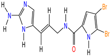
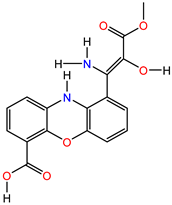
Our investigation focused on examining the inhibitory potential of five compounds (Table 1) sourced from the PubChem database in sdf format. These compounds were tested against six neuroblastoma (NB) targets [43], namely Ageladine A (Mol1), Oroidine (Mol2), Strepoxazine A (Mol3), Cyclooroidine (Mol4), and Taurodispacamide A (Mol5). The obtained Md results were analyzed using Discovery Studio Visualizer (DSV), v 20.0 [44].
The NB targets were obtained from the Protein Data Bank (PDB) [49], and were prepared following the step I explained above. The targets used for docking, along with their corresponding parameters, are presented in Table 2.

2.4. Normal Mode Analysis

Normal mode analysis (NMA) was carried out to check the stability of the best complexes resulting from the molecular docking. The iMODS (http://imods.chaconlab.org/ accessed on 12 December 2023) [25,50,51] is a fast easy webserver, user-friendly, and effective NMA tool, it can be used efficiently to investigate the structural dynamics of studied complexes. It was used to calculate the values of structural deformability, B-factor (disorder of atoms), eigenvalue, variance, covariance map, and elastic network.

3. Results and Discussion

3.1. Pass Study

The generated results from the PASS analysis for our proposed inhibitors are presented in Table 3. The primary PASS results indicate moderate-predicted biological activity numbers (PBAn) for all inhibitors, ranging from 68 to 901. Each inhibitor obtained at least one PBAn value of 0.9. Mol1 and Mol2 share the same PASS profile, particularly in terms of the first three Pa/Pi values, although Mol1 achieved a higher PBAn value. Furthermore, all compounds except Mol3 were identified as MAP kinase 1 inhibitors, while Mol3 was classified as a MAP kinase stimulant with a Pa value of 0.253. Mol3 demonstrated the highest PBAn score, while Mol2 and Mol5 obtained the lowest PBAn scores. No significant predicted activities related to our selected targets were found; they scored low Pa values for a few of them. Only Mol1 exhibited low FAK inhibition activity, with a Pa value of 0.190. We did not observe any Caspase-3 inhibition activity; however, Mol3 demonstrated Caspase 8 stimulant properties with a Pa value of 0.290.

3.2. ADMT

The ADMT parameters predicted for all compounds are presented in Table 4. The results indicate low Caco-2 cell permeability, ranging from 3.93 nm/s to 6.45 nm/s, for Mol1 and Mol3 suggesting difficulties in crossing this restrictive membrane. However, Mol2, Mol4, and Mol5 indicate moderate permeability. Additionally, the compounds exhibit different water solubility. On the other hand, Mol1, Mol2, and Mol4 demonstrate high human intestinal absorption, ranging from 79 to 86%, except for Mol5 and Mol3, the latter has a low absorption value of 26%. This indicates favorable absorption characteristics for the majority of the compounds. Furthermore, P-glycoprotein inhibition was not detected in all compounds, which helps pump these compounds out of cells in case of their harmful effects.
The distribution profile of our predicted compounds indicates challenges in crossing the blood-brain barrier (BBB), with scores ranging from 0.03 to 0.28. These scores are comparatively low when compared to the standard values (>2, indicating easy crossing of the BBB). Consequently, their potential neurological effects are diminished due to the low likelihood of crossing this barrier. However, all the molecules exhibit the ability to pass through the skin easily.
The percentage of plasma protein binding was consistently low for all molecules, indicating weak binding to plasma proteins such as albumin. This suggests that our compounds have a weak interaction with these proteins, which in turn requires lower doses to reach their intended targets. The compounds’ binding fraction should not saturate all the available binding sites on albumin before they are released once more.
Metabolic profile analysis revealed that Mol2 and Mol5 inhibited CYP 2C19, while the other compounds were either weak or substrates for cytochromes including CYP 2C9, 2D6 and 3A4. These findings offer insights into the efficacy and toxicity of xenobiotics. If xenobiotics are rapidly metabolized, they may not remain in the bloodstream for a sufficient duration to exert their desired effects. Conversely, if they are metabolized slowly, they may accumulate and lead to toxicity, although they may also demonstrate effectiveness.
The toxicity profile of the compounds indicates consistent outcomes in terms of mutagenicity in the Ames tests (as shown in Table 4). The majority of the compounds exhibited negative results for mouse carcinogenicity, however, most of them were positive for rat carcinogenicity, which is more closely related to human genes, indicating a high potential for carcinogenicity, particularly with high doses and prolonged exposure. The inhibition of human ether-related gene channels posed a moderate risk, except for Mol3. Consequently, it is crucial to adjust drug doses, administration routes, and treatment durations to address certain weak pharmacokinetic parameters.

3.3. Molecular Docking

We investigated the inhibition potential of five Agelas sp. alkaloids, such as Ageladine A (Mol1), Oroidine (Mol2), Strepoxazine A (Mol3), Cyclooroidine (Mol4), and Taurodispacamide A (Mol5), to inhibit six neuroblastoma targets, including: the focal adhesion kinase 1 (FAK), the caspase-3 (ca3), the phosphatidylinositol 4,5-bisphosphate 3-kinase catalytic subunit gamma isoform (PI3K), the telomerase reverse transcriptase (TERT), the osm-9-like TRP channel 1 (TRPV1), and the RAC-alpha serine/threonine-protein kinase (AKT1), as well as the type of interactions the involved amino acids. The results are presented in Table 5.

3.3.1. The Focal Adhesion Kinase 1

Focal Adhesion Kinase 1 or FAK is a non-specific protein-tyrosine kinase located in the cytoplasm. It is part of the focal adhesion family, which consists of significant macromolecular complexes. It can be triggered into activation by integrins, growth factors, cytokines, and hormones which play a critical role in transmitting signals that regulate a wide range of cellular processes such as proliferation, differentiation, and migration [52]. Dysregulation of FAK has the potential to contribute to cancer development [53], particularly in relation to the proliferation of neuroblastoma.
Its functioning as a transferase enzyme facilitates the phosphorylation of tyrosine residues in cytoskeletal proteins (Ckp) initiated by the adhesion properties of integrins by utilizing adenosine-5′-triphosphate (ATP), resulting in the production of adenosine-5′-diphosphate (ADP) and O-phospho-L-tyrosyl-[Ckp]. Its crystal structure was found complexed with ATP in chain A and ADP in chain B, the docking validation shows that ATP was bound in the active site and formed mostly hydrogen bonds with several residues, including the important of them: Gln432, Lys454, and Cys502, with the support of Arg550, Gly429, Gly431, Ile428, Met499, Val436, Ala452, and Leu553. The ATP purine moiety forms several hydrophobic interactions with Ala452, Cys502, and Leu553. These interactions are crucial for phosphorylation.
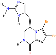
Mol1 is a pyrrole-imidazole alkaloid comprising an imidazo[4,5-c]pyridin group attached to a dibromo-pyrrole moiety. It demonstrates the lowest PLPchem score compared to other compounds targeting FAK (Table 5), which explains its reduced stability and suggests it may not serve as the perfect model for FAK. Mol1 binds to FAK through a crucial conventional hydrogen bond with Cys502, along with seven other residues involved in eight hydrophobic interaction types, including: Ile428, Met499, Val436, Ala452, and Leu553. Among these interactions, the imidazo[4,5-c]pyridin side of Mol1 plays the most significant role.
Mol2, Mol3, and Mol4 exhibited nearly identical PLPchem scores (Table 5). Among them, Mol4 performed the best, securing the second rank among all compounds. Its superiority over Mol2, which ranked third, can be attributed to the presence of a dibromo-pyrrole ring. This highly reactive feature allowed Mol4 to position itself deeply within the ATP binding site, enabling crucial binding interactions with two essential residues, Cys502 and Lys454. These residues typically interact with the pyrrole and phosphorus moieties of ATP, respectively, playing a critical role in the phosphorylation process. Mol4 also engaged in other significant hydrophobic interactions, such as pi-alkyl interactions with Ile428, Val436, and Leu553. The cyclisation in Mol4 provided a structural advantage, enhancing its affinity for FAK compared to Mol2. Notably, Mol2’s structure underwent a 90° rotation compared to Mol4, leading it to bind only to Cys502 along with other residues, resulting in a reduced PLPchem score (Table 5).
Mol3 ranked fourth among the compounds targeting FAK. It formed important interactions with three key residues involved in phosphorylation, namely: Cys502, Gln432, and Lys454. Despite this binding, Mol3 achieved a lower PLPchem score compared to Mol2 and Mol4. We hypothesize that Mol3’s stability can be attributed to its phenoxazine ring located in its central core. This ring engaged in crucial hydrophobic interactions, contributing to its overall stability.
Mol5 was the best compared to all other compounds (Figure 1), as all its functions involved bonds/interactions similar to those observed in ATP. Mol5’s conformation was docked vertically, resembling ATP, and its sulfonic acid side effectively interacted with Gln432 and Lys454. This precise positioning within the ATP binding site facilitated multiple conventional hydrogen bonds and hydrophobic interactions. Consequently, Mol5 effectively blocked the phosphorylation process, making it an ideal model for FAK inhibition.

3.3.2. The Caspase-3

Caspase-3, commonly referred to as ca3, belongs to the family of cysteine-aspartic acid proteases. It is present in the form of procaspases, which are precursor enzymes. Caspase-3 plays a significant role in actively breaking down proteins through proteolysis, particularly in the process of apoptosis [54], Apoptosis is a crucial physiological mechanism responsible for regulated cell death, essential for proper organismal development. Ca3 specifically targets vital structural proteins, cell cycle proteins, and DNase proteins like poly(ADP-ribose) polymerase, gelsolin, and DNA-dependent kinase [55]. Nevertheless, caspases have been found to have roles beyond apoptosis. Research has revealed that ca3, in particular, can facilitate the growth of cancer cells under stress, as well as enhance cellular migration, invasiveness, and tumor angiogenesis [56,57,58,59]. As a result, inhibiting ca3 has been shown to improve the effectiveness of chemotherapy and reduce the occurrence of spontaneous tumor development [60].
Upon activation, following apoptotic signaling events, ca3 focuses on cleaves proteins that possess the Asp-Glu-Val-Asp (DEVD) sequence motif [61]. This process is facilitated by specific residues, namely: Cys163, Gly238, and His121, which stabilize the peptide bond cleavage of the target protein within the active site. The crystal structures of ca3 have revealed its binding affinity to the (1S)-2-oxo-1-phenyl-2-[(1,3,4-trioxo-1,2,3,4-tetrahydroisoquinolin-5-yl)amino]ethyl acetate (RXB) inhibitor. RXB has the ability to irreversibly inhibit it, with its effectiveness dependent on the presence of 1,4-dithiothreitol (DTT) in vitro and dihydrolipoic acid in vivo, as well as oxygen through redox cycling. Docking validation has demonstrated that RXB binds to the active site located between chains C and A and primarily engages in hydrophobic interactions with Leu168 and Phe256 from chain A. The isoquinoline moiety of RXB exhibited the highest reactivity.
Mol1 exhibits a moderate PLPchem score compared to other compounds that target Ca3, thereby providing a better explanation for its moderate stability when compared to the case of the FAK target. This suggests that Mol1 could serve as a model for Ca3. Notably, Mol1 forms crucial interactions with key residues involved in proteolysis, forming three hydrogen bonds with Thr166 and Leu168, as well as five hydrophobic interactions with Phe256, Tyr204, and Leu168. Additionally, a Pi-sulfur interaction was observed with Cys170. The interactions observed were mainly with residues located in chain A, except for Tyr204, which is from chain C. These interactions confirm Mol1’s affinity for Ca3, and all its functions are involved.
Mol2 outperformed Mol4 in terms of their PLPchem scores (Table 5), favoring Mol2 as it secured the third rank among all compounds. Mol2 bound closely to multiple key residues from both chain C and Chain A, giving it a significant advantage over Mol4. The stabilization of Mol2 primarily relied on hydrophobic interactions, as it formed bonds between its amine imidazole and dibromo-pyrrole rings with the two-leucine residues from both chain A and C, along with two other residues from chain C (Thr166 and Phe256). These interactions caused Mol2 to adopt an angle shape of 120°, effectively linking the two chains and blocking the proteolysis site. On the other hand, Mol4 is also bound to Leu168 from chain A and three other residues from chain C (Phe256, His121, and Tyr204). However, the positioning of its 5,6-dibromo pyrrole pyrimidine ring near His121 led to an unfavorable cyclization in the dibromo-pyrrole, potentially impacting its PLPchem score and overall stability. Nonetheless, Mol4 still represents a good model for Ca3, as it interacts with the necessary key residues involved in proteolysis.
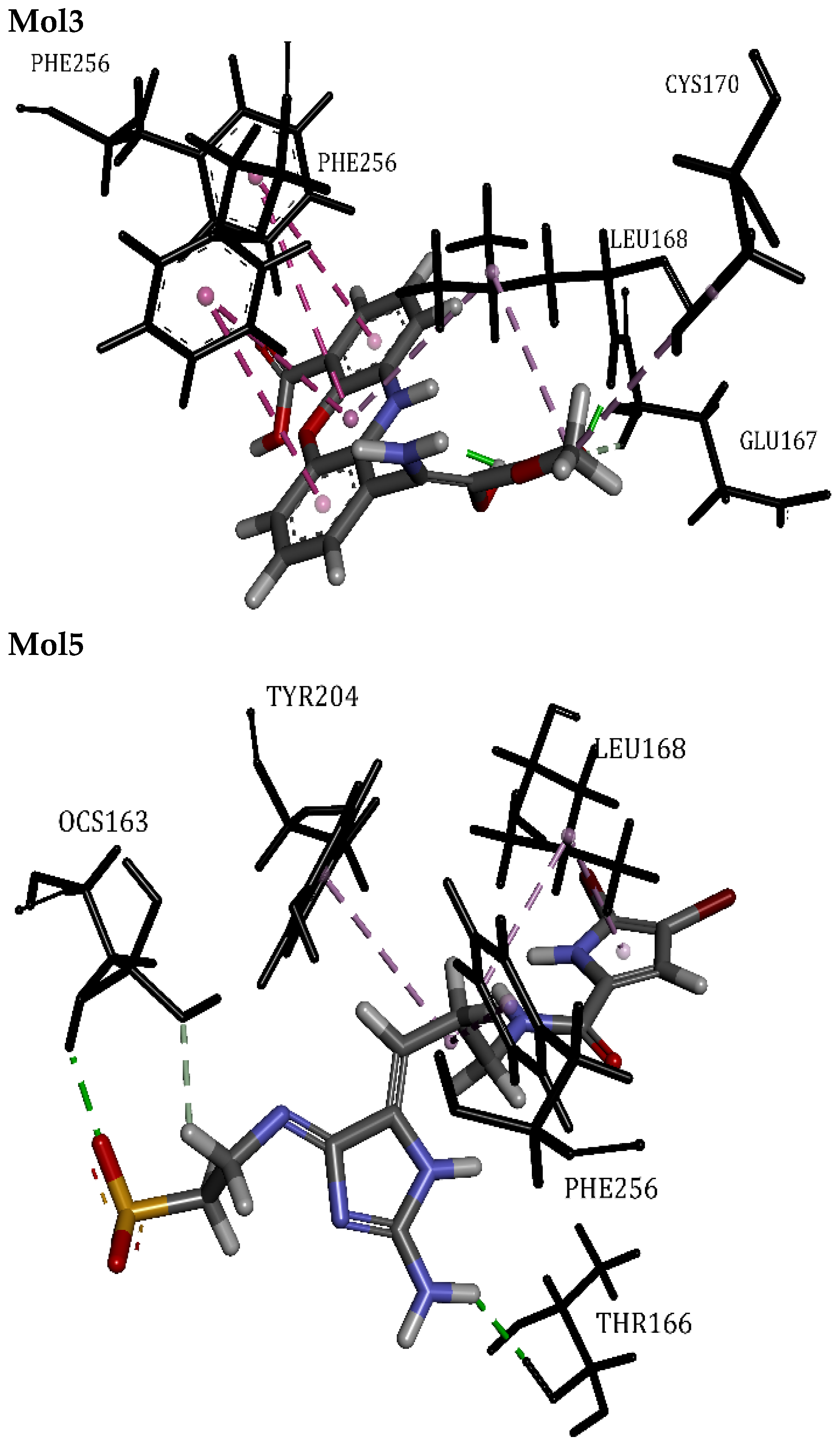
Mol5 and Mol3 achieved the first and second rankings (Figure 2), respectively, among the compounds targeting Ca3. Both compounds exhibited nearly similar PLPchem scores, which were better than the control RXB (Table 5). They shared the same inhibition mechanism, adopting a vertical conformation as the most favorable arrangement that allowed them to bind to key residues and maintain essential carbon-hydrogen bonds. Mol3 showed a preference for binding with chain A residues to block Ca3, with mostly hydrophobic interactions involving Leu168, Cys170, and Phe256 from chain A. Notably, it formed four Pi-Pi T-shaped interactions and a carbon-hydrogen bond with Glu167. On the other hand, Mol5 displayed a preference for binding with residues from both chains A and C to block Ca3. The interactions involved were a combination of hydrophobic and hydrogen bonds with Leu168, Tyr204, Thr166, Phe256, and Ocs163 (L-cysteic acid). The latter played a role in two hydrogen bonds, one of which was the carbon-hydrogen bond.

3.3.3. Phosphatidylinositol 4,5-Bisphosphate 3-Kinase Catalytic Subunit Gamma Isoform

The phosphatidylinositol 4,5-bisphosphate 3-kinase catalytic subunit gamma isoform, also known as PI3K-C2α, belongs to the family of phosphoinositide 3-lipid kinases, which consists of eight distinct PI3-kinase catalytic subunits [62]. In humans, these eight isoforms have been categorized into three functional classes [63] based on their protein domain structure, lipid substrate specificity, and associated regulatory subunits. The class I enzymes include p110α, p110β, p110δ, and p110γ. The class II enzymes consist of PI3K-C2α, PI3K-C2β, and PI3K-C2γ. Lastly, the class III enzyme is known as Vps34.
PI3K, known for its capability to phosphorylate the hydroxyl group at the 3 position of phosphatidylinositol lipids [62], plays a crucial role in transducing signals from various growth factors and cytokines. This transduction process involves the generation of phospholipids that activate the serine/threonine kinase AKT and other downstream effector pathways [64]. Extensive research has highlighted the involvement of PI3K in diverse cellular processes such as cell growth, survival, differentiation, motility, proliferation, and intracellular trafficking [63]. Consequently, inhibitors targeting these enzymes are considered potential therapeutic targets for cancer treatment.
As a transferase enzyme, it plays a crucial role in facilitating the phosphorylation of 1,2-diacyl-sn-glycero-3-phospho-(1D-myo-inositol-4,5-bisphosphate) using ATP. This enzymatic process leads to the generation of ADP and 1,2-diacyl-sn-glycero-3-phospho-(1D-myo-inositol-3,4,5-trisphosphate). Its crystal structure contains one single chain named A and was found complexed with 0TA ligand or (2-Amino-8-Cyclopentyl-4-Methyl-6-(1h-Pyrazol-4-Yl) pyrido [2,3-D]pyrimidin-7(8h). The docking validation shows that OTA was bound in the active site with mostly hydrophobic interactions with several residues, including Met953, Met804, Phe961, Ile963, Ile831, Ile879, Ile881, Val882, and Tyr867. In addition to a Pi-sulfur interaction with Met953. OTA establishes a crucial carbon-hydrogen bond with Lys833 and two conventional hydrogen bonds with Val882 through its aminopyrimidine function. These interactions were deemed critical for the inhibition mechanism [65].
Mol1 and Mol2 exhibit moderate PLPchem scores and are ranked lower compared to other compounds targeting PI3K-C2α, indicating their moderate stability. Notably, both Mol1 and Mol2 share the same mode of action toward this target, forming crucial interactions with key residues involved in phosphorylation. They both establish hydrogen bonds with Val882 and engage in multiple hydrophobic interactions, including alkyl and Pi-alkyl interactions with Ile963, Met804, Pro810, and Ile831. Additionally, a Pi-sulfur interaction was observed with Met953 for both compounds. These findings suggest that Mol1 and Mol2 can serve as a model for PI3K-C2α and confirm their strong affinity to it.
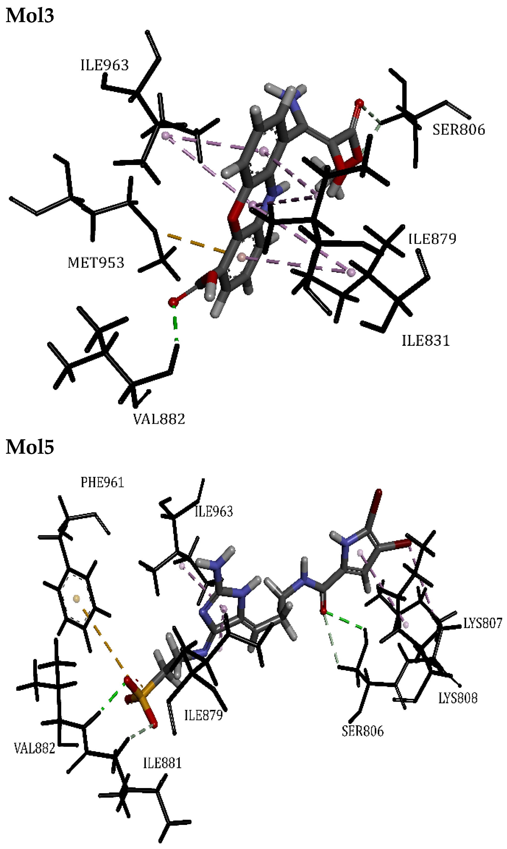
Mol3, Mol4, and Mol5 displayed nearly identical PLPchem scores (Table 5). Among them, Mol3 performed the best with a slight advantage over Mol5 (Figure 3), despite their significant difference in molecular weight. Mol5, on the other hand, ranked second among all compounds and demonstrated high stability in two targets, FAK and Ca3. Both Mol3 and Mol5 were found to be closely integrated within the binding site, adopting the same mode of action toward PI3K-C2α and forming crucial interactions with key phosphorylation-involved residues. They established two carbon-hydrogen bonds, one with Ser806 and the other with Ile881, indicative of high stability. Additionally, they engaged in multiple hydrophobic interactions, including alkyl and Pi-alkyl interactions with Ile963, Ile879, Pro810, and Ile831. Furthermore, Mol3 displayed a Pi-sulfur interaction with Met953, while Mol5 exhibited one with Phe961. Interestingly, Mol3 showed a preference for hydrophobic interactions to achieve stabilization, while Mol5 relied equally on both hydrogen bonds and hydrophobic interactions.
Mol4 secured the third rank and proved to be a promising model, along with all the other tested compounds for this target. Its mode of action differed significantly from other compounds, as it binds to PI3K-C2α away from Val882. Mol4 formed four conventional hydrogen bonds with residues, including Tyr867 and Asp964, saving three. Nevertheless, it maintained interactions with Ile881, Met953, Ile879, and Ile963, allowing it to engage in hydrophobic interactions of alkyl and Pi-alkyl types.

3.3.4. Telomerase Reverse Transcriptase

Telomerase reverse transcriptase, referred to as TERT, is a catalytic subunit of the telomerase enzyme. It is derived from Tribolium castaneum and belongs to the family of ribonucleoprotein polymerases. TERT’s primary function is to maintain the ends of telomeres by adding the telomere repeat TTAGGG [66]. Telomerase is responsible for lengthening the telomere sequences at the ends of chromosomes, which helps protect the genomic DNA. It plays a crucial role in determining the maximum number of times a cell can divide and thus plays a significant part in the immortality of cell lines, including cancer cells.
TERT, functioning as a transferase enzyme, has a vital role in enabling the phosphorylation of 2′-deoxyribonucleoside 5′-triphosphate using DNA (n). This enzymatic activity results in the formation of diphosphate and DNA (n + 1). The crystal structure of TERT comprises a single chain referred to as A, which has been observed to form complexes with the G2P ligand or (phosphor-methyl-phosphonic acid guanylate ester). The docking validation reveals that G2P predominantly engages in hydrogen bonds with several key residues, such as Ala195, Asp301, Gly309, Gln308, Asp254, and Arg194. These interactions are primarily facilitated by the G2P phosphorus tail moiety and are deemed essential for the inhibition mechanism.
Mol1 demonstrates a lower PLPchem score when compared to other compounds targeting TERT, which helps explain its reduced stability. This observation raises doubts about Mol1 being an ideal model. Specifically, Mol1 only forms a single interaction with Gln308 involved in phosphorylation and binds at a considerable distance. Additionally, its dibromo-pyrrole ring is attracted to Tyr256, leading to a Pi-Pi Stacked interaction that adversely affects its overall binding with G2P. This steric motion forces Mol1 to interact with other residues, resulting in most of the saved interactions or bonds. Consequently, Mol1’s optimal conformation appears to compromise its stability and may interfere with phosphorylation processes.
Mol2 outperformed Mol4 in terms of their PLPchem scores (Table 5), making Mol2 the third-ranked compound among all tested compounds. Both Mol2 and Mol4 closely interacted with important residues such as Asp254 and Ala195, while Mol4 also had remarkable interactions with Arg194 and Ala195. Both compounds formed Pi-Pi Stacked and Pi-Alkyl interactions with Tyr256, respectively. In contrast to Mol1, the Pi-Pi Stacked interaction did not negatively impact Mol2’s overall stability. This was because Mol2’s tail was flexible enough to adjust near Ala255 and Asp254, providing it with a significant advantage over Mol4. Mol2’s stabilization primarily relied on hydrophobic interactions, particularly with the dibromo-pyrrole ring and Tyr256, along with crucial carbon-hydrogen bonds with Asp254 that facilitated multiple conventional hydrogen bonds. On the other hand, Mol4’s binding to Tyr256 through a Pi-Alkyl interaction caused a 90° torsion, leading it to adopt a pose close to Arg194 as its best conformation. This slightly affected its PLPchem score, positioning it in the fourth rank due to having the lowest number of interactions, only seven in total. Nevertheless, Mol4’s positioning of its imidazole ring near Arg194 and Ala195 still makes it a potential inhibitor candidate for TERT.
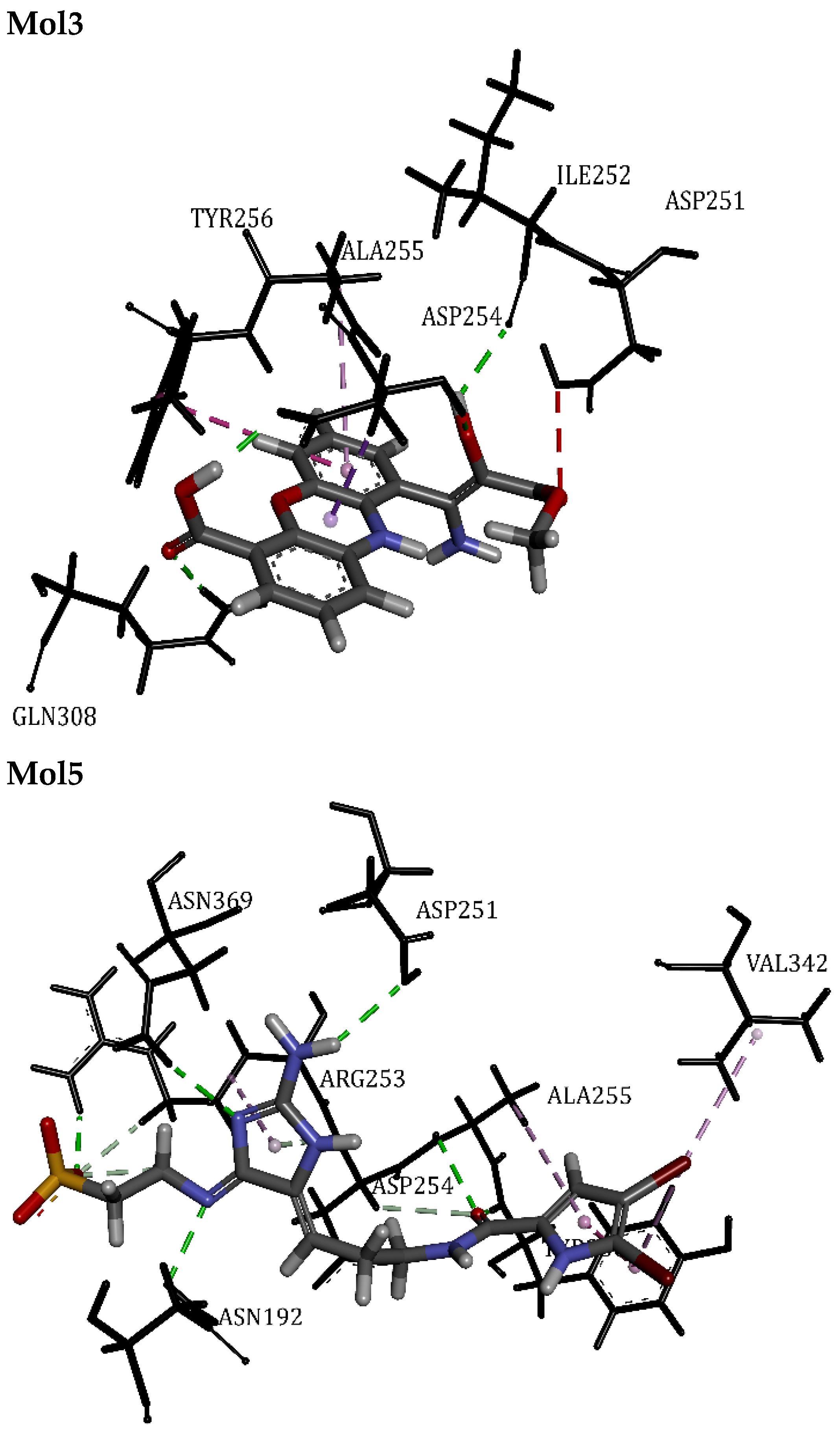
Mol5 and Mol3 secured the top two rankings among the compounds targeting TERT (Figure 4), as indicated in the PLPchem scores table. They employed distinct inhibition mechanisms. Notably, Mol5 interacted with a key residue same as G2P, which served as the reference inhibitor. It successfully blocked the binding site with 15 interactions, surpassing all other compounds in interaction efficiency with TERT. Mol5 shared a similar mechanism with Mol2, involving a Pi-Pi Stacked interaction with Tyr256 and its dibromo-pyrrole ring. This suggested the importance of this interaction in defining the behavior of all compounds with TERT. Moreover, Mol5’s tail displayed flexibility, allowing it to adjust near Asp254 and five other residues (Asp251, Asn369, Asn192, Arg253, and Ala255), providing a significant advantage over Mol3. The stabilization of Mol5 primarily relied on multiple hydrogen bonds, including three carbon-hydrogen bonds, five conventional hydrogen bonds, and a Pi-donor hydrogen bond, in addition to hydrophobic interactions. This combination suggested an irreversible blocking effect on the TERT binding site. In contrast, Mol3 exhibited a preference for binding with two key residues, Gln308 and Asp254, to block TERT, with a predominant reliance on conventional hydrogen bonds. Notably, it formed a Pi-Pi T-shaped interaction with Tyr256, which caused it to interact unfavorably with Asp251, affecting its PLPchem score slightly. However, its phenoxazine ring successfully blocked the binding site horizontally, where the necessary phosphorylation process occurs.

3.3.5. Osm-9-like TRP Channel 1

TRPV1, which stands for Osm-9-like Transient Receptor Potential Vanilloid channel 1, is a type of transmembrane protein that functions as a heat sensor. It consists of two parts: the cap domain (gate entrance), which is located in the outer part of the membrane, and the C-terminus domain, which is located in the inner part of the membrane (gate tip), between which the individual chains are located. It enables sensory nerve endings to detect high temperatures through ion channels belonging to the TRP family. The TRP superfamily consists of 28 cation-permeable channels, categorized into six subfamilies based on their sequence similarities: TRPC (Canonical), TRPV (Vanilloid), TRPM (Melastatin), TRPA (Ankyrin), TRPML (Mucolipin), and TRPP (Polycystic) [67]. TRPV1 specifically responds to stimuli such as capsaicin, protons, toxins, and temperatures within the noxious range (>42 °C). This channel serves as a polymodal molecular sensor involved in numerous physiological processes and has been implicated in various human diseases [68].
TRPV1, belonging to the category of cationic channels, functions as a signal transducer by modulating the membrane potential or intracellular calcium (Ca2+) concentration. It achieves this by allowing the passage of Ca2+ through a pore formed by specific segments within the membrane. These segments, namely transmembrane segments 5 and 6 (S5 and S6), form a flexible selectivity filter due to the absence of hydrogen bonding within and between their pore helices. Additionally, the pore-forming P loop, consisting of segments (S1–S4), remains relatively stable regardless of the channel’s state be it closed, partially open, or fully open. The movement of Ca2+ is regulated by a dual gating mechanism that involves upper gate and lower gate regions.
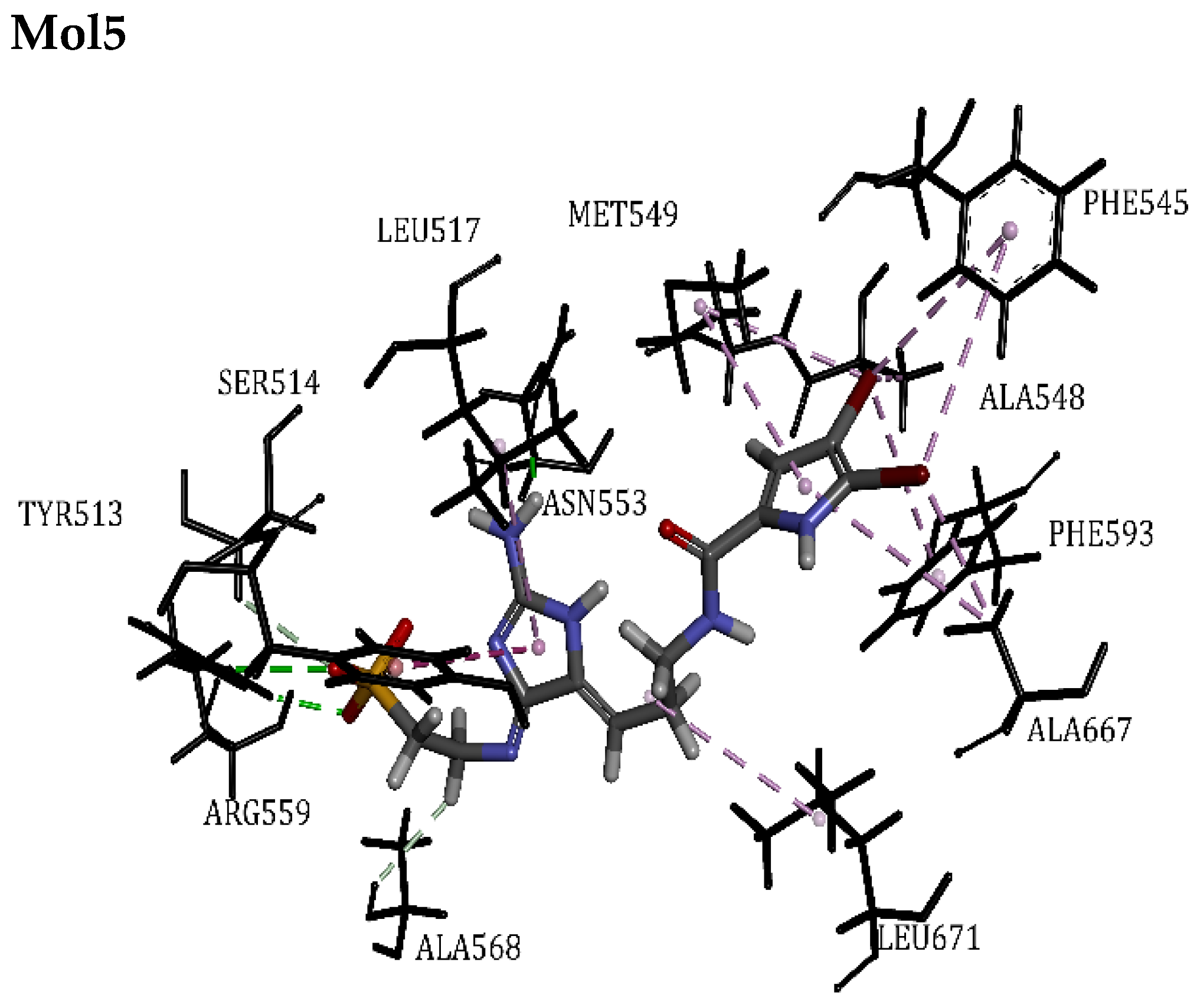
The crystal structure of TRPV1 consists of four chains and has been observed to form acomplex with the Resiniferatoxin (RTX) ligand. The docking validation shows that RTX binds to the vanilloid site (VS) coordinated by residues including Arg559, Leu555, Phe593, Ile575, Ile571, Asn553, Tyr513, Leu517, Ala548, and Met549 based on hydrophobic interactions and carbon-hydrogen bonds. These interactions are crucial for the channel inhibition mechanism.
Mol1 exhibits a lower PLPchem score when compared to other compounds that target TRPV1. However, this disparity in scores does not affect its efficacy. Notably, Mol1 establishes significant interactions with specific residues, such as Leu517, Leu555, Ile517, and Met549, which are crucial for channel opening. Particularly, its dibromo-pyrrole ring forms an attraction with Leu671 through a carbon-hydrogen bond, which is a residue located in S6. These interactions at the VS position Mol1 as a promising candidate for TRPV1 inhibition.
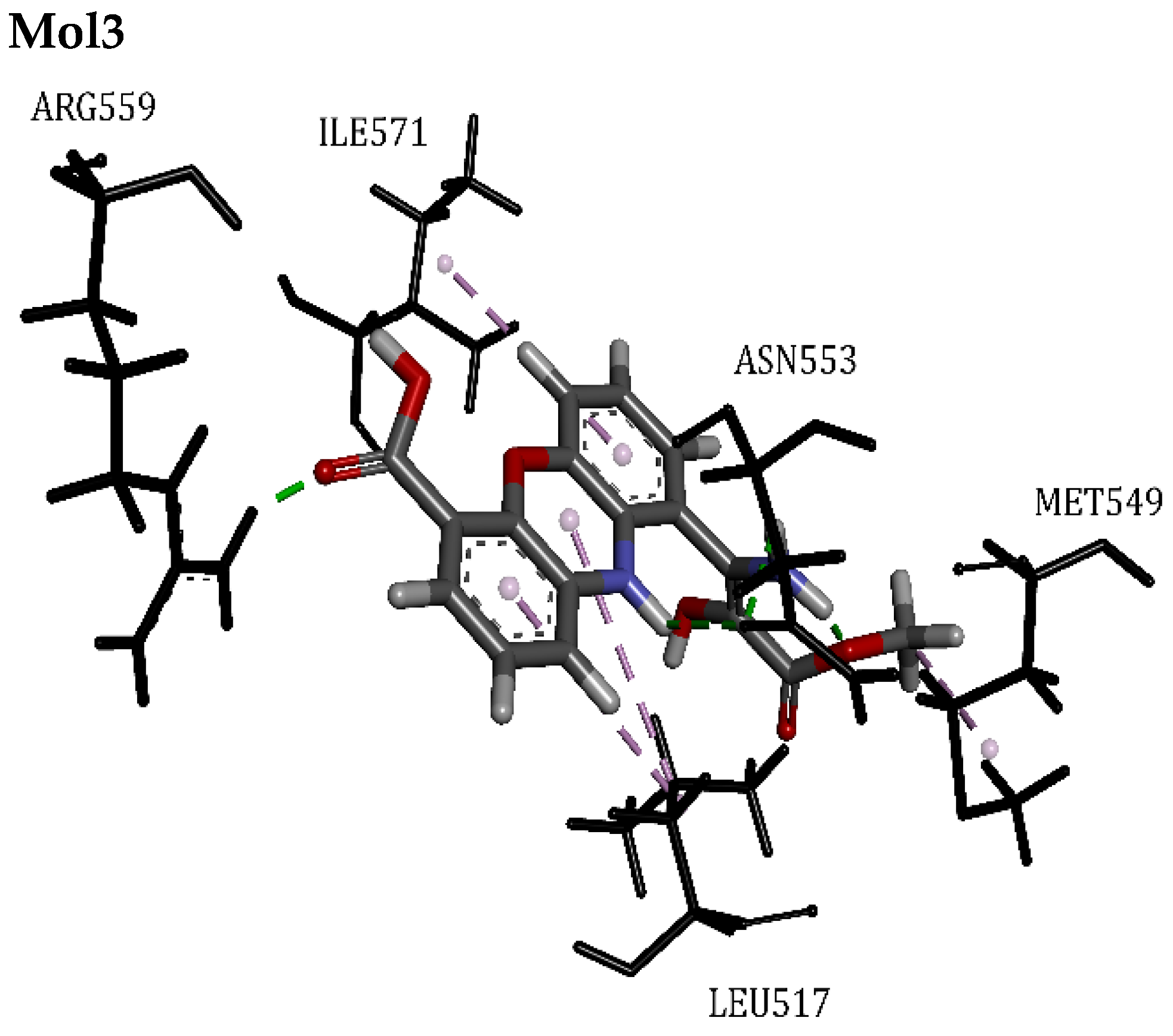
Mol2, Mol3, Mol4, and Mol5 achieved top rankings based on their PLPchem scores, as shown in Table 5, with a score ranging from 43 to 49, Mol3 ranked first, however, Mol5 emerged as the best candidate, securing the second rank and boasting the highest number of interactions, surpassing all other compounds. All the compounds primarily relied on hydrophobic interactions, particularly Pi-Alkyl, to stabilize in the VS. Notably, Mol2, Mol3, and Mol4 exhibited similar results, each establishing seven interactions with specific residues like Met549 and Leu517, along with other key residues located close to the VS, suggesting the possibility of distinct inhibition mechanisms for each. Remarkably, Mol5 adopted a unique docking orientation (Figure 5), positioning itself vertically with respect to the cap domain and aligning with the S3–S4 and S5–S6 segments. Its dibromo-pyrrole ring formed linkages with residues from both S5 and S6, while its tail connected S3 with S4. In this configuration, Mol5 engaged with twelve different residues, including those critical for the proper functioning of the channel, effectively occupying the entire VS space. As a result, Mol5 emerged as the most promising model for inhibiting TRPV1.

3.3.6. RAC-Alpha Serine/Threonine-Protein Kinase

AKT1, also known as RAC-alpha serine/threonine-protein kinase or “AKR mouse strain Thymoma”, is one isoform of the human AKT protein, alongside AKT2 and AKT3. All three isoforms share a similar structure, including a pleckstrin homology (PH) domain at the N-terminal, an inter-domain linker, a kinase domain, and a hydrophobic motif (HM) (from Ser463 to Ser477) at the C-terminal [69]. AKT1 activation requires two steps: translocation and phosphorylation. In its normal state, AKT1 is found to be inactive, adopting the PH-in conformation. However, multiple triggers can activate AKT1, including the presence of phospholipids produced in the PI3K pathway discussed earlier in this study. The PH domain assists in positioning AKT1 at the cell membrane by binding to PI3K products. This positioning facilitates the phosphorylation of AKT1’s Thr308 located in the activation loop region by the phosphoinositide-dependent kinase-1 (PDK1). An additional phosphorylation occurring at Ser473, within the HM, collaborates synergistically to achieve full activation of AKT1 [70]. This activation process utilizes ATP, leading to the production of ADP and O-phospho-L-threonyl-AKT1. Once activated, it phosphorylates various proteins involved in promoting growth signals, angiogenesis, tumor development, metabolism, and cell survival. Additionally, AKT1 inhibits apoptotic processes, making it an appealing target for cancer therapy [71].
Its crystal structure represents the active form, also known as the PH-out conformation. It consists of two chain complexes, each one bound with an ANP ligand or Phosphoaminophosphonic acid-Adenylate ester in the ATP binding site. The docking validation shows that ANP form equal hydrogen bonds and hydrophobic interactions with several residues, including: Ser7, Thr5, Leu156, Val164, Ala177, Lys179, Glu228, Tyr229, Ala230, and Met281. The ANP amino purine moiety forms key hydrophobic interactions with ATP binding site residues like Glu228, Tyr229, and Ala230. These interactions play a vital role in inhibiting ATP usage, resulting in the cessation of ADP production and the subsequent inactivation of AKT1.
Mol1 exhibits a lower PLPchem score compared to other compounds that target AKT1, and this finding is consistent across all the targets examined in this study. Despite the lower PLPchem score, Mol1 stands out as an ideal model due to its significant interactions with AKT1, ranking second in terms of the number of interactions formed (17 interactions/bonds). Notably, ten of these interactions involve its dibromo-pyrrole ring. The lower PLPchem score can be explained by the fact that the dibromo-pyrrole ring is rotated by approximately 20° to adopt the optimal conformation required for forming all these interactions. Specifically, Mol1 forms interactions with crucial residues in the ATP binding site, such as Ala230, Tyr229, and Glu228, with the latter forming a halogen interaction with one of Mol1’s bromine atoms. Additionally, it interacts with residues from the PH domain, including Leu156, Val164, Ala177, and Lys179, which are vital for AKT1’s active-inactive cycle. Moreover, Mol1 engages in Pi-Sulfur interactions with Met281 and Met227, potentially leading to an irreversible linkage that blocks ATP usage.
Mol4 outperformed Mol2, as evident from their PLPchem scores (Table 5). Mol4 stood out with a significantly higher number of interactions, almost double that of Mol2 (12 interactions). Interestingly, none of these interactions occurred near any of the ATP binding site residues. Instead, both Mol4 and Mol2 bound to residues within the PH domain, such as Leu156, Val164, Ala177, and Lys179, primarily through hydrophobic interactions, including alkyl and Pi-alkyl types. These findings suggest that Mol4 and Mol2 employ a distinct inhibition mechanism by interacting with the PH domain residues, effectively preventing the conformational shift that would release the kinase domain and expose Thr308 and Ser473 for phosphorylation. Consequently, the ATP has a high chance of being fixed at its binding site, however, remains unconverted to ADP due to the kinase domain’s inactivation.
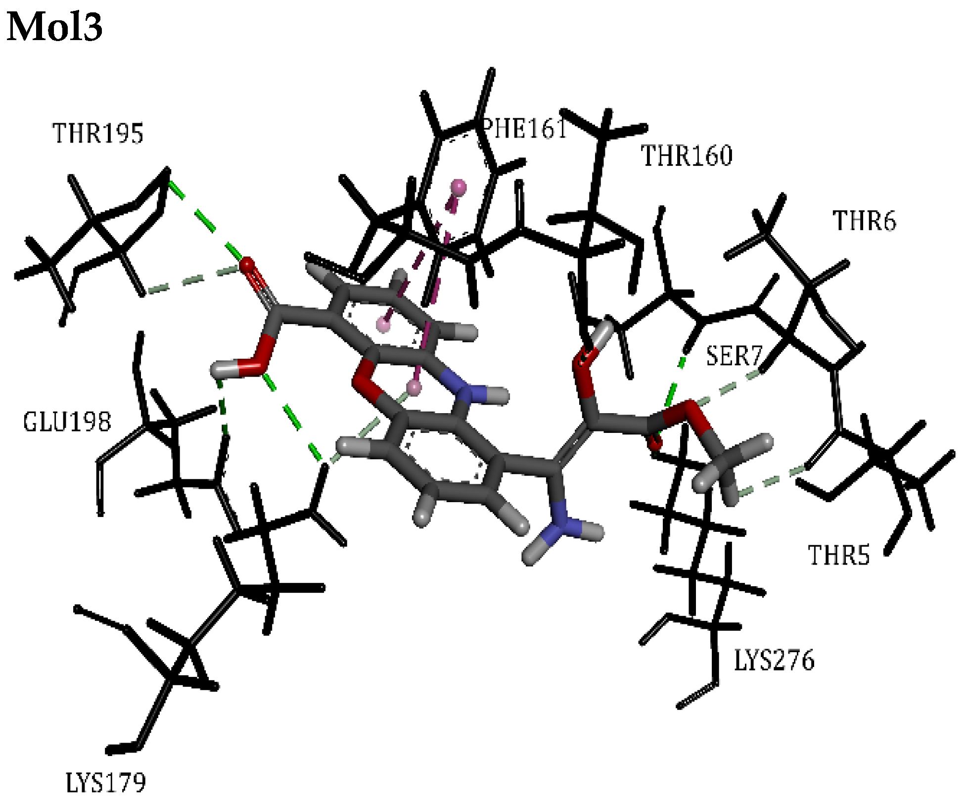
Mol5 and Mol3 emerged as the top two ranked compounds targeting AKT1 (Figure 6), a consistent finding across all the targets examined in this study, as evidenced by their PLPchem scores (Table 5). These compounds employed distinct inhibition mechanisms. Notably, Mol5 exhibited the highest number of interactions (19 interactions) compared to Mol3. One intriguing aspect of Mol5 is that it formed interactions with both ATP binding site residues and PH domain residues, making it stand out as an ideal model. These interactions included various types such as conventional hydrogen bonds, carbon-hydrogen bonds, halogen interactions, and hydrophobic interactions like Pi-sulfur, Pi-alkyl, and Alkyl. Eleven of these interactions were specifically attributed to its dibromo-pyrrole ring, ensuring stable binding with AKT1. Given its unique combination of interactions, Mol5 presents itself as a promising candidate for further study as a potential AKT1 inhibitor. On the other hand, Mol3’s interactions were primarily with different PH domain residues compared to those observed with the ANP ligand or other compounds. It was docked at the entrance of the PH domain, forming interactions with Thr160, Phe161, Thr195, Lys276, and Lys179, following a mode similar to that of Mol4 and Mol2. However, the stability of Mol3’s binding might be inferior to Mol5, despite its high PLPchem score. Therefore, we ranked Mol3 second due to the possibility of its binding being less stable compared to Mol5.

3.4. Normal Mode Analysis

Normal mode analysis (NMA) was conducted utilizing the iMODS server to assess the stability and mobility of atoms within the complex. The deformability of the target under investigation relies on the potential for deformation at any of its amino acid residues. Each normal mode is linked to an eigenvalue that characterizes the rigidity of motion, with its magnitude directly linked to the energy needed to alter the structure. Lower eigenvalues indicate greater ease of deformation [25,32,51,72,73]. The B-factor corresponds to the root mean square deviation (RMSD) because B-factors reflect how different parts of the structure vibrate relative to each other. Low B-factors for atoms signify their membership in the stable regions of the structure, while atoms in the flexible portions exhibit higher B-factors [74,75]. The NMA results are shown in the figure below. The variance is indicated by purple-colored bars and cumulative variance by green-colored bars, it is inversely related to the eigenvalue. The outcomes for both Mol3/Mol5/RXB-Ca3 docking complexes can be seen in Figure 7A. Figure 7B displays the deformability graph for these complexes, with the peaks on the graph corresponding to areas of deformability within the complexes.
Significantly, both Mol3 and Mol5 yielded comparable outcomes when interacting with the Ca3 target. Therefore, only one set of results is presented for both in Figure 7. In Figure 7C, the B-factor of the complex provides an average root mean square value, reflecting the motion of individual atoms within the complex. The majority of Ca3 atoms, in the presence of Mol3 and Mol5, exhibited B-factor values around 0.2, indicating a higher likelihood of rigid segments and affinity compared to RXB, where the B-factor line exceeded 0.2. The eigenvalues for the RXB-Ca3 complex were notably higher in comparison to those of the Mol3/Mol5-Ca3 complexes, as shown in Figure 7E. This suggests that more energy would be required to deform the RXB-Ca3 complex when compared to the Mol3/Mol5-Ca3 complexes. This finding is a positive indicator, suggesting a potential swift clearance of Mol3 and Mol5 from the body. The variance map exhibited minimal variances at the individual level and substantial cumulative variances for both scenarios. In the complex depicted in Figure 7G, the covariance map reveals the interactions between pairs of residues, indicating whether they undergo correlated (red), uncorrelated (white), or anti-correlated (blue) motions. It isevident that the Mol3/Mol5-Ca3 complexes exhibit a higher degree of correlation in comparison to the RXB case. The elastic network model establishes connections between atom pairs using springs, with each dot on the graph symbolizing a spring connecting a specific pair of atoms. These dots are shaded to reflect their stiffness, with darker grays indicating stiffer springs, as seen in Figure 7F. The findings from the elastic network analysis reveal a greater number of strongly connected atom pairs in comparison to the RXB case. The results from the iMODS simulation imply that the Mol3/Mol5-Ca3 complexes exhibit stability.

4. Study Limitation

While our study successfully identified promising PIAs inhibitors targeting key pathways involved in neuroblastoma, several limitations must be acknowledged. Although Md and NMA simulations were employed to confirm stability, the accuracy of these findings can still be influenced by the quality of the target protein structures and the inherent assumptions of the docking algorithms. Additionally, the study’s reliance on computational predictions may not fully capture the complexities of biological systems, including cellular environments and potential off-target effects. To address these limitations in future research, it is essential to complement in silico findings with extensive experimental validation, including in vitro and in vivo studies to assess the efficacy and safety of the identified inhibitors.

5. Conclusions

In this computer-based study, we present novel findings regarding the capacity of five molecules classified as PIAs originating from Agelas oroides S. and Agelas nakamurai H. These molecules demonstrate, for the first time, their potential to inhibit targets associated with neuroblastoma. The results of the molecular docking study, which were confirmed by the normal mode analysis using iMODS server, are promising. The five ligands showed significant affinity for these enzymes. Notably, Mol3 and Mol5 exhibited potency across all targets, coupled with a favorable ADMT profile. This implies their viability as a valuable natural therapeutic avenue for quelling inflammation and alleviating pain, all without causing harm to vital organs such as the kidneys. Furthermore, they have the potential to enhance the efficacy of chemotherapy. Based on the data presented in this paper, we suggest further in vitro and in vivo tests of these compounds to investigate their anti-cancer effects and to develop new potent drugs to prevent neuroblastoma.

Author Contributions

Conceptualization, A.L. and K.B.; methodology, K.B.; software, T.S.; validation, A.L., B.Y.A., L.B.-S. and K.B.; formal analysis, S.B.; investigation, L.B.-S.; resources, B.l.H.; data curation, S.B.; writing—original draft preparation, A.L.; writing—review and editing, A.L.; visualization, B.l.H.; supervision, K.B.; project administration, K.B.; funding acquisition, B.Y.A. All authors have read and agreed to the published version of the manuscript.

Funding

The Researchers would like to thank the Deanship of Graduate Studies and Scientific Research at Qassim University for the financial support (QU-APC-2024-9/1).

Institutional Review Board Statement

Not applicable.

Informed Consent Statement

Not applicable.

Data Availability Statement

The original contributions presented in the study are included in the article, further inquiries can be directed to the corresponding author.

Conflicts of Interest

The authors declare no conflicts of interest.

References

  1. Segura, M.F.; Soriano, A.; Roma, J.; Piskareva, O.; Jiménez, C.; Boloix, A.; Fletcher, J.I.; Haber, M.; Gray, J.C.; Cerdá-Alberich, L. Methodological advances in the discovery of novel neuroblastoma therapeutics. Expert Opin. Drug Discov. 2022, 17, 167–179. [Google Scholar] [CrossRef] [PubMed]
  2. Capasso, M.; Diskin, S.J. Genetics and genomics of neuroblastoma. In Cancer Genetics; Springer: Boston, MA, USA, 2010; pp. 65–84. [Google Scholar]
  3. Russo, C.; Cohn, S.L.; Petruzzi, M.J.; de Alarcon, P.A. Long-term neurologic outcome in children with opsoclonus-myoclonus associated with neuroblastoma: A report from the Pediatric Oncology Group. Med. Pediatr. Oncol. Off. J. SIOP-Int. Soc. Pediatr. Oncol. (Soc. Int. D’oncologie Pédiatrique) 1997, 28, 284–288. [Google Scholar] [CrossRef]
  4. Franks, L.M.; Bollen, A.; Seeger, R.C.; Stram, D.O.; Matthay, K.K. Neuroblastoma in adults and adolescents: An indolent course with poor survival. Cancer Interdiscip. Int. J. Am. Cancer Soc. 1997, 79, 2028–2035. [Google Scholar] [CrossRef]
  5. Maris, J.M. Recent advances in neuroblastoma. N. Engl. J. Med. 2010, 362, 2202–2211. [Google Scholar] [CrossRef] [PubMed]
  6. Tomolonis, J.A.; Agarwal, S.; Shohet, J.M. Neuroblastoma pathogenesis: Deregulation of embryonic neural crest development. Cell Tissue Res. 2018, 372, 245–262. [Google Scholar] [CrossRef]
  7. Fukuta, M.; Nakai, Y.; Kirino, K.; Nakagawa, M.; Sekiguchi, K.; Nagata, S.; Matsumoto, Y.; Yamamoto, T.; Umeda, K.; Heike, T. Derivation of mesenchymal stromal cells from pluripotent stem cells through a neural crest lineage using small molecule compounds with defined media. PLoS ONE 2014, 9, e112291. [Google Scholar] [CrossRef]
  8. Lasorella, A.; Noseda, M.; Beyna, M.; Iavarone, A. Id2 is a retinoblastoma protein target and mediates signalling by Myc oncoproteins. Nature 2000, 407, 592–598. [Google Scholar] [CrossRef]
  9. Alborzinia, H.; Flórez, A.F.; Kreth, S.; Brückner, L.M.; Yildiz, U.; Gartlgruber, M.; Odoni, D.I.; Poschet, G.; Garbowicz, K.; Shao, C.; et al. MYCN mediates cysteine addiction and sensitizes neuroblastoma to ferroptosis. Nat. Cancer 2022, 3, 471–485. [Google Scholar] [CrossRef]
  10. Johnsen, J.I.; Lindskog, M.; Ponthan, F.; Pettersen, I.; Elfman, L.; Orrego, A.; Sveinbjörnsson, B.; Kogner, P. NSAIDs in neuroblastoma therapy. Cancer Lett. 2005, 228, 195–201. [Google Scholar] [CrossRef]
  11. Bou-Salah, L.; Benarous, K.; Linani, A.; Rabhi, F.; Chaib, K.; Chine, I.; Bensaidane, H.; Yousfi, M. Anti-inflammatory drugs as new inhibitors to xanthine oxidase: In vitro and in silico approach. Mol. Cell. Probes 2021, 58, 101733. [Google Scholar] [CrossRef]
  12. Weinstein, J.L.; Katzenstein, H.M.; Cohn, S.L. Advances in the Diagnosis and Treatment of Neuroblastoma. Oncologist 2003, 8, 278–292. [Google Scholar] [CrossRef] [PubMed]
  13. Strother, D.R.; London, W.B.; Schmidt, M.L.; Brodeur, G.M.; Shimada, H.; Thorner, P.; Collins, M.H.; Tagge, E.; Adkins, S.; Reynolds, C.P. Outcome after surgery alone or with restricted use of chemotherapy for patients with low-risk neuroblastoma: Results of Children’s Oncology Group study P9641. J. Clin. Oncol. 2012, 30, 1842. [Google Scholar] [CrossRef]
  14. Zafar, A.; Wang, W.; Liu, G.; Wang, X.; Xian, W.; McKeon, F.; Foster, J.; Zhou, J.; Zhang, R. Molecular targeting therapies for neuroblastoma: Progress and challenges. Med. Res. Rev. 2021, 41, 961–1021. [Google Scholar] [CrossRef] [PubMed]
  15. Zafar, A.; Wang, W.; Liu, G.; Xian, W.; McKeon, F.; Zhou, J.; Zhang, R. Targeting the p53-MDM2 pathway for neuroblastoma therapy: Rays of hope. Cancer Lett. 2021, 496, 16–29. [Google Scholar] [CrossRef] [PubMed]
  16. Yang, X.; Li, J.; Yang, J. Promising Molecular Targets and Novel Therapeutic Approaches in Neuroblastoma. Curr. Pharmacol. Rep. 2023, 9, 43–58. [Google Scholar] [CrossRef]
  17. Jin, W.; Zhang, Y.; Zhao, Z.; Gao, M. Developing targeted therapies for neuroblastoma by dissecting the effects of metabolic reprogramming on tumor microenvironments and progression. Theranostics 2024, 14, 3439–3469. [Google Scholar] [CrossRef]
  18. Müller, W.E.G.; Batel, R.; Schröder, H.C.; Müller, I.M. Traditional and modern biomedical prospecting: Part I—The history. Evid.-Based Complement. Altern. Med. 2004, 1, 71–82. [Google Scholar] [CrossRef]
  19. Liang, J.; She, J.; Fu, J.; Wang, J.; Ye, Y.; Yang, B.; Liu, Y.; Zhou, X.; Tao, H. Advances in Natural Products from the Marine-Sponge-Associated Microorganisms with Antimicrobial Activity in the Last Decade. Mar. Drugs 2023, 21, 236. [Google Scholar] [CrossRef]
  20. Choi, C.; Cho, Y.; Son, A.; Shin, S.-W.; Lee, Y.-J.; Park, H.C. Therapeutic Potential of (−)-Agelamide D, a Diterpene Alkaloid from the Marine Sponge Agelas sp., as a Natural Radiosensitizer in Hepatocellular Carcinoma Models. Mar. Drugs 2020, 18, 500. [Google Scholar] [CrossRef]
  21. Chu, M.-J.; Li, M.; Ma, H.; Li, P.-L.; Li, G.-Q. Secondary metabolites from marine sponges of the genus Agelas: A comprehensive update insight on structural diversity and bioactivity. RSC Adv. 2022, 12, 7789–7820. [Google Scholar] [CrossRef]
  22. Lee, S.; Tanaka, N.; Takahashi, S.; Tsuji, D.; Kim, S.-Y.; Kojoma, M.; Itoh, K.; Kobayashi, J.i.; Kashiwada, Y. Agesasines A and B, Bromopyrrole Alkaloids from Marine Sponges Agelas spp. Mar. Drugs 2020, 18, 455. [Google Scholar] [CrossRef] [PubMed]
  23. Khalifa, S.A.M.; Elias, N.; Farag, M.A.; Chen, L.; Saeed, A.; Hegazy, M.F.; Moustafa, M.S.; Abd El-Wahed, A.; Al-Mousawi, S.M.; Musharraf, S.G.; et al. Marine Natural Products: A Source of Novel Anticancer Drugs. Mar. Drugs 2019, 17, 491. [Google Scholar] [CrossRef] [PubMed]
  24. Jones, G.; Willett, P.; Glen, R.C.; Leach, A.R.; Taylor, R. Development and validation of a genetic algorithm for flexible docking11Edited by F. E. Cohen. J. Mol. Biol. 1997, 267, 727–748. [Google Scholar] [CrossRef] [PubMed]
  25. López-Blanco, J.R.; Aliaga, J.I.; Quintana-Ortí, E.S.; Chacón, P. iMODS: Internal coordinates normal mode analysis server. Nucleic Acids Res. 2014, 42, W271–W276. [Google Scholar] [CrossRef] [PubMed]
  26. Goel, R.K.; Singh, D.; Lagunin, A.; Poroikov, V. PASS-assisted exploration of new therapeutic potential of natural products. Med. Chem. Res. 2011, 20, 1509–1514. [Google Scholar] [CrossRef]
  27. Linani, A.; Benarous, K.; Bou-salah, L.; Yousfi, M. Hispidin, Harmaline, and Harmine as potent inhibitors of bovine xanthine oxidase: Gout treatment, in vitro, ADMET prediction, and SAR studies. Bioorg. Chem. 2021, 112, 104937. [Google Scholar] [CrossRef]
  28. Lee, S.; Lee, I.; Kim, H.; Chang, G.; Chung, J.; No, K. The PreADME Approach: Web-based program for rapid prediction of physico-chemical, drug absorption and drug-like properties. In EuroQSAR Designing Drugs and Crop Protectants: Processes, Problems and Solutions; John Wiley & Sons: Hoboken, NJ, USA, 2003; pp. 418–420. [Google Scholar]
  29. Garg, S.; Roy, A. In silico analysis of selected alkaloids against main protease (Mpro) of SARS-CoV-2. Chem.-Biol. Interact. 2020, 332, 109309. [Google Scholar] [CrossRef]
  30. Jamkhande, P.G.; Wattamwar, A.S.; Pekamwar, S.S.; Chandak, P.G. Antioxidant, antimicrobial activity and in silico PASS prediction of Annona reticulata Linn. root extract. Beni-Suef Univ. J. Basic Appl. Sci. 2014, 3, 140–148. [Google Scholar] [CrossRef]
  31. Khurana, N.; Ishar, M.P.S.; Gajbhiye, A.; Goel, R.K. PASS assisted prediction and pharmacological evaluation of novel nicotinic analogs for nootropic activity in mice. Eur. J. Pharmacol. 2011, 662, 22–30. [Google Scholar] [CrossRef]
  32. Linani, A.; Serseg, T.; Benarous, K.; Bou-salah, L.; Yousfi, M.; Alama, M.N.; Ashraf, G.M. Cupressus sempervirens L. flavonoids as potent inhibitors to xanthine oxidase: In vitro, molecular docking, ADMET and PASS studies. J. Biomol. Struct. Dyn. 2023, 41, 7055–7068. [Google Scholar] [CrossRef]
  33. van Breemen, R.B.; Li, Y. Caco-2 cell permeability assays to measure drug absorption. Expert Opin. Drug Metab. Toxicol. 2005, 1, 175–185. [Google Scholar] [CrossRef] [PubMed]
  34. Yan, A.; Wang, Z.; Cai, Z. Prediction of human intestinal absorption by GA feature selection and support vector machine regression. Int. J. Mol. Sci. 2008, 9, 1961–1976. [Google Scholar] [CrossRef] [PubMed]
  35. Lefauconnier, J.M.; Hauw, J.J. The blood-brain barrier. II. Physiological data (conclusion). Rev. Neurol. 1984, 140, 89–109. [Google Scholar] [PubMed]
  36. Luo, S.; Kansara, V.S.; Zhu, X.; Mandava, N.K.; Pal, D.; Mitra, A.K. Functional characterization of sodium-dependent multivitamin transporter in MDCK-MDR1 cells and its utilization as a target for drug delivery. Mol. Pharm. 2006, 3, 329–339. [Google Scholar] [CrossRef]
  37. Nation, R.L.; Theuretzbacher, U.; Tsuji, B.T. Concentration-dependent plasma protein binding: Expect the unexpected. Eur. J. Pharm. Sci. Off. J. Eur. Fed. Pharm. Sci. 2018, 122, 341–346. [Google Scholar] [CrossRef]
  38. Lynch, T.; Price, A. The effect of cytochrome P450 metabolism on drug response, interactions, and adverse effects. Am. Fam. Physician 2007, 76, 391–396. [Google Scholar]
  39. Ames, B.N.; Durston, W.E.; Yamasaki, E.; Lee, F.D. Carcinogens are mutagens: A simple test system combining liver homogenates for activation and bacteria for detection. Proc. Natl. Acad. Sci. USA 1973, 70, 2281–2285. [Google Scholar] [CrossRef]
  40. Hedley, P.L.; Jørgensen, P.; Schlamowitz, S.; Wangari, R.; Moolman-Smook, J.; Brink, P.A.; Kanters, J.K.; Corfield, V.A.; Christiansen, M. The genetic basis of long QT and short QT syndromes: A mutation update. Hum. Mutat. 2009, 30, 1486–1511. [Google Scholar] [CrossRef]
  41. Todorov, L.; Saso, L.; Benarous, K.; Traykova, M.; Linani, A.; Kostova, I. Synthesis, Structure and Impact of 5-Aminoorotic Acid and Its Complexes with Lanthanum(III) and Gallium(III) on the Activity of Xanthine Oxidase. Molecules 2021, 26, 4503. [Google Scholar] [CrossRef]
  42. HyperChem(TM) Professional, version 8.0; Hypercube Inc.: Gainesville, FL, USA, 2009.
  43. Kim, S.; Thiessen, P.A.; Bolton, E.E.; Chen, J.; Fu, G.; Gindulyte, A.; Han, L.; He, J.; He, S.; Shoemaker, B.A.; et al. PubChem Substance and Compound databases. Nucleic Acids Res. 2016, 44, D1202–D1213. [Google Scholar] [CrossRef]
  44. Dassault Systèmes BIOVIA. BIOVIA Workbook, Release 2017; BIOVIA Pipeline Pilot; Dassault Systèmes: San Diego, CA, USA, 2017. [Google Scholar]
  45. Magoulas, G.E. Ageladine A, a Bromopyrrole Alkaloid from the Marine Sponge Agelas nakamurai. Compounds 2023, 3, 107–121. [Google Scholar] [CrossRef]
  46. Varijakzhan, D.; Loh, J.-Y.; Yap, W.-S.; Yusoff, K.; Seboussi, R.; Lim, S.-H.E.; Lai, K.-S.; Chong, C.-M. Bioactive compounds from marine sponges: Fundamentals and applications. Mar. Drugs 2021, 19, 246. [Google Scholar] [CrossRef] [PubMed]
  47. Elissawy, A.M.; Soleiman Dehkordi, E.; Mehdinezhad, N.; Ashour, M.L.; Mohammadi Pour, P. Cytotoxic alkaloids derived from marine sponges: A comprehensive review. Biomolecules 2021, 11, 258. [Google Scholar] [CrossRef] [PubMed]
  48. Fattorusso, E.; Taglialatela-Scafati, O. Two novel pyrrole-imidazole alkaloids from the Mediterranean sponge Agelas oroides. Tetrahedron Lett. 2000, 41, 9917–9922. [Google Scholar] [CrossRef]
  49. Burley, S.K.; Berman, H.M.; Kleywegt, G.J.; Markley, J.L.; Nakamura, H.; Velankar, S. Protein Data Bank (PDB): The single global macromolecular structure archive. Methods Mol. Biol. 2017, 1607, 627–641. [Google Scholar]
  50. Kovacs, J.A.; Chacón, P.; Abagyan, R. Predictions of protein flexibility: First-order measures. Proteins 2004, 56, 661–668. [Google Scholar] [CrossRef]
  51. Lopéz-Blanco, J.R.; Garzón, J.I.; Chacón, P. iMod: Multipurpose normal mode analysis in internal coordinates. Bioinformatics 2011, 27, 2843–2850. [Google Scholar] [CrossRef]
  52. Zamir, E.; Geiger, B. Focal Adhesions. In Encyclopedia of Biological Chemistry; Lennarz, W.J., Lane, M.D., Eds.; Elsevier: New York, NY, USA, 2004; pp. 128–133. [Google Scholar] [CrossRef]
  53. Tan, H.; Liu, Y.; Gong, C.; Zhang, J.; Huang, J.; Zhang, Q. Synthesis and evaluation of FAK inhibitors with a 5-fluoro-7H-pyrrolo [2,3-d]pyrimidine scaffold as anti-hepatocellular carcinoma agents. Eur. J. Med. Chem. 2021, 223, 113670. [Google Scholar] [CrossRef]
  54. Eskandari, E.; Eaves, C.J. Paradoxical roles of caspase-3 in regulating cell survival, proliferation, and tumorigenesis. J. Cell Biol. 2022, 221, e202201159. [Google Scholar] [CrossRef]
  55. Ponder, K.G.; Boise, L.H. The prodomain of caspase-3 regulates its own removal and caspase activation. Cell Death Discov. 2019, 5, 56. [Google Scholar] [CrossRef]
  56. Feng, X.; Yu, Y.; He, S.; Cheng, J.; Gong, Y.; Zhang, Z.; Yang, X.; Xu, B.; Liu, X.; Li, C.-Y. Dying glioma cells establish a proangiogenic microenvironment through a caspase 3 dependent mechanism. Cancer Lett. 2017, 385, 12–20. [Google Scholar] [CrossRef] [PubMed]
  57. Mukai, M.; Kusama, T.; Hamanaka, Y.; Koga, T.; Endo, H.; Tatsuta, M.; Inoue, M. Cross talk between apoptosis and invasion signaling in cancer cells through caspase-3 activation. Cancer Res. 2005, 65, 9121–9125. [Google Scholar] [CrossRef] [PubMed]
  58. Lauber, K.; Bohn, E.; Kröber, S.M.; Xiao, Y.-j.; Blumenthal, S.G.; Lindemann, R.K.; Marini, P.; Wiedig, C.; Zobywalski, A.; Baksh, S. Apoptotic cells induce migration of phagocytes via caspase-3-mediated release of a lipid attraction signal. Cell 2003, 113, 717–730. [Google Scholar] [CrossRef] [PubMed]
  59. Jonges, L.E.; Nagelkerke, J.F.; Ensink, N.G.; van der Velde, E.A.; Tollenaar, R.A.E.M.; Fleuren, G.J.; van de Velde, C.J.H.; Morreau, H.; Kuppen, P.J.K. Caspase-3 activity as a prognostic factor in colorectal carcinoma. Lab. Investig. 2001, 81, 681–688. [Google Scholar] [CrossRef]
  60. Bernard, A.; Chevrier, S.; Beltjens, F.; Dosset, M.; Viltard, E.; Lagrange, A.; Derangère, V.; Oudot, A.; Ghiringhelli, F.; Collin, B.; et al. Cleaved Caspase-3 Transcriptionally Regulates Angiogenesis-Promoting Chemotherapy Resistance. Cancer Res. 2019, 79, 5958–5970. [Google Scholar] [CrossRef]
  61. Eckhart, L.; Ballaun, C.; Hermann, M.; VandeBerg, J.L.; Sipos, W.; Uthman, A.; Fischer, H.; Tschachler, E. Identification of novel mammalian caspases reveals an important role of gene loss in shaping the human caspase repertoire. Mol. Biol. Evol. 2008, 25, 831–841. [Google Scholar] [CrossRef]
  62. Foster, F.M.; Traer, C.J.; Abraham, S.M.; Fry, M.J. The phosphoinositide (PI) 3-kinase family. J. Cell Sci. 2003, 116, 3037–3040. [Google Scholar] [CrossRef]
  63. Fry, M.J. Structure, regulation and function of phosphoinositide 3-kinases. Biochim. Biophys. Acta (BBA)-Mol. Basis Dis. 1994, 1226, 237–268. [Google Scholar] [CrossRef]
  64. Liu, P.; Cheng, H.; Roberts, T.M.; Zhao, J.J. Targeting the phosphoinositide 3-kinase pathway in cancer. Nat. Rev. Drug Discov. 2009, 8, 627–644. [Google Scholar] [CrossRef]
  65. Le, P.T.; Cheng, H.; Ninkovic, S.; Plewe, M.; Huang, X.; Wang, H.; Bagrodia, S.; Sun, S.; Knighton, D.R.; LaFleur Rogers, C.M.; et al. Design and synthesis of a novel pyrrolidinyl pyrido pyrimidinone derivative as a potent inhibitor of PI3Kα and mTOR. Bioorg. Med. Chem. Lett. 2012, 22, 5098–5103. [Google Scholar] [CrossRef]
  66. Schaich, M.A.; Sanford, S.L.; Welfer, G.A.; Johnson, S.A.; Khoang, T.H.; Opresko, P.L.; Freudenthal, B.D. Mechanisms of nucleotide selection by telomerase. eLife 2020, 9, e55438. [Google Scholar] [CrossRef] [PubMed]
  67. Samanta, A.; Hughes, T.E.T.; Moiseenkova-Bell, V.Y. Transient Receptor Potential (TRP) Channels. Sub-Cell. Biochem. 2018, 87, 141–165. [Google Scholar] [CrossRef]
  68. Nadezhdin, K.D.; Neuberger, A.; Nikolaev, Y.A.; Murphy, L.A.; Gracheva, E.O.; Bagriantsev, S.N.; Sobolevsky, A.I. Extracellular cap domain is an essential component of the TRPV1 gating mechanism. Nat. Commun. 2021, 12, 2154. [Google Scholar] [CrossRef] [PubMed]
  69. Coffer, P.J.; Jin, J.; Woodgett, J.R. Protein kinase B (c-Akt): A multifunctional mediator of phosphatidylinositol 3-kinase activation. Biochem. J. 1998, 335, 1–13. [Google Scholar] [CrossRef] [PubMed]
  70. Yang, J.; Cron, P.; Thompson, V.; Good, V.M.; Hess, D.; Hemmings, B.A.; Barford, D. Molecular mechanism for the regulation of protein kinase B/Akt by hydrophobic motif phosphorylation. Mol. Cell 2002, 9, 1227–1240. [Google Scholar] [CrossRef]
  71. Lin, K.; Lin, J.; Wu, W.I.; Ballard, J.; Lee, B.B.; Gloor, S.L.; Vigers, G.P.; Morales, T.H.; Friedman, L.S.; Skelton, N.; et al. An ATP-site on-off switch that restricts phosphatase accessibility of Akt. Sci. Signal. 2012, 5, ra37. [Google Scholar] [CrossRef]
  72. Awan, F.M.; Obaid, A.; Ikram, A.; Janjua, H.A. Mutation-Structure-Function Relationship Based Integrated Strategy Reveals the Potential Impact of Deleterious Missense Mutations in Autophagy Related Proteins on Hepatocellular Carcinoma (HCC): A Comprehensive Informatics Approach. Int. J. Mol. Sci. 2017, 18, 139. [Google Scholar] [CrossRef]
  73. Prabhakar, P.K.; Srivastava, A.; Rao, K.K.; Balaji, P.V. Monomerization alters the dynamics of the lid region in Campylobacter jejuni CstII: An MD simulation study. J. Biomol. Struct. Dyn. 2016, 34, 778–791. [Google Scholar] [CrossRef]
  74. Balendiran, G.K.; Pandian, J.R.; Drake, E.; Vinayak, A.; Verma, M.; Cascio, D. B-factor Analysis and Conformational Rearrangement of Aldose Reductase. Curr. Proteom. 2014, 11, 151–160. [Google Scholar] [CrossRef]
  75. Carugo, O. How large B-factors can be in protein crystal structures. BMC Bioinform. 2018, 19, 61. [Google Scholar] [CrossRef]
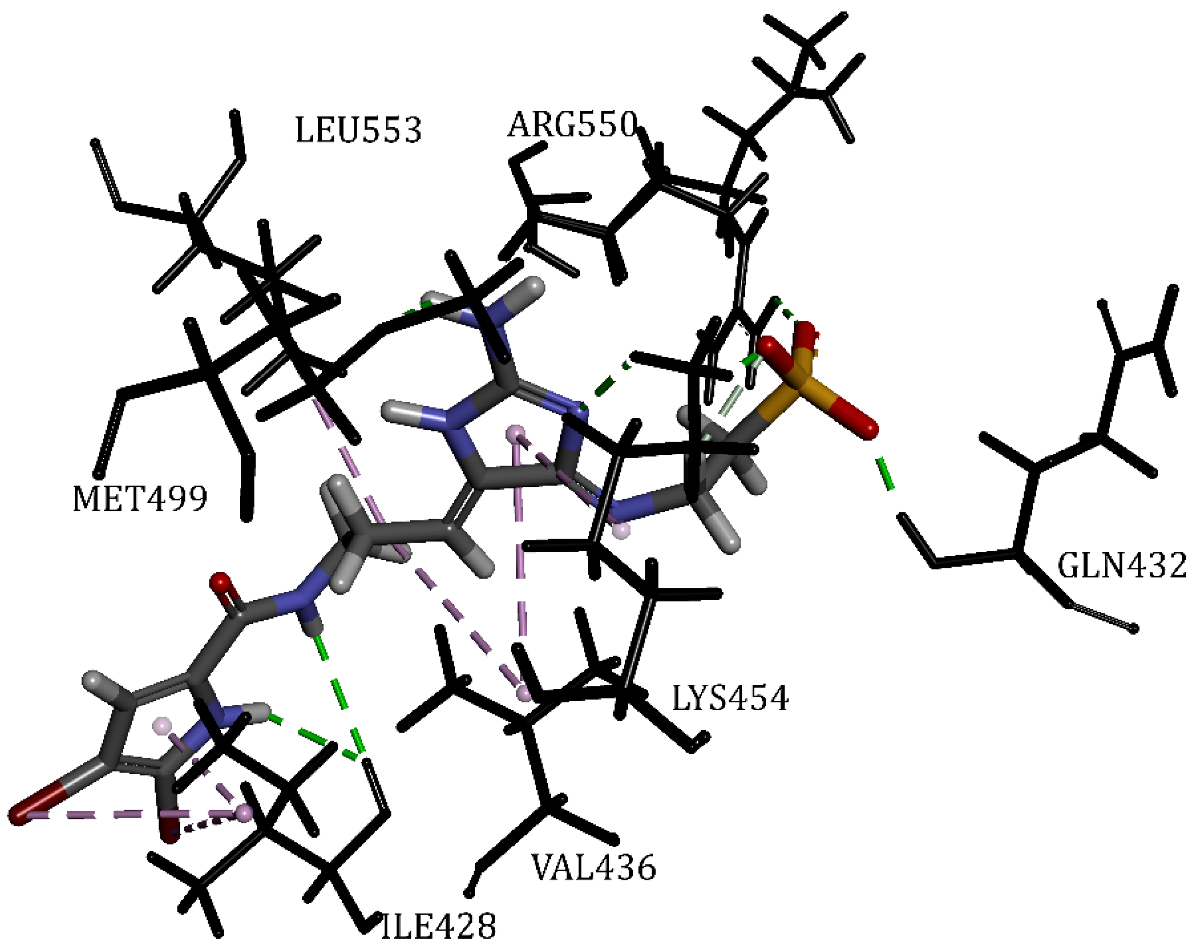
Figure 1. The 3D structure shows the most favorable FAK complex after docking, with the inhibitor (Mol5) displayed in stick form and colored using default atom colors.
Applsci 14 09306 g001
Figure 2. The 3D structure shows the most favorable Ca3 complexes after docking, with the inhibitors (Mol3 and Mol5) displayed in stick form and colored using default atom colors.
Applsci 14 09306 g002
Figure 3. The 3D structure shows the most favorable PI3K-C2α complexes after docking, with the inhibitors (Mol3 and Mol5) displayed in stick form and colored using default atom colors.
Applsci 14 09306 g003
Figure 4. The 3D structure shows the most favorable TERT complexes after docking, with the inhibitors (Mol3 and Mol5) displayed in stick form and colored using default atom colors.
Applsci 14 09306 g004
Figure 5. The 3D structure shows the most favorable TRPV1 complexes after docking, with the inhibitors (Mol3 and Mol5) displayed in stick form and colored using default atom colors.
Applsci 14 09306 g005aApplsci 14 09306 g005b
Figure 6. The 3D structure shows the most favorable AKT1 complexes after docking, with the inhibitors (Mol3 and Mol5) displayed in stick form and colored using default atom colors.
Applsci 14 09306 g006aApplsci 14 09306 g006b
Figure 7. Normal mode analysis of Mol3/Mol5-Ca3 and RXB-Ca3 complexes. (A) docking complexes. (B) Main-chain deformability. (C) B-factor values. (D) Variance. (E) eigenvalue. (F) Elastic network. (G) Covariance map.
Applsci 14 09306 g007aApplsci 14 09306 g007b
Table 1. The chemical properties of the studied compounds and their structures.
Code2D StructureSource
Mol1
(Ageladine A)
PubID 1: 10089677
MW 2: 357.00 g/mol
F 3: C10H7Br2N5
Applsci 14 09306 i001Agelas nakamurai H. [45]
Mol2
(Oroidin)
PubID: 6312649
MW: 389.05 g/mol
F: C11H11Br2N5O
Applsci 14 09306 i002Agelas oroides S.
[46,47]
Mol3
(Strepoxazine A)
PubID: 132526082
MW: 342.30 g/mol
F: C17H14N2O6
Applsci 14 09306 i003
Mol4
(Cyclooroidin)
PubID: 10739060
MW: 389.05 g/mol
F: C11H11Br2N5O
Applsci 14 09306 i004Agelas oroides S. [48]
Mol5
(Taurodispacamide A)
PubID: 135501141
MW: 512.18 g/mol
F: C13H16Br2N6O4S
Applsci 14 09306 i005
1 Pubchem ID, 2 Molecular weight, 3 Formula.
Table 2. Target enzymes and their associated codes.
PDB ID2IJM3DEI4FA66USR7LQZ4EKK
CodeFAKCa3PI3KTERTTRPV1AKT1
TargetFocal Adhesion Kinase 1Caspase-3Phosphatidylinositol 4,5-bisphosphate 3-kinase catalytic subunit gamma isoformTelomerase reverse transcriptaseOsm-9-like TRP channel 1RAC-alpha serine/threonine-protein kinase
Table 3. The predicted biological activities for the four best inhibitors in three targets.
MoleculesPa 1Pi 2PBA 3PBAn 4
Mol10.9640.001MAP kinase 1 inhibitor383
0.9030.002Protein kinase inhibitor
0.8960.002Raf kinase inhibitor
Mol20.9600.001MAP kinase 1 inhibitor68
0.8830.002Protein kinase inhibitor
0.8780.002Raf kinase inhibitor
Mol30.7720.010Fusarinine-C ornithinesterase inhibitor901
0.6860.030Pro-opiomelanocortin converting enzyme inhibitor
0.6430.016Preneoplastic conditions treatment
Mol40.9170.002MAP kinase 1 inhibitor109
0.8250.003Mycothiol-S-conjugate amidase inhibitor
0.6990.003Antineoplastic (brain cancer)
Mol50.9530.001MAP kinase 1 inhibitor68
0.8070.002Coenzyme-B sulfoethylthiotransferase inhibitor
0.8050.003Mycothiol-S-conjugate amidase inhibitor
1 probable activity, 2 probable inactivity, 3 predicted biological activity, 4 number.
Table 4. The ADMT properties of the five studied compounds.
PharmacokineticsMol1Mol2Mol3Mol4Mol5
Absorption
Caco-2 cell 1 permeability (nm/s) > 203.9319.776.4519.2319.67
Human intestinal absorption
(HIA %) 70 to 100%
86.0379.4526.8884.7340.14
Water solubility (g/L)55.970.15451.3827.244.20
P-glycoprotein inhibition
a substrate of it indicates high levels of absorption
NonNonNonNonNon
Distribution
Blood-brain barrier penetration
(C.brain/C.blood)
>2 cross the blood–brain barrier easily
0.280.080.030.240.04
MDCK 2 cell permeability (nm/s)
Low (<1 nm/s), moderate (1–10 nm/s), and high (>10 nm/s)
0.550.460.570.570.49
Plasma protein binding (%)
80 to 100% is considered high, 50 to 80% (moderate), <50% (low)
49.0160.1432.4338.7056.46
Skin permeability
(logKp 3, cm/hour)
<−2.5 considered high permeable
−5.19−5.02−5.12−5.20−4.82
Metabolism
Cytochrome P450 2C19 inhibitionNonInhibitorNonNonInhibitor
Cytochrome P450 2C9 inhibitionNonNonNonNonNon
Cytochrome P450 2D6 inhibitionInhibitorInhibitorInhibitorInhibitorInhibitor
Cytochrome P450 2D6 substrateSubstrateSubstrateSubstrateSubstrateSubstrate
Cytochrome P450 3A4 inhibitionNonInhibitorNonNonNon
Cytochrome P450 3A4 substrateWeaklyWeaklyWeaklyWeaklyWeakly
Toxicity
Ames testMutagenMutagennon-mutagenMutagenMutagen
Carcinogenicity (Mouse)NegativeNegativeNegativeNegativeNegative
Carcinogenicity (Rat)PositivePositiveNegativePositiveNegative
HERG 4_inhibitionMedium riskMedium risklow_riskMedium riskAmbiguous
TA100 (+S9)PositivePositiveNegativePositiveNegative
TA100 (−S9)NegativeNegativeNegativeNegativeNegative
Ames TA1535 (+S9)PositiveNegativeNegativePositiveNegative
Ames TA1535 (−S9)PositivePositiveNegativePositivePositive
1 Human colorectal carcinoma, 2 Mandin Darby Canine Kidney, 3 skin permeability constant, 4 Human ether-related gene channel.
Table 5. Molecular docking results of the five alkaloids with different targets.
LigandPLPchem ScoreClosest ResiduesInteractions
Type
Length (Å)Fav/Unfav 1
Bond
FAK
ATP61.64Cys502, Leu553, Met499, Ala452, Glu506, Arg550, Gln432, Gly431,
Val436, Lys454, Ile428
Hydrogen Bond1.6021/0
Pi-Sigma2.64
Pi Alkyl>3.4
Pi-Sulfur>5.0
Mol138.60Cys502, Met499, Leu553, Ile428, Val436, Ala452,Hydrogen Bond2.1410/0
Pi-Alkyl>3.4
Alkyl>3.4
Pi-Sulfur>5.0
Mol246.40Cys502, Met499, Leu553, Ile428, Val436, Ala452, Leu501Hydrogen Bond1.9510/0
Pi-Alkyl>3.4
Alkyl>3.4
Pi-Sulfur>5.0
Mol344.93Cys502, Leu553, Met499, Ala452, Val436, Lys454, Gln432Hydrogen Bond2.0211/1
Pi-Alkyl>3.4
Pi-Sulfur>5.0
Mol449.30Cys502, Leu553, Val436, Lys454, Ile428Hydrogen Bond1.548/0
Pi-Alkyl>3.4
Alkyl>3.4
Mol551.42Leu553, Met499, Arg550, Gln432, Val436, Lys454, Ile428Hydrogen Bond1.6715/0
Pi-Alkyl>3.4
Alkyl>3.4
Ca3
RXB50.30Phe256, Leu168Hydrogen Bond2.053/0
Pi-Alkyl>3.4
Pi-Pi T-shaped>3.4
Mol141.04Phe256, Leu168, Cys170, Thr166, Tyr204Hydrogen Bond1.7510/0
Pi-Alkyl>3.4
Pi-Sulfur>5.0
Mol246.86Phe256, Leu168, Thr166, Tyr204Hydrogen Bond2.625/0
Pi-Sigma2.87
Pi Alkyl>3.4
Mol360.49Phe256, Leu168, Cys170, Glu167Hydrogen Bond1.6010/0
Pi-Alkyl>3.4
Alkyl>3.4
Pi-Pi T-shaped>3.4
Mol438.22Phe256, Leu168, Tyr204, Glu167, His121Hydrogen Bond1.887/0
Pi-Alkyl>3.4
Pi-Pi T-shaped>3.4
Mol561.14Phe256, Leu168, Thr166, Tyr204, Ocs163Hydrogen Bond2.047/0
Pi-Alkyl>3.4
Alkyl>3.4
PI3K
OTA71.97Met804, Met953, Ile831, Ile879, Ile881, Ile963, Lys833, Val882, Tyr867, Phe961Hydrogen Bond2.0816/0
Alkyl>3.4
Pi-Alkyl>3.4
Pi-Sulfur>5.0
Mol144.15Met804, Met953, Ile831, Ile881, Ile963, Val882, Pro810Hydrogen Bond1.6312/0
Alkyl>3.4
Pi-Alkyl>3.4
Pi-Sulfur> 5.0
Mol249.42Met804, Met953, Ile831, Ile879, Ile881, Ile963, Val882, Pro810, Ser806Hydrogen Bond2.0613/0
Alkyl>3.4
Pi-Alkyl>3.4
Pi-Sulfur>5.0
Mol356.06Ile831, Ile879, Ile963, Met953, Val882, Ser806Hydrogen Bond2.0610/0
Pi-Alkyl>3.4
Pi-Sulfur>5.0
Mol453.06Ile831, Ile879, Ile963, Met953, Tyr867, Trp812, Asp964Hydrogen Bond1.8310/0
Alkyl>3.4
Pi-Alkyl>3.4
Mol555.92Ile879, Ile881, Ile963, Lys807, Lys808, Val882, Phe961, Ser806Hydrogen Bond2.059/0
Alkyl>3.4
Pi-Alkyl>3.4
Pi-Sulfur>5.0
TERT
G2P58.02Gly309, Gln308, Asp310, Ala195, Arg194, Asp254Hydrogen Bond1.839/1
Pi-Sigma2.88
Mol138.59Asp251, Ile252, Gln308, Tyr256, Val342, Ala255Hydrogen Bond1.999/0
Alkyl>3.4
Pi-Alkyl>3.4
Pi-Pi Stacked>3.4
Mol247.58Asp254, Tyr256, Val342, Ala255, Ala195, Phe193Hydrogen Bond1.7612/0
Alkyl>3.4
Pi-Alkyl>3.4
Pi-Pi Stacked>3.4
Pi-Pi T-shaped>3.4
Mol353.74Asp254, Asp251, Gln308, Tyr256, Ile252Hydrogen Bond1.747/1
Pi-Sigma2.70
Pi-Alkyl>3.4
Pi-Pi T-shaped>3.4
Mol442.93Tyr256, Ala255, Ala195, Arg194Hydrogen Bond3.036/0
Alkyl>3.4
Pi-Alkyl>3.4
Mol556.74Asp254, Asp251, Tyr256, Asn192, Val342, Ala255, Asn369, Arg253Hydrogen Bond1.9315/0
Alkyl>3.4
Pi-Alkyl>3.4
Pi-Pi Stacked>3.4
TRPV1
RTX83.61Tyr513, Arg559, Ile571, Ile575, Met549, Leu517, Leu555, Asn553, Met516, Phe593, Ala548Hydrogen Bond2.4914/1
Pi-Alkyl>3.4
Alkyl>3.4
Pi-Pi T-shaped>3.4
Mol139.66Leu517, Leu555, Leu671, Ala568, Met549, Ile571Hydrogen Bond2.978/0
Pi-Alkyl>3.4
Alkyl>3.4
Mol245.95Ala548, Tyr513, Tyr556, Met549, Leu517, Phe593Hydrogen Bond2.469/0
Pi-Alkyl>3.4
Alkyl>3.4
Mol349.18Met549, Leu517, Asn553, Ile571, Arg559Hydrogen Bond1.608/0
Pi-Alkyl>3.4
Alkyl>3.4
Mol443.47Leu517, Leu555, Ala568, Tyr513, Phe518, Ile571, Arg559Hydrogen Bond1.7210/0
Pi-Alkyl>3.4
Alkyl>3.4
Pi-Pi T-shaped>3.4
Mol547.68Ser514, Ala667, Phe545, Ala568, Asn553, Leu671, Leu517, Tyr513, Phe593, Ala548, Met549, Arg559Hydrogen Bond1.8417/0
Pi-Alkyl>3.4
Alkyl>3.4
Pi-Sulfur>5.0
Pi-Pi T-shaped>3.4
AKT1
ANP70.98Lys276, Lys179, Val164, Met281, Leu156, Ala177, Ala230, Glu228, Tyr229, Ser7, Thr5Hydrogen Bond1.8514/0
Pi-Alkyl>3.4
Pi-Sulfur>5.0
Mol137.66Phe438, Leu156, Val164, Met281, Met227, Lys179, Ala177, Glu228, Thr291, Tyr229, Ala230,Hydrogen Bond1.8417/0
Pi-Alkyl>3.4
Alkyl>3.4
Pi-Sulfur>5.0
Halogen (Br)2.48
Mol242.74Leu156, Val164, Met281, Leu181, Phe161, Asn279Hydrogen Bond2.737/0
Pi-Alkyl>3.4
Alkyl>3.4
Pi-Sulfur>5.0
Mol357.59Lys276, Lys179, Thr160, Phe161, Thr195, Glu198, Thr5, Thr6, Ser7Hydrogen Bond2.0812/0
Pi-Pi T-shaped>3.4
Mol446.66Lys276, Lys179, Val164, Met281, Leu156, Ala177,
Met227, Asp292, Phe438,
Hydrogen Bond2.0712/2
Pi-Alkyl>3.4
Alkyl>3.4
Pi-Sulfur>5.0
Mol551.47Thr160, Gly159, Ala177, Val164, Met281, Met227, Phe438, Thr291, Tyr229, Glu228, Leu156,
Arg4, Thr5, Thr6, Ser7
Hydrogen Bond2.2019/0
Pi-Alkyl>3.4
Alkyl>3.4
Pi-Sulfur>5.0
Halogen (Br)3.03
1 Favorable/Unfavorable.
Disclaimer/Publisher’s Note: The statements, opinions and data contained in all publications are solely those of the individual author(s) and contributor(s) and not of MDPI and/or the editor(s). MDPI and/or the editor(s) disclaim responsibility for any injury to people or property resulting from any ideas, methods, instructions or products referred to in the content.