

Advanced Medical Image Segmentation Enhancement: A Particle-Swarm-Optimization-Based Histogram Equalization Approach


Abstract
1. Introduction
2. Related Works
2.1. Traditional Segmentation Methods
2.2. Enhancement Techniques
2.3. Optimization-Based Approaches
2.4. Particle Swarm Optimization (PSO) in Image Analysis
2.5. Fusion of Optimization and Image Enhancement
3. Materials and Methods
3.1. Dataset Selection
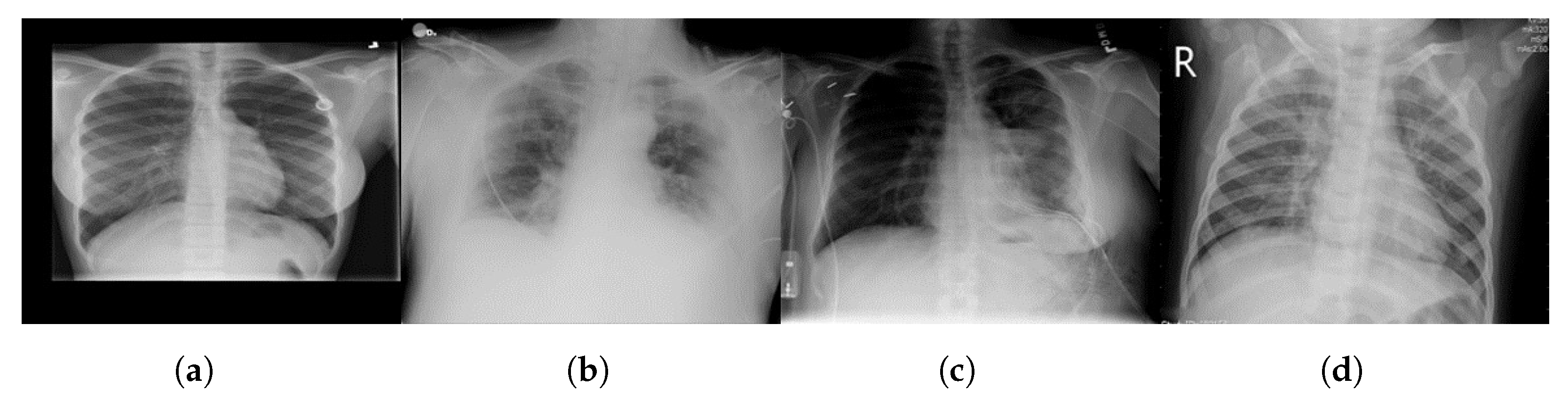
3.1.1. Lung CT Scan Dataset [81]
3.1.2. Chest X-ray (COVID-19) Dataset [82]
3.1.3. Ground-Truth Annotations
3.2. Dataset Preprocessing
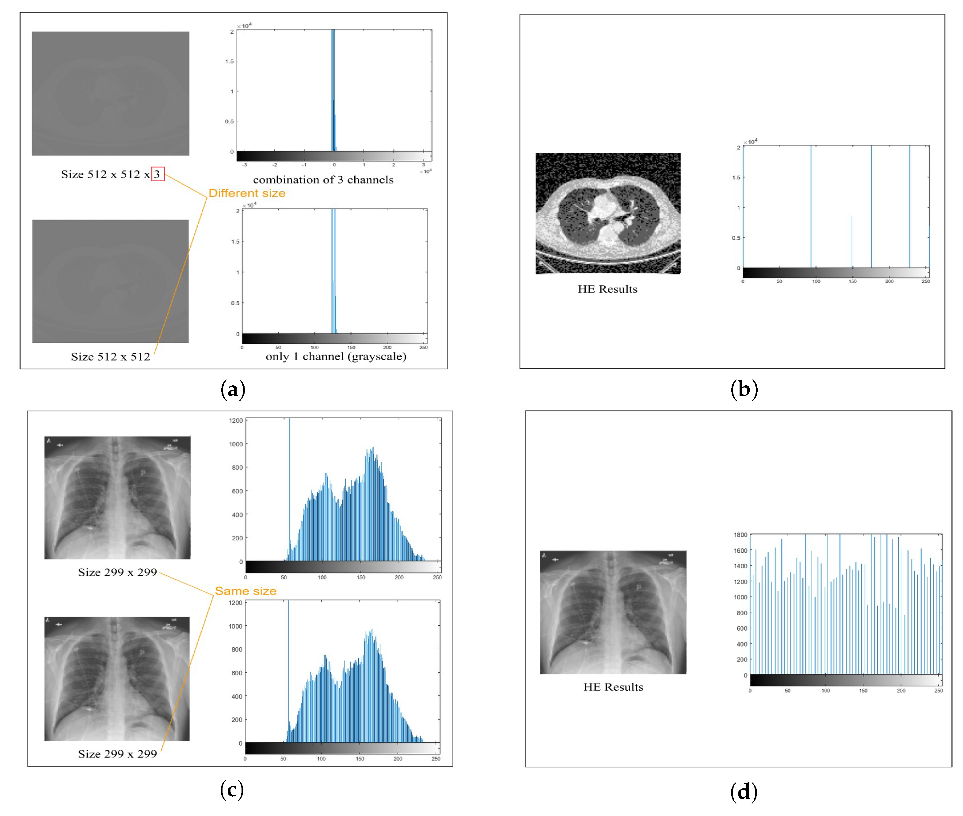
3.2.1. Image Conversion to 8-bit
3.2.2. Grayscaling
3.2.3. Image Enhancement with Histogram Equalization (HE)
3.2.4. Image Adjustment
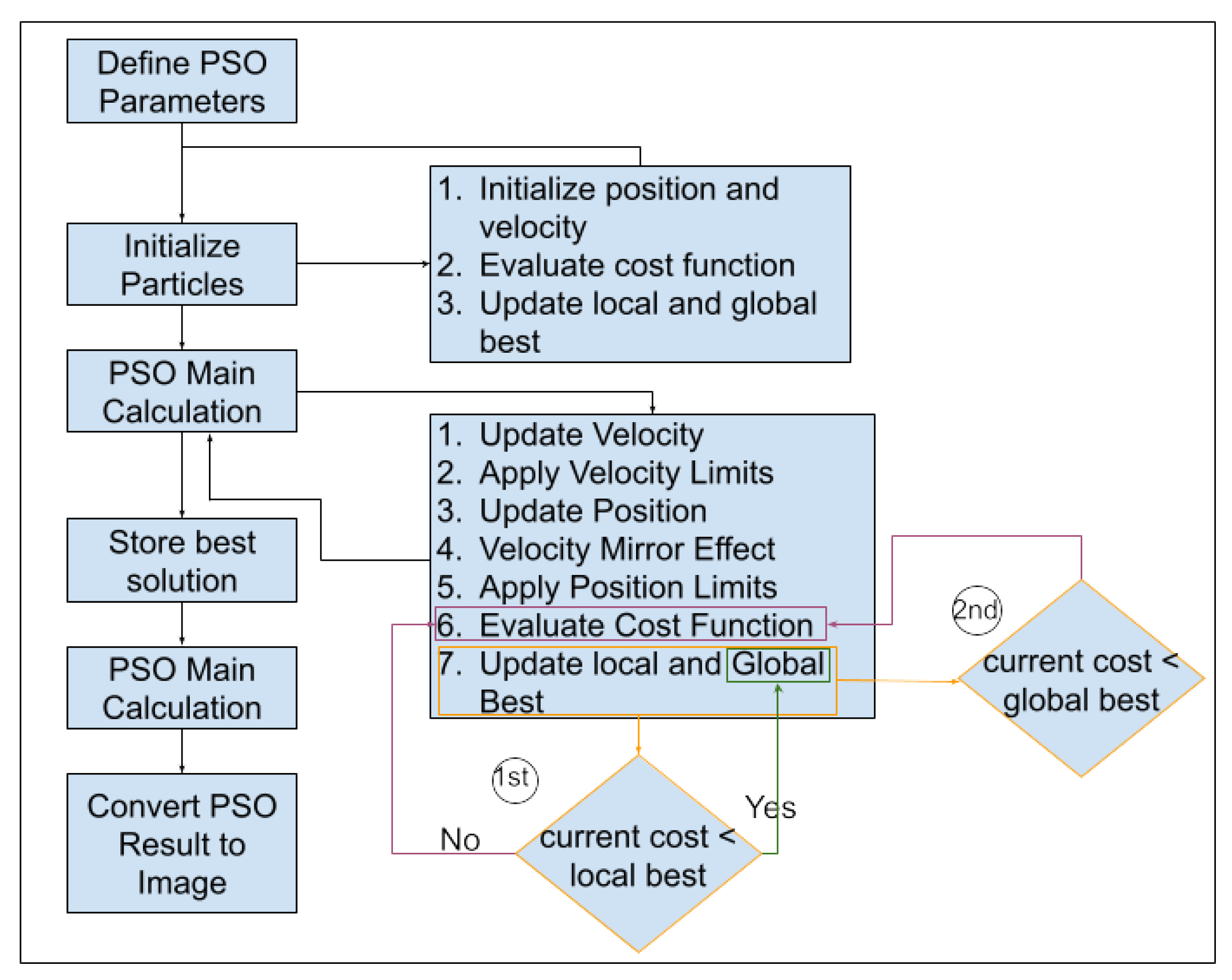
3.3. Particle Swarm Optimization (PSO)
- (a)
- Particle Initialization: We initialize a swarm of particles, wherein each particle represents a potential segmentation solution. The particles are assigned positions in the search space corresponding to potential image partitions. In the context of medical image segmentation, each partition represents a potential delineation between regions of interest within the image.
- (b)
- Objective Function: We define an objective function (Equation (1)) that quantifies the quality of a given image partition. The objective function considers factors such as intensity, gradient information, and region connectivity. The goal is to find the partition that minimizes this function—effectively identifying the optimal image segmentation.
- Intensity Component: The intensity term () gauges the distribution and variation of pixel intensities within the image partition. Higher values amplify the emphasis on the intensity, making it a pivotal factor for distinguishing regions based on pixel brightness. This ensures that the segmentation process prioritizes areas with distinct intensity characteristics.
- Gradient Information Component: The gradient information term ( Gradient(X)) delves into the spatial variations in pixel intensities, emphasizing edges and boundaries. A higher amplifies the influence of gradient information in the objective function, steering the segmentation towards regions with pronounced transitions in pixel values. This is crucial for preserving fine details and outlining distinct structures within the image.
- Region Connectivity Component: The region connectivity term ( Connectivity(X)) assesses the coherence and connectivity between adjacent pixels within the partition. Increasing intensifies the significance of region connectivity in the overall objective function, guiding the segmentation process towards creating contiguous and meaningful partitions. This ensures that segmented regions exhibit a natural flow and connectivity, enhancing the overall structural integrity of the partition.
The terms , , and are weights assigned to each measure to adjust their influence on the overall objective function. The optimization goal is to find the optimal partition by minimizing . - (c)
- Optimization Iterations: The PSO algorithm iteratively updates the positions of particles based on their previous best positions and the best positions found by neighboring particles. Particles adjust their positions in search of the optimal image partition. The optimization continues until convergence criteria are met or a specified number of iterations is reached. Each particle keeps track of two pivotal fitness values: “local best (pbest)” (Equation (2)) and “global best (gbest)” (Equation (3)) [95]. The term “pbest” signifies the best value achieved by an individual particle throughout the optimization process and is recalculated iteratively at each time step. In contrast, “global best” represents the overarching best value attained among all particles’ “pbest” values up to that specific time step.In optimization [98], i represents the particle’s index (1 to N), t is the current iteration, f is the objective function to be minimized, x is the position vector, and N is the total number of particles in the swarm.
- (d)
- Optimal Partition Extraction: The final result of PSO is the image partition that minimizes the objective function. This partition represents segmentation of the input medical image into distinct regions of interest. The partition is chosen based on the collective behavior of particles, which adapt and explore the search space to find the best segmentation. Particle positions and velocities evolve dynamically based on Equations (4) and (5).
3.4. Integrating PSO and HE for Image Segmentation
3.4.1. Algorithmic Integration and Parameter Dynamics
3.4.2. Rationale, Benefits, and Experimental Rigor
3.4.3. Applications and Considerations
4. Results and Discussion
4.1. Experimental Setup
4.1.1. Preprocessing Parameters
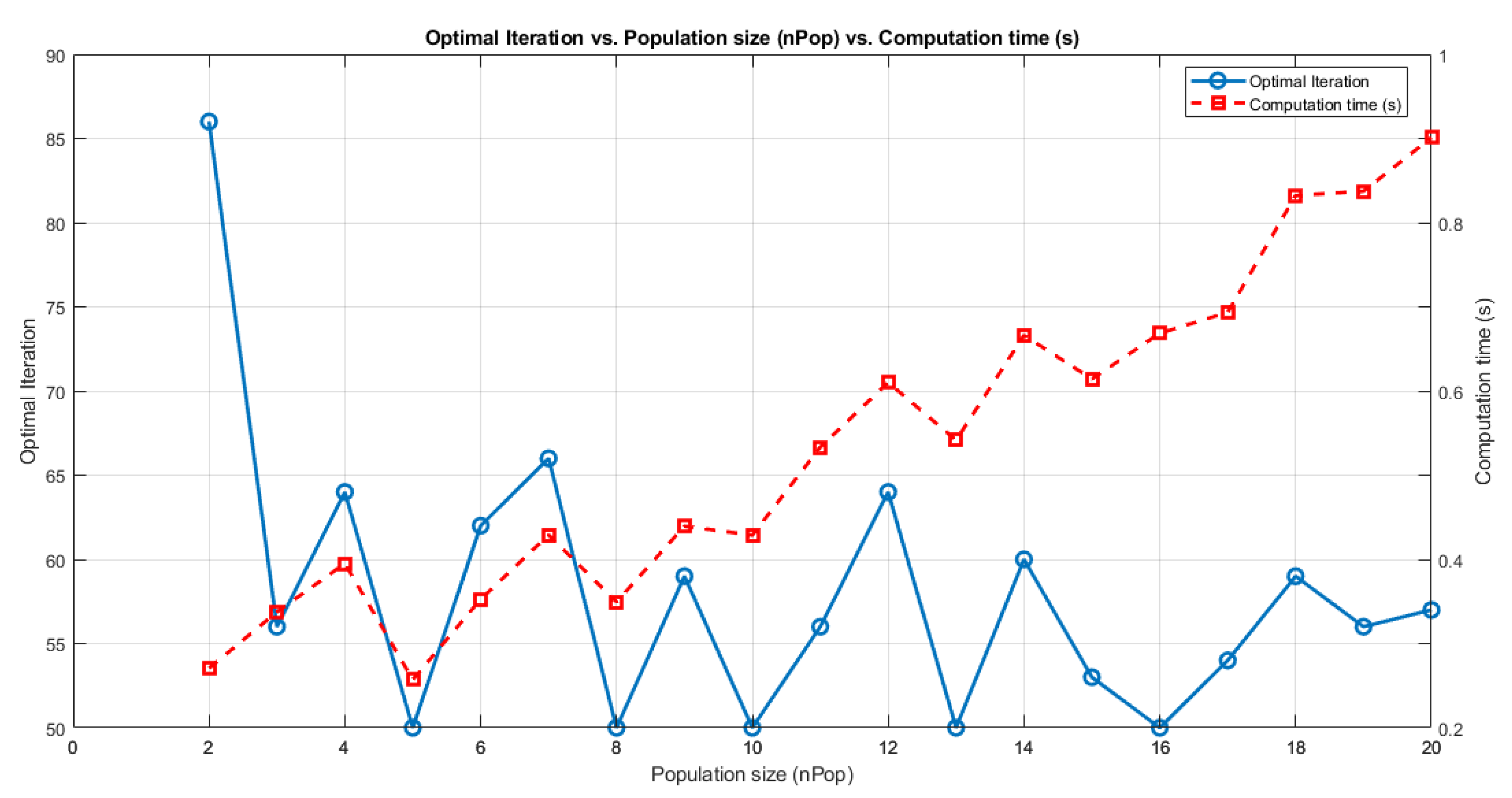
4.1.2. PSO Segmentation Parameters
4.1.3. Evaluation Metrics
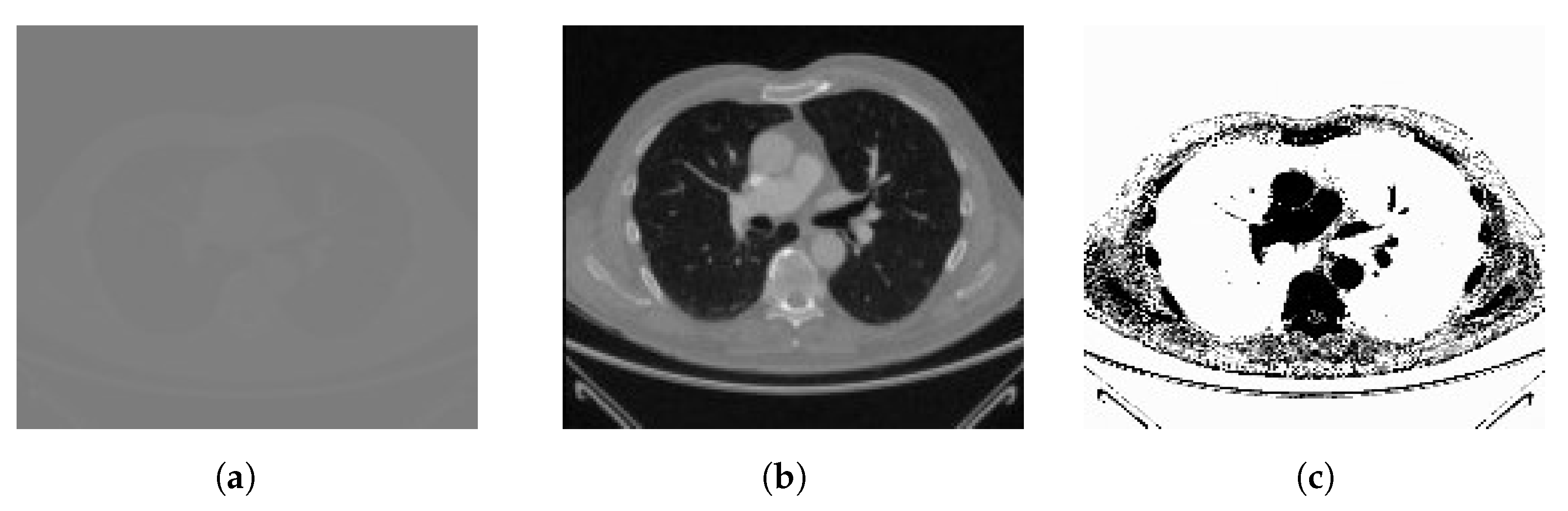
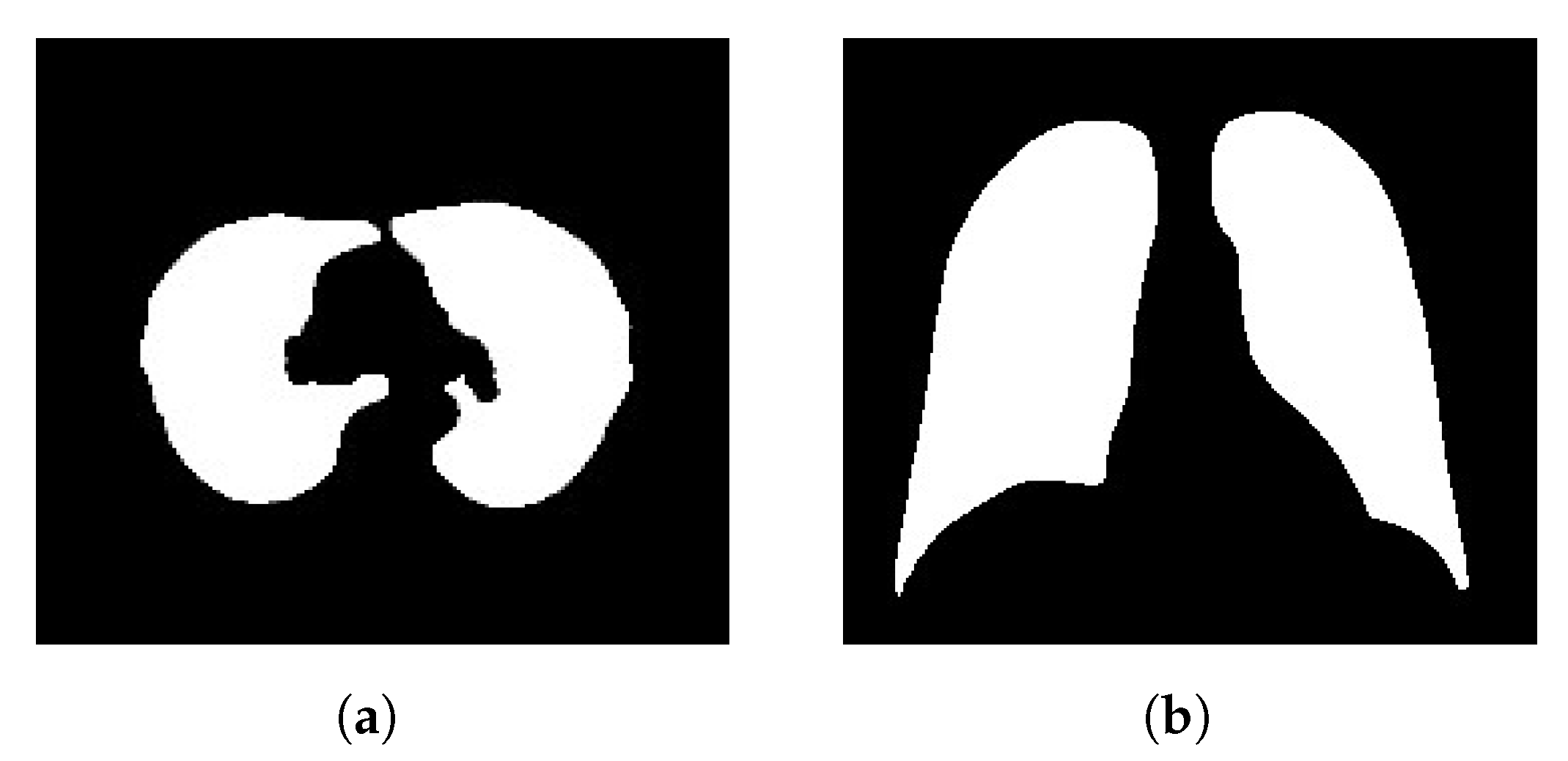
4.2. Segmentation Performance
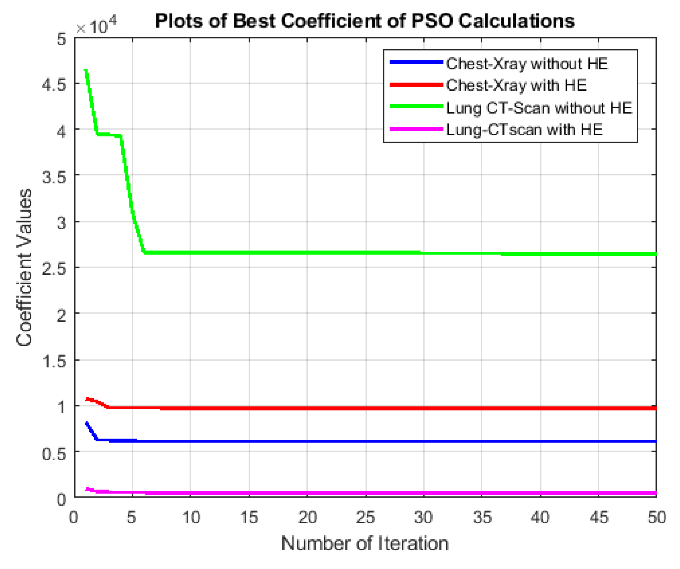
4.2.1. Analysis of Best-Cost Values
- Chest X-ray SegmentationWithout HE, the best-cost values exhibit fluctuations within a relatively narrow range. This indicates that the PSO algorithm converges effectively to a stable solution. The consistency of best-cost values can be attributed to the nature of chest X-ray images, which already possess a certain level of contrast and a grayscale distribution suitable for segmentation. When HE is applied, the best-cost values slightly increase. This is likely due to HE redistribution of pixel intensities and emphasizing the overall contrast, which can create additional complexity in the segmentation process. However, despite the slight increase, the best-cost values remain relatively stable.
- Lung CT Scan SegmentationIn the case of lung CT scan images, the best-cost values demonstrate more significant variations. Without HE, the algorithm exhibits periodic dips and peaks. The complexity in these images, characterized by intricate anatomical structures like lung parenchyma, blood vessels, and airways, contributes to the periodic fluctuations. These variations might indicate the algorithm’s challenges in settling on a precise segmentation solution. With HE preprocessing, the best-cost values exhibit a notable reduction in fluctuations. The redistribution of pixel intensities through HE improves the overall contrast, resulting in a smoother convergence pattern. It is important to note that in this context, a lower best-cost value reflects a more accurate segmentation.The comparison between chest X-ray and lung CT scan datasets highlights the significance of dataset characteristics in influencing the convergence behavior of the PSO algorithm. Moreover, the impact of HE on the optimization process is more pronounced in lung CT scan images, which emphasizes the need for preprocessing methods tailored to the dataset’s characteristics.
4.2.2. Analysis of Evaluation Metrics
4.3. Segmentation Results Based on Object Cropping
4.3.1. Quality Enhancement with Preprocessing
4.3.2. Comparison with Other Segmentation Methods
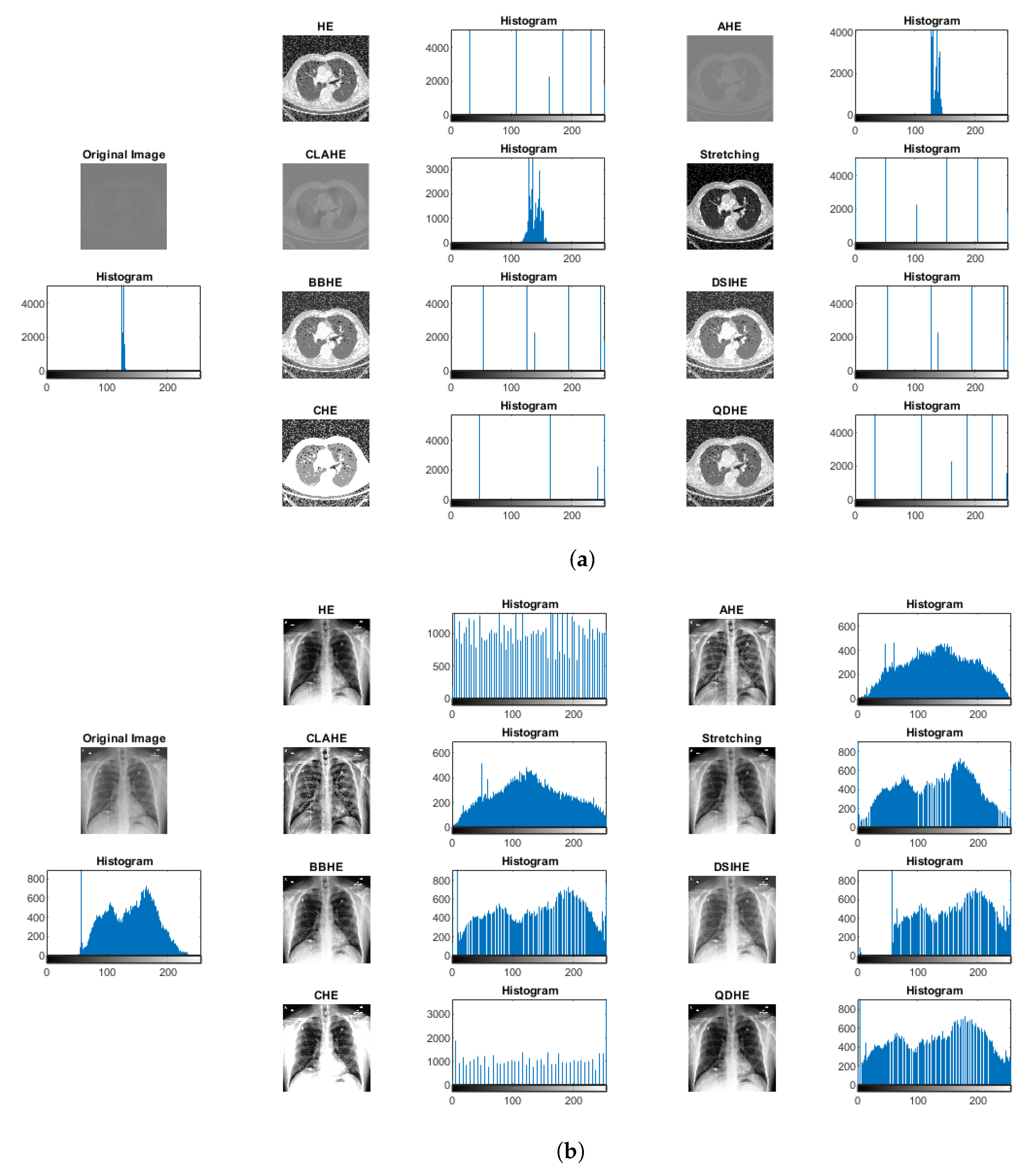
4.3.3. Comparison of Our Method with Other Image Preprocessing Approaches Based on Histograms
4.4. Limitations
Collaborative Adaptability for Overlapping Structures
5. Conclusions
Author Contributions
Funding
Institutional Review Board Statement
Informed Consent Statement
Data Availability Statement
Conflicts of Interest
References
- Karagiannis, S.; Magkos, E.; Ntantogian, C.; Cabecinha, R.; Fotis, T. Cybersecurity and Medical Imaging: A Simulation-Based Approach to DICOM Communication. Appl. Sci. 2023, 13, 10072. [Google Scholar] [CrossRef]
- Waili, A.R.A. Using Convolutional Neural Networks for Edge Detection in Medical Images to Determine Surgery Instrument Tools. J. Artif. Intell. Mach. Learn. Neural Netw. 2023, 3, 13–25. [Google Scholar] [CrossRef]
- Hao, X.; Yin, L.; Li, X.; Zhang, L.; Yang, R. A Multi-Objective Semantic Segmentation Algorithm Based on Improved U-Net Networks. Remote Sens. 2023, 15, 1838. [Google Scholar] [CrossRef]
- Ansari, M.Y.; Abdalla, A.; Ansari, M.Y.; Ansari, M.I.; Malluhi, B.; Mohanty, S.; Mishra, S.; Singh, S.S.; Abinahed, J.; Al-Ansari, A.; et al. Practical utility of liver segmentation methods in clinical surgeries and interventions. BMC Med. Imaging 2022, 22, 97. [Google Scholar] [CrossRef]
- Urban, R.; Haluzová, S.; Strunga, M.; Surovková, J.; Lifková, M.; Tomášik, J.; Thurzo, A. AI-Assisted CBCT Data Management in Modern Dental Practice: Benefits, Limitations and Innovations. Electronics 2023, 12, 1710. [Google Scholar] [CrossRef]
- Asiri, A.F.; Altuwalah, A.S. The role of neural artificial intelligence for diagnosis and treatment planning in endodontics: A qualitative review. Saudi Dent. J. 2022, 34, 270–281. [Google Scholar] [CrossRef]
- Vobugari, N.; Raja, V.; Sethi, U.; Gandhi, K.; Raja, K.; Surani, S.R. Advancements in Oncology with Artificial Intelligence—A Review Article. Cancers 2022, 14, 1349. [Google Scholar] [CrossRef]
- KV, R.; Prasad, K.; Peralam Yegneswaran, P. Segmentation and Classification Approaches of Clinically Relevant Curvilinear Structures: A Review. J. Med. Syst. 2023, 47, 40. [Google Scholar] [CrossRef]
- Iqbal, S.; Khan, T.M.; Naveed, K.; Naqvi, S.S.; Nawaz, S.J. Recent trends and advances in fundus image analysis: A review. Comput. Biol. Med. 2022, 151, 106277. [Google Scholar] [CrossRef]
- Wu, R.; Liang, C.; Li, Y.; Shi, X.; Zhang, J.; Huang, H. Self-supervised transfer learning framework driven by visual attention for benign–malignant lung nodule classification on chest CT. Expert Syst. Appl. 2023, 215, 119339. [Google Scholar] [CrossRef]
- Kaur, C.; Garg, U. Artificial intelligence techniques for cancer detection in medical image processing: A review. Mater. Today Proc. 2023, 81, 806–809. [Google Scholar] [CrossRef]
- Liu, F.; Chen, L.; Lu, L.; Ahmad, A.; Jeon, G.; Yang, X. Medical image fusion method by using Laplacian pyramid and convolutional sparse representation. Concurr. Comput. Pract. Exp. 2020, 32, e5632. [Google Scholar] [CrossRef]
- Junn, J.C.; Soderlund, K.A.; Glastonbury, C.M. Imaging of Head and Neck Cancer With CT, MRI, and US. Semin. Nucl. Med. 2021, 51, 3–12. [Google Scholar] [CrossRef] [PubMed]
- Dara, O.A.; Lopez-Guede, J.M.; Raheem, H.I.; Rahebi, J.; Zulueta, E.; Fernandez-Gamiz, U. Alzheimer’s Disease Diagnosis Using Machine Learning: A Survey. Appl. Sci. 2023, 13, 8298. [Google Scholar] [CrossRef]
- Chakraborty, S.; Chatterjee, S.; Das, A.; Mali, K. Penalized Fuzzy C-Means Enabled Hybrid Region Growing in Segmenting Medical Images. Stud. Comput. Intell. 2020, 841, 41–65. [Google Scholar] [CrossRef]
- Ramesh, K.; Kumar, G.; Swapna, K.; Datta, D.; Rajest, S. A Review of Medical Image Segmentation Algorithms. EAI Endorsed Trans. Pervasive Health Technol. 2021, 7, 169184. [Google Scholar] [CrossRef]
- Yang, C.; Weng, G.; Chen, Y. Active contour model based on local Kullback–Leibler divergence for fast image segmentation. Eng. Appl. Artif. Intell. 2023, 123, 106472. [Google Scholar] [CrossRef]
- Oulefki, A.; Agaian, S.; Trongtirakul, T.; Kassah Laouar, A. Automatic COVID-19 lung infected region segmentation and measurement using CT-scans images. Pattern Recognit. 2021, 114, 107747. [Google Scholar] [CrossRef]
- Shaikh, F.; Dehmeshki, J.; Bisdas, S.; Roettger-Dupont, D.; Kubassova, O.; Aziz, M.; Awan, O. Artificial Intelligence-Based Clinical Decision Support Systems Using Advanced Medical Imaging and Radiomics. Curr. Probl. Diagn. Radiol. 2021, 50, 262–267. [Google Scholar] [CrossRef]
- Mall, P.K.; Singh, P.K.; Srivastav, S.; Narayan, V.; Paprzycki, M.; Jaworska, T.; Ganzha, M. A comprehensive review of deep neural networks for medical image processing: Recent developments and future opportunities. Healthc. Anal. 2023, 4, 100216. [Google Scholar] [CrossRef]
- Faragallah, O.S.; El-Hoseny, H.; El-Shafai, W.; El-Rahman, W.A.; El-Sayed, H.S.; El-Rabaie, E.S.M.; El-Samie, F.E.A.; Geweid, G.G.N. A Comprehensive Survey Analysis for Present Solutions of Medical Image Fusion and Future Directions. IEEE Access 2021, 9, 11358–11371. [Google Scholar] [CrossRef]
- Shang, H.; Zhao, S.; Du, H.; Zhang, J.; Xing, W.; Shen, H. A new solution model for cardiac medical image segmentation. J. Thorac. Dis. 2020, 12, 7298–7312. [Google Scholar] [CrossRef] [PubMed]
- Saifullah, S.; Dreżewski, R. Enhanced Medical Image Segmentation using CNN based on Histogram Equalization. In Proceedings of the 2023 2nd International Conference on Applied Artificial Intelligence and Computing (ICAAIC), Salem, India, 4–6 May 2023; pp. 121–126. [Google Scholar] [CrossRef]
- Allioui, H.; Sadgal, M.; Elfazziki, A. Optimized control for medical image segmentation: Improved multi-agent systems agreements using Particle Swarm Optimization. J. Ambient Intell. Humaniz. Comput. 2021, 12, 8867–8885. [Google Scholar] [CrossRef]
- El-Khatib, S.; Skobtsov, Y.; Rodzin, S. Improved Particle Swarm Medical Image Segmentation Algorithm for Decision Making. Stud. Comput. Intell. 2020, 869, 437–442. [Google Scholar] [CrossRef]
- Eisham, Z.K.; Haque, M.M.; Rahman, M.S.; Nishat, M.M.; Faisal, F.; Islam, M.R. Chimp optimization algorithm in multilevel image thresholding and image clustering. Evol. Syst. 2023, 14, 605–648. [Google Scholar] [CrossRef]
- Vijh, S.; Sharma, S.; Gaurav, P. Brain Tumor Segmentation Using OTSU Embedded Adaptive Particle Swarm Optimization Method and Convolutional Neural Network. Lect. Notes Data Eng. Commun. Technol. 2020, 32, 171–194. [Google Scholar] [CrossRef]
- Shehanaz, S.; Daniel, E.; Guntur, S.R.; Satrasupalli, S. Optimum weighted multimodal medical image fusion using particle swarm optimization. Optik 2021, 231, 166413. [Google Scholar] [CrossRef]
- Saifullah, S.; Dreżewski, R. Modified Histogram Equalization for Improved CNN Medical Image Segmentation. Procedia Comput. Sci. 2023, 225, 3021–3030. [Google Scholar] [CrossRef]
- Lan, K.; Zhou, J.; Jiang, X.; Wang, J.; Huang, S.; Yang, J.; Song, Q.; Tang, R.; Gong, X.; Liu, K.; et al. Group theoretic particle swarm optimization for multi-level threshold lung cancer image segmentation. Quant. Imaging Med. Surg. 2023, 13, 1312–1322. [Google Scholar] [CrossRef]
- Naidu, S.; Quadros, A.; Natekar, A.; Parvatkar, P.; Chaman Kumar, K.; Aswale, S. Enhancement of X-ray images using various Image Processing Approaches. In Proceedings of the 2021 International Conference on Technological Advancements and Innovations (ICTAI), Tashkent, Uzbekistan, 10–12 November 2021; pp. 115–120. [Google Scholar] [CrossRef]
- Elyan, E.; Vuttipittayamongkol, P.; Johnston, P.; Martin, K.; McPherson, K.; Moreno-García, C.F.; Jayne, C.; Mostafa Kamal Sarker, M. Computer vision and machine learning for medical image analysis: Recent advances, challenges, and way forward. Artif. Intell. Surg. 2022, 2, 24–25. [Google Scholar] [CrossRef]
- Abualigah, L.; Habash, M.; Hanandeh, E.S.; Hussein, A.M.; Shinwan, M.A.; Zitar, R.A.; Jia, H. Improved Reptile Search Algorithm by Salp Swarm Algorithm for Medical Image Segmentation. J. Bionic Eng. 2023, 20, 1766–1790. [Google Scholar] [CrossRef] [PubMed]
- Khaniabadi, S.M.; Ibrahim, H.; Huqqani, I.A.; Khaniabadi, F.M.; Sakim, H.A.M.; Teoh, S.S. Comparative Review on Traditional and Deep Learning Methods for Medical Image Segmentation. In Proceedings of the 2023 IEEE 14th Control and System Graduate Research Colloquium (ICSGRC), Shah Alam, Malaysia, 5 August 2023; pp. 45–50. [Google Scholar] [CrossRef]
- Jardim, S.; António, J.; Mora, C. Image thresholding approaches for medical image segmentation - short literature review. Procedia Comput. Sci. 2023, 219, 1485–1492. [Google Scholar] [CrossRef]
- Feng, Y.; Liu, Y.; Liu, Z.; Liu, W.; Yao, Q.; Zhang, X. A Novel Interval Iterative Multi-Thresholding Algorithm Based on Hybrid Spatial Filter and Region Growing for Medical Brain MR Images. Appl. Sci. 2023, 13, 1087. [Google Scholar] [CrossRef]
- Xie, Y.; Zhang, Z.; Chen, S.; Qiu, C. Detect, Grow, Seg: A weakly supervision method for medical image segmentation based on bounding box. Biomed. Signal Process. Control 2023, 86, 105158. [Google Scholar] [CrossRef]
- Jaglan, P.; Dass, R.; Duhan, M. A Comparative Analysis of Various Image Segmentation Techniques. Lect. Notes Netw. Syst. 2019, 46, 359–374. [Google Scholar] [CrossRef]
- Sarhan, A.; Rokne, J.; Alhajj, R. Glaucoma detection using image processing techniques: A literature review. Comput. Med. Imaging Graph. 2019, 78, 101657. [Google Scholar] [CrossRef]
- Bennai, M.T.; Guessoum, Z.; Mazouzi, S.; Cormier, S.; Mezghiche, M. A stochastic multi-agent approach for medical-image segmentation: Application to tumor segmentation in brain MR images. Artif. Intell. Med. 2020, 110, 101980. [Google Scholar] [CrossRef]
- Biratu, E.S.; Schwenker, F.; Ayano, Y.M.; Debelee, T.G. A Survey of Brain Tumor Segmentation and Classification Algorithms. J. Imaging 2021, 7, 179. [Google Scholar] [CrossRef]
- Biratu, E.S.; Schwenker, F.; Debelee, T.G.; Kebede, S.R.; Negera, W.G.; Molla, H.T. Enhanced Region Growing for Brain Tumor MR Image Segmentation. J. Imaging 2021, 7, 22. [Google Scholar] [CrossRef]
- Naveen, H.M.; Naveena, C.; Manjunath Aradhya, V.N. An approach for classification of lung nodules. Tumor Discov. 2023, 2, 317. [Google Scholar] [CrossRef]
- Azouz, Z.; Honarvar Shakibaei Asli, B.; Khan, M. Evolution of Crack Analysis in Structures Using Image Processing Technique: A Review. Electronics 2023, 12, 3862. [Google Scholar] [CrossRef]
- Kheradmandi, N.; Mehranfar, V. A critical review and comparative study on image segmentation-based techniques for pavement crack detection. Constr. Build. Mater. 2022, 321, 126162. [Google Scholar] [CrossRef]
- Aldoury, R.S.; Al-Saidi, N.M.; Ibrahim, R.W.; Kahtan, H. A new X-ray images enhancement method using a class of fractional differential equation. MethodsX 2023, 11, 102264. [Google Scholar] [CrossRef] [PubMed]
- Pradeep Kumar, B.P.; Rangaiah, P.K.B.; Augustine, R. Enhancing Medical Image Reclamation for Chest Samples using B-Coefficients, DT-CWT and EPS Algorithm. IEEE Access 2023, 11, 113360–113375. [Google Scholar] [CrossRef]
- Zebari, D.A.; Zeebaree, D.Q.; Abdulazeez, A.M.; Haron, H.; Hamed, H.N.A. Improved Threshold Based and Trainable Fully Automated Segmentation for Breast Cancer Boundary and Pectoral Muscle in Mammogram Images. IEEE Access 2020, 8, 203097–203116. [Google Scholar] [CrossRef]
- Zhang, W.; Zhuang, P.; Sun, H.H.; Li, G.; Kwong, S.; Li, C. Underwater Image Enhancement via Minimal Color Loss and Locally Adaptive Contrast Enhancement. IEEE Trans. Image Process. 2022, 31, 3997–4010. [Google Scholar] [CrossRef] [PubMed]
- Majeed, S.H.; Isa, N.A.M. Adaptive Entropy Index Histogram Equalization for Poor Contrast Images. IEEE Access 2021, 9, 6402–6437. [Google Scholar] [CrossRef]
- Fan, X.; Sun, Z.; Tian, E.; Yin, Z.; Cao, G. Medical image contrast enhancement based on improved sparrow search algorithm. Int. J. Imaging Syst. Technol. 2023, 33, 389–402. [Google Scholar] [CrossRef]
- Jawdekar, A.; Dixit, M. A.; Dixit, M. A Review of Image Enhancement Techniques in Medical Imaging. In Machine Intelligence and Smart Systems. Algorithms for Intelligent Systems; Agrawal, S., Kumar Gupta, K., H. Chan, J., Agrawal, J., Gupta, M., Eds.; Springer: Singapore, 2021; pp. 25–33. [Google Scholar] [CrossRef]
- Islam, S.M.; Mondal, H.S. Image Enhancement Based Medical Image Analysis. In Proceedings of the 2019 10th International Conference on Computing, Communication and Networking Technologies (ICCCNT), Kanpur, India, 6–8 July 2019; pp. 1–5. [Google Scholar] [CrossRef]
- Wang, S.; Cao, G.; Wang, Y.; Liao, S.; Wang, Q.; Shi, J.; Li, C.; Shen, D. Review and Prospect: Artificial Intelligence in Advanced Medical Imaging. Front. Radiol. 2021, 1, 781868. [Google Scholar] [CrossRef]
- Farshi, T.R.; Drake, J.H.; Özcan, E. A multimodal particle swarm optimization-based approach for image segmentation. Expert Syst. Appl. 2020, 149, 113233. [Google Scholar] [CrossRef]
- Shi, M.; Chen, C.; Liu, L.; Kuang, F.; Zhao, D.; Chen, X. A grade-based search adaptive random slime mould optimizer for lupus nephritis image segmentation. Comput. Biol. Med. 2023, 160, 106950. [Google Scholar] [CrossRef] [PubMed]
- Khosla, T.; Verma, O.P. Optimal threshold selection for segmentation of Chest X-Ray images using opposition-based swarm-inspired algorithm for diagnosis of pneumonia. Multimed. Tools Appl. 2023. [Google Scholar] [CrossRef]
- Lakshman Narayana, V.; Lakshmi Patibandla, R.S.M.; Pavani, V.; Radhika, P. Optimized Nature-Inspired Computing Algorithms for Lung Disorder Detection. Stud. Comput. Intell. 2023, 1066, 103–118. [Google Scholar] [CrossRef]
- de Albuquerque, V.H.C.; Gupta, D.; De Falco, I.; Sannino, G.; Bouguila, N. Special issue on Bio-inspired optimization techniques for Biomedical Data Analysis: Methods and applications. Appl. Soft Comput. 2020, 95, 106672. [Google Scholar] [CrossRef]
- Shehab, M.; Abualigah, L.; Al Hamad, H.; Alabool, H.; Alshinwan, M.; Khasawneh, A.M. Moth–flame optimization algorithm: Variants and applications. Neural Comput. Appl. 2020, 32, 9859–9884. [Google Scholar] [CrossRef]
- Yu, Y.; Wang, C.; Fu, Q.; Kou, R.; Huang, F.; Yang, B.; Yang, T.; Gao, M. Techniques and Challenges of Image Segmentation: A Review. Electronics 2023, 12, 1199. [Google Scholar] [CrossRef]
- Zhang, L.; Lim, C.P. Intelligent optic disc segmentation using improved particle swarm optimization and evolving ensemble models. Appl. Soft Comput. 2020, 92, 106328. [Google Scholar] [CrossRef]
- Mandave, D.D.; Patil, L.V. Bio-inspired computing algorithms in dementia diagnosis—A application-oriented review. Results Control Optim. 2023, 12, 100276. [Google Scholar] [CrossRef]
- Nayak, J.; Swapnarekha, H.; Naik, B.; Dhiman, G.; Vimal, S. 25 Years of Particle Swarm Optimization: Flourishing Voyage of Two Decades. Arch. Comput. Methods Eng. 2023, 30, 1663–1725. [Google Scholar] [CrossRef]
- Dhal, K.G.; Ray, S.; Das, A.; Das, S. A Survey on Nature-Inspired Optimization Algorithms and Their Application in Image Enhancement Domain. Arch. Comput. Methods Eng. 2019, 26, 1607–1638. [Google Scholar] [CrossRef]
- Kavitha, A.; Chellamuthu, C. Brain tumour detection using self-adaptive learning PSO-based feature selection algorithm in MRI images. Int. J. Bus. Intell. Data Min. 2019, 15, 71. [Google Scholar] [CrossRef]
- Sarvamangala, D.R.; Kulkarni, R.V. A Comparative Study of Bio-inspired Algorithms for Medical Image Registration. Stud. Comput. Intell. 2019, 687, 27–44. [Google Scholar] [CrossRef]
- Kate, V.; Shukla, P. Image Segmentation of Breast Cancer Histopathology Images Using PSO-Based Clustering Technique. Lect. Notes Netw. Syst. 2020, 100, 207–216. [Google Scholar] [CrossRef]
- Zhao, Y.; Yu, X.; Wu, H.; Zhou, Y.; Sun, X.; Yu, S.; Yu, S.; Liu, H. A Fast 2-D Otsu lung tissue image segmentation algorithm based on improved PSO. Microprocess. Microsyst. 2021, 80, 103527. [Google Scholar] [CrossRef]
- Chakraborty, R.; Sushil, R.; Garg, M.L. An Improved PSO-Based Multilevel Image Segmentation Technique Using Minimum Cross-Entropy Thresholding. Arab. J. Sci. Eng. 2019, 44, 3005–3020. [Google Scholar] [CrossRef]
- Öztürk, c.; Ahmad, R.; Akhtar, N. Variants of Artificial Bee Colony algorithm and its applications in medical image processing. Appl. Soft Comput. 2020, 97, 106799. [Google Scholar] [CrossRef]
- Guo, J.; Ma, J.; García-Fernández, Á.F.; Zhang, Y.; Liang, H. A survey on image enhancement for Low-light images. Heliyon 2023, 9, e14558. [Google Scholar] [CrossRef]
- Liu, L.; Zhao, D.; Yu, F.; Heidari, A.A.; Ru, J.; Chen, H.; Mafarja, M.; Turabieh, H.; Pan, Z. Performance optimization of differential evolution with slime mould algorithm for multilevel breast cancer image segmentation. Comput. Biol. Med. 2021, 138, 104910. [Google Scholar] [CrossRef]
- Alnazer, I.; Bourdon, P.; Urruty, T.; Falou, O.; Khalil, M.; Shahin, A.; Fernandez-Maloigne, C. Recent advances in medical image processing for the evaluation of chronic kidney disease. Med. Image Anal. 2021, 69, 101960. [Google Scholar] [CrossRef]
- Saini, M.; Susan, S. Tackling class imbalance in computer vision: A contemporary review. Artif. Intell. Rev. 2023, 56, 1279–1335. [Google Scholar] [CrossRef]
- Huang, Q.; Ding, H.; Razmjooy, N. Oral cancer detection using convolutional neural network optimized by combined seagull optimization algorithm. Biomed. Signal Process. Control 2024, 87, 105546. [Google Scholar] [CrossRef]
- Karsa, A.; Punwani, S.; Shmueli, K. An optimized and highly repeatable MRI acquisition and processing pipeline for quantitative susceptibility mapping in the head-and-neck region. Magn. Reson. Med. 2020, 84, 3206–3222. [Google Scholar] [CrossRef]
- Hadjiiski, L.; Cha, K.; Chan, H.; Drukker, K.; Morra, L.; Näppi, J.J.; Sahiner, B.; Yoshida, H.; Chen, Q.; Deserno, T.M.; et al. AAPM task group report 273: Recommendations on best practices for AI and machine learning for computer-aided diagnosis in medical imaging. Med. Phys. 2023, 50, e1–e24. [Google Scholar] [CrossRef] [PubMed]
- Maier-Hein, L.; Eisenmann, M.; Sarikaya, D.; März, K.; Collins, T.; Malpani, A.; Fallert, J.; Feussner, H.; Giannarou, S.; Mascagni, P.; et al. Surgical data science—From concepts toward clinical translation. Med. Image Anal. 2022, 76, 102306. [Google Scholar] [CrossRef] [PubMed]
- Rehman, M.U.; Akhtar, S.; Zakwan, M.; Mahmood, M.H. Novel architecture with selected feature vector for effective classification of mitotic and non-mitotic cells in breast cancer histology images. Biomed. Signal Process. Control 2022, 71, 103212. [Google Scholar] [CrossRef]
- Scott Mader, K. Finding and Measuring Lungs in CT Data. 2017. Available online: https://www.kaggle.com/datasets/kmader/finding-lungs-in-ct-data (accessed on 23 June 2023).
- Rahman, T.; Chowdhury, M.; Khandakar, A. COVID-19 Radiography Database. 2022. Available online: https://www.kaggle.com/datasets/tawsifurrahman/covid19-radiography-database (accessed on 23 June 2023).
- Saifullah, S.; Yuwono, B.; Rustamaji, H.C.; Saputra, B.; Dwiyanto, F.A.; Dreżewski, R. Detection of Chest X-ray Abnormalities Using CNN Based on Hyperparameter Optimization. Eng. Proc. 2023, 56, 223. [Google Scholar] [CrossRef]
- Song, Y.; Ren, S.; Lu, Y.; Fu, X.; Wong, K.K. Deep learning-based automatic segmentation of images in cardiac radiography: A promising challenge. Comput. Methods Programs Biomed. 2022, 220, 106821. [Google Scholar] [CrossRef] [PubMed]
- Saifullah, S.; Drezewski, R.; Khaliduzzaman, A.; Tolentino, L.K.; Ilyos, R. K-Means Segmentation Based-on Lab Color Space for Embryo Detection in Incubated Egg. J. Ilm. Tek. Elektro Komput. Dan Inform. 2022, 8, 175–185. [Google Scholar] [CrossRef]
- Rehman, M.U.; Ryu, J.; Nizami, I.F.; Chong, K.T. RAAGR2-Net: A brain tumor segmentation network using parallel processing of multiple spatial frames. Comput. Biol. Med. 2023, 152, 106426. [Google Scholar] [CrossRef]
- Masoudi, S.; Harmon, S.A.; Mehralivand, S.; Walker, S.M.; Raviprakash, H.; Bagci, U.; Choyke, P.L.; Turkbey, B. Quick guide on radiology image pre-processing for deep learning applications in prostate cancer research. J. Med. Imaging 2021, 8, 010901. [Google Scholar] [CrossRef]
- Aumann, S.; Donner, S.; Fischer, J.; Müller, F. Optical Coherence Tomography (OCT): Principle and Technical Realization. In High Resolution Imaging in Microscopy and Ophthalmology; Springer: Cham, Switzerland, 2019; pp. 59–85. [Google Scholar] [CrossRef]
- El-Shafai, W.; Almomani, I.; Ara, A.; Alkhayer, A. An optical-based encryption and authentication algorithm for color and grayscale medical images. Multimed. Tools Appl. 2023, 82, 23735–23770. [Google Scholar] [CrossRef]
- Hoque, M.Z.; Keskinarkaus, A.; Nyberg, P.; Seppänen, T. Stain normalization methods for histopathology image analysis: A comprehensive review and experimental comparison. Inf. Fusion 2024, 102, 101997. [Google Scholar] [CrossRef]
- Nazir, N.; Sarwar, A.; Saini, B.S.; Shams, R. A Robust Deep Learning Approach for Accurate Segmentation of Cytoplasm and Nucleus in Noisy Pap Smear Images. Computation 2023, 11, 195. [Google Scholar] [CrossRef]
- Saifullah, S.; Drezewski, R.; Yudhana, A.; Pranolo, A.; Kaswijanti, W.; Suryotomo, A.P.; Putra, S.A.; Khaliduzzaman, A.; Prabuwono, A.S.; Japkowicz, N. Nondestructive chicken egg fertility detection using CNN-transfer learning algorithms. J. Ilm. Tek. Elektro Komput. Dan Inform. (JITEKI) 2023, 9, 854–871. [Google Scholar]
- Saifullah, S.; Dreżewski, R. Non-Destructive Egg Fertility Detection in Incubation Using SVM Classifier Based on GLCM Parameters. Procedia Comput. Sci. 2022, 207, 3254–3263. [Google Scholar] [CrossRef]
- Okwu, M.O.; Tartibu, L.K. Particle Swarm Optimisation. In Metaheuristic Optimization: Nature-Inspired Algorithms Swarm and Computational Intelligence, Theory and Applications; Springer: Cham, Switzerland, 2021; Volume 927, pp. 5–13. [Google Scholar] [CrossRef]
- Gad, A.G. Particle Swarm Optimization Algorithm and Its Applications: A Systematic Review. Arch. Comput. Methods Eng. 2022, 29, 2531–2561. [Google Scholar] [CrossRef]
- Djemame, S.; Batouche, M.; Oulhadj, H.; Siarry, P. Solving reverse emergence with quantum PSO application to image processing. Soft Comput. 2019, 23, 6921–6935. [Google Scholar] [CrossRef]
- Narayan, V.; Faiz, M.; Mall, P.K.; Srivastava, S. A Comprehensive Review of Various Approach for Medical Image Segmentation and Disease Prediction. Wirel. Pers. Commun. 2023, 132, 1819–1848. [Google Scholar] [CrossRef]
- He, M.; Liu, M.; Wang, R.; Jiang, X.; Liu, B.; Zhou, H. Particle swarm optimization with damping factor and cooperative mechanism. Appl. Soft Comput. 2019, 76, 45–52. [Google Scholar] [CrossRef]
- Papazoglou, G.; Biskas, P. Review and Comparison of Genetic Algorithm and Particle Swarm Optimization in the Optimal Power Flow Problem. Energies 2023, 16, 1152. [Google Scholar] [CrossRef]
- Piotrowski, A.P.; Napiorkowski, J.J.; Piotrowska, A.E. Population size in Particle Swarm Optimization. Swarm Evol. Comput. 2020, 58, 100718. [Google Scholar] [CrossRef]
- Chen, H.l.; Yang, B.; Wang, S.j.; Wang, G.; Liu, D.y.; Li, H.z.; Liu, W.b. Towards an optimal support vector machine classifier using a parallel particle swarm optimization strategy. Appl. Math. Comput. 2014, 239, 180–197. [Google Scholar] [CrossRef]
- Han, W.; Yang, P.; Ren, H.; Sun, J. Comparison study of several kinds of inertia weights for PSO. In Proceedings of the 2010 IEEE International Conference on Progress in Informatics and Computing, Shanghai, China, 10–12 December 2010. [Google Scholar] [CrossRef]
- Taha, A.A.; Hanbury, A. Metrics for evaluating 3D medical image segmentation: Analysis, selection, and tool. BMC Med. Imaging 2015, 15, 29. [Google Scholar] [CrossRef] [PubMed]


| Parameter | Description |
|---|---|
| k | Number of segments (set to 2 in experiments): determines image partitioning. |
| Objective function guiding PSO: minimizes image partitioning quality. | |
| Decision variable matrix size, : indicates the matrix for decision variables. | |
| Number of decision variables: calculated from and represents total variables. | |
| Lower and upper bounds of decision variables based on image data. | |
| Maximum iterations (50): controls optimization duration. | |
| Population size (5): aligned with PSO swarm size. | |
| w | Inertia weight (initially 1): balances particle velocity and best-known position. |
| Inertia weight damping (0.99): regulates inertia weight decrease. | |
| Personal and global learning coefficients (1.5 and 2.5): influence particle movement. | |
| Velocity limits: preserve particle velocities within bounds. |
| Parameters | Experiments | ||||||
|---|---|---|---|---|---|---|---|
| 1 | 2 | 3 | 4 | 5 | 6 | 7 | |
| Weight (w) | 1 | 0.9 | 0.8 | 0.7 | 0.6 | 0.5 | 0.4 |
| Best Cost | 93.3202 | 93.3203 | 93.3203 | 93.3204 | 93.3205 | 93.3984 | 93.3725 |
| Medical Images | HE | Evaluation Metrics | |||||
|---|---|---|---|---|---|---|---|
| Accuracy | Precision | Recall | Specificity | Dice | Jaccard | ||
| PSO Segmentation Approach | |||||||
| Lung CT | No | 0.91893 | 0.90036 | 0.97545 | 0.88041 | 0.9364 | 0.82984 |
| Lung CT | Yes | 0.9563 | 0.97529 | 0.96811 | 0.93322 | 0.967 | 0.9361 |
| Chest X-ray | No | 0.9065 | 0.9350 | 0.93162 | 0.87751 | 0.90202 | 0.82153 |
| Chest X-ray | Yes | 0.90363 | 0.86891 | 0.93714 | 0.87369 | 0.90173 | 0.82105 |
| Otsu’s Approach | |||||||
| Lung CT | No | 0.91323 | 0.9422 | 0.928 | 0.88283 | 0.93504 | 0.87801 |
| Lung CT | Yes | 0.91893 | 0.90036 | 0.97545 | 0.88041 | 0.9364 | 0.82984 |
| Chest X-ray | No | 0.91796 | 0.88379 | 0.95155 | 0.88786 | 0.91642 | 0.84573 |
| Chest X-ray | Yes | 0.90796 | 0.92473 | 0.89751 | 0.91947 | 0.91091 | 0.8364 |
| Watershed Approach | |||||||
| Lung CT | No | 0.87057 | 0.88893 | 0.91348 | 0.79259 | 0.90104 | 0.8199 |
| Lung CT | Yes | 0.9035 | 0.94483 | 0.91266 | 0.88346 | 0.92847 | 0.86649 |
| Chest X-ray | No | 0.89371 | 0.89279 | 0.89778 | 0.88954 | 0.89528 | 0.81041 |
| Chest X-ray | Yes | 0.88755 | 0.92746 | 0.86203 | 0.91842 | 0.89355 | 0.80758 |
| K-means Approach | |||||||
| Lung CT | No | 0.82512 | 0.8654 | 0.87008 | 0.73812 | 0.86773 | 0.76636 |
| Lung CT | Yes | 0.9132 | 0.94216 | 0.92799 | 0.88276 | 0.93502 | 0.87798 |
| Chest X-ray | No | 0.91754 | 0.88928 | 0.94544 | 0.84587 | 0.9165 | 0.89192 |
| Chest X-ray | Yes | 0.90794 | 0.92473 | 0.89749 | 0.83638 | 0.9109 | 0.91947 |
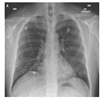
| Medical Images | Preprocessed | Ground Truth | Segmented | Cropped Comparison | ||
|---|---|---|---|---|---|---|
| Yes/No | Result | Result | Ground Truth | |||
 | Yes |  |  |  |  |  |
 | No |  |  |  |  |  |
 | Yes | 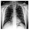 |  | 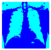 |  |  |
 | No | 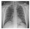 |  | 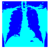 |  |  |
| No. | Ground Truth | HE | PSO Proposed | Comparison with Other Methods | ||
|---|---|---|---|---|---|---|
| Otsu | Watershed | K-Means | ||||
| 1 |  | Yes |  | 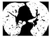 |  |  |
| 2 |  | No | 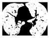 |  |  |  |
| 3 |  | Yes |  | 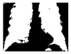 |  |  |
| 4 |  | No |  |  |  |  |
| Medical Image | Pre-Processing | Evaluation Metrics | |||||
|---|---|---|---|---|---|---|---|
| Accuracy | Precision | Recall | Specificity | Dice | Jaccard | ||
| Proposed HE | 0.9563 | 0.97529 | 0.96811 | 0.93322 | 0.967 | 0.9361 | |
| HE | 0.94959 | 0.95632 | 0.90652 | 0.92529 | 0.93075 | 0.87048 | |
| CLAHE | 0.94785 | 0.95 | 0.96363 | 0.91195 | 0.95922 | 0.92164 | |
| AHE | 0.94905 | 0.94589 | 0.96446 | 0.90638 | 0.95996 | 0.92301 | |
| Lung | Stretching | 0.94959 | 0.94589 | 0.95529 | 0.90652 | 0.96037 | 0.92376 |
| CT | BBHE | 0.95553 | 0.95829 | 0.96244 | 0.92594 | 0.96531 | 0.93295 |
| DSIHE | 0.95572 | 0.95858 | 0.96245 | 0.92642 | 0.96547 | 0.93324 | |
| CHE | 0.88164 | 0.8371 | 0.91621 | 0.76431 | 0.90132 | 0.82036 | |
| QDHE | 0.92889 | 0.91219 | 0.93613 | 0.89228 | 0.94308 | 0.85702 | |
| Proposed HE | 0.9065 | 0.93504 | 0.93714 | 0.94691 | 0.90173 | 0.89648 | |
| HE | 0.90614 | 0.9342 | 0.86086 | 0.94627 | 0.89603 | 0.81165 | |
| CLAHE | 0.75458 | 0.69369 | 0.72702 | 0.77399 | 0.70996 | 0.55034 | |
| AHE | 0.82245 | 0.84244 | 0.7694 | 0.87027 | 0.80426 | 0.67261 | |
| Chest | Stretching | 0.88782 | 0.92061 | 0.80864 | 0.92351 | 0.88225 | 0.78931 |
| X-ray | BBHE | 0.88718 | 0.92101 | 0.80745 | 0.92381 | 0.88171 | 0.78844 |
| DSIHE | 0.97381 | 0.93413 | 0.85782 | 0.94512 | 0.89618 | 0.81189 | |
| CHE | 0.88837 | 0.92938 | 0.81013 | 0.92249 | 0.88263 | 0.78992 | |
| QDHE | 0.90619 | 0.9321 | 0.85905 | 0.94839 | 0.89638 | 0.81222 | |
Disclaimer/Publisher’s Note: The statements, opinions and data contained in all publications are solely those of the individual author(s) and contributor(s) and not of MDPI and/or the editor(s). MDPI and/or the editor(s) disclaim responsibility for any injury to people or property resulting from any ideas, methods, instructions or products referred to in the content. |
© 2024 by the authors. Licensee MDPI, Basel, Switzerland. This article is an open access article distributed under the terms and conditions of the Creative Commons Attribution (CC BY) license (https://creativecommons.org/licenses/by/4.0/).
Share and Cite
Saifullah, S.; Dreżewski, R. Advanced Medical Image Segmentation Enhancement: A Particle-Swarm-Optimization-Based Histogram Equalization Approach. Appl. Sci. 2024, 14, 923. https://doi.org/10.3390/app14020923
Saifullah S, Dreżewski R. Advanced Medical Image Segmentation Enhancement: A Particle-Swarm-Optimization-Based Histogram Equalization Approach. Applied Sciences. 2024; 14(2):923. https://doi.org/10.3390/app14020923
Chicago/Turabian StyleSaifullah, Shoffan, and Rafał Dreżewski. 2024. "Advanced Medical Image Segmentation Enhancement: A Particle-Swarm-Optimization-Based Histogram Equalization Approach" Applied Sciences 14, no. 2: 923. https://doi.org/10.3390/app14020923
APA StyleSaifullah, S., & Dreżewski, R. (2024). Advanced Medical Image Segmentation Enhancement: A Particle-Swarm-Optimization-Based Histogram Equalization Approach. Applied Sciences, 14(2), 923. https://doi.org/10.3390/app14020923