Article

Effects of Photobiomodulation Using Near-Infrared Light on the Dentin and Periodontal Ligament in a Beagle Model

1
Department of Biosystems & Biomaterials Science and Engineering, Seoul National University, Seoul 08826, Republic of Korea
2
Medical Engineering Research Center, The Standard Co., Ltd., Gunpo 15880, Republic of Korea
3
Department of Electrical and Biological Physics, Kwangwoon University, Seoul 01897, Republic of Korea
4
Department of Oral and Maxillofacial Surgery, Dental Research Institute, School of Dentistry, Seoul National University, Seoul 03080, Republic of Korea
*
Authors to whom correspondence should be addressed.
Appl. Sci. 2024, 14(2), 724; https://doi.org/10.3390/app14020724
Submission received: 17 December 2023 / Revised: 9 January 2024 / Accepted: 12 January 2024 / Published: 15 January 2024

Abstract

:
In this study, we investigated the effect of photobiomodulation (PBM) using near-infrared light on the dentin and periodontal ligament in a beagle model. We utilized a specific PBM device to irradiate NIR light with a wavelength of 810 nm and an energy density of 80.22 mJ/cm2. The device’s settings were optimized for a frequency of 300 Hz and a 30% duty cycle, allowing precise and controlled light exposure. Through a comprehensive analysis involving micro-computed tomography, scanning electron microscopy, and hematoxylin and eosin staining, we demonstrated increased odontoblast activity at the pulp–dentin interface in PBM-treated samples. This increased activity may be postulated to potentially contribute to alleviating dental hypersensitivity through the differentiation of dental pulp stem cells and the promotion of vascular development within the odontoblast layer. Moreover, our observations also indicated an improvement in the strength and integrity of fibrous connective tissue within the periodontal ligament. These findings highlight the potential of PBM with specific parameters applied using NIR as a valuable treatment method for tooth tissue regeneration. It shows particular promise in the treatment of dental diseases associated with dentin and periodontal ligament damage and offers a new perspective in the management of tooth hypersensitivity and other related dental diseases.

1. Introduction

Recent advances in dentistry have placed increasing emphasis on the transition from the use of conventional inert materials to regenerative technologies [1]. Common dental conditions, such as adult caries and pulp trauma, often require endodontic treatment, in which the damaged pulp is replaced with a composite material. However, these artificial substitutes are often insufficient to restore the full function of the tissue, mainly due to the lack of vascular and neural elements [2]. The rapidly growing field of regenerative medicine is exploring the potential of dental stem cells, known for their multifaceted differentiation capacity, including osteogenesis, dentinogenesis, adipogenesis, and chondrogenesis [3,4,5,6,7]. Biomaterials acting as scaffolds create a cellular microenvironment that attracts exogenous or endogenous stem cells, promoting innate healing mechanisms that lead to cell proliferation and tissue repair. Although introducing exogenously cultured stem cells using these biomaterials carries a risk of contamination, utilizing endogenous stem cells reduces immune response complications and avoids pitfalls associated with cell transplantation [8]. Nonetheless, achieving the necessary angiogenesis and minimizing off-target effects remains a significant challenge.
To address these challenges, our research focuses on photobiomodulation (PBM) using near-infrared (NIR), recognized for its tissue regenerative capabilities [9]. NIR light achieves deeper tissue penetration due to its unique properties of reduced scattering and absorption by water molecules, thereby affecting key biological processes such as stem cell proliferation and differentiation [10,11,12,13,14,15]. However, the direct impact of NIR-mediated PBM on dentin and fibrous connective tissue generation, especially through innate stem cell stimulation, has not yet been fully investigated. Resident stem cells are in specific niches in dental tissues. These niches are highly dynamic and composed of multiple cell types. A stem cell niche typically contains solid components, including cells and extracellular matrix (ECM), and soluble factors, such as growth factors [16]. Among the possible dental niches, we focused on the dental pulp niche with vascularization through the blood supply from the apical foramen, containing pulp stem cells that differentiate into odontoblasts and bioactive molecules that release to initiate cellular events, leading to the regeneration of pulp and dentin, including the fibrous connective tissue of periodontal ligaments [17]. Such stem cells can be physiologically activated by external cues (physical and chemical cues), and NIR PBM can be one of them.
To establish a fundamental understanding of these effects, our study initially focused on applying NIR light to normal, healthy dental tissue in a beagle in vivo model. This approach is an important step for establishing foundational knowledge before introducing the complexities associated with diseased tissue in the future. This systematic approach allows us to carefully evaluate the efficacy and safety of NIR PBM in a controlled environment before applying these findings to more complex scenarios involving diseased or damaged dental tissues. Therefore, the purpose of this study is to reveal the potential of NIR PBM in dental tissue regeneration, focusing on whether it can activate regeneration of dentin or periodontal ligament. We utilized the optimized PBM condition found in previous in vitro experiments and analyzed the changes in tissue structures using micro-computed tomography, hematoxylin and eosin staining, and scanning electron microscopy. This fundamental study can provide basic information for the use of PBM in dentistry.

2. Materials and Methods

2.1. Animal

This study was conducted on a male beagle weighing 12 kg. The experimental protocol was approved by the Animal Research Ethics Committee at Seoul National University Dental Hospital (Seoul, Korea; permit number SNU-120427-2-2). All procedures implemented complied with relevant guidelines and regulations. The beagle was maintained under standard conditions: temperature 22 ± 2 °C, relative humidity 55 ± 10%, and a 12 h alternating light–dark cycle. The animal was fed with ground commercial dog food, and water was provided ad libitum. All experimental procedures on the animal were conducted under general anesthesia via the intravenous injection of zolazepam (0.3 mL/kg, Zoletil 50, Virbac, Carros, France) together with 2% Rompun (0.15 cc/kg, xylazine HCL, Bayer, Berlin, Germany). To investigate the effect of PBM, we compared left and right premolar teeth. We assumed that the tissues on both sides would be almost identical, and we confirmed this by comparing the volume of each tooth at the same locations. The volume difference between the left and right premolar teeth was about 3.2 ± 4.6%. The premolar teeth at the upper and lower left were irradiated by NIR (PBM), and the un-treated premolar teeth at the upper and lower right were not treated (Control), as shown in Figure 1a.

2.2. Photobiomodulation (PBM)

The beagle’s teeth were exposed to NIR light from a 9-LED array. This array consisted of LEDs (MTE2081-OH5, Marktech Optoelectronics, Latham, NY, USA) with a central wavelength of 810 nm. The half-intensity beam angle for each LED was ±7 degrees. The full width at half maximum was 40 nm. A 100 Ω resistor was integrated to protect against excessive current. The LED array was operated in pulsed wave (PW) mode at a frequency of 300 Hz and a 30% duty cycle. This setup was managed by FET (Si2305, P-Channel MOSFET, Vishay, PA, USA) under the management of an 8-bit microcontroller (MC95FG308, ABOV semiconductor, Cheongju, Korea) as shown in Figure 1b [18]. The selected radiated power density averaged 1.91 mW/cm2, producing an energy density of 80.22 mJ/cm2 towards the target over 42 s. Power density was confirmed using a power meter (PM-SUB-100, Thorlabs, NJ, USA) [18]. Light intensity was checked before running each experiment. The LED array was manually oriented to be perpendicular to the mucosal surface and in direct contact with the gingival tissue and teeth (Figure 1c). The beagle’s gingival tissue and teeth were exposed to this light regimen every other day for 8 weeks to stimulate neodentin formation.

2.3. Micro-Computed Tomography (Micro-CT)

After 8 weeks of light exposure, the beagle was humanely euthanized using a lethal dose of sodium pentothal. The mandibular blocks were then extracted and immersed in a 5% formaldehyde solution at pH 7.0. The upper and lower jaws were incised separately and immediately fixed in a 4% formaldehyde solution for 2 weeks. The mandibular blocks were then examined using a micro-CT scanner (SkyScan 1172 X-ray Microtomography, Bruker, Antwerp, Belgium). The scanner is equipped with an X-ray tube operated at settings of 80 kV and 125 μA. It has the ability to adjust specimen orientation and is complemented by a 12-bit digital CCD camera. This camera’s pixel resolution is 11.2–11.6 μm and is amplified using optical fiber. The specified distance maintained was 105.95 mm from the specimen to the X-ray source and 217.9 mm between the camera and the light source. A 0.5 mm thick aluminum filter was integrated during the scanning process.
The mandibular block was mounted within a 7 mm diameter plastic container on the scanning platform, as detailed in a previous study [19]. The X-ray beam was rotated 180° in 0.4° increments throughout the scanning process. It took approximately 60 min to scan each sample. Raw scan data were converted to digital images using reconstruction software (NRecon Server, Ver 2.0). Initially, images were captured with standard settings. The histogram was then adjusted to enhance the contrast in the region of interest to ensure optimal image processing. Cross-sectional images were reconstructed using a modified Feldkamp cone-beam algorithm, producing images with an average pixel size of 11.6 μm and dimensions of 900 × 900 pixels.

2.4. Histological Procedure

After micro-CT analysis, preserved samples were decalcified employing the rapid decalcifier, Shandon TBD-1TM (Thermo Scientific, Waltham, MA, USA). This step took place over 8 days, during which time, the specimens were continuously agitated at 10 RPM on a shaker. After decalcification, a stepwise dehydration procedure was initiated using ethyl alcohol, the concentration of which was systematically increased from 70% to 100%. Once optimal dehydration was reached, specimens were embedded in paraffin. This was then cut to produce slices with a thickness of 6 µm. These sections were then stained with hematoxylin–eosin (H&E) to highlight tissue architecture and cellular details.

2.5. Scanning Electron Microscopy (SEM)

After decalcification, samples were subjected to a systematic dehydration process for scanning electron microscopy (SEM) analysis. This involved sequential immersion in ethanol solutions with increasing concentrations of 30, 50, and 80, reaching 100%. Once fully dehydrated, the samples were sputter-coated with a fine layer of gold to improve electronic conductivity, making them ready for optimal imaging. High-resolution images of these samples were acquired using a Zeiss scanning electron microscope (Munich, Germany) at an acceleration voltage of 15 kV.

3. Results

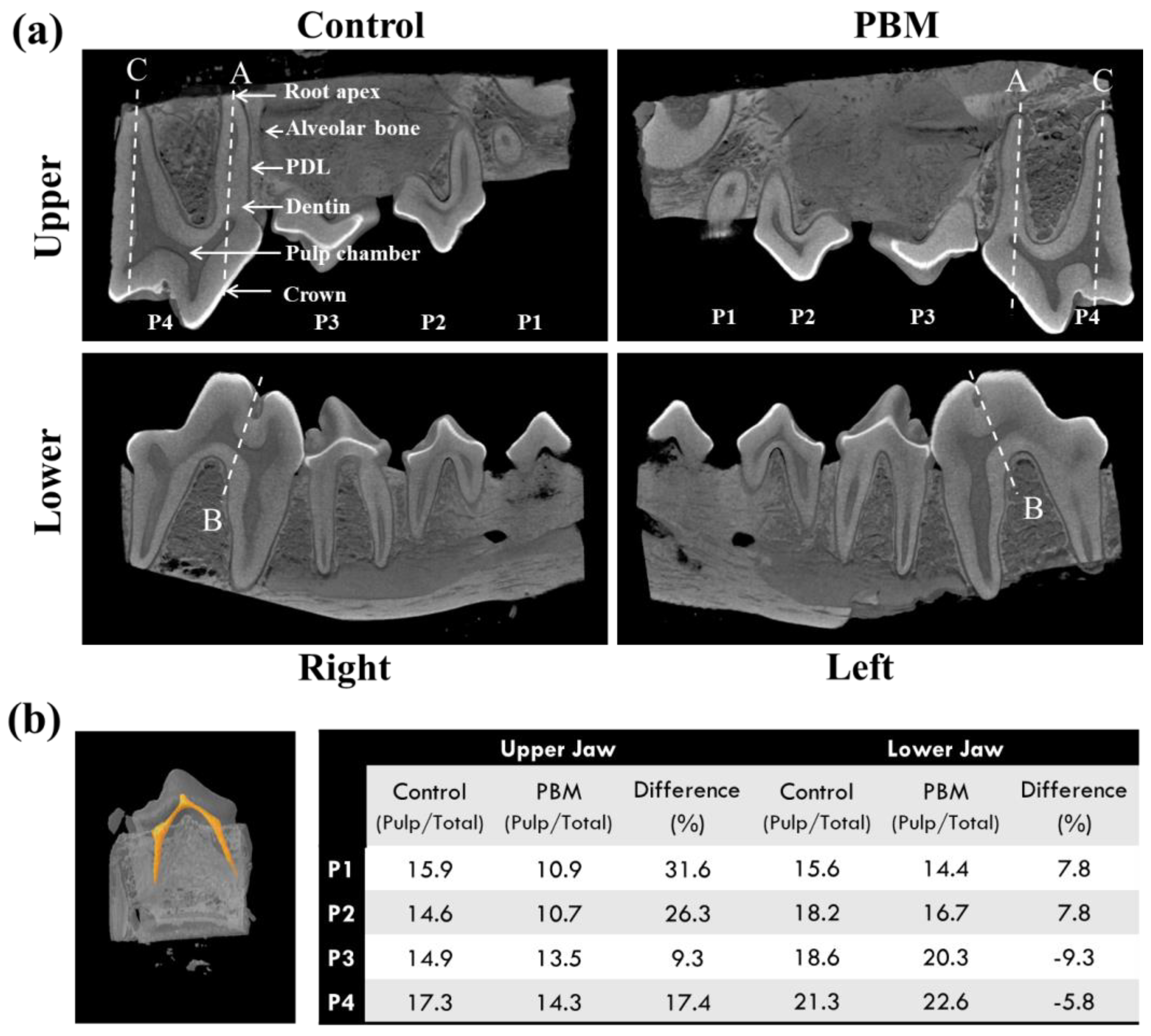
3.1. Micro-Computed Tomography (CT)

In this animal model, the premolars (P1, P2, P3, P4) of the lower and upper jaw of both the Control right and PBM-treated left were observed through micro-CT analysis after 8 weeks of PBM. In Figure 2a, we can see that the crown, dentin, pulp chamber, periodontal ligament, and root apex were densely packed. Since notable structural differences were not observed between the Control and PBM groups in micro-CT images, we analyzed the volume ratio (pulp chamber/total tooth) of each tooth. A reduction in the pulp chamber is known to imply secondary dentin formation. Three-dimensional reconstruction was performed via a modified Feldkamp cone-beam algorithm, and Figure 2b shows the representative image of the reconstructed pulp chamber of a P2 tooth. The volume ratios of the pulp chamber to the total tooth were calculated, and the differences between the Control and PBM ((Control − PBM)/Control × 100) were analyzed. Average increases of 21.1 and 0.1% were calculated for the upper and the lower jaw, respectively. We can assume that the PBM promoted the production of dentin compared to the Control.

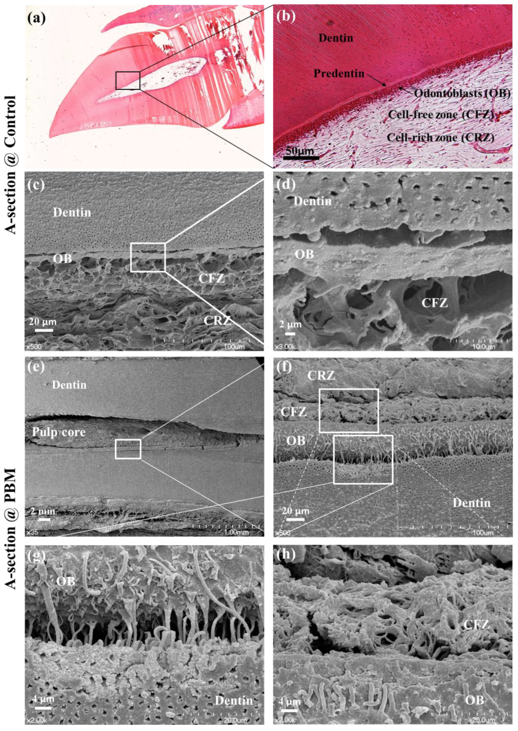
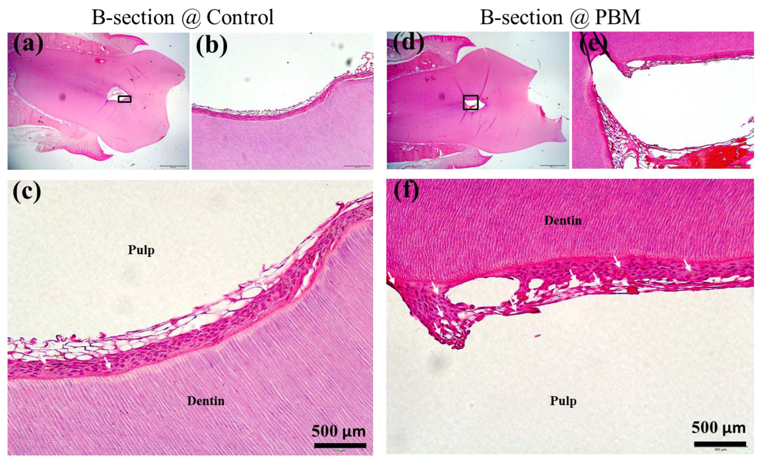
3.2. Structural Analysis of Dentin-Pulp Interface

We then conducted a structural analysis of the dentin–pulp interface to confirm photobiomodulation-induced neodentin formation. Premolar P4 was cut from both sides of the upper jaw and sectioned along the line ‘A’, as shown in Figure 2b. H&E staining of these sections demonstrated that Control and PBM-treated premolars were histologically similar in all sections. The pulp chamber was composed of dentin, predentin, odontoblasts, and pulp stem cells (Figure 3a,b).
SEM analysis also revealed that both Control and PBM-treated teeth exhibited distinct dentin layers, odontoblasts (OB), cell-free zones (CFZs), and cell-rich zones (CRZs) (Figure 3c,h). Upon magnification, odontoblasts in PBM-treated samples appeared more clearly adjacent to predentin (Figure 3g,h) than those in Control samples (Figure 3d). This indicates the increased activity of odontoblasts in dentin formation after PBM treatment [20].
The vascular system plays an important role in supporting dentinogenesis by odontoblasts. H&E staining (Figure 4) was conducted on B-sections of P4 premolars to examine vascular development. In particular, a great number of vascular structures were observed in PBM-treated tissue (Figure 4f), whereas few were observed in Control tissue (Figure 4c). The vasculature appeared in the odontoblast layer of PBM-irradiated samples in contrast to the Control sample.

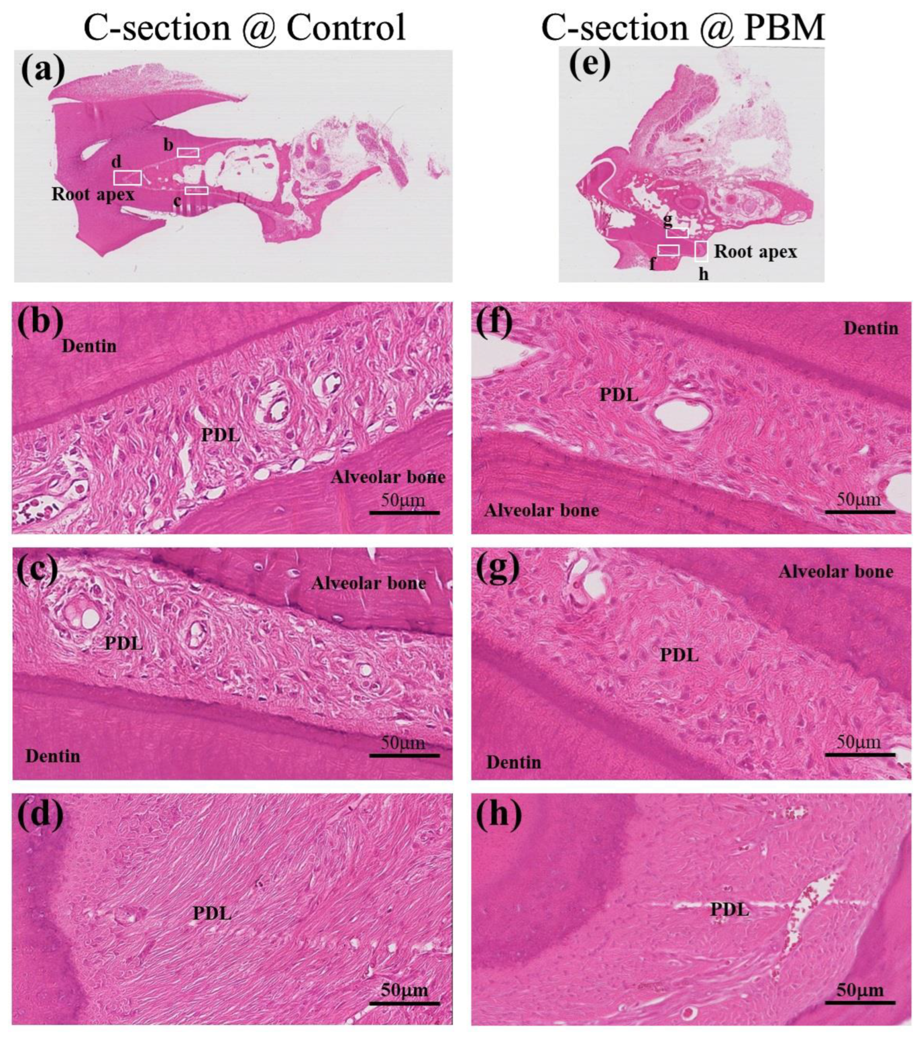
3.3. Structural Analysis of Periodontal Ligament (PDL)

To assess the impact of photobiomodulation on the fibrous connective tissue development of the periodontal ligament, histological analysis was performed using H&E staining on C-sections of Control and PBM-treated teeth. We observed three regions in both samples: two regions were periodontal ligament (PDL) regions at the side of roots, and one region was PDL at the root apex (Figure 5a–h). Similar areas of Control and PBM-treated teeth were compared. Overall, microscopic examination revealed that the PBM-treated sample had denser fibrous connective tissue in the PDL than Control teeth (Figure 5f–h).

4. Discussion

Our image analysis revealed that NIR-PBM induced structural changes in the pulp–dentin interface. We could observe a PBM-activated population of odontoblasts making a stack of odontoblasts (Figure 3). The odontoblast becomes increasingly crowded at the pulp–dentin interface when the dentin layer forms [20]. The dentin deposition after tooth structural completion is called the deposition of secondary dentin, which is in many cases related to tooth eruption or irritation. This secondary dentin deposition is the process of recovery from the diseased states, but the process is too slow (~0.4 μm/day) to reach full recovery when necessary [21]. Therefore, if secondary dentin formation is accelerated by external physical cues, the modality may help protect teeth from deteriorating. External biophysical cues may be kinds of irritation. Such external stimulation can induce the enhancement of the activity of odontoblasts by up to 50% or more, higher than that of odontoblasts of fully matured, healthy teeth [22]. Dentinal hypersensitivity [23,24], commonly associated with dental procedures such as gingival recession, caries removal, and prosthetic placement, is a prevalent concern which underscores the need for advanced treatment strategies in dentistry [25]. Despite maintaining normal tooth morphology, patients frequently experience discomfort due to exposed tooth roots and changes in dentin [24]. Traditional treatments, ranging from non-invasive desensitizers to invasive procedures like root canal treatment, often do not fully resolve these symptoms. Our results suggest that NIR PBM is one of the regenerative technologies which can be used over conventional inert materials [26,27,28]. Our findings also highlight PBM’s effect on the fibrous connective tissue of the periodontal ligament and demonstrate active proliferation and densification, indicating the comprehensive benefits of PBM on dental health. This is consistent with the broader goal of regenerative dentistry to harness the potential of stem cells and biomaterials to create an environment conducive to innate healing and tissue repair. The impact of PBM goes beyond hypersensitivity and plays a pivotal role in the overall health of dental tissues, including dentin and periodontal ligament [29,30,31]. The continuous remodeling of dentin, characterized by an organic matrix within a permeable tubular structure and an apatite crystalline structure, was reported to be greatly enhanced by PBM [29,32,33].
An activated odontoblast might be from the dental pulp stem cell differentiation induced by PBM. The activation of stem cells was also confirmed by the reduction in the pulp chamber volume ratio (Figure 2) and by the increased vasculature in the odontoblast layer (Figure 4). The differentiation pathway into odontoblasts is mediated by the activation of transforming growth factor-beta 1 (TGF-b1) in pulp tissue. This activation cascade may be initiated by reactive oxygen species (ROS), which originate from the mitochondrial electron transport chain system triggered by PBM [34]. The underlying mechanism of PBM-mediated dentinogenesis through stem cell differentiation was further demonstrated in studies using PBM pulsed wave [18,34,35], which demonstrated superior ROS generation compared to their continuous counterparts. The exact mechanism of pulp stem cell differentiation by PBM in vivo should be elucidated in the future.
In this study, we used a pulsed wave (PW) approach to PBM, which distinguishes it from continuous wave (CW) methods in the application of optical energy. Unlike CW, which operates at higher power densities, typically in the range of 0.5 to 48 J/cm2 [36], PW uses lower power densities, delivering photon energy in pulses. In this case, the PW setting of the power density of 1.91 mW/cm2 at the duty cycle of 30% was used to produce an energy density of 80.22 mJ/cm2. This selection was informed by previous studies showing the superiority of PW in differentiating pulp stem cells into odontoblast-like cells compared to CW [18,34,35]. Despite its lower energy density, PW was observed to be more effective than CW in stimulating tooth tissue regeneration, as evidenced in our study by the enhanced activity of odontoblasts and the improved condition of fibrous connective tissue within the periodontal ligament.
However, our study has a few limitations. One significant limitation is the scope of the study model, which was confined to a single animal subject. This limits the breadth of the data and raises questions about the generalizability of the results. Additionally, comprehensive data on the long-term effectiveness and safety of PBM are lacking, which are crucial for understanding its viability as a long-term treatment option. Furthermore, although positive outcomes have been observed, the exact biological mechanisms by which PBM promotes tooth tissue regeneration are only partially understood. An in-depth exploration of these mechanisms is necessary to fully comprehend and optimize the use of PBM in dental health applications.
In summary, the application of PBM using 300 Hz-NIR can be a promising tool in dental tissue regeneration. This not only addresses the challenge of managing dental hypersensitivity but also plays a crucial role in the regeneration and health of tooth tissue. While acknowledging certain limitations, our research has contributed significantly to the evolving landscape of regenerative dentistry, paving the way for innovative treatment strategies in the field of dental tissue management and regeneration.

5. Conclusions

Using 300 Hz-NIR PBM, odontoblast activity was improved in a beagle model, and fibrous connective tissue within the periodontal ligament was strengthened. These findings provide potential avenues for therapeutic strategies aimed at optimizing dentin and periodontal ligament health.

Author Contributions

Conceptualization, H.B.K. and M.H.K.; methodology, H.B.K.; validation, H.B.K. and K.Y.B.; formal analysis, H.B.K. and K.Y.B.; investigation, H.B.K.; resources, J.H.C.; data curation, K.Y.B.; writing—original draft preparation, H.B.K.; writing—review and editing, K.Y.B.; visualization, H.B.K.; supervision, J.H.C.; project administration, J.H.C.; funding acquisition, H.B.K. All authors have read and agreed to the published version of the manuscript.

Funding

This research was funded by National Research Foundation of Korea (NRF) (No. 2022R1F1A1075102 and 2022R1F1A1076242), and partially by Kwangwoon University (2023).

Institutional Review Board Statement

The study was conducted in accordance with the Declaration of Helsinki and approved by the Animal Research Ethics Committee of Seoul National University Dental Hospital (protocol code SNU-120427-2-2).

Informed Consent Statement

Not applicable.

Data Availability Statement

The data presented in this study are available in the article.

Conflicts of Interest

Author Hong Bae Kim was employed by the company Medical Engineering Research Center, The Standard Co., Ltd. The remaining authors declare that the research was conducted in the absence of any commercial or financial relationships that could be construed as a potential conflict of interest.

References

  1. Sun, H.H.; Jin, T.; Yu, Q.; Chen, F.M. Biological approaches toward dental pulp regeneration by tissue engineering. J. Tissue Eng. Regen. Med. 2011, 5, e1–e16. [Google Scholar] [CrossRef] [PubMed]
  2. Murray, P.E.; Garcia-Godoy, F.; Hargreaves, K.M. Regenerative endodontics: A review of current status and a call for action. J. Endod. 2007, 33, 377–390. [Google Scholar] [CrossRef] [PubMed]
  3. Gay, I.C.; Chen, S.; MacDougall, M. Isolation and characterization of multipotent human periodontal ligament stem cells. Orthod. Craniofac Res. 2007, 10, 149–160. [Google Scholar] [CrossRef]
  4. Kerkis, I.; Kerkis, A.; Dozortsev, D.; Stukart-Parsons, G.C.; Gomes Massironi, S.M.; Pereira, L.V.; Caplan, A.I.; Cerruti, H.F. Isolation and characterization of a population of immature dental pulp stem cells expressing OCT-4 and other embryonic stem cell markers. Cells Tissues Organs 2006, 184, 105–116. [Google Scholar] [CrossRef]
  5. Morsczeck, C.; Moehl, C.; Gotz, W.; Heredia, A.; Schaffer, T.E.; Eckstein, N.; Sippel, C.; Hoffmann, K.H. In vitro differentiation of human dental follicle cells with dexamethasone and insulin. Cell Biol. Int. 2005, 29, 567–575. [Google Scholar] [CrossRef] [PubMed]
  6. Seo, B.M.; Miura, M.; Gronthos, S.; Bartold, P.M.; Batouli, S.; Brahim, J.; Young, M.; Robey, P.G.; Wang, C.Y.; Shi, S. Investigation of multipotent postnatal stem cells from human periodontal ligament. Lancet 2004, 364, 149–155. [Google Scholar] [CrossRef]
  7. Miura, M.; Gronthos, S.; Zhao, M.; Lu, B.; Fisher, L.W.; Robey, P.G.; Shi, S. SHED: Stem cells from human exfoliated deciduous teeth. Proc. Natl. Acad. Sci. USA 2003, 100, 5807–5812. [Google Scholar] [CrossRef]
  8. Arany, P.R.; Cho, A.; Hunt, T.D.; Sidhu, G.; Shin, K.; Hahm, E.; Huang, G.X.; Weaver, J.; Chen, A.C.; Padwa, B.L.; et al. Photoactivation of endogenous latent transforming growth factor-beta1 directs dental stem cell differentiation for regeneration. Sci. Transl. Med. 2014, 6, 238ra69. [Google Scholar] [CrossRef]
  9. Mester, E.; Ludany, G.; Sellyei, M.; Szende, B. On the biologic effect of laser rays. Bull. Soc. Int. Chir. 1968, 27, 68–73. [Google Scholar]
  10. Yang, D.; Yi, W.; Wang, E.; Wang, M. Effects of light-emitting diode irradiation on the osteogenesis of human umbilical cord mesenchymal stem cells in vitro. Sci. Rep. 2016, 6, 37370. [Google Scholar] [CrossRef]
  11. Tuby, H.; Maltz, L.; Oron, U. Induction of autologous mesenchymal stem cells in the bone marrow by low-level laser therapy has profound beneficial effects on the infarcted rat heart. Lasers Surg. Med. 2011, 43, 401–409. [Google Scholar] [CrossRef]
  12. Hashmi, J.T.; Huang, Y.Y.; Sharma, S.K.; Kurup, D.B.; De Taboada, L.; Carroll, J.D.; Hamblin, M.R. Effect of pulsing in low-level light therapy. Lasers Surg. Med. 2010, 42, 450–466. [Google Scholar] [CrossRef] [PubMed]
  13. Mvula, B.; Mathope, T.; Moore, T.; Abrahamse, H. The effect of low level laser irradiation on adult human adipose derived stem cells. Lasers Med. Sci. 2008, 23, 277–282. [Google Scholar] [CrossRef] [PubMed]
  14. Hou, J.F.; Zhang, H.; Yuan, X.; Li, J.; Wei, Y.J.; Hu, S.S. In vitro effects of low-level laser irradiation for bone marrow mesenchymal stem cells: Proliferation, growth factors secretion and myogenic differentiation. Lasers Surg. Med. 2008, 40, 726–733. [Google Scholar] [CrossRef] [PubMed]
  15. Stein, A.; Benayahu, D.; Maltz, L.; Oron, U. Low-level laser irradiation promotes proliferation and differentiation of human osteoblasts in vitro. Photomed. Laser Surg. 2005, 23, 161–166. [Google Scholar] [CrossRef]
  16. Jones, D.L.; Wagers, A.J. No place like home: Anatomy and function of the stem cell niche. Nat. Rev. Mol. Cell Biol. 2008, 9, 11–21. [Google Scholar] [CrossRef]
  17. Gurtner, G.C.; Werner, S.; Barrandon, Y.; Longaker, M.T. Wound repair and regeneration. Nature 2008, 453, 314–321. [Google Scholar] [CrossRef]
  18. Kim, H.B.; Baik, K.Y.; Choung, P.H.; Chung, J.H. Pulse frequency dependency of photobiomodulation on the bioenergetic functions of human dental pulp stem cells. Sci. Rep. 2017, 7, 15927. [Google Scholar] [CrossRef]
  19. Verma, P.; Love, R.M. A micro CT study of the mesiobuccal root canal morphology of the maxillary first molar tooth. Int. Endod. J. 2011, 44, 210–217. [Google Scholar] [CrossRef]
  20. Egbuniwe, O.; Idowu, B.D.; Funes, J.M.; Grant, A.D.; Renton, T.; Di Silvio, L. P16/p53 expression and telomerase activity in immortalized human dental pulp cells. Cell Cycle 2011, 10, 3912–3919. [Google Scholar] [CrossRef]
  21. Massler, M.; Schour, I. The appositional life span of the enamel and dentin-forming cells; human deciduous teeth and first permanent molars; introduction. J. Dent. Res. 1946, 25, 145–150. [Google Scholar] [CrossRef] [PubMed]
  22. Mitsiadis, T.A.; De Bari, C.; About, I. Apoptosis in developmental and repair-related human tooth remodeling: A view from the inside. Exp. Cell Res. 2008, 314, 869–877. [Google Scholar] [CrossRef] [PubMed]
  23. Jeon, M.J.; Park, J.W.; Seo, D.G. Intratubular crystal formation in the exposed dentin from nano-sized calcium silicate for dentin hypersensitivity treatment. Sci. Rep. 2023, 13, 14243. [Google Scholar] [CrossRef]
  24. Abuzinadah, S.H.; Alhaddad, A.J. A randomized clinical trial of dentin hypersensitivity reduction over one month after a single topical application of comparable materials. Sci. Rep. 2021, 11, 6793. [Google Scholar] [CrossRef] [PubMed]
  25. West, N.X.; Lussi, A.; Seong, J.; Hellwig, E. Dentin hypersensitivity: Pain mechanisms and aetiology of exposed cervical dentin. Clin. Oral. Investig. 2013, 17, S9–S19. [Google Scholar] [CrossRef]
  26. Zheng, C.; Chen, J.; Liu, S.; Jin, Y. Stem cell-based bone and dental regeneration: A view of microenvironmental modulation. Int. J. Oral. Sci. 2019, 11, 23. [Google Scholar] [CrossRef] [PubMed]
  27. He, L.; Zhou, J.; Chen, M.; Lin, C.S.; Kim, S.G.; Zhou, Y.; Xiang, L.; Xie, M.; Bai, H.; Yao, H.; et al. Parenchymal and stromal tissue regeneration of tooth organ by pivotal signals reinstated in decellularized matrix. Nat. Mater. 2019, 18, 627–637. [Google Scholar] [CrossRef]
  28. Yoshida, S.; Ohshima, H. Distribution and organization of peripheral capillaries in dental pulp and their relationship to odontoblasts. Anat. Rec. 1996, 245, 313–326. [Google Scholar] [CrossRef]
  29. Mir, M.; Mojahedi, S.M.; Tuner, J.; Shabani, M.; Darabi, F.; Rohban, A. The effectiveness of home-use photobiomodulation toothbrush for treating dentin hypersensitivity: A pilot study. Laser Ther. 2019, 28, 193–198. [Google Scholar] [CrossRef]
  30. Barros, A.P.O.; de Melo Alencar, C.; de Melo Pingarilho Carneiro, A.; da Silva Pompeu, D.; Barbosa, G.M.; Araujo, J.L.N.; Silva, C.M. Combination of two desensitizing protocols to control dentin hypersensitivity in non-carious lesions: A randomized, double-blind clinical trial. Clin. Oral. Investig. 2022, 26, 1299–1307. [Google Scholar] [CrossRef]
  31. Fossati, A.L.; Sobral, A.P.T.; Bruno, M.L.L.H.; Viarengo, N.O.; Sertaje, M.R.F.; Santos, E.M.; Gonçalves, M.L.L.; Ferrari, R.A.M.; Fernandes, K.P.S.; Horliana, A.C.R.T.; et al. Photobiomodulation and glass ionomer sealant as complementary treatment for hypersensitivity in molar incisor hypomineralisation in children: Protocol for a blinded randomised clinical trial. BMJ Open 2023, 13, e068102. [Google Scholar] [CrossRef] [PubMed]
  32. Yong, J.; Groger, S.; Wu, Z.; Ruf, S.; Ye, Y.; Chen, X. Photobiomodulation Therapy and Pulp-Regenerative Endodontics: A Narrative Review. Bioengineering 2023, 10, 371. [Google Scholar] [CrossRef] [PubMed]
  33. Alkhudhairy, F.; Vohra, F.; Naseem, M.; Ahmad, Z.H. Adhesive bond integrity of dentin conditioned by photobiomodulation and bonded to bioactive restorative material. Photodiagn. Photodyn. Ther. 2019, 28, 110–113. [Google Scholar] [CrossRef] [PubMed]
  34. Kim, H.B.; Baik, K.Y.; Seonwoo, H.; Jang, K.J.; Lee, M.C.; Choung, P.H.; Chung, J.H. Effects of pulsing of light on the dentinogenesis of dental pulp stem cells in vitro. Sci. Rep. 2018, 8, 2057. [Google Scholar] [CrossRef]
  35. Kim, H.B.; Kang, M.H.; Baik, K.Y.; Kim, J.E.; Park, S.B.; Choung, P.H.; Chung, J.H. Integration of blue light with near-infrared irradiation accelerates the osteogenic differentiation of human dental pulp stem cells. J. Photochem. Photobiol. B 2023, 245, 112752. [Google Scholar] [CrossRef]
  36. Chung, H.; Dai, T.; Sharma, S.K.; Huang, Y.Y.; Carroll, J.D.; Hamblin, M.R. The nuts and bolts of low-level laser (light) therapy. Ann. Biomed. Eng. 2012, 40, 516–533. [Google Scholar] [CrossRef]
Figure 1. Experimental setup demonstrating PBM in the beagle model. (a) PBM was performed on the left teeth and compared with untreated right teeth (Control). (b) FET (Si2305; P-Channel 1.25-W, 1.8-V MOSFET) was employed in the switch mechanism. (c) An LED array system consisting of nine LEDs was placed between the gingival tissue and teeth to cover most areas of the teeth.
Applsci 14 00724 g001
Figure 2. Micro-CT images and analysis of the premolar teeth of the upper and lower jaws of the beagle model after PBM. (a) Micro-CT images clearly show the crown, pulp chamber, dentin, PDL, alveolar bone, and root apex areas of each tooth. The dotted lines marked from A to C in P4 teeth represent the sectioned planes for histological examinations. (b) A representative reconstructed image shows the pulp chamber in a P2 tooth. The volume ratios of the pulp chamber to the total tooth were calculated, and the differences of between the Control and PBM ((Control − PBM)/Control × 100) were analyzed.
Applsci 14 00724 g002
Figure 3. Histology of A-sectioned P4 teeth, as marked in Figure 2. (ad) present images of the Control teeth, and (eh) present images of PBM-treated teeth. (a,b) present H&E staining images, and (ch) present SEM images. (b,d,fh) are enlargements of the inserted square areas in (a,c,e,f). (fh) exhibit odontoblasts activated for enhanced tubular dentin formation in PBM-treated teeth.
Applsci 14 00724 g003
Figure 4. H&E staining images of B-plane sectioned teeth, as shown in Figure 2. (ac) are for Control, and (df) are for PBM-treated teeth. (b,c) are enlargements of the inserted square area in (a), and (e,f) are enlargements of this inserted square area in (d). White arrows in (c,f) point to the tubular epithelia of the blood vessels.
Applsci 14 00724 g004
Figure 5. H&E staining images of C-plane sectioned teeth, as shown in Figure 2. (ad) are for Control, and (eh) are for PBM-treated teeth. (bd) are enlargements of the inserted square areas in (a), and (fh) are enlargements of the inserted square areas in (e). The locations in images (b,c,f,g) represent the oblique PDL, and locations in images (d,h) represent the apical PDL. Photobiomodulation resulted in the microscopic appearance of a higher density of fibrous connective tissue within the periodontal ligament compared to the Control.
Applsci 14 00724 g005
Disclaimer/Publisher’s Note: The statements, opinions and data contained in all publications are solely those of the individual author(s) and contributor(s) and not of MDPI and/or the editor(s). MDPI and/or the editor(s) disclaim responsibility for any injury to people or property resulting from any ideas, methods, instructions or products referred to in the content.

