Abstract
The cornea relies on a healthy endothelium to maintain transparency, and damage to endothelial cells can result in corneal oedema and vision loss. Current treatments, which often involve the use of donor corneas, face significant limitations due to a shortage of donor tissue. Although human corneal endothelial cells (HCECs) can be cultured and transplanted, their low attachment rates limit the effectiveness of these treatments. In this review, we examined studies that explore the use of magnetic nanoparticles (MNPs) to enhance the attachment of HCECs to the cornea. We evaluated the effectiveness, cell viability, and safety of this approach. Findings indicate that MNPs facilitate the targeted delivery of HCECs under a magnetic field, resulting in improved corneal clarity and reduced oedema in animal models. Cell viability remained high, and no significant safety concerns were identified. MNPs present a promising strategy to enhance HCEC transplantation. However, further research, including ongoing clinical trials, is necessary to confirm the safety and efficacy of this approach before it can be adopted for widespread clinical use.
1. Introduction
The cornea, a transparent avascular tissue of the anterior eye, transmits incoming light while also providing the necessary anterior refractive surface, contributing to approximately two-thirds of the eye’s total refractive power [1]. The cornea is traditionally anatomically segmented into five layers, progressing from the outermost epithelium, Bowman’s layer, the stroma, Descemet’s membrane, to the innermost endothelium; however, it is important to acknowledge that the cornea’s anatomy is, in reality, more nuanced (Figure 1) [2,3]. The corneal endothelium, a single layer of cells lining the posterior corneal surface, regulates water flow, maintains tissue hydration at approximately 70%, and sustains clear vision by coordinating ionic pumps and tight junction barriers [4]. Among the key components of these tight junctions is ZO-1, a submembranous protein located along the apical side of the cells that contributes to structural integrity by forming hexagonal shapes and aiding in signal transduction [5,6,7].
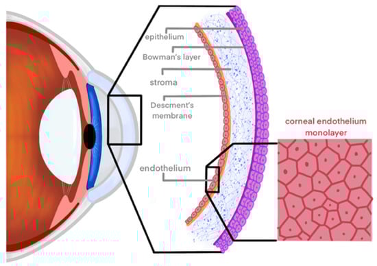
Figure 1.
Cornea anatomy. Layers of the cornea depicted from inner to outer: endothelium, Descemet’s membrane, stroma, Bowman’s layer, and epithelium. The endothelium is highlighted to show its hexagonal monolayer arrangement of cells, crucial for maintaining corneal transparency and hydration balance.
While the epithelium is characterised by self-renewal and hosts a resident stem cell population in the Palisades of Vogt, human corneal endothelial cells (HCECs) typically maintain a quiescent state and demonstrate negligible in vivo proliferation [8]. By the 6th week of human gestation, most HCECs are arrested in the G1 phase of the cell due to the suppressive effects of TGF-β2 present in the aqueous humour, inhibiting their entry into the S-phase [9,10]. On average, the adult healthy human endothelium is approximately 4 μm in thickness and has a cell density of 3000 cells/mm2, with a gradual 0.5–0.6% decrease in central endothelial cell density annually [11,12,13]. Although HCECs can repair minor damage or aging-related issues by elongating and migrating, their limited in vivo proliferation capability suggests that complete repair of the endothelium following larger trauma may be unattainable [14,15].
Factors like trauma or genetic endotheliopathies can further accelerate the decline of endothelial cell density [16,17]. In Fuchs’ dystrophy, the most common primary corneal disease, excessive abnormal Descemet’s membrane secretion by enlarged endothelial cells leads to cornea guttata, impaired pump function, and corneal oedema [18]. Conditions like posterior polymorphous dystrophy, congenital hereditary endothelial dystrophy, and iridocorneal endothelial syndrome exhibit endothelial cells with epithelial-like characteristics, causing congenital corneal cloudiness and forming membranes that affect the iris and anterior chamber angle [19,20,21]. Additionally, damage from contact lens wear or surgical procedures can lead to endothelial cell loss, increased polymegethism and pleomorphism, and decreased transparency [22,23,24].
Currently, treatments for moderate corneal endothelial dysfunction and failure include transplantation of donor allo-endothelium (Table 1). Endothelial keratoplasty (EK) is a surgical procedure designed to address corneal endothelial diseases by selectively transplanting the Descemet membrane and endothelium from a donor cornea to the recipient’s eye [25]. This approach aims to restore corneal clarity and enhance visual acuity by replacing dysfunctional endothelial cells, crucial for maintaining corneal deturgescence [26].

Table 1.
Overview of current treatments for corneal endothelial diseases. This table summarises the various treatments available for common corneal endothelial diseases, highlighting both conservative and surgical options.
Selective endothelial replacement, initially introduced clinically by Gerrit Melles in 1998, presented a notably more challenging procedure compared to the traditional standard of penetrating keratoplasty (PK), which entails the complete replacement of the corneal thickness [27]. Modern techniques, such as Descemet’s stripping endothelial keratoplasty (DSEK) and Descemet membrane endothelial keratoplasty (DMEK), were introduced in the early 2000s [28]. These methods vary in the thickness of the transplanted tissue and surgical approach, refining the approach and offering faster visual recovery compared to traditional penetrating keratoplasty [29,30,31].
Despite advancements, EK poses risks including early endothelial cell loss, graft detachment, and challenges in restoring corneal shape and clarity [25]. Studies report average endothelial cell losses of 15–36% at 1 year and 47–55% at 5 years post-DSAEK [32]. Moreover, EK procedures, notably DMEK with its ultra-thin tissue (≤20 μm), demand precise donor tissue manipulation, recipient Descemet membrane dissection, and meticulous graft positioning and adherence [33]. Furthermore, the global demand for corneal transplants poses a considerable challenge, a situation exacerbated by the COVID-19 pandemic and prolonged life expectancy [29,34].
1.1. Magnetic Nanotechnology
The field of nanotechnology has demonstrated significant potential for enhanced and targeted drug delivery to the cornea [35]. Nanoparticles, ranging in size from 1 to 100 nm, exhibit unique interactions with cells and tissues at the molecular level, offering advantages such as sustained drug release and precise targeting capabilities [36]. Magnetic nanoparticles (MNPs) possess the distinct ability to align their magnetic spins with an external magnetic field, thereby achieving magnetisation. Originally recognised for their application in magnetic resonance imaging (MRI) for diagnostic purposes, MNPs have experienced substantial growth in biomedical applications over the past few decades [37]. This versatile class of nanoparticles includes metallic, bimetallic, and superparamagnetic iron oxide nanoparticles (SPIONs) [38]. MNPs serve as effective drug delivery systems due to their reactive surfaces, which allow for functionalisation with biocompatible coatings, bioactive molecules, or targeting moieties, thereby enhancing specificity for cellular targets while reducing interactions with healthy tissues [39,40]. Furthermore, MNPs can be manipulated using non-contact forces, thereby extending their potential beyond simple molecular transport [41].
When comparing nanoparticles to microparticles (typically around 4 μm), Raju et al. showed that nanoparticles demonstrated significantly lower toxicity, as evidenced by higher corneal endothelial cell (CEC) counts and reduced iron deposition compared to microparticles [42]. Additionally, nanoparticles generated fewer reactive oxygen species (ROS), which minimises oxidative stress and potential cellular damage. These properties contribute to their lower toxicity profile and improved biocompatibility, making nanoparticles particularly promising for ophthalmic applications [42].
While MNPs are proposed for therapeutic injection of CECs, they have also demonstrated the ability to target the posterior segment of the eye. In a study by Giannaccini et al., MNPs were functionalised with vascular endothelial growth factor (VEGF), a bioactive molecule known for its transcytosis capability from the retinal pigment epithelium (RPE) to deeper ocular layers, effectively directing the MNPs to the choroid [43]. These results highlight the potential of MNPs combined with various bioactive molecules to enhance targeted cell delivery within the eye.
In cancer therapy MNPs, especially iron oxide nanoparticles, are used in cancer treatment by catalysing the Fenton reaction to produce toxic ROS, causing tumour cell death [44,45]. In drug and gene delivery, MNPs improve specificity by guiding therapeutic agents directly to disease sites. For instance, poly-L-lysine-coated Fe3O4@FePt nanoparticles target glioblastoma cells, enhancing chemotherapy efficiency [46]. In gene delivery, magnetofection directs viral vectors to lung tissue, improving gene transfection and therapeutic outcomes [47,48,49]. Moreover, the utilisation of MNPs as carriers has expanded into the realm of in vivo corneal regeneration, with studies evaluating the feasibility of directly delivering nanoparticle-loaded CEC into the anterior chamber and maintaining cellular viability under various loading conditions [35,50].
1.2. Culturing CECs
Research on culturing CECs began in 1979 with Baum et al.’s successful cultivation of HCECs [51]. Techniques for producing Good Manufacturing Practice (GMP)-grade cells have since evolved, with the peel-and-digest method by Peh et al. emerging as a leading approach for isolating HCECs using collagenase to preserve cell junctions and enhance viability and proliferation [52,53].
CEC culture media include maintenance media without growth factors and proliferation media with growth factors. A dual media approach combining both prevents endothelial-mesenchymal transformation (EnMT) and better maintains cell characteristics than proliferation media alone [54,55]. Supplements like L-ascorbic acid 2-phosphate (A-2P) and basic fibroblast growth factor (FGF-2) enhance CEC growth, with rho-kinase inhibitors (ROCKi) like Y-27632 widely used to promote proliferation and inhibit apoptosis [56,57,58,59,60,61,62,63,64]. MNPs are added to the cell culture medium at various concentrations, often expressed as ratios of particles to cells or as μg/mL concentrations. The MNPs are typically left in the culture media with the cells for a specified exposure time, often 24–72 h [65].
Culturing CECs presents challenges such as donor variability, endothelial-to-mesenchymal transition (EnMT), and the need to optimise culture conditions. Essential considerations include maintaining cell viability, implementing effective cryopreservation methods, and translating research findings into clinical trials [66].
1.3. Options for Delivery of Cultured CECs
At the moment, there are two methods of cell delivery described in the literature—tissue engineered endothelial keratoplasty (TE-EK) and direct corneal endothelial cell injection (CE-CI) [67]. The TE-EK approach builds upon established EK surgeries, such as DSEK and DSAEK, for graft delivery. Firstly, the cultured CECs are grown to confluence, dissociated into single-cell suspension, and seeded densely onto a thin biological scaffold carrier at a density of 3000 cells/mm2 [67]. After allowing the TE-EK graft to stabilise for up to 7 days, it is transplanted into the eye [68]. As for, CE-CI approach, after dissociation into a single-cell suspension, cultivated CECs are administered via direct injection into the recipient’s anterior chamber [69].
In a study comparing the techniques for treating bullous keratopathy in a rabbit model, animals receiving TE-EK grafts initially exhibited an increase in central corneal thickness (CCT) to >1000 μm post-operatively. However, from week 1 onwards, improvements in corneal clarity were observed, with CCT measuring 484.3 ± 73.7 μm by week 3. In contrast, rabbits treated with CE-CI maintained corneal clarity throughout the study period, with CCT measuring 582.5 ± 171.5 μm at week 3. These findings underscore the efficacy of both TE-EK and CE-CI therapies in restoring corneal clarity and function in the rabbit model, with CE-CI showing a quicker initial response [67]. Due to its simpler procedure and reduced invasiveness compared to TE-EK, direct injection of cells into the anterior chamber is often preferred.
Numerous animal studies have demonstrated the potential of CE-CI, often in combination with ROCKi, to restore corneal endothelial function in models of endothelial dysfunction [70]. The effectiveness of this approach has been demonstrated in multiple models, including rabbits and monkeys; however, complete functional recovery of the corneal endothelium could not be clearly demonstrated following CE-CI in in a feline model, possibly due to species-specific differences and variations in the techniques used to induce CEC injury and culture the cells [67,71,72,73]. In a rabbit model, a study investigating CE-CI for corneal endothelial deficiency revealed challenges in cell attachment. Injected cells, contrary to previous findings, did not effectively reduce corneal oedema in an upright eye position; rather, they adhered to the posterior corneal surface only when the eye was positioned downward, relying on gravitational forces for attachment [74,75].
The promising findings from preclinical studies have laid the groundwork for the progression of the cell injection technique to human clinical trials. In 2018, Kinoshita et al. released an influential study demonstrating the efficacy of injecting 300 μL HCECs cultured from donor cornea supplemented with ROCKi in 11 patients with bullous keratopathy [69]. All 11 eyes achieved a CEC density greater than 500 cells/mm2 at 24 weeks (100%; 95% confidence interval [CI], 72 to 100). Corneal thickness decreased significantly to less than 630 μm in 10 out of 11 patients (91%; 95% CI, 59 to 100). Visual acuity improved in most cases, with an improvement in best-corrected visual acuity of two lines or more recorded in 9 of the 11 treated eyes (82%; 95% CI, 48 to 98). A follow-up study confirmed the safety and efficacy of cultured HCEC injection therapy for up to 5 years after surgery with a mean endothelial cell density of 1257 cells/mm2 [76]. Following this pioneering research, other research groups are aiming to re-create this work. AurionBiotech is the first clinical trial currently underway trying to recreate these findings outside of Japan [77].
Kinoshita et al.’s procedural method implemented two key precautions aimed at maximising endothelial cell attachment: maintaining patients in a prone position for 3 h post-injection and supplementing cells with ROCKi, which has been previously shown to enhance cellular attachment and adhesion [78]. Despite these efforts to enhance accurate cell attachment to the damaged cornea, the procedure led to a notable loss of injected cells. Authors hypothesised that the cells likely entered systemic circulation via the trabecular meshwork, raising concerns about potential complications such as increased intraocular pressure (IOP) due to blockage of aqueous outflow or, more critically, ectopic tumour formation from cell migration through the bloodstream.
To address the issue of ensuring accurate cell attachment, the use of MNPs presents a promising solution to improve CEC transplantation and overcome the challenges of precise cell localisation (Figure 2). By injecting MNPs into the culture media, the transplanted CECs can be guided and targeted to the desired location within the cornea. The integration of nanotechnology represents an innovative approach to address the key issues in CEC transplantation and improve the outcomes of this pioneering procedure for restoring corneal health and visual function [79,80].
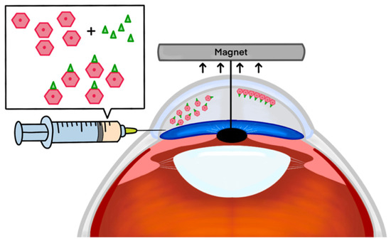
Figure 2.
Magnetic nanoparticle-guided delivery of corneal endothelial cells for ocular regeneration. Depiction of the general method for targeted delivery of corneal endothelial cells using magnetic nanoparticles. Corneal endothelial cells are combined with magnetic nanoparticles and injected into the anterior chamber of the eye. An external magnet is utilised to localise the cell–nanoparticle complex precisely to the central area of damaged endothelial cells. This approach aims to enhance the efficiency and efficacy of cell therapy for corneal regeneration, offering precise targeting for the treatment of corneal dysfunction.
This review offers a succinct overview of recent advancements in employing MNPs to facilitate CEC attachment. It evaluates the efficacy of diverse MNPs and cell injection techniques, assesses cell viability, and explores procedural challenges and safety considerations. In doing so, this review provides a comprehensive exploration of current developments in magnetic nanotechnology integrated with HCEC injection for addressing corneal dysfunction.
2. Discussion
2.1. Effectiveness of Magnetic Nanoparticles in Enhanced Delivery of HCECs
The effectiveness of MNPs in enhancing the delivery of HCECs has been investigated through various experimental approaches, aiming to optimise targeted cell transplantation while preserving cell morphology and function (Table 2). An essential focus across these studies was to demonstrate that the use of MNPs enhances cell delivery efficiency beyond gravitational forces alone, while ensuring procedural safety. Moysidis et al. explored the impact of 50 nm diameter MNPs on the delivery of cadaveric donor HCECs, revealing a significant 2.4-fold increase in cell density compared to conventional gravity-driven methods [80]. This enhancement was found to correlate logarithmically with the strength of the magnetic field and the initial concentration of nanoparticles within the cells, indicating a dose-dependent relationship for enhancing cell delivery efficiency with MNPs. Additionally, treatment with higher concentrations of nanoparticles also resulted in higher velocity of cells. Nevertheless, there was no notable change in the number of cells whose path was affected by the electromagnet at the volume of 10 µL nanoparticles or higher. This indicates a saturation effect where the magnetic guidance system’s efficacy plateaued despite higher nanoparticle concentrations, suggesting an optimal concentration range for effective cell delivery under magnetic guidance.
When conducting histological analysis, Moysidis et al. discovered that, at the target location, the magnetic HCECs formed tight junctions, arranging themselves into a functional monolayer, thus replicating a significant function of endogenous HCECs in vivo [80]. Therefore, these data indicate the potential effectiveness of cultured HCECs in replacing deficient or repopulating damaged corneal endothelium. Xia et al. observed similar histological findings when utilising MNPs to guide injected HCECs in rabbit models with corneal endothelial dysfunction [81]. They reported preserved corneal morphology, maintenance of cellular tight junction protein expression including ZO-1 and neural cell adhesion marker (NCAM), and functional integration into a monolayer in the innermost corneal layer. Therefore, following transplantation in vivo, donor cells exhibited markers indicative of the essential functions of HCECs.
Regarding the restoration of corneal clarity and function, the research conducted by Xia et al. evidenced improved post-operative corneal clarity and reduced thickness relative to control groups administered with balanced salt solution plus (BSS+) [81]. Optical coherence tomography (OCT) imaging confirmed thinner corneal profiles and highlighted distinct endothelial layers in eyes treated with magnetic HCECs, indicative of successful transplantation and integration into the host tissue. All eight eyes treated with BSS+ had corneal thickness measurements that exceeded the upper limit of the pachymeter’s range (>1500 μm), whilst six of the twelve eyes injected with magnetic HCECs had measurable corneal thickness values that were below the pachymeter’s range limit (<1500 μm). Transplanted magnetic HCECs were successfully delivered to the posterior corneal surface, with no migration of cells to the iris or trabecular meshwork and no acute fluctuations in IOP. This research paper concluded that magnetic HCECs showed superior performance compared to control HCECs guided solely by gravity, demonstrating that targeted magnetic guidance may enhance cell delivery and integration at the host corneal endothelium.
While the studies mentioned demonstrate promising outcomes of MNPs in CE-CI, their short follow-up periods limit our understanding of the long-term implications of this technique. In a related investigation, Mimura et al. explored the long-term outcomes of transplanting cultured rabbit CECs (RCECs) [82]. Cultured RCECs were labelled with iron nanoparticles and transplanted into the anterior chamber of rabbits with corneal endothelial dysfunction induced by cryo-injury. A cryo-injury group with normal injection of RCECs and a cryo-injury group with no treatment injection were used as controls. At 24 h post-injection the corneal thickness was 1000 μm in all groups and at 2 weeks the iron–RCEC-treated group had a mean corneal thickness of 492 μm, whilst the cryo-only and the cryo-RCEC groups showed limited variability at 1016 μm and 1004 μm, respectively. Throughout the 12 months, corneal thickness in the iron–RCEC group remained around 400 μm. Data in both control groups were unable to be collected from the second month onwards due to post-operative corneal oedema. Histological examination at 12 months post-operatively showed that in the iron–RCEC group, RCECs were found to completely cover Descemet’s membrane across the entire cornea. Conversely, RCECs were only present in the periphery and midperiphery in both of the other groups, but not in the central region. The results showed that the magnetic attraction method facilitated the delivery and retention of the transplanted CECs to Descemet’s membrane. The transplanted cells maintained high viability and functionality over the observation period, with evidence of CEC layer regeneration and improvement in corneal transparency. Consistent with the aforementioned study, there was no increase in IOP observed during the 12-month observation period, no migration of iron powder into ocular tissues, and no recorded ocular complications [81]. Although the study’s focus on long-term results is promising, the study’s limited sample size and follow-up duration may inhibit the generalisability of the findings.
Zhao et al.’s study contributes to the existing literature on magnetic assisted CEC transplantation, providing a novel approach using injectable magnetic hyaluronic acid (HA) gel [83]. HA is a natural polysaccharide widely investigated in tissue engineering due to its adequate biocompatibility and clearance in the body [84]. Endogenous HA has been found to support the migration of epithelial cells and promote wound healing [84,85]. Recent studies focusing on the ability of HA hydrogels to aid in the delivery of corneal epithelial cells in the context of corneal regeneration have reported favourable outcomes, but its use in endothelial cell delivery is underexplored [85,86,87]. Zhao et al. incorporated superparamagnetic iron oxide nanoparticles into a HA gel matrix which was used to encapsulate donor HCECs. After the rabbit corneal endothelium injury model was established by mechanical destruction, the magnetic HA gel formulation with 100,000 CECs was then introduced into rabbit corneas and guided by an external neodymium magnetic to the target location [83]. The same injury model was used for the control group as well as the same concentration of MNPs and quantity of CECs; however, instead of HA gel, the magnetic formulation contained sterile phosphate-buffered saline (PBS). As the gel gradually degraded, HCECs were continuously released and delivered to the injured corneal endothelium. The quantity of HCECs delivered was higher in the HA gel group at each tested checkpoint compared to the control. The magnetic HA gel facilitated precise localisation of the transplanted cells whilst maintaining high cell viability. However, HCECs signals were not equally dispersed onto the corneal endothelium in fluorescent imaging. This inconsistency may be a result of irregular cellular distribution or a possible discrepancy between gel degradation and CEC release, which may result in HA gel degrading prior to the release and adhesion of all the cells. Furthermore, the injected HCECs demonstrated functional characteristics, including cell morphology and expression of endothelial markers, providing relevant insights into the functional integration of cells into the corneal endothelium, signalling a successful transplantation. The magnetic HA gel matrix ensured stable localisation and release of the cells in a controlled environment, which may be crucial for long-term graft survival and function. However, HA is used in ophthalmic surgery as a viscoelastic agent to raise IOP by increasing the volume of the aqueous humour, therefore avoiding anterior chamber collapse, and aiding in the manipulation of ocular structures [88,89]. Zhao et al. did not investigate the effect of the HA gel formulation on IOP, which is a vital parameter to evaluate in the context of adverse events that could inform future safety practices. Nevertheless, this study underscores significant implications for future research by elucidating the potential versatility of an HA system platform, delineating its capability to integrate and enhance functionality through the incorporation of adhesion-promoting peptides, particularly ROCK inhibitors.
Table 2.
Overview of the key studies exploring MNP-based therapies. This table provides a detailed comparative summary of the scientific studies investigating different MNP systems for HCECs delivery. NA—No adverse events were noted as this is an in vitro study and did not affect any other anatomical structures of physiological processes.
| Moysidis et al. [80] | Xia et al. [81] | Zhao et al. [83] | |
|---|---|---|---|
| Cell type | Cadaveric donor HCECs—50,000 | Cadaveric donor primary HCECs—200,000–600,000 | Donor CECs (origin not specified)—100,000 |
| Experimental Model | In vitro (contact lens model) | Rabbit model (corneal endothelial dysfunction; endothelial cell or Descemet stripping) | Rabbit model (corneal endothelium injury by mechanical destruction) |
| MNPs used | 50 nm diameter superparamagnetic nanoparticles | 50 nm diameter superparamagnetic nanoparticles | Superparamagnetic Fe3O4 nanoparticles in a HA gel matrix (size not specified) |
| Magnet used | Custom made magnet | External neodymium magnet (diameter = 12 mm and height 20 mm) | External neodymium magnet (diameter = 5 mm and height = 20 mm) |
| Control | HCECs without nanoparticles | BSS+ solution | Magnetic PBS solution |
| Results | 2.4-fold increase in cell density compared to gravity | Improved post-operative corneal clarity and reduced corneal thickness | Increased delivery efficiency with HA gel, challenges in uniform distribution |
| Histological findings | Tight junction formation (ZO-1) and functional integration into a monolayer (preserved corneal morphology of transplanted cells) | Tight junction protein expression (ZO-1 and NCAM) and functional integration into a monolayer (preserved corneal morphology of transplanted cells) | Functional integration, irregular cellular distribution |
| Adverse events | N/A as in vitro study | No migration of cells to the iris or trabecular meshwork and no acute fluctuations in IOP | IOP was not investigated as an adverse event |
| Challenges | Saturation effect at higher | Limited follow-up period | Gel degradation impacting cell distribution, impact on IOP not reported |
Comparative analysis across these studies underscores the diverse methodologies and outcomes associated with MNP-assisted HCEC transplantation. Moysidis et al. highlighted the logarithmic correlation between magnetic field strength and delivered HCEC density, emphasising the importance of nanoparticle concentration in optimising cell delivery efficiency [80]. Xia et al. provided quantitative OCT data showing reduced corneal thickness and improved clarity following magnetic HCEC transplantation, corroborated by histological findings of preserved endothelial tight junctions and marker expression [81]. Mimura et al. extended these findings with long-term observations demonstrating sustained corneal transparency and cellular integration post-MNP-labelled RCEC transplantation, further supporting the therapeutic potential of MNPs in corneal endothelial regeneration [82]. Zhao et al.’s use of magnetic HA gel matrices highlighted controlled cell release dynamics and functional HCEC integration despite challenges in achieving uniform distribution [83]. These studies collectively underscore the potential of MNPs in enhancing targeted HCEC delivery and integration, while emphasising the need for continued research to address validate clinical applicability.
Despite promising preclinical outcomes of magnetic HCEC transplants, significant scientific and clinical hurdles remain before widespread clinical adoption. These include ensuring the safety and biocompatibility of MNPs, optimising targeted delivery efficiency, and conducting rigorous clinical trials to thoroughly evaluate long-term safety and efficacy. Ongoing phase I human trials are assessing the safety and tolerance of single intracameral injections of magnetic HCECs in patients with corneal oedema, with or without endothelial brushing or Descemet Stripping [90,91]. Initial results indicate no significant adverse events; however, comprehensive post-trial data will provide conclusive insights into safety profiles and secondary outcomes, including changes in corneal thickness and visual acuity.
2.2. Limitations, Safety and Challenges
Cell viability was examined in the majority of studies, using a variety of exposure checkpoints and concentrations. Moysidis et al. reported no statistically significant change in cell viability and function 24 h post-exposure to 10, 20, 100, or 1000 µL of MNPs in vitro compared to controls [80]. Similarly, Park et al. investigated cell viability in immortalised and primary HCECs after 48 and 72 h of exposure to magnetic micro and nanoparticles (average size 100–1000 nm) [65]. They found that exposure to low concentrations (up to 20 µg/mL) over 48 and 72 h did not significantly affect viability, while higher concentrations (40 µg/mL and 80 µg/mL) reduced viability in immortalised HCECs; primary HCECs showed reduced viability only at the highest concentration of 80 µg/mL after 48 h. In a comparative study where bovine CEC viability was assessed in vitro, Cornell et al. found that cell viability remains unaffected until the concentration of iron oxide MNPs per cell reaches 100 × 106; moreover, the study showed that the cytoskeletal integrity, measured by actin filament structure, was unaffected in the course of the study [79]. Although differences between human and animal CECs hinder direct comparisons, these results may indicate a dose-dependent toxic effect on cell viability.
Zhao et al. monitored cell viability over a 7-day period and found no significant differences in viability after 1 day regardless of HA, MNP concentration, or magnet use [83]. By day 4, magnets adversely affected cell viability in non-HA controls at higher MNP concentrations (around 50 and 100 µg/mL), possibly due to increased MNP retention via endocytosis [80,92]. Conversely, HA groups showed minimal impact on cell viability at 100 µg/mL and no significant effects at other concentrations; this protective effect became more pronounced with longer cell culture. By day 7, magnets began to detrimentally affect cell viability at concentrations as low as 25 µg/mL in the non-HA group, with effects intensifying at higher MNP concentrations. In contrast, cell viability in HA groups remained stable at 25 and 50 µg/mL, with significant impact only observed at 100 µg/mL, albeit to a lesser extent compared to the non-HA control. Furthermore, at the 7-day mark, cells cultured with 25 µg/mL MNPs in the HA group exhibited expression of tight junction marker ZO-1 and maintained F-actin structure between cells, indicating that cellular morphology of CECs remained unaffected by external magnetic fields and increasing MNP concentrations, consistent with earlier findings [65,79,80,81].
Concerns over iron toxicity have been explored in Mimura et al. in vitro [82]. Concentrations of iron powder of 10 μM or above decreased viability of rabbit CECs by over 50%. Therefore, the concentration was kept at a maximum of 5 μM for in vivo experiments. Toxicity of magnetic micro and nanoparticles in rats was studied by Raju et al., who found 50 nm-sized nanoparticles containing 55–59% of iron oxide had no statistically significant toxic effect on CECs; however, injection of 4 µm-sized microparticles caused a reduction in CEC density after 1 week and 5 months [93]. These data introduced the consideration of long-term toxicity. When circulating systemically, nanoparticles have demonstrated potential toxic effects, such as triggering inflammatory responses and oxidative stress [94]. For instance, iron oxide nanoparticles induced cardiac oxidative stress and DNA damage in mice at remarkably low doses ranging from 0.4 to 10 μg/kg [95]. Similarly, nickel nanoparticles induced cardiac toxicity and organ damage in rats at a dose of 20 mg/kg, resulting in arrhythmias and damage to the liver, spleen, and lungs [96]. However, it is noteworthy that the utilisation of nanoparticles within ocular contexts has not been associated with identifiable toxic effects [97].
While all the aforementioned studies did not exhibit toxic effects on the CECs, for up to 4 µm-sized microparticles, it is vital to consider long-term implications of utilising MNPs. In vitro cells cultured with nanoparticles for longer periods of time (30 days) have been shown to have significantly lower amounts of intracellular nanoparticles, suggesting that after being endocytosed, nanoparticles may be removed from the cell by exocytosis or lysosomal degradation [80,98]. If an equivalent process takes place in vivo, it may permit MNPs to be dissociated from the transplanted HCECs, and either affect trabecular meshwork function, or exhibit intraocular or systemic toxicity.
Another critical safety consideration regarding the use of MNPs, particularly iron-based ones in ocular applications, pertains to magnetic hyperthermia. This phenomenon involves MNPs acting as nano heaters, inducing localised heat in ocular tissues when exposed to an alternating magnetic field [99]. This is of particular concern due to the high sensitivity and vulnerability of ocular tissues to heat and rapid temperature fluctuations [100]. Furthermore, MNPs activated during magnetic hyperthermia can generate ROS, leading to DNA and mitochondrial damage, as well as protein oxidation [99]. In magnetic hyperthermia, the oscillation of MNPs in response to an alternating magnetic field converts absorbed energy into heat within targeted tissues, such as tumours. While this therapeutic approach has demonstrated efficacy in treating cancers like retinoblastomas, with minimal impact on non-tumour cells, its application in ocular settings necessitates careful consideration due to the eye’s sensitivity [101]. Importantly, the reviewed studies did not explicitly discuss magnetic hyperthermia as an adverse effect. Therefore, proactive measures are essential to anticipate and mitigate potential risks. These include precise monitoring and characterisation of MNP distribution and concentration, as well as meticulous control of magnetic field application to avoid unintended heat generation and mitigate the risk of tissue damage.
Immunogenicity concerns related to MNPs derived from animal proteins highlight potential risks in human applications, necessitating thorough evaluation to ensure compatibility and safety. In certain investigations, animal immunoglobulins were utilised, as illustrated in the protocol by Myosidis et al., which employed rat anti-mouse IgG1 superparamagnetic MACS MicroBeads (Miltenyi Biotec, Bergisch Gladbach, Germany). Notably, no immunogenic reactions were observed in these studies [80,81]. However, the transfer of non-human-derived protein to human subjects raises concerns about increased risk of immunogenic reaction to the HCECs.
Although the aforementioned studies noted that ROS production following MNP cellular uptake did not affect cell viability in the corneal endothelium, it is crucial to consider the potential impact of ROS generation on nearby sensitive tissues, such as the lens and retina [102]. Research indicates that exposure of retinal cells to silver and gold nanoparticles elevates ROS levels, thereby triggering microglial activation within retinal tissue [103,104]. Söderstjerna et al. demonstrated that exposure to 80 nm silver nanoparticles resulted in a significant 30% increase in activated microglia, while exposure to 20 nm and 80 nm gold nanoparticles led to increases of 15% and 11% in activated microglia, respectively [103]. This phenomenon is of particular concern due to its association with photoreceptor apoptosis and the progression to retinal degeneration [104]. Within the lens, cerium oxide nanoparticles (nanoceria) demonstrate antioxidative properties that alleviate oxidative stress and inhibit protein glycation in human lens epithelial cells (HLECs) [105]. Nanoceria effectively permeate HLECs, thereby enhancing the reduced to oxidised glutathione ratio (GSH/GSSG) and attenuating glucose-induced protein glycation, both pivotal mechanisms in cataract formation [105]. Nevertheless, a dose–response investigation revealed that whilst concentrations up to 200 µg/mL of nanoceria do not interfere with basal levels of ROS, higher concentrations (400 µg/mL) of nanoceria induce ROS production, resulting in mitochondrial membrane depolarisation, DNA damage, and activation of the caspase cascade, characteristic of apoptotic pathways [106]. These findings underscore the complexity of nanoparticle interactions within ocular tissues, suggesting a critical need for further nuanced investigation into ROS-mediated effects to fully comprehend their implications for ocular health and potential therapeutic applications.
Accurately replicating in vivo conditions presents multifaceted challenges, encompassing factors such as the dynamics of aqueous fluid and patient-specific variations like immune responses, which complicate the extrapolation of data to humans. Moreover, conducting human trials necessitates specialised laboratory facilities and skilled personnel to obtain and propagate HCECs while adhering to rigorous regulatory safety standards. Despite these challenges, the favourable safety profile observed with MNPs in conjunction with CECs, alongside the global demand for innovative treatments for endothelial diseases, highlights the need for further exploration and potential implementation of this approach in human trials. Ensuring the biocompatibility of nanomaterials, including a thorough evaluation of dose-dependent effects, remains crucial despite the widespread adoption of new nanotechnologies. There is a significant gap between the rapid integration of these materials into various applications and the limited scope of comprehensive nanotoxicological assessments. This emphasises the necessity for meticulous evaluation to guide decisions regarding their safe and effective use in clinical settings.
3. Conclusions and Future Directions
MNPs offer a promising strategy for targeted delivery and improved efficacy of CE-CI. Preclinical studies have shown that this approach can enhance cell attachment and restore corneal clarity. Combining MNPs with biocompatible materials such as hyaluronic acid gels could further optimise cell viability and controlled release.
However, several challenges must be addressed before widespread clinical use is feasible. The variation of methods across studies, including the origin of CECs, animal models, magnet types, injury models, culture mediums, and negative controls, hinders the generalisability of study results and limits their application to clinical practice. Further research is needed to develop standardised protocols for the large-scale, high-yield culture of functional CECs with magnetic properties. Additionally, alternative injectable biomaterials should be explored to improve cell survival and targeting efficiency. Current studies mainly focus on short-term outcomes, making long-term follow-up essential to evaluate the durability of the treatment and potential delayed side effects. It is also crucial to investigate potential immune responses to MNPs, particularly those utilising animal immunoglobulin material. Future data considering all significant parameters will inform the development of protocols in compliance with GMP guidelines.
Despite these challenges, ongoing phase I human trials and promising preclinical data indicate that MNPs have significant potential to improve the treatment of corneal endothelial dysfunction.
Author Contributions
V.V., P.T., M.C., M.W., P.S. and H.R. have made substantial contributions to the conception or design of the work; or the acquisition, analysis, or interpretation of data for the work; and drafting the work or revising it critically for important intellectual content. All authors have read and agreed to the published version of the manuscript.
Funding
This research received no external funding.
Data Availability Statement
Not applicable.
Conflicts of Interest
The authors declare no conflicts of interest.
References
- Sridhar, M.S. Anatomy of cornea and ocular surface. Indian J. Ophthalmol. 2018, 66, 190–194. [Google Scholar] [CrossRef]
- DelMonte, D.W.; Kim, T. Anatomy and physiology of the cornea. J. Cataract Refract. Surg. 2011, 37, 588–598. [Google Scholar] [CrossRef]
- Dua, H.S.; Faraj, L.A.; Said, D.G.; Gray, T.; Lowe, J. Human Corneal Anatomy Redefined: A Novel Pre-Descemet’s Layer (Dua’s Layer). Ophthalmology 2013, 120, 1778–1785. [Google Scholar] [CrossRef]
- Klyce, S.D. 12. Endothelial pump and barrier function. Exp. Eye Res. 2020, 198, 108068. [Google Scholar] [CrossRef]
- He, Z.; Forest, F.; Gain, P.; Rageade, D.; Bernard, A.; Acquart, S.; Peoc’h, M.; Defoe, D.M.; Thuret, G. 3D map of the human corneal endothelial cell. Sci. Rep. 2016, 6, 29047. [Google Scholar] [CrossRef]
- Okumura, N.; Hirano, H.; Numata, R.; Nakahara, M.; Ueno, M.; Hamuro, J.; Kinoshita, S.; Koizumi, N. Cell surface markers of functional phenotypic corneal endothelial cells. Investig. Ophthalmol. Vis. Sci. 2014, 55, 7610–7618. [Google Scholar] [CrossRef]
- Parekh, M.; Peh, G.; Mehta, J.S.; Ahmad, S.; Ponzin, D.; Ferrari, S. Effects of corneal preservation conditions on human corneal endothelial cell culture. Exp. Eye Res. 2019, 179, 93–101. [Google Scholar] [CrossRef]
- Polisetti, N.; Joyce, N.C. The culture of limbal stromal cells and corneal endothelial cells. Corneal Regen. Med. Methods Protoc. 2013, 1014, 131–139. [Google Scholar]
- Joyce, N.C.; Meklir, B.; Joyce, S.J.; Zieske, J.D. Cell cycle protein expression and proliferative status in human corneal cells. Investig. Ophthalmol. Vis. Sci. 1996, 37, 645–655. [Google Scholar]
- Chen, K.H.; Harris, D.L.; Joyce, N.C. TGF-beta2 in aqueous humor suppresses S-phase entry in cultured corneal endothelial cells. Investig. Ophthalmol. Vis. Sci. 1999, 40, 2513–2519. [Google Scholar]
- Murphy, C.; Alvarado, J.; Juster, R.; Maglio, M. Prenatal and postnatal cellularity of the human corneal endothelium. A quantitative histologic study. Investig. Ophthalmol. Vis. Sci. 1984, 25, 312–322. [Google Scholar]
- Age-Related Changes and Diseases of the Ocular Surface and Cornea |IOVS| ARVO Journals. Available online: https://iovs.arvojournals.org/article.aspx?articleid=2127383 (accessed on 5 May 2024).
- Stiemke, M.M.; Edelhauser, H.F.; Geroski, D.H. The developing corneal endothelium: Correlation of morphology, hydration and Na/K ATPase pump site density. Curr. Eye Res. 1991, 10, 145–156. [Google Scholar] [CrossRef]
- Bourne, W.M. Cellular changes in transplanted human corneas. Cornea 2001, 20, 560–569. [Google Scholar] [CrossRef]
- Zhu, C.; Joyce, N.C. Proliferative Response of Corneal Endothelial Cells from Young and Older Donors. Investig. Opthalmol. Vis. Sci. 2004, 45, 1743. [Google Scholar] [CrossRef]
- Feizi, S. Corneal endothelial cell dysfunction: Etiologies and management. Ther. Adv. Ophthalmol. 2018, 10, 2515841418815802. [Google Scholar] [CrossRef]
- Vaiciuliene, R.; Rylskyte, N.; Baguzyte, G.; Jasinskas, V. Risk factors for fluctuations in corneal endothelial cell density (Review). Exp. Ther. Med. 2022, 23, 129. [Google Scholar] [CrossRef]
- Adamis, A.P.; Filatov, V.; Tripathi, B.J. Fuchs’ endothelial dystrophy of the cornea. Surv. Ophthalmol. 1993, 38, 149–168. [Google Scholar] [CrossRef]
- Krachmer, J.H. Posterior polymorphous corneal dystrophy: A disease characterized by epithelial-like endothelial cells which influence management and prognosis. Trans. Am. Ophthalmol. Soc. 1985, 83, 413–475. [Google Scholar]
- Chan, C.C.; Green, W.R.; Barraquer, J.; Barraquer-Somers, E.; de la Cruz, Z.C. Similarities between Posterior Polymorphous and Congenital Hereditary Endothelial Dystrophies: A Study of 14 Buttons of 11 Cases. Cornea 1982, 1, 155. [Google Scholar] [CrossRef]
- Shields, M.B. Progressive essential iris atrophy, Chandler’s syndrome, and the iris nevus (Cogan-Reese) syndrome: A spectrum of disease. Surv. Ophthalmol. 1979, 24, 3–20. [Google Scholar] [CrossRef]
- Carlson, K.H.; Ilstrup, D.M.; Bourne, W.M.; Dyer, J.A. Effect of silicone elastomer contact lens wear on endothelial cell morphology in aphakic eyes. Cornea 1990, 9, 45–47. [Google Scholar] [CrossRef]
- Bourne, W.M.; Hodge, D.O.; McLaren, J.W. Estimation of corneal endothelial pump function in long-term contact lens wearers. Investig. Ophthalmol. Vis. Sci. 1999, 40, 603–611. [Google Scholar]
- Bourne, W.M.; Nelson, L.R.; Hodge, D.O. Continued endothelial cell loss ten years after lens implantation. Ophthalmology 1994, 101, 1014–1022; discussion 1022–1023. [Google Scholar] [CrossRef]
- Price, M.O.; Feng, M.T.; Price, F.W.J. Endothelial Keratoplasty Update 2020. Cornea 2021, 40, 541. [Google Scholar] [CrossRef]
- Price, M.O.; Price, F.W., Jr. Endothelial keratoplasty—A review. Clin. Exp. Ophthalmol. 2010, 38, 128–140. [Google Scholar] [CrossRef]
- Melles, G.R.; Eggink, F.A.; Lander, F.; Pels, E.; Rietveld, F.J.; Beekhuis, W.H.; Binder, P.S. A surgical technique for posterior lamellar keratoplasty. Cornea 1998, 17, 618–626. [Google Scholar] [CrossRef]
- Melles, G.R.J.; Wijdh, R.H.J.; Nieuwendaal, C.P. A technique to excise the descemet membrane from a recipient cornea (descemetorhexis). Cornea 2004, 23, 286–288. [Google Scholar] [CrossRef]
- Gain, P.; Jullienne, R.; He, Z.; Aldossary, M.; Acquart, S.; Cognasse, F.; Thuret, G. Global Survey of Corneal Transplantation and Eye Banking. JAMA Ophthalmol. 2016, 134, 167–173. [Google Scholar] [CrossRef]
- Stuart, A.J.; Romano, V.; Virgili, G.; Shortt, A.J. Descemet’s membrane endothelial keratoplasty (DMEK) versus Descemet’s stripping automated endothelial keratoplasty (DSAEK) for corneal endothelial failure. Cochrane Database Syst. Rev. 2018, 6, CD012097. [Google Scholar] [CrossRef]
- Grottone, G.T.; Pereira, N.C.; Gomes, J.Á.P. Endothelial keratoplasty: Evolution and horizons. Arq. Bras. Oftalmol. 2012, 75, 439–446. [Google Scholar] [CrossRef]
- Lee, W.B.; Jacobs, D.S.; Musch, D.C.; Kaufman, S.C.; Reinhart, W.J.; Shtein, R.M. Descemet’s stripping endothelial keratoplasty: Safety and outcomes: A report by the American Academy of Ophthalmology. Ophthalmology 2009, 116, 1818–1830. [Google Scholar] [CrossRef]
- Hurley, D.J.; Murtagh, P.; Guerin, M. Ultrathin Descemet Stripping Automated Endothelial Keratoplasty (UT-DSAEK) versus Descemet Membrane Endothelial Keratoplasty (DMEK)—A systematic review and meta-analysis. Eye 2023, 37, 3026–3032. [Google Scholar] [CrossRef]
- Zafar, S.; Parker, J.S.; de Kort, C.; Melles, G.; Sikder, S. Perceived difficulties and barriers to uptake of Descemet’s membrane endothelial keratoplasty among surgeons. Clin. Ophthalmol. 2019, 13, 1055–1061. [Google Scholar] [CrossRef]
- Weng, Y.; Liu, J.; Jin, S.; Guo, W.; Liang, X.; Hu, Z. Nanotechnology-based strategies for treatment of ocular disease. Acta Pharm. Sin. B 2017, 7, 281–291. [Google Scholar] [CrossRef]
- Chaurasia, S.S.; Lim, R.R.; Lakshminarayanan, R.; Mohan, R.R. Nanomedicine Approaches for Corneal Diseases. J. Funct. Biomater. 2015, 6, 277. [Google Scholar] [CrossRef]
- Maldonado-Camargo, L.; Unni, M.; Rinaldi, C. Magnetic Characterization of Iron Oxide Nanoparticles for Biomedical Applications. Biomed. Nanotechnol. Methods Protoc. 2017, 1570, 47–71. [Google Scholar]
- Sun, C.; Lee, J.S.H.; Zhang, M. Magnetic Nanoparticles in MR Imaging and Drug Delivery. Adv. Drug Deliv. Rev. 2008, 60, 1252–1265. [Google Scholar] [CrossRef]
- Gupta, A.K.; Naregalkar, R.R.; Vaidya, V.D.; Gupta, M. Recent advances on surface engineering of magnetic iron oxide nanoparticles and their biomedical applications. Nanomedicine 2007, 2, 23–39. [Google Scholar] [CrossRef]
- Misra, R.D.K. Magnetic nanoparticle carrier for targeted drug delivery: Perspective, outlook and design. Mater. Sci. Technol. 2008, 24, 1011–1019. [Google Scholar] [CrossRef]
- Edelman, E.R.; Langer, R. Optimization of release from magnetically controlled polymeric drug release devices. Biomaterials 1993, 14, 621–626. [Google Scholar] [CrossRef]
- Raju, H.B.; Hu, Y.; Vedula, A.; Dubovy, S.R.; Goldberg, J.L. Evaluation of magnetic micro- and nanoparticle toxicity to ocular tissues. PLoS ONE 2011, 6, e17452. [Google Scholar] [CrossRef]
- Giannaccini, M.; Pedicini, L.; De Matienzo, G.; Chiellini, F.; Dente, L.; Raffa, V. Magnetic nanoparticles: A strategy to target the choroidal layer in the posterior segment of the eye. Sci. Rep. 2017, 7, 43092. [Google Scholar] [CrossRef]
- Yu, S.; Zhang, H.; Zhang, S.; Zhong, M.; Fan, H. Ferrite Nanoparticles-Based Reactive Oxygen Species-Mediated Cancer Therapy. Front. Chem. 2021, 9, 651053. [Google Scholar] [CrossRef]
- Zhang, D.; Zhao, Y.X.; Gao, Y.J.; Gao, F.P.; Fan, Y.S.; Li, X.J.; Duan, Z.Y.; Wang, H. Anti-bacterial and in vivo tumor treatment by reactive oxygen species generated by magnetic nanoparticles. J. Mater. Chem. B 2013, 1, 5100–5107. [Google Scholar] [CrossRef]
- Pandey, A.; Singh, K.; Subramanian, S.; Korde, A.; Singh, R.; Sawant, K. Heterogeneous surface architectured pH responsive Metal-Drug Nano-conjugates for mitochondria targeted therapy of Glioblastomas: A multimodal intranasal approach. Chem. Eng. J. 2020, 394, 124419. [Google Scholar] [CrossRef]
- Pankhurst, Q.; Connolly, J.; Jones, S.; Dobson, J. TOPICAL REVIEW: Applications of magnetic nanoparticles in biomedicine. J. Phys. Appl. Phys. 2003, 36. [Google Scholar] [CrossRef]
- Plank, C.; Scherer, F.; Schillinger, U.; Bergemann, C.; Anton, M. Magnetofection: Enhancing and targeting gene delivery with superparamagnetic nanoparticles and magnetic fields. J. Liposome Res. 2003, 13, 29–32. [Google Scholar] [CrossRef]
- Bono, N.; Ponti, F.; Mantovani, D.; Candiani, G. Non-Viral in Vitro Gene Delivery: It is Now Time to Set the Bar! Pharmaceutics 2020, 12, 183. [Google Scholar] [CrossRef]
- Bartakova, A.; Kunzevitzky, N.J.; Goldberg, J.L. Regenerative Cell Therapy for Corneal Endothelium. Curr. Ophthalmol. Rep. 2014, 2, 81–90. [Google Scholar] [CrossRef]
- Baum, J.L.; Niedra, R.; Davis, C.; Yue, B.Y. Mass culture of human corneal endothelial cells. Arch. Ophthalmol. 1979, 97, 1136–1140. [Google Scholar] [CrossRef]
- Peh, G.S.L.; Toh, K.P.; Wu, F.Y.; Tan, D.T.; Mehta, J.S. Cultivation of human corneal endothelial cells isolated from paired donor corneas. PLoS ONE 2011, 6, e28310. [Google Scholar] [CrossRef]
- Frausto, R.F.; Swamy, V.S.; Peh, G.S.L.; Boere, P.M.; Hanser, E.M.; Chung, D.D.; George, B.L.; Morselli, M.; Kao, L.; Azimov, R.; et al. Phenotypic and functional characterization of corneal endothelial cells during in vitro expansion. Sci. Rep. 2020, 10, 7402. [Google Scholar] [CrossRef]
- Peh, G.S.L.; Chng, Z.; Ang, H.P.; Cheng, T.Y.D.; Adnan, K.; Seah, X.Y.; George, B.L.; Toh, K.P.; Tan, D.T.; Yam, G.H.F.; et al. Propagation of human corneal endothelial cells: A novel dual media approach. Cell Transplant. 2015, 24, 287–304. [Google Scholar] [CrossRef]
- Bartakova, A.; Kuzmenko, O.; Alvarez-Delfin, K.; Kunzevitzky, N.J.; Goldberg, J.L. A Cell Culture Approach to Optimized Human Corneal Endothelial Cell Function. Investig. Ophthalmol. Vis. Sci. 2018, 59, 1617–1629. [Google Scholar] [CrossRef]
- Takamizawa, S.; Maehata, Y.; Imai, K.; Senoo, H.; Sato, S.; Hata, R.I. Effects of ascorbic acid and ascorbic acid 2-phosphate, a long-acting vitamin C derivative, on the proliferation and differentiation of human osteoblast-like cells. Cell Biol. Int. 2004, 28, 255–265. [Google Scholar] [CrossRef] [PubMed]
- Schweigerer, L.; Neufeld, G.; Friedman, J.; Abraham, J.A.; Fiddes, J.C.; Gospodarowicz, D. Capillary endothelial cells express basic fibroblast growth factor, a mitogen that promotes their own growth. Nature 1987, 325, 257–259. [Google Scholar] [CrossRef] [PubMed]
- Shima, N.; Kimoto, M.; Yamaguchi, M.; Yamagami, S. Increased proliferation and replicative lifespan of isolated human corneal endothelial cells with L-ascorbic acid 2-phosphate. Investig. Ophthalmol. Vis. Sci. 2011, 52, 8711–8717. [Google Scholar] [CrossRef]
- Lee, J.G.; Jung, E.; Heur, M. Fibroblast growth factor 2 induces proliferation and fibrosis via SNAI1-mediated activation of CDK2 and ZEB1 in corneal endothelium. J. Biol. Chem. 2018, 293, 3758–3769. [Google Scholar] [CrossRef]
- Ko, M.K.; Kay, E.P. Regulatory role of FGF-2 on type I collagen expression during endothelial mesenchymal transformation. Investig. Ophthalmol. Vis. Sci. 2005, 46, 4495–4503. [Google Scholar] [CrossRef]
- Engelmann, K.; Böhnke, M.; Friedl, P. Isolation and long-term cultivation of human corneal endothelial cells. Investig. Ophthalmol. Vis. Sci. 1988, 29, 1656–1662. [Google Scholar]
- Okumura, N.; Ueno, M.; Koizumi, N.; Sakamoto, Y.; Hirata, K.; Hamuro, J.; Kinoshita, S. Enhancement on primate corneal endothelial cell survival in vitro by a ROCK inhibitor. Investig. Ophthalmol. Vis. Sci. 2009, 50, 3680–3687. [Google Scholar] [CrossRef] [PubMed]
- Narumiya, S.; Ishizaki, T.; Uehata, M. Use and properties of ROCK-specific inhibitor Y-27632. Methods Enzymol. 2000, 325, 273–284. [Google Scholar] [PubMed]
- Ishizaki, T.; Uehata, M.; Tamechika, I.; Keel, J.; Nonomura, K.; Maekawa, M.; Narumiya, S. Pharmacological properties of Y-27632, a specific inhibitor of rho-associated kinases. Mol. Pharmacol. 2000, 57, 976–983. [Google Scholar] [PubMed]
- Park, J.H.; Lee, K.; Park, C.Y. Effect of Magnetic Microparticles on Cultivated Human Corneal Endothelial Cells. Transl. Vis. Sci. Technol. 2023, 12, 14. [Google Scholar] [CrossRef] [PubMed]
- Wongvisavavit, R.; Parekh, M.; Ahmad, S.; Daniels, J.T. Challenges in corneal endothelial cell culture. Regen. Med. 2021, 16, 871–891. [Google Scholar] [CrossRef] [PubMed]
- Peh, G.S.L.; Ong, H.S.; Adnan, K.; Ang, H.P.; Lwin, C.N.; Seah, X.Y.; Lin, S.J.; Mehta, J.S. Functional Evaluation of Two Corneal Endothelial Cell-Based Therapies: Tissue-Engineered Construct and Cell Injection. Sci. Rep. 2019, 9, 6087. [Google Scholar] [CrossRef]
- Zhang, Y.; Hu, Z.; Qu, J.; Xie, H.; Zhao, J.; Fan, T.; Liu, X.; Zhang, M. Tissue-Engineered Corneal Endothelial Sheets Using Ultrathin Acellular Porcine Corneal Stroma Substrates for Endothelial Keratoplasty. ACS Biomater. Sci. Eng. 2022, 8, 1301–1311. [Google Scholar] [CrossRef]
- Kinoshita, S.; Koizumi, N.; Ueno, M.; Okumura, N.; Imai, K.; Tanaka, H.; Yamamoto, Y.; Nakamura, T.; Inatomi, T.; Bush, J.; et al. Injection of Cultured Cells with a ROCK Inhibitor for Bullous Keratopathy. N. Engl. J. Med. 2018, 378, 995–1003. [Google Scholar] [CrossRef]
- Park, S.; Leonard, B.C.; Raghunathan, V.K.; Kim, S.; Li, J.Y.; Mannis, M.J.; Murphy, C.J.; Thomasy, S.M. Animal models of corneal endothelial dysfunction to facilitate development of novel therapies. Ann. Transl. Med. 2021, 9, 1271. [Google Scholar] [CrossRef]
- Okumura, N.; Koizumi, N.; Ueno, M.; Sakamoto, Y.; Takahashi, H.; Tsuchiya, H.; Hamuro, J.; Kinoshita, S. ROCK Inhibitor Converts Corneal Endothelial Cells into a Phenotype Capable of Regenerating In Vivo Endothelial Tissue. Am. J. Pathol. 2012, 181, 268–277. [Google Scholar] [CrossRef]
- Okumura, N.; Sakamoto, Y.; Fujii, K.; Kitano, J.; Nakano, S.; Tsujimoto, Y.; Nakamura, S.-I.; Ueno, M.; Hagiya, M.; Hamuro, J.; et al. Rho kinase inhibitor enables cell-based therapy for corneal endothelial dysfunction. Sci. Rep. 2016, 6, 26113. [Google Scholar] [CrossRef] [PubMed]
- Bostan, C.; Thériault, M.; Forget, K.J.; Doyon, C.; Cameron, J.D.; Proulx, S.; Brunette, I. In Vivo Functionality of a Corneal Endothelium Transplanted by Cell-Injection Therapy in a Feline Model. Investig. Ophthalmol. Vis. Sci. 2016, 57, 1620–1634. [Google Scholar] [CrossRef]
- Mimura, T.; Shimomura, N.; Usui, T.; Noda, Y.; Kaji, Y.; Yamgami, S.; Amano, S.; Miyata, K.; Araie, M. Magnetic attraction of iron-endocytosed corneal endothelial cells to Descemet’s membrane. Exp. Eye Res. 2003, 76, 745–751. [Google Scholar] [CrossRef]
- Mimura, T.; Yamagami, S.; Yokoo, S.; Yanagi, Y.; Usui, T.; Ono, K.; Araie, M.; Amano, S. Sphere Therapy for Corneal Endothelium Deficiency in a Rabbit Model. Investig. Ophthalmol. Vis. Sci. 2005, 46, 3128–3135. [Google Scholar] [CrossRef][Green Version]
- Numa, K.; Imai, K.; Ueno, M.; Kitazawa, K.; Tanaka, H.; Bush, J.D.; Teramukai, S.; Okumura, N.; Koizumi, N.; Hamuro, J.; et al. Five-Year Follow-up of First 11 Patients Undergoing Injection of Cultured Corneal Endothelial Cells for Corneal Endothelial Failure. Ophthalmology 2021, 128, 504–514. [Google Scholar] [CrossRef]
- Aurion Biotech. CLARA: A Phase 1/2 Multi-Center, Randomized, Double-Masked, Prospective, Parallel-Arm Study of AURN001 in Subjects with Corneal Edema Secondary to Corneal Endothelial Dysfunction (ABA-1). 2024; Report No.: NCT06041256. Available online: https://clinicaltrials.gov/study/NCT06041256 (accessed on 1 January 2024).
- Worthylake, R.A.; Burridge, K. RhoA and ROCK Promote Migration by Limiting Membrane Protrusions. J. Biol. Chem. 2003, 278, 13578–13584. [Google Scholar] [CrossRef]
- Cornell, L.E.; Wehmeyer, J.L.; Johnson, A.J.; Desilva, M.N.; Zamora, D.O. Magnetic Nanoparticles as a Potential Vehicle for Corneal Endothelium Repair. Mil. Med. 2016, 181 (Suppl. S5), 232–239. [Google Scholar] [CrossRef]
- Moysidis, S.N.; Alvarez-Delfin, K.; Peschansky, V.J.; Salero, E.; Weisman, A.D.; Bartakova, A.; Raffa, G.A.; Merkhofer, R.M.; Kador, K.E.; Kunzevitzky, N.J.; et al. Magnetic field-guided cell delivery with nanoparticle-loaded human corneal endothelial cells. Nanomed. Nanotechnol. Biol. Med. 2015, 11, 499–509. [Google Scholar] [CrossRef]
- Xia, X.; Atkins, M.; Dalal, R.; Kuzmenko, O.; Chang, K.C.; Sun, C.B.; Benatti, C.A.; Rak, D.J.; Nahmou, M.; Kunzevitzky, N.J.; et al. Magnetic Human Corneal Endothelial Cell Transplant: Delivery, Retention, and Short-Term Efficacy. Investig. Ophthalmol. Vis. Sci. 2019, 60, 2438–2448. [Google Scholar] [CrossRef]
- Mimura, T.; Yamagami, S.; Usui, T.; Ishii, Y.; Ono, K.; Yokoo, S.; Funatsu, H.; Araie, M.; Amano, S. Long-term outcome of iron-endocytosing cultured corneal endothelial cell transplantation with magnetic attraction. Exp. Eye Res. 2005, 80, 149–157. [Google Scholar] [CrossRef]
- Zhao, S.; Hou, S.; Li, D.; Li, L.; Ding, X.; Huang, Y.; Li, Y.; Ji, J.; Wang, L.; Fan, Y. Injectable magnetic hyaluronic acid gel for corneal endothelial cells efficient delivery and retention. Appl. Mater. Today 2024, 37, 102090. [Google Scholar] [CrossRef]
- Sun, X.; Song, W.; Teng, L.; Huang, Y.; Liu, J.; Peng, Y.; Lu, X.; Yuan, J.; Zhao, X.; Zhao, Q.; et al. MiRNA 24-3p-rich exosomes functionalized DEGMA-modified hyaluronic acid hydrogels for corneal epithelial healing. Bioact. Mater. 2023, 25, 640–656. [Google Scholar] [CrossRef] [PubMed]
- Fernandes-Cunha, G.M.; Jeong, S.H.; Logan, C.M.; Le, P.; Mundy, D.; Chen, F.; Chen, K.M.; Kim, M.; Lee, G.-H.; Na, K.-S.; et al. Supramolecular host-guest hyaluronic acid hydrogels enhance corneal wound healing through dynamic spatiotemporal effects. Ocul. Surf. 2022, 23, 148–161. [Google Scholar] [CrossRef] [PubMed]
- Koivusalo, L.; Kauppila, M.; Samanta, S.; Parihar, V.S.; Ilmarinen, T.; Miettinen, S.; Oommen, O.P.; Skottman, H. Tissue adhesive hyaluronic acid hydrogels for sutureless stem cell delivery and regeneration of corneal epithelium and stroma. Biomaterials 2019, 225, 119516. [Google Scholar] [CrossRef] [PubMed]
- Koivusalo, L.; Karvinen, J.; Sorsa, E.; Jönkkäri, I.; Väliaho, J.; Kallio, P.; Ilmarinen, T.; Miettinen, S.; Skottman, H.; Kellomäki, M. Hydrazone crosslinked hyaluronan-based hydrogels for therapeutic delivery of adipose stem cells to treat corneal defects. Mater. Sci. Eng. C Mater. Biol. Appl. 2018, 85, 68–78. [Google Scholar] [CrossRef] [PubMed]
- Seo, H.; Hong, Y.M.; Chung, W.G.; Park, W.; Lee, J.; Kim, H.K.; Byeon, S.H.; Kim, D.W.; Park, J.-U. Real-time in vivo monitoring of intraocular pressure distribution in the anterior chamber and vitreous chamber for diagnosis of glaucoma. Sci. Adv. 2024, 10, eadk7805. [Google Scholar] [CrossRef] [PubMed]
- Benozzi, J.; Nahum, L.P.; Campanelli, J.L.; Rosenstein, R.E. Effect of hyaluronic acid on intraocular pressure in rats. Investig. Ophthalmol. Vis. Sci. 2002, 43, 2196–2200. [Google Scholar]
- Emmecell. A Phase 1, Prospective, Multi-Center, Open-Label, Dose-Escalation Study to Assess the Safety, and Tolerability of EO2002 with and without Endothelial Brushing or Descemet Stripping in the Treatment of Corneal Edema (EMME-001). 2024; Report No.: NCT04894110. Available online: https://clinicaltrials.gov/study/NCT04894110 (accessed on 1 January 2024).
- Asociación para Evitar la Ceguera en México. Phase 1, Multiple Dose, Open-Label Study to Assess the Safety and Tolerability of EO2002 Intracameral Injections with or without Topical Ripasudil in the Treatment of Corneal Edema. 2022. Report No.: NCT05636579. Available online: https://clinicaltrials.gov/study/NCT05636579 (accessed on 1 January 2024).
- Markides, H.; Rotherham, M.; Haj, A. Biocompatibility and Toxicity of Magnetic Nanoparticles in Regenerative Medicine. J. Nanomater. 2012, 2012, 614094. [Google Scholar] [CrossRef]
- Raju, H.B.; Hu, Y.; Padgett, K.R.; Rodriguez, J.E.; Goldberg, J.L. Investigation of nanoparticles using magnetic resonance imaging after intravitreal injection. Clin. Exp. Ophthalmol. 2012, 40, 100–107. [Google Scholar] [CrossRef]
- Yamashita, K.; Yoshioka, Y.; Higashisaka, K.; Mimura, K.; Morishita, Y.; Nozaki, M.; Yoshida, T.; Ogura, T.; Nabeshi, H.; Nagano, K.; et al. Silica and titanium dioxide nanoparticles cause pregnancy complications in mice. Nat. Nanotechnol. 2011, 6, 321–328. [Google Scholar] [CrossRef]
- Gaharwar, U.S.; Meena, R.; Rajamani, P. Iron oxide nanoparticles induced cytotoxicity, oxidative stress and DNA damage in lymphocytes. J. Appl. Toxicol. JAT 2017, 37, 1232–1244. [Google Scholar] [CrossRef] [PubMed]
- Magaye, R.R.; Yue, X.; Zou, B.; Shi, H.; Yu, H.; Liu, K.; Lin, X.; Xu, J.; Yang, C.; Zhao, J.; et al. Acute toxicity of nickel nanoparticles in rats after intravenous injection. Int. J. Nanomed. 2014, 9, 1393–1402. [Google Scholar]
- Yang, C.; Yang, J.; Lu, A.; Gong, J.; Yang, Y.; Lin, X.; Li, M.; Xu, H. Nanoparticles in ocular applications and their potential toxicity. Front. Mol. Biosci. 2022, 9, 931759. [Google Scholar] [CrossRef] [PubMed]
- Sakhtianchi, R.; Minchin, R.F.; Lee, K.B.; Alkilany, A.M.; Serpooshan, V.; Mahmoudi, M. Exocytosis of nanoparticles from cells: Role in cellular retention and toxicity. Adv. Colloid Interface Sci. 2013, 201–202, 18–29. [Google Scholar] [CrossRef] [PubMed]
- Salunkhe, A.B.; Khot, V.M.; Pawar, S.H. Magnetic Hyperthermia with Magnetic Nanoparticles: A Status Review. Curr. Top. Med. Chem. 2014, 14, 572–594. [Google Scholar] [CrossRef] [PubMed]
- Mirnezami, S.A.; Rajaei Jafarabadi, M.; Abrishami, M. Temperature Distribution Simulation of the Human Eye Exposed to Laser Radiation. J. Lasers Med. Sci. 2013, 4, 175–181. [Google Scholar] [PubMed]
- Demirci, H.; Slimani, N.; Pawar, M.; Kumon, R.E.; Vaishnava, P.; Besirli, C.G. Magnetic Hyperthermia in Y79 Retinoblastoma and ARPE-19 Retinal Epithelial Cells: Tumor Selective Apoptotic Activity of Iron Oxide Nanoparticle. Transl. Vis. Sci. Technol. 2019, 8, 18. [Google Scholar] [CrossRef]
- Shu, D.Y.; Chaudhary, S.; Cho, K.S.; Lennikov, A.; Miller, W.P.; Thorn, D.C.; Yang, M.; McKay, T.B. Role of Oxidative Stress in Ocular Diseases: A Balancing Act. Metabolites 2023, 13, 187. [Google Scholar] [CrossRef] [PubMed]
- Söderstjerna, E.; Bauer, P.; Cedervall, T.; Abdshill, H.; Johansson, F.; Johansson, U.E. Silver and Gold Nanoparticles Exposure to In Vitro Cultured Retina—Studies on Nanoparticle Internalization, Apoptosis, Oxidative Stress, Glial- and Microglial Activity. PLoS ONE 2014, 9, e105359. [Google Scholar] [CrossRef]
- Langmann, T. Microglia activation in retinal degeneration. J. Leukoc. Biol. 2007, 81, 1345–1351. [Google Scholar] [CrossRef]
- Hanafy, B.I.; Cave, G.W.V.; Barnett, Y.; Pierscionek, B.K. Nanoceria Prevents Glucose-Induced Protein Glycation in Eye Lens Cells. Nanomaterials 2021, 11, 1473. [Google Scholar] [CrossRef] [PubMed]
- Hanafy, B.I.; Cave, G.W.V.; Barnett, Y.; Pierscionek, B. Treatment of Human Lens Epithelium with High Levels of Nanoceria Leads to Reactive Oxygen Species Mediated Apoptosis. Molecules 2020, 25, 441. [Google Scholar] [CrossRef] [PubMed]


Disclaimer/Publisher’s Note: The statements, opinions and data contained in all publications are solely those of the individual author(s) and contributor(s) and not of MDPI and/or the editor(s). MDPI and/or the editor(s) disclaim responsibility for any injury to people or property resulting from any ideas, methods, instructions or products referred to in the content. |
© 2024 by the authors. Licensee MDPI, Basel, Switzerland. This article is an open access article distributed under the terms and conditions of the Creative Commons Attribution (CC BY) license (https://creativecommons.org/licenses/by/4.0/).