Review

Promising Electrode Surfaces, Modified with Nanoparticles, in the Sensitive and Selective Electroanalytical Determination of Antibiotics: A Review

by
Christina Sarakatsanou
,
Sophia Karastogianni
and
Stella Girousi
*
Analytical Chemistry Laboratory, School of Chemistry, Faculty of Sciences, Aristotle University of Thessaloniki, 54124 Thessaloniki, Greece
*
Author to whom correspondence should be addressed.
Appl. Sci. 2023, 13(9), 5391; https://doi.org/10.3390/app13095391
Submission received: 7 March 2023 / Revised: 10 April 2023 / Accepted: 19 April 2023 / Published: 26 April 2023

Abstract

:
This review highlights the importance of monitoring the levels of antibiotics in different matrices and the need for sensitive and selective detection methods of antibiotic residues in various samples. Additionally, this article discusses the use of modified electrodes, specifically those modified with nanoparticles, for the detection of antibiotics using electroanalytical techniques. These modified electrodes have been found to have advantages over unmodified electrodes, such as enhanced sensitivity, selectivity, and stability. Nanoparticles such as graphene, carbon nanotubes, metal nanoparticles, and metal oxides have been used to modify electrodes because of their excellent properties, such as their large electroactive surfaces. The review provides examples of nanoparticle-modified electrodes that have been used successfully for the determination of a broad range of antibiotics.

1. Introduction

In 1910, the first antibiotic, salvarsan, was used. In just over a century, antibiotics have transformed modern medicine and have increased the average life expectancy by 23 years. Penicillin’s discovery in 1928 ushered in a great era of discovery for antibiotics derived from natural products, which reached a peak in the mid-1950s [1]. Ever since, the current antimicrobial resistance crisis has resulted from a gradual decline in antibiotic discovery and development, as well as the evolution of drug resistance in many human pathogens [1,2]. The future of antibiotic discovery is bright, as new technologies such as genome mining and editing are used to discover new natural products with diverse bioactivities [3].
It is worth mentioning that antibiotics are essential for the treatment of infections caused by pathogenic microorganisms. Antibiotics, though, used to address infectious diseases, enabled many modern medical procedures, such as cancer treatment, organ transplants, and open-heart surgery [4]. Overuse of these substances, nevertheless, has led to a rapid increase in antimicrobial resistance (AMR), with a few infections presently being effectively incurable [5]. Therefore, the widespread use of antibiotics in food production and healthcare has led to concerns about the accumulation of these compounds in the environment [6,7].
Antibiotics are considered “pseudopersistent” contaminants because they are continually being introduced into the ecosystem [8]. Antibiotics are introduced into the environment through a variety of sources. The most common are medical and veterinary use. In other words, antibiotics are widely used in the treatment of human and animal diseases, and the excretion of these drugs can end up in wastewater, which can then enter rivers, lakes, and other bodies of water [7,8]. Another source is in agriculture, where antibiotics are also utilized to prevent and treat bacterial infections in livestock and can end up in the environment through animal waste, which is often used as fertilizer [9]. On the other hand, antibiotics are used in aquaculture to prevent and treat bacterial infections in fish and other aquatic organisms and can therefore enter the environment through fish waste and wastewater from aquaculture facilities [10]. Furthermore, some industries, such as pharmaceutical manufacturing, produce wastewater that contains high levels of antibiotics, which can enter the ecosystem through discharge into waterways or through land application [11]. Nonetheless, antibacterial soaps and cleaning products can also introduce antibiotics into the ecosystem, as they can be washed down the drain and end up in waterways [12]. Figure 1 shows the release of antibiotics into the environment and their subsequent impact on the trophic chain.
On the other hand, antibiotic use has increased significantly, owing to their specific activity against bacteria or fungi in human and animal hosts, as well as their capacity to increase growth rates and improve feed efficiency [13] in the field of domesticated animals. Antibiotics involve natural molecules generated by bacteria and fungi (for example, benzylpenicillin and gentamicin), semi-synthetic antibiotics (natural antibiotics that have been chemically modified to increase their stability), and synthetic products. Antibiotics are classified into several groups based on their chemical structure: beta-lactams (e.g., penicillin), macrolides (erythromycin), tetracyclines, quinolones (ciprofloxacin), aminoglycosides (e.g., kanamycin), sulfonamides (e.g., sulfadiazine), glycopeptides, and oxazolidinones [14].
Monitoring the levels of antibiotics in the environment is, therefore, crucial to assessing their potential impacts on human health and the environment. Therefore, there is a need for sensitive and selective methods for the determination of antibiotic residues in various samples. Analyzing antibiotics can be challenging due to the complexity of the samples being analyzed and the typically low concentrations at which these substances are found in these kinds of samples. This requires the use of highly sensitive analytical methods for monitoring these compounds at low concentration levels.
In this context, sensitive analytical methods are required, not only for the quantitative determination of antibiotics in food and the environment but also for the detection of bacteria, screening compounds with antibacterial activity, and antibiotic susceptibility testing. Several separation methods exist, such as liquid chromatography (LC) [15], where detection limits have been obtained in the range of 1–10 ng/L in treated water, 0.8–10 ng/L in groundwater, and 0.8–25 ng/L in river water. Capillary electrophoresis (CE) [16] has also been used and low limits of detection of 4.1–6.3 ng/mL were obtained. Thin-layer chromatography (TLC) [17] is often used in conjunction with diode array detectors (DAD) [18] and mass spectrometry (MS) detectors [19] to accurately determine the presence of antibiotics, where the detection limits were found to be sufficiently low.
It is worth mentioning that preconcentration techniques, such as solid-phase extraction [15,19] and liquid–liquid extraction (LLE) [20], are often used with liquid chromatography, capillary electrophoresis, and other separation techniques before detection in order to lower the detection limits of antibiotics in complex matrices and improve the sensitivity of the assay. This step aimed at separating the target analytes from interferences is generally time-consuming and costly. In addition, preconcentration techniques are likely to eliminate other compounds whose analysis may be critical to the current or future evaluation of the sample (e.g., the presence of other contaminants, chemical tracers, metabolites, etc.). Alternatively, direct injection without complex sample preparation has been introduced for the rapid analysis of trace compounds in environmental and food matrices into high-performance liquid chromatography systems and became a widely utilized technique [21]. For instance, Bayen et al. [22] studied a direct injection method for the detection of veterinary antibiotics in freshwater and seawater using liquid chromatography-electrospray ionization mass spectrometry (LC-ESI-MS).
Although chromatography is a useful technique for determining the presence of antibiotics because of its automation, accurate quantification, simultaneous detection, and high specificity based on the structural information of the analytes, it is limited by several factors, including the high cost of time and equipment. Other precise methods for detecting antibiotics, such as capillary electrophoresis, fluorescence immunoassay, direct assay, and enzyme-linked immunosorbent assay, also require skilled personnel and complex sample preparation, which limits their widespread use [23].
Therefore, there is a pressing need for more accurate, simple, and cost-effective methods. Electrochemical sensors could potentially be used as a screening tool for quickly estimating antibiotic contamination in various samples. Electroanalytical chemistry is a well-established technique that offers high sensitivity and selectivity for the determination of antibiotics in complex matrices. In order to develop effective electrochemical sensors for therapeutic drug monitoring (TDM) that are also low-cost, we must carefully choose the electrode materials and coating layer materials for functionalization [24].
Due to their physical properties, nanoparticles have been incorporated into various matrices in order to expand their utility in nanomaterials and biomedical sensor applications as electrode surface modifiers, making them excellent candidates in fabricating nanoscale devices. Thus, the present review will cover different types of nanoparticles, such as metallic, semiconducting, and carbon-based nanoparticles, and their applications in various electroanalytical techniques, including voltammetry and amperometry. The review will also discuss the advantages and limitations of using nanoparticle-modified electrodes for the determination of antibiotics and provide perspectives for future developments in this field. There have been several published reviews on the determination of specific groups of pharmaceuticals in various samples [25,26,27]. These reviews aim to summarize the recent progress in the development of promising electrode surfaces modified with nanoparticles for the sensitive and selective determination of antibiotics. Thus, Hong et al. [25] summarize carbon and graphene nanomaterials and their derivatives to establish electrochemical methods for the analysis of antibiotics. In this review [25], biosensors were reported to detect tetracyclines and streptogramins in human plasma, kanamycin, and other antibiotics. Furthermore, Wang et al. [26] report the use of carbon nanomaterials, metal and metal oxide nanomaterials, and quantum dots in the electrochemical detection of antibiotics such as chloramphenicol, tetracycline, norfloxacin, and other antibiotics. Alsaiari et al. [27] report the use of quantum dots, metal–organic frameworks, metal nanoparticles, magnetic nanoparticles, carbon nanomaterials, graphene-based nanomaterials, carbon nanotubes, and other carbon nanomaterials using cyclic voltammetry, impedance spectroscopy, and other voltammetric techniques to detect antibiotics such as tetracycline, chloramphenicol, kanamycin, etc.

1.1. Modified Electrode Types

Electrodes play a crucial role in electroanalytical techniques, as they provide the interface between the analyte and the electrochemical cell. Electrochemical sensors have emerged as a promising approach for the detection of antibiotics, due to their low cost, high sensitivity, and ease of use [26,28].
Modified electrodes have been applied in various fields such as energy production and analytical chemistry. Modified electrodes have been particularly successful in the electrochemical determination of trace levels of amino acids, peptides, proteins, alcohols, and sugars, but they have also been used to study inorganic ions. However, modified electrodes can be limited by long-term stability and the ability to restore activity after surface contamination [29]. Compared to modified electrodes, the surface properties of bare solid electrodes have certain limitations, such as smaller specific surface areas, fewer surface functional groups, and poorer specificity [30]. These shortcomings are not conducive to the adsorption and catalytic oxidation of the substance to be tested on the electrode surface. Therefore, researchers have focused more on chemical or chemically modified electrodes [31,32]. It can be expressed as a solid electrode with a specific microstructure after the surface has been artificially modified and has a significant performance improvement [26].
Some common types of modified electrodes include:
  • Carbon paste electrodes: These electrodes are made by mixing carbon particles with a binder material to form a paste, which is then applied to the surface of a conducting substrate. Carbon paste electrodes are often used in potentiometric and amperometric measurements.
  • Electrodes modified with enzymes: Enzymes can be attached to the surface of an electrode to create a biosensor. The enzyme is chosen based on its ability to catalyze a specific reaction with a specific analyte.
  • Electrodes modified with nanoparticles: Nanoparticles can be attached to the surface of an electrode to create a modified electrode with improved catalytic activity or sensitivity.
  • Microelectrodes: These are small electrodes with dimensions in the order of micrometers. They are often used to study electrochemical processes at the microscopic scale.
  • Electrodes modified with chemical modifiers: Chemical modifiers can be used to modify the surface of an electrode to make it reactive with certain analytes.

1.1.1. Chemically Modified Electrodes [27,33]

Chemically modified electrodes are electrodes that have been modified with chemicals that can bind to specific species in solution [34]. These chemicals are often referred to as “functional groups,” and they can be tailored to bind to a particular species of interest. In particular, chemically modified electrodes are conducting or semiconducting materials that have been covered with a film made of a single molecule, multiple molecules, ions, or polymers. This film changes the electrochemical, optical, and other characteristics of the interface. The conductive and semiconductive substrates are typically made from standard electrode materials, while the adlayers can come from a variety of sources and have diverse properties. This diversity expands the range and capabilities of electrochemical techniques [35]. Molecules that are electrochemically reactive are the most useful for attaching to functionalized electrodes. For instance, ruthenium complexes with carboxylic acid groups can be coupled to an alkylamine-silanized Pt oxide surface. By applying a potential to a Pt electrode, the immobilized Ru(II) complex can be made to undergo the same oxidation and reduction reactions as it would in solutions with electron acceptors or donor reagents. In cyclic voltammetry, this results in an anodic current peak at +0.79 V when the potential is swept linearly. The Ru(III) complex product remains attached to the Pt and can be reduced in a reverse potential sweep, resulting in a cathodic current peak [36].
Chemically modified electrodes are often used in electroanalytical chemistry, where they are used to measure the concentration of various species in solution. They can also be used in sensors and other types of analytical instruments. One advantage of chemically modified electrodes is that they can be highly selective for a particular species, which makes them useful for detecting trace amounts of a specific analyte. However, they can also be prone to fouling and instability, which can limit their performance [34].
Any conventional electrode material can be used as a substrate, but some materials are easier to modify. The most commonly modified conductive metals are Au, Ag, and Pt, as these surfaces can be easily reproduced and kept clean in the laboratory. Carbon electrodes are also frequently used for similar reasons. Other metals and semiconductors are used less often but are still important materials [34].
In antibiotics detection, different electrochemical sensing strategies were achieved by the combination of bio-recognition molecules (antibodies, enzymes, and aptamers) with the unique sensitivity of metal nanoparticles (NPs) [27]. Therefore, daunomycin was selectively and sensitively detected using a label-free electrochemical aptasensing method [37]. Here, Au NPs were functionalized with 2, 2′:5′2″-terthiophene-3′ (p-benzoic acid) used for the effective co-immobilization of aptamers and phosphatidylserine. The authors tested the effect of different factors such as reaction time, temperature, immobilized aptamer concentration, and pH to obtain the maximal sensitivity; a detection limit of 0.053 nM over a concentration range of 0.1 to 61 nM toward the antibiotic daunomycin was found. The fabricated sensor provided brilliant selectivity, sensitivity, and stability toward daunomycin detection in real samples of human urine. Similarly, kanamycin was detected using another fabricated label-free electrochemical aptasensor constructed by covalent immobilization of a kanamycin-specific DNA aptamer over Au NPs previously modified with poly–[2,5-di(2-thienyl)-1H-pyrrole-1-(p-benzoic acid)] [38]. Using LSV (linear sweep voltammetry) as a transduction method, this fabricated aptasensor provided a detection limit of 9 nM over a concentration range of 0.06 to 10 μM.

1.1.2. Carbon Nanotube Electrodes [39]

Carbon nanotube (CNT) electrodes are electrodes that are made by coating a conductive substrate with a layer of carbon nanotubes. Carbon nanotubes are small, tube-like structures made of carbon atoms that are arranged in a hexagonal lattice. They have a high aspect ratio (length-to-diameter ratio) and a high surface area, which makes them highly conductive and gives them unique electrical and mechanical properties.
Carbon nanotube electrodes are often used in electroanalytical chemistry, where they are used to measure the concentration of various species in solution. They are also used in sensors, fuel cells, and other electrochemical devices. One advantage of carbon nanotube electrodes is that they have a high surface area, which can enhance the sensitivity of the electrode. They are also highly stable and resistant to fouling, which makes them useful for long-term measurements. However, they can be difficult to fabricate and may be expensive to produce on a large scale.
Fang’s group, for example, has reported several CNT-based DNA electrochemical sensors for the determination of sequence-specific DNA over the last few years [40]. Improved hybridization responses of covalent DNA immobilized on CNT electrodes were also observed by Wang et al. [41] and Kerman et al. [42] using methylene blue and Escherichia coli single-strand binding protein (SSB) as the indicators, respectively.

1.1.3. Carbon Paste Glassy Carbon Electrodes [33]

A carbon paste electrode is a type of electrochemical electrode that is composed of a mixture of graphite or other carbon materials and a liquid binder, such as oil or water. The paste is then packed onto the surface of a metal or conductive substrate, which serves as the current collector for the electrode. The carbon paste serves as the working electrode in electrochemical experiments, and the electrode can be modified with various chemical additives to give it specific properties or functions.
Carbon paste electrodes are often used in electroanalytical chemistry, where they are used to measure the concentration of various species in solution. One advantage of carbon paste electrodes is that they are relatively easy to prepare and can be easily modified to suit a particular application. However, they can be prone to fouling and instability, which can limit their performance.
In addition, glassy carbon (GCE) is widely used in the detection of various analytes. This is due to the following significant advantages:
(1)
CGE offers an attractive electrochemical reactivity, negligible porosity, and good mechanical rigidity;
(2)
It has a low background current, wide potential window, and chemical inertness, and it is low cost and suitable for various sensing and detection applications. Among the carbon family, glassy carbon is the most popular electrode material that offers attractive electrochemical reactivity, negligible porosity, and good mechanical rigidity. Based on these advantages, glassy carbon microparticles were first introduced by Wang et al. [22] as electrode materials to fabricate glassy carbon paste electrodes.
Meanwhile, electrochemistry is a relatively clean chemical system that is easy to control and can be performed in aprotic and aqueous solutions, allowing the evaluation of the behavior of free radicals generated in biological systems. Thus, voltammetry is an extremely sensitive electrochemical technique for measuring trace amounts of pharmaceutically active compounds either in dosage forms or in biological samples using glassy carbon or carbon paste electrodes, selectively detecting these pharmaceutically active components. Since pharmaceutical products are biologically active chemicals with functional groups that can undergo redox processes, their redox activity can be detected by cyclic voltammetry and other voltammetric techniques [33,43]. An example can be given dealing with the application of such electrodes for trace iron(III) determination in compounds of pharmaceutical significance [43].

1.1.4. Nanoparticle-Modified Electrodes [27,44]

Recently, the trend in electrochemical sensing is the utilization of nanomaterials, because they can offer the following advantages in electrode surface modification:
(i)
Enhanced surface kinetics;
(ii)
Large electroactive surface area and therefore accelerated electrochemical reactions;
(iii)
Enhancement of analyte adsorption on the electrode surface and, consequently, lowered detection limits;
(iv)
Nanoparticles give effective active functionalization sites towards analytes and usually have good stability as supporting platforms providing better selectivity than conventional electrodes.
Electrochemical analysis based on screen-printed electrodes (SPEs) also shows an excellent miniaturized and portable alternative to conventional methods due to their disposable character, good reproducibility, and low-cost commercial availability [45,46]. SPEs’ applications have been widely extended, which makes it important to design functionalization strategies to improve their analytical response [45,46]. In this sense, different types of nanoparticles (NPs) have been used to enhance the electrochemical features of SPEs. NPs’ sizes (1–100 nm) provide them with unique optical, mechanical, electrical, and chemical properties that give the modified SPEs increased electrode surface area, increased mass-transport rate, and faster electron transfer [45,46]. Recent progress in nanoscale material science has led to the creation of reproducible, customizable, and simple synthetic procedures to obtain a wide variety of shaped NPs [45,46]. The success of SPCE fabrication is highly dependent on the composition of conductive ink, which consists of conductive materials, binders, and solvents; the substrate; and the fabrication techniques. Among the carbon-based materials, the most widely used for SPCE fabrication are graphite, graphene, and carbon nanotubes [45]. The solvents used for SPCE fabrication are varied and include water and various organic solvents. The main characteristics of the SPCE substrate should be inert in order to avoid any interferences during electrochemical measurements. Screen printing and inkjet printing techniques are preferred for SPCE fabrication due to their simplicity and the possibility for mass production [45,46].
Thus, there has been increasing interest in modifying electrode surfaces with nanoparticles to enhance their performance in the determination of antibiotics. These modified electrodes can be used in various electroanalytical techniques, such as voltammetry and amperometry, to detect a wide range of antibiotics.
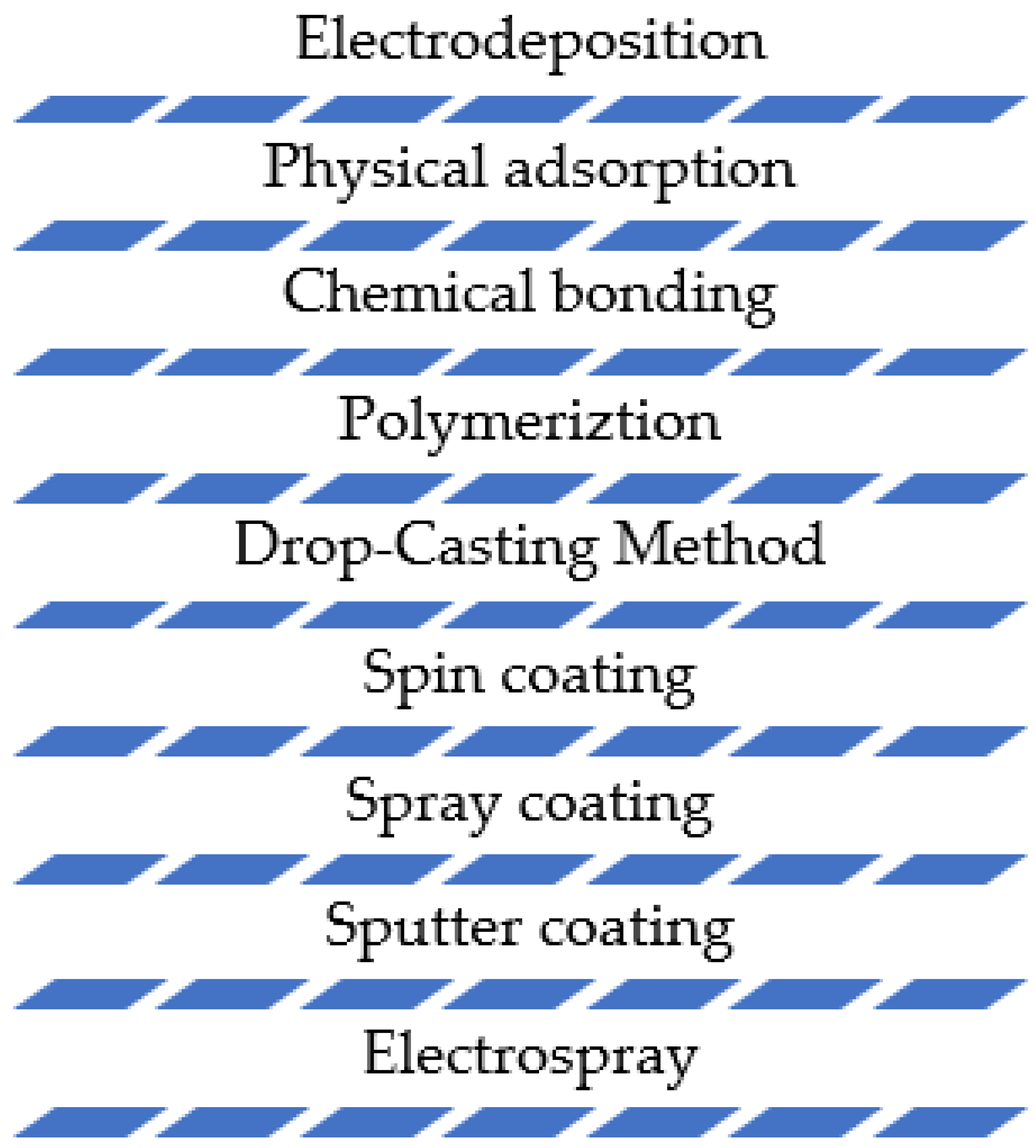
Despite the fact that nanomaterials can provide the above-mentioned advantages in the electrochemical sensing of antibiotics, their immobilization on the electrode surfaces and further functionalization with desired moieties is a quite challenging task. The success of these steps is basically a key factor in improving the electrode’s performance. Additionally, electrochemical signal interferences from other compounds with similar chemical structures can limit the selectivity of these sensors. The methods that are currently being employed for electrode modification with nanomaterials include electrodeposition, physical adsorption, polymerization, through chemical bonding, etc., as seen in Figure 2.
In recent years, there has been increasing interest in modifying electrode surfaces with nanoparticles to enhance their performance in the determination of antibiotics. These modified electrodes can be used in various electroanalytical techniques, such as voltammetry and amperometry, to detect a wide range of antibiotics. Thus, nanoparticle-modified electrodes have been shown to be effective for the detection of many different antibiotics, including penicillins, cephalosporins, tetracyclines, and quinolones. Some of the most promising nanoparticle materials that have been used for electrode modifications include graphene, carbon nanotubes, metal nanoparticles, and various types of metal oxides. Nanoparticles can provide additional active sites for the adsorption of antibiotics, increase the electrochemical surface area, and improve the electron transfer between the analyte and the electrode; thus, they can provide enhanced sensitivity, selectivity, and stability compared to unmodified electrodes.
Despite the many advantages of nanoparticle-modified electrodes, there are also some challenges and limitations to their use. For instance, the synthesis and functionalization of nanoparticles can be difficult and time-consuming, and the stability of these materials can vary depending on the conditions of the electroanalytical measurement. Additionally, electrochemical signal interferences from other compounds with similar chemical structures can limit the selectivity of these sensors. Conclusively, Figure 3 summarizes some of the most common materials and nanomaterials used in electrode modification [47].
In the case of antibiotics, Kanamycin was sensed using a label-free electrochemical immunosensor that was fabricated as a nanocomposite of thionine (TH)-functionalized graphene sheets (GS), Ag NPs, and magnetic NPs [48]. In this study, graphite powder was used to synthesize graphene oxide through modified Hummer’s method followed by the reduction of graphene oxide to produce graphene sheets. Then, the thionine as a mediator was adsorbed on the graphene sheets by π–π stacking to produce TH-GS, which was used to modify the GCE surface. Afterward, the synthesized Ag@Fe3O4 NPs were mixed with kanamycin antibodies for one day to produce Ag@Fe3O4–Ab. The glutaraldehyde (GA) solution as a bifunctional linker was added to the electrode surface to save aldehyde groups for the combination between TH-GS and Ag@Fe3O4–Ab. Finally, an electrode modified by TH-GS/GA/Ag@Fe3O4–Ab as a label-free immunosensor was used for kanamycin detection. Each component of this TH-GS/GA/Ag@Fe3O4–Ab label-free immunosensor has a role in the sensitive detection of kanamycin. Thus, the sensitivity toward the antibiotic has been enhanced in two ways, enlarging responding electrochemical signals and increasing the loading capacities of antibodies. In this study, kanamycin was accurately detected over the concentration range of 0.1 to 34 nM and a detection limit of 0.04 nM using the SWV technique.

2. Nanoparticle-Modified Electrodes

Nanoparticle-modified electrodes have shown promising results in the sensitive and selective electroanalytical determination of antibiotics. In recent papers, nanomaterials such as carbon nanotubes [49] and graphene oxide [50] have been used to modify electrodes, resulting in improved electrocatalytic properties and enhanced sensitivity and selectivity in electrochemical sensors. For example, Feizollahi at al. [49] developed a new method for detecting sulfamethazine (SMZ) in cow’s milk using a glassy carbon electrode modified with graphene oxide and decorated with Cu-Ag core–shell nanoparticles.
Electrodes modified with nanoparticles could be utilized in multiple electroanalytical applications of a broad range of antibiotics, including chloramphenicol [51], doxorubicin [52], pyrazinamide [53], streptomycin [54], and amoxicillin [55]. Some examples of electrode surfaces modified with nanoparticles that have been used in the determination of antibiotics are shown in Table 1.
These are just a few examples of nanoparticle-modified electrode surfaces that have been used for the electroanalytical determination of antibiotics. There are more examples of electrode surfaces modified with nanoparticles for the sensitive and selective electroanalytical determination of antibiotics. For instance, Shun Liu et al. developed a sensor using a nanocomposite of reduced graphene oxide and silver nanoparticles to detect the antibiotic chloramphenicol. This sensor was proved to be reproducible, stable, and selective over similar interfering substances in order to accurately detect chloramphenicol in milk samples [61]. Cesarino et al. also reported the preparation of a paraffin composite electrode based on multi-walled carbon nanotubes (MWCNT) modified with antimony nanoparticles (SbNPs). The sensor was used to detect two antibiotics, sulfamethoxazole and trimethoprim, using differential pulse voltammetry in natural water samples. The sensor’s structure and electrochemical properties were studied using field emission gun scanning electron microscopy and cyclic voltammetry, respectively [48].
These examples demonstrate the potential of using nanoparticles to modify electrode surfaces for the sensitive and selective electroanalytical determination of antibiotics, but there are many more. In one study, the development of an electrochemical sensor using platinum nanoparticles on carbon (PtNPs/C) for the detection of tetracycline is described. The sensor was first synthesized and characterized using X-ray diffraction and transmission electron microscopy. The researchers then studied how different experimental conditions, such as the number of platinum nanoparticles and the pH of the solution, affected the sensor’s behavior. They used cyclic voltammetry and differential pulse voltammetry to investigate how tetracycline is electro-oxidized on the sensor and determined that the sensor was able to accurately detect tetracycline over a wide range of concentrations in urine samples. This suggests that the sensor may have the potential for use in clinical analysis and quality control [62].
In another study, Zhu et al. developed a copper nanoparticle-based film incorporating a cationic surfactant and graphene for determining the presence of gatifloxacin and perflocacin using differential pulse stripping voltammetry. The film’s surface was analyzed using SEM and EDS, and it was found to have improved electrocatalytic properties due to the combination of copper nanoparticles, graphene, and CTAB surfactants. The modified electrode showed good performance in detecting the drugs, with linear responses at concentrations between 0.02–40 µM and 0.04–20 µM and detection limits of 0.0021 µM and 0.0025 µM, respectively. The sensors were also used to detect the drugs in shrimp and animal serum with successful results, suggesting that the film is a promising catalyst for electrocatalysis [65].
Over the past few decades, various studies have reported on the use of electrochemical methods for accurately measuring the presence of antibiotics. These methods have proven to be effective in detecting these substances in a quantitative manner. Primarily, according to Dai et al. [66], gold nanoparticles (∼30–60 nm in diameter) were deposited onto the surface of glassy carbon microspheres. Later, Wang et al. [67] reported the use of a tetracycline sensor, using a molecularly imprinted, polymer-modified carbon nanotube–gold nanoparticles electrode. Moreover, Bagheri Hashkavayi et al. [68] developed a label-free electrochemical aptasensor for the determination of chloramphenicol based on gold nanocube-modified, screen-printed gold electrodes. Giribabu et al. [69] developed an electrochemical sensor for chloramphenicol determination, using a glassy carbon electrode modified with dendrite-like Fe3O4 nanoparticles, while Prado et al. [70] prepared an electrochemical sensor modified with ruthenium nanoparticles on reduced graphene oxide for the simultaneous determination of amoxicillin. In addition, M. K. L. Da Silva et al. [71] synthesized a sensor with reduced graphene oxide, modified with antimony and copper nanoparticles for levofloxacin oxidation. Guaraldo et al. [72] introduced a glassy carbon (GC) electrode modified with Printex L6 carbon black (PC) and copper (II) phthalocyanine (CuPh) films for the electrochemical determination of the antibiotic trimethoprim. In 2020, Sanz et al. [73] constructed a multi-walled carbon nanotube (CNT) and gold nanoparticle (AuNP)-modified glassy carbon electrode (GCE) for the sensitive determination of cefadroxil antibiotic, while W. da Silva et al. [74] developed a poly(methylene green) film on a Fe2O3 magnetic nanoparticle-modified electrode in sulfuric acid-doped ethaline deep eutectic solvent for the antibiotics dapsone and Vajdle et al. [75] used a carbon paste electrode modified with gold nanoparticles for selected macrolide antibiotics determination as standard and in pharmaceutical preparations. Moreover, in 2021, Feizollahi et al. [49] developed a determination method of sulfamethazine (SMZ) in cow’s milk using a glassy carbon electrode modified with graphene oxide decorated with Cu–Ag core–shell nanoparticles, Olugbenga Osikoya and Poomani Govender [76] developed a benzene-sourced graphene–gold nanoparticle sensor for the detection of tetracycline. Mahmoudpour et al. [77] used reduced graphene oxide and a nanogold-functionalized poly(amidoamine) dendrimer for ciprofloxacin determination in real samples, and Zeb et al. [78] used a magnetic nanoparticles/MIP-based electrochemical sensor for the quantification of tetracycline in milk samples. Table 2 provides a summary of the above-mentioned studies on using nanoparticle-modified electrodes for measuring antibiotic levels in different types of samples.
When using solid electrodes in electrochemical applications, limitations such as a narrow potential range, electrode fouling, low detection limits, poor sensitivity, and reproducibility are often encountered. To address these issues, various strategies have been developed to modify the surfaces of electrodes using different redox-active molecules, perm-selective polymers, and electrode activation methods. These modifications result in improved electrode activity and sensitivity towards the target material, faster electron transfer between the analyte and electrode, increased electrocatalytic activity with a larger surface area, faster diffusion at the electrode surface, less interference, and reduced fouling [79,80].
In addition to their electrocatalytic properties, these nanoparticle-modified electrodes have the advantages of low cost, high sensitivity, and convenient operability, making them very promising to overcome potential interferences from analogs with similar chemical structures and to improve the stability and selectivity of these sensors.
Recently, two-dimensional nanomaterials have received great attention in the field of electrochemical applications because of their outstanding physicochemical properties [81]. Therefore, vanadium carbide entrapped on graphitic carbon nitride nanosheets (VC/g-CN NSs) was used as a highly efficient electrocatalyst for the determination of furazolidone (FZD) in biological samples. The electrochemical performance of the VC/g-CN NSs-modified glassy carbon electrode (GCE) for FZD detection was studied using cyclic voltammetry and amperometric methods. Thus, the proposed electrode offered active sites with a large electron-transfer rate and a rapid mass transport ability to enhance the electrochemical activity toward FZD detection. Under the optimization conditions, the VC/g-CN NSs modified electrode showed a wide detection range (0.004–141 μM), a very low detection limit (0.5 nM), and excellent selectivity and reproducibility.

3. Conclusions and Future Challenges

The present review highlighted the use of nanomaterials as electrode surface modifiers in the detection of antibiotics. It discusses the most recent and novel trends in electrochemical sensing and the use of nanoparticles in the detection of antibiotics.
Furthermore, the sensitivity and practicality of electrochemically sensing antibiotics can be enhanced by further focusing on the fabrication of nanoelectrodes or multi-nanoelectrode arrays. The combination of various advanced nanomaterials to generate a nanoelectrode can lead to an ultra-sensitive electrode surface with far greater potential implementations, notably for in vivo applications.
As the need for smaller, quicker, cost-effective, and ultrasensitive sample qualification and quantification grows, electrochemical methods and nanoparticles offer a viable path to the next generation of electrochemical sensors. Further advances in the synthesis of nanomaterials and the fabrication of nanoelectrodes have the potential to benefit society not only in the detection of antibiotics but also in a wide range of areas, with the prospect of real-time, point-of-care analyte detection slowly becoming a reality. The requirement for a reproducible, low-cost production process for nanomaterial-based devices on an economically beneficial scale while maintaining the accuracy achieved in a laboratory environment remains a major challenge for the commercial development of such applications.
Overall, nanoparticle-modified electrodes have demonstrated great potential for the sensitive and selective determination of antibiotic residues in various samples. This field will continue to evolve as new and better methods for analyzing the surface of electrodes are developed. However, further research is needed in order to improve the stability of nanoparticle-modified electrodes. The stability of nanoparticle-modified electrodes can vary depending on the conditions of the electroanalytical measurement. Finally, different strategies need to be developed to overcome interferences from other compounds in order to improve the accuracy of the measurements.

Author Contributions

Conceptualization, S.K. and S.G.; methodology, S.K. and S.G.; software, S.K. and C.S.; validation, S.K., S.G. and C.S.; formal analysis, S.K. and C.S.; investigation, S.K. and C.S.; resources, S.K.; data curation, S.K., C.S. and S.G.; writing—original draft preparation, S.K., C.S. and S.G.; writing—review and editing, S.K., C.S. and visualization, S.K., C.S.; supervision, S.G.; project administration, S.K., C.S. and S.G.; funding acquisition, S.K., C.S. and S.G. All authors have read and agreed to the published version of the manuscript.

Funding

This research received no external funding.

Institutional Review Board Statement

Not applicable.

Informed Consent Statement

Not applicable.

Data Availability Statement

Not applicable.

Conflicts of Interest

The authors declare no conflict of interest.

References

  1. Aminov, R.I. A Brief History of the Antibiotic Era: Lesson Learned and Challenges for the Future. Front. Microbiol. 2010, 1, 134. [Google Scholar] [CrossRef] [PubMed]
  2. Adedeji, W.A. The Treasure Called Antibiotics. Ann. Ib. Postgrad. Med. 2016, 14, 56–57. [Google Scholar]
  3. Hutchings, M.; Truman, A.; Wilkinson, B. Antibiotics: Past, present and future. Curr. Opin. Microbiol. 2019, 51, 72–80. [Google Scholar] [CrossRef] [PubMed]
  4. Prestinaci, F.; Pezzotti, P.; Pantosti, A. Antimicrobial resistance: A global multifaceted phenomenon. Pathog. Glob. Health 2015, 109, 309–318. [Google Scholar] [CrossRef]
  5. Foulston, L. Genome mining and prospects for antibiotic discovery. Curr. Opin. Microbiol. 2019, 51, 1–8. [Google Scholar] [CrossRef]
  6. Li, Z.; Sobek, A.; Radke, M. Fate of Pharmaceuticals and Their Transformation Products in Four Small European Rivers Receiving Treated Wastewate. Environ. Sci. Technol. 2016, 50, 5614–5621. [Google Scholar] [CrossRef]
  7. Chen, Y.; Jiang, C.; Wang, Y.; Song, R.; Tan, Y.; Yang, Y.; Zhang, Z. Sources, Environmental Fate, and Ecological Risks of Antibiotics in Sediments of Asia’s Longest River: A Whole-Basin Investigation. Environ. Sci. Technol. 2022, 56, 14439–14451. [Google Scholar] [CrossRef]
  8. Seifrtová, M.; Nováková, L.; Lino, C.; Pena, A.; Solich, P. An Overview of Analytical Methodologies for the Determination of Antibiotics in Environmental Waters. Anal. Chim. Acta 2009, 649, 158–179. [Google Scholar] [CrossRef] [PubMed]
  9. Stockwell, V.O.; Duffy, B. Use of antibiotics in plant agriculture. Rev. Sci. Tech. Off. Int. Des. Epizoot. 2012, 31, 199–210. [Google Scholar] [CrossRef]
  10. Chen, J.; Sun, R.; Pan, C.; Sun, Y.; Mai, B.; Li, Q.X. Antibiotics and Food Safety in Aquaculture. J. Agric. Food Chem. 2020, 68, 11908–11919. [Google Scholar] [CrossRef]
  11. Akhil, D.; Lakshmi, D.; Kumar, P.S.; Vo, D.-V.N.; Kartik, A. Occurrence and removal of antibiotics from industrial wastewater. Environ. Chem. Lett. 2021, 19, 1477–1507. [Google Scholar] [CrossRef]
  12. McNulty, C.A.M.; Collin, S.M.; Cooper, E.; Lecky, D.M.; Butler, C.C. Public understanding and use of antibiotics in England: Findings from a household survey in 2017. BMJ Open 2019, 9, e030845. [Google Scholar] [CrossRef] [PubMed]
  13. Ur Rehman, M.S.; Rashid, N.; Ashfaq, M.; Saif, A.; Ahmad, N.; Han, J.I. Global risk of pharmaceutical contamination from highly populated developing countries. Chemosphere 2015, 138, 1045–1055. [Google Scholar] [CrossRef]
  14. Adzitey, F. Antibiotic classes and antibiotic susceptibility of bacterial isolates from selected poultry; a mini review. World Vet. J. 2015, 5, 36–41. [Google Scholar] [CrossRef]
  15. Mirzaei, R.; Yunesian, M.; Nasseri, S.; Gholami, M.; Jalilzadeh, E.; Shoeibi, S.; Bidshahi, H.S.; Mesdaghinia, A. An optimized SPE-LC-MS/MS method for antibiotics residue analysis in ground, surface and treated water samples by response surface methodology- central composite design. J. Environ. Health Sci. Engineer. 2017, 15, 21. [Google Scholar] [CrossRef]
  16. Semail, N.F.; Abdul Keyon, A.S.; Saad, B.; Kamaruzaman, S.; Mohamad Zain, N.N.; Lim, V.; Miskam, M.; Wan Abdullah, W.N.; Yahaya, N.; Chen, D.D.Y. Simultaneous Preconcentration and Determination of Sulfonamide Antibiotics in Milk and Yoghurt by Dynamic PH Junction Focusing Coupled with Capillary Electrophoresis. Talanta 2022, 236, 122833. [Google Scholar] [CrossRef] [PubMed]
  17. Senthilkumar, M.; Amaresan, N.; Sankaranarayanan. Detection of Pyoluteorin by Thin Layer Chromatography. In Plant-Microbe Interactions. Springer Protocols Handbooks; Humana: New York, NY, USA, 2021. [Google Scholar] [CrossRef]
  18. Barzallo, D.; Palacio, E.; March, J.; Ferrer, L. 3D Printed Device Coated with Solid-Phase Extraction Resin for the on-Site Extraction of Seven Sulfonamides from Environmental Water Samples Preceding HPLC-DAD Analysis. Environ. Anal. Chem. Gr. 2022, 25, 108609. [Google Scholar] [CrossRef]
  19. Shen, F.; Xu, Y.J.; Wang, Y.; Chen, J.; Wang, S. Rapid and Ultra-Trace Levels Analysis of 33 Antibiotics in Water by on-Line Solid-Phase Extraction with Ultra-Performance Liquid Chromatography-Tandem Mass Spectrometry. J. Chromatogr. A 2022, 1677, 463304. [Google Scholar] [CrossRef]
  20. Khatibi, S.A.; Hamidi, S.; Siahi-Shadbad, M.R. Application of Liquid-Liquid Extraction for the Determination of Antibiotics in the Foodstuff: Recent Trends and Developments. Crit. Rev. Anal. Chem. 2022, 52, 327–342. [Google Scholar] [CrossRef]
  21. Eyken, A.; Furlong, D.; Arooni, S.; Butterworth, F.; Roy, J.F.; Zweigenbaum, J.; Bayen, S. Direct injection high performance liquid chromatography coupled to data independent acquisition mass spectrometry for the screening of antibiotics in honey. J. Food Drug Anal. 2019, 27, 679–691. [Google Scholar] [CrossRef]
  22. Bayen, S.; Yi, X.Z.; Segovia, E.; Zhou, Z.; Kelly, B.C. Analysis of selected antibiotics in surface freshwater and seawater using direct injection in liquid chromatography electrospray ionization tandem mass spectrometry. J. Chromatogr. A 2014, 1338, 38–43. [Google Scholar] [CrossRef] [PubMed]
  23. Tran, T.T.T.; Do, M.N.; Dang, T.N.H.; Tran, Q.H.; Le, V.T.; Dao, A.Q.; Vasseghian, Y. A State-of-the-Art Review on Graphene-Based Nanomaterials to Determine Antibiotics by Electrochemical Techniques. Environ. Res. 2022, 208, 112744. [Google Scholar] [CrossRef] [PubMed]
  24. Materón, E.M.; Wong, A.; Freitas, T.A.; Faria, R.C.; Oliveira, O.N. A Sensitive Electrochemical Detection of Metronidazole in Synthetic Serum and Urine Samples Using Low-Cost Screen-Printed Electrodes Modified with Reduced Graphene Oxide and C60. J. Pharm. Anal. 2021, 11, 646–652. [Google Scholar] [CrossRef] [PubMed]
  25. Hong, J.; Su, M.; Zhao, K.; Zhou, Y.; Wang, J.; Zhou, S.-F.; Lin, X. A Minireview for Recent Development of Nanomaterial-Based Detection of Antibiotics. Biosensors 2023, 13, 327. [Google Scholar] [CrossRef] [PubMed]
  26. Wang, Q.; Xue, Q.; Chen, T.; Li, J.; Liu, W.; Shan, X.; Liu, F.; Jia, J. Recent advances in electrochemical sensors for antibiotics and their applications. Chin. Chem. Lett. 2021, 32, 609–619. [Google Scholar] [CrossRef]
  27. Alsaiari, N.S.; Katubi, K.M.M.; Alzahrani, F.M.; Siddeeg, S.M.; Tahoon, M.A. The Application of Nanomaterials for the Electrochemical Detection of Antibiotics: A Review. Micromachines 2021, 12, 308. [Google Scholar] [CrossRef]
  28. Simoska, O.; Gaffney, E.M.; Minteer, S.D.; Franzetti, A.; Cristiani, P.; Grattieri, M.; Santoro, C. Recent trends and advances in microbial electrochemical sensing technologies: An overview. Curr. Opin. Electrochem. 2021, 30, 100762. [Google Scholar] [CrossRef]
  29. Cox, J.A.; Tess, M.E.; Cummings, T.E. Electroanalytical Methods Based on Modified Electrodes: A Review of Recent Advances. Rev. Anal. Chem. 1996, 15, 173–223. [Google Scholar] [CrossRef]
  30. Gan, T.; Shi, Z.X.; Sun, J.Y.; Liu, Y.M. Simple and novel electrochemical sensor for the determination of tetracycline based on iron/zinc cations–exchanged montmorillonite catalyst. Talanta 2014, 121, 187–193. [Google Scholar] [CrossRef]
  31. Zhang, P.P.; Zhang, N.N.; Jing, L.J.; Hu, B.B.; Yang, X.D.; Ma, X.L. Silver Nanoparticles/Carboxylic Short-Chain Multi-Wall Carbon Nanotubes as Electrochemical Sensor for Ultrasensitive Detection of Chloramphenicol in Food. Int. J. Electrochem. Sci. 2019, 14, 9337–9346. [Google Scholar] [CrossRef]
  32. Rouhbakhsh, Z.; Verdian, A.; Rajabzadeh, G. Design of a liquid crystal-based aptasensing platform for ultrasensitive detection of tetracycline. Talanta 2020, 206, 120246. [Google Scholar] [CrossRef] [PubMed]
  33. Brett, C.M.A.; Brett, A.M.O. Electrochemistry: Principles, Methods, and Applications; Oxford University Press: Oxford, UK, 2009. [Google Scholar]
  34. Fujihira, M. Modified Electrodes. Top. Org. Electrochem. 1986, 10, 255–294. [Google Scholar] [CrossRef]
  35. Edwards, G.A.; Bergren, A.J.; Porter, M.D. Chemically Modified Electrodes. Handb. Electrochem. 2007, 8, 295–327. [Google Scholar] [CrossRef]
  36. Murray, R.W. Chemically Modified Electrodes for Electroanalysis. Philos. Trans. R. Soc. Lond. 1981, 302, 135–141. [Google Scholar]
  37. Chandra, P.; Noh, H.-B.; Won, M.-S.; Shim, Y.-B. Detection of daunomycin using phosphatidylserine and aptamer co-immobilized on Au nanoparticles deposited conducting polymer. Biosens. Bioelectron. 2011, 26, 4442–4449. [Google Scholar] [CrossRef]
  38. Hu, Y.; Chandra, P.; Song, K.-M.; Ban, C.; Shim, Y.-B. Label-Free detection of kanamycin based on the aptamer-functionalized conducting polymer/gold nanocomposite. Biosens. Bioelectron. 2012, 36, 29–34. [Google Scholar]
  39. Hu, C.; Hu, S. Carbon Nanotube-Based Electrochemical Sensors: Principles and  Applications  in  Biomedical  Systems. J. Sens. 2009, 2009, 187615. [Google Scholar] [CrossRef]
  40. Cheng, G.; Zhao, J.; Tu, Y.; He, P.; Fang, Y. A sensitive DNA electrochemical biosensor based on magnetite with a glassy carbon electrode modified by muti-walled carbon nanotubes in polypyrrole. Anal. Chim. Acta 2005, 533, 11–16. [Google Scholar] [CrossRef]
  41. Wang, S.G.; Wang, R.; Sellin, P.J.; Zhang, Q. DNA biosensors based on self-assembled carbon nanotubes. Biochem. Biophys. Res. Commun. 2004, 325, 1433–1437. [Google Scholar] [CrossRef]
  42. Kerman, K.; Morita, Y.; Takamura, Y.; Tamiya, E. Escherichia coli single-strand binding protein-DNA interactions on carbon nanotube-modified electrodes from a label-free electrochemical hybridization sensor. Anal. Bioanal. Chem. 2005, 381, 1114–1121. [Google Scholar] [CrossRef]
  43. Komersová, A.; Bartoš, M.; Kalcher, K.; Vytřas, K. Trace iron determination in aminoisophthalic acid using differential-pulse cathodic stripping voltammetry at carbon paste electrodes. J. Pharm. Biomed. Anal. 1998, 16, 1373–1379. [Google Scholar] [CrossRef] [PubMed]
  44. Oyama, M. Recent Nanoarchitectures in Metal Nanoparticle-modified Electrodes for Electroanalysis. Anal. Sci. 2010, 26, 1–12. [Google Scholar] [CrossRef] [PubMed]
  45. Torres-Rivero, K.; Florido, A.; Bastos-Arrieta, J. Recent Trends in the Improvement of the Electrochemical Response of Screen-Printed Electrodes by Their Modification with Shaped Metal Nanoparticles. Sensors 2021, 21, 2596. [Google Scholar] [CrossRef] [PubMed]
  46. Yu, S.; Wei, Q.; Du, B.; Wu, D.; Li, H.; Yan, L.; Ma, H.; Zhang, Y. Label-Free immunosensor for the detection of kanamycin using Ag@ Fe3O4 nanoparticles and thionine mixed graphene sheet. Biosens. Bioelectron. 2013, 48, 224–229. [Google Scholar] [CrossRef]
  47. Baig, N.; Sajid, M.; Saleh, T.A. Recent trends in nanomaterial-modified electrodes for electroanalytical applications. TrAC Trends Anal. Chem. 2019, 111, 47–61. [Google Scholar] [CrossRef]
  48. Cesarino, I.; Cesarino, V.; Lanza, M.R.V. Carbon Nanotubes Modified with Antimony Nanoparticles in a Paraffin Composite Electrode: Simultaneous Determination of Sulfamethoxazole and Trimethoprim. Sens. Actuators B Chem. 2013, 188, 1293–1299. [Google Scholar] [CrossRef]
  49. Feizollahi, A.; Rafati, A.A.; Assari, P.; Asadpour Joghani, R. Development of an Electrochemical Sensor for the Determination of Antibiotic Sulfamethazine in Cow Milk Using Graphene Oxide Decorated with Cu-Ag Core-Shell Nanoparticles. Anal. Methods 2021, 13, 910–917. [Google Scholar] [CrossRef]
  50. Kokulnathan, T.; Sharma, T.S.K.; Chen, S.M.; Chen, T.W.; Dinesh, B. Ex-Situ Decoration of Graphene Oxide with Palladium Nanoparticles for the Highly Sensitive and Selective Electrochemical Determination of Chloramphenicol in Food and Biological Samples. J. Taiwan Inst. Chem. Eng. 2018, 89, 26–38. [Google Scholar] [CrossRef]
  51. Alavi-Tabari, S.A.R.; Khalilzadeh, M.A.; Karimi-Maleh, H. Simultaneous Determination of Doxorubicin and Dasatinib as Two Breast Anticancer Drugs Uses an Amplified Sensor with Ionic Liquid and ZnO Nanoparticle. J. Electroanal. Chem. 2018, 811, 84–88. [Google Scholar] [CrossRef]
  52. Simioni, N.B.; Silva, T.A.; Oliveira, G.G.; Fatibello-Filho, O. A Nanodiamond-Based Electrochemical Sensor for the Determination of Pyrazinamide Antibiotic. Sens. Actuators B Chem. 2017, 250, 315–323. [Google Scholar] [CrossRef]
  53. Ghanbari, K.; Roushani, M. A Novel Electrochemical Aptasensor for Highly Sensitive and Quantitative Detection of the Streptomycin Antibiotic. Bioelectrochemistry 2018, 120, 43–48. [Google Scholar] [CrossRef] [PubMed]
  54. Nosuhi, M.; Nezamzadeh-Ejhieh, A. Comprehensive Study on the Electrocatalytic Effect of Copper—Doped Nano-Clinoptilolite towards Amoxicillin at the Modified Carbon Paste Electrode—Solution Interface. J. Colloid Interface Sci. 2017, 497, 66–72. [Google Scholar] [CrossRef] [PubMed]
  55. Afkhami, A.; Soltani-Felehgari, F.; Madrakian, T. Gold Nanoparticles Modified Carbon Paste Electrode as an Efficient Electrochemical Sensor for Rapid and Sensitive Determination of Cefixime in Urine and Pharmaceutical Samples. Electrochim. Acta 2013, 103, 125–133. [Google Scholar] [CrossRef]
  56. Manjunatha, P.; Nayaka, Y.A. Cetyltrimethylammonium Bromide-Gold Nanoparticles Composite Modified Pencil Graphite Electrode for the Electrochemical Investigation of Cefixime Present in Pharmaceutical Formulations and Biology. Chem. Data Collect. 2019, 21, 100217. [Google Scholar] [CrossRef]
  57. Kumar, N.; Goyal, R.N. Gold-Palladium Nanoparticles Aided Electrochemically Reduced Graphene Oxide Sensor for the Simultaneous Estimation of Lomefloxacin and Amoxicillin. Sens. Actuators B Chem. 2017, 243, 658–668. [Google Scholar] [CrossRef]
  58. Zhai, H.; Liang, Z.; Chen, Z.; Wang, H.; Liu, Z.; Su, Z.; Zhou, Q. Simultaneous Detection of Metronidazole and Chloramphenicol by Differential Pulse Stripping Voltammetry Using a Silver Nanoparticles/Sulfonate Functionalized Graphene Modified Glassy Carbon Electrode. Electrochim. Acta 2015, 171, 105–113. [Google Scholar] [CrossRef]
  59. Cheng, S.; Liu, H.; Zhang, H.; Chu, G.; Guo, Y.; Sun, X. Ultrasensitive Electrochemiluminescence Aptasensor for Kanamycin Detection Based on Silver Nanoparticle-Catalyzed Chemiluminescent Reaction between Luminol and Hydrogen Peroxide. Sens. Actuators B Chem. 2020, 304, 127367. [Google Scholar] [CrossRef]
  60. Atif, S.; Baig, J.A.; Afridi, H.I.; Kazi, T.G.; Waris, M. Novel Nontoxic Electrochemical Method for the Detection of Cefadroxil in Pharmaceutical Formulations and Biological Samples. Microchem. J. 2020, 154, 104574. [Google Scholar] [CrossRef]
  61. Liu, S.; Lai, G.; Zhang, H.; Yu, A. Amperometric Aptasensing of Chloramphenicol at a Glassy Carbon Electrode Modified with a Nanocomposite Consisting of Graphene and Silver Nanoparticles. Microchim. Acta 2017, 184, 1445–1451. [Google Scholar] [CrossRef]
  62. Kushikawa, R.T.; Silva, M.R.; Angelo, A.C.D.; Teixeira, M.F.S. Construction of an Electrochemical Sensing Platform Based on Platinum Nanoparticles Supported on Carbon for Tetracycline Determination. Sens. Actuators B Chem. 2016, 228, 207–213. [Google Scholar] [CrossRef]
  63. Karimi-Maleh, H.; Tahernejad-Javazmi, F.; Gupta, V.K.; Ahmar, H.; Asadi, M.H. A Novel Biosensor for Liquid Phase Determination of Glutathione and Amoxicillin in Biological and Pharmaceutical Samples Using a ZnO/CNTs Nanocomposite/Catechol Derivative Modified Electrode. J. Mol. Liq. 2014, 196, 258–263. [Google Scholar] [CrossRef]
  64. Velusamy, V.; Palanisamy, S.; Kokulnathan, T.; Chen, S.W.; Yang, T.C.K.; Banks, C.E.; Pramanik, S.K. Novel Electrochemical Synthesis of Copper Oxide Nanoparticles Decorated Graphene-β-Cyclodextrin Composite for Trace-Level Detection of Antibiotic Drug Metronidazole. J. Colloid Interface Sci. 2018, 530, 37–45. [Google Scholar] [CrossRef] [PubMed]
  65. Zhu, M.; Li, R.; Lai, M.; Ye, H.; Long, N.; Ye, J.; Wang, J. Copper Nanoparticles Incorporating a Cationic Surfactant-Graphene Modified Carbon Paste Electrode for the Simultaneous Determination of Gatifloxacin and Pefloxacin. J. Electroanal. Chem. 2020, 857, 113730. [Google Scholar] [CrossRef]
  66. Dai, X.; Wildgoose, G.G.; Salter, C.; Crossley, A.; Compton, R.G. Electroanalysis Using Macro-, Micro-, and Nanochemical Architectures on Electrode Surfaces. Bulk Surface Modification of Glassy Carbon Microspheres with Gold Nanoparticles and Their Electrical Wiring Using Carbon Nanotubes. Anal. Chem. 2006, 78, 6102–6108. [Google Scholar] [CrossRef]
  67. Wang, H.; Zhao, H.; Quan, X.; Chen, S. Electrochemical Determination of Tetracycline Using Molecularly Imprinted Polymer Modified Carbon Nanotube-Gold Nanoparticles Electrode. Electroanalysis 2011, 23, 1863–1869. [Google Scholar] [CrossRef]
  68. Bagheri Hashkavayi, A.; Bakhsh Raoof, J.; Ojani, R.; HamidiAsl, E. Label-Free Electrochemical Aptasensor for Determination of Chloramphenicol Based on Gold Nanocubes-Modified Screen-Printed Gold Electrode. Electroanalysis 2015, 27, 1449–1456. [Google Scholar] [CrossRef]
  69. Giribabu, K.; Jang, S.C.; Haldorai, Y.; Rethinasabapathy, M.; Oh, S.Y.; Rengaraj, A.; Han, Y.K.; Cho, W.S.; Roh, C.; Huh, Y.S. Electrochemical Determination of Chloramphenicol Using a Glassy Carbon Electrode Modified with Dendrite-like Fe3O4 Nanoparticles. Carbon Lett. 2017, 23, 38–47. [Google Scholar] [CrossRef]
  70. Prado, T.M.; Cincotto, F.H.; Moraes, F.C.; Machado, S.A.S. Electrochemical Sensor-Based Ruthenium Nanoparticles on Reduced Graphene Oxide for the Simultaneous Determination of Ethinylestradiol and Amoxicillin. Electroanalysis 2017, 29, 1278–1285. [Google Scholar] [CrossRef]
  71. Da Silva, M.K.L.; Plana Simões, R.; Cesarino, I. Evaluation of Reduced Graphene Oxide Modified with Antimony and Copper Nanoparticles for Levofloxacin Oxidation. Electroanalysis 2018, 30, 2066–2076. [Google Scholar] [CrossRef]
  72. Guaraldo, T.T.; Goulart, L.A.; Moraes, F.C.; Lanza, M.R.V. Carbon Black Nanospheres Modified with Cu (II)-Phthalocyanine for Electrochemical Determination of Trimethoprim Antibiotic. Appl. Surf. Sci. 2019, 470, 555–564. [Google Scholar] [CrossRef]
  73. Sanz, C.G.; Serrano, S.H.P.; Brett, C.M.A. Electroanalysis of Cefadroxil Antibiotic at Carbon Nanotube/Gold Nanoparticle Modified Glassy Carbon Electrodes. ChemElectroChem 2020, 7, 2151–2158. [Google Scholar] [CrossRef]
  74. da Silva, W.; Queiroz, A.C.; Brett, C.M.A. Poly(Methylene Green)—Ethaline Deep Eutectic Solvent/Fe2O3 Nanoparticle Modified Electrode Electrochemical Sensor for the Antibiotic Dapsone. Sens. Actuators B Chem. 2020, 325, 128747. [Google Scholar] [CrossRef]
  75. Vajdle, O.; Šekuljica, S.; Guzsvány, V.; Nagy, L.; Kónya, Z.; Avramov Ivić, M.; Mijin, D.; Petrović, S.; Anojčić, J. Use of Carbon Paste Electrode and Modified by Gold Nanoparticles for Selected Macrolide Antibiotics Determination as Standard and in Pharmaceutical Preparations. J. Electroanal. Chem. 2020, 873, 114324. [Google Scholar] [CrossRef]
  76. Olugbenga Osikoya, A.; Poomani Govender, P. Electrochemical Detection of Tetracycline on Highly Sensitive Benzene Sourced CVD Graphene-Gold Nanoparticles Nanointerfaces. Electroanalysis 2021, 33, 412–420. [Google Scholar] [CrossRef]
  77. Mahmoudpour, M.; Kholafazad-kordasht, H.; Nazhad Dolatabadi, J.E.; Hasanzadeh, M.; Rad, A.H.; Torbati, M. Sensitive Aptasensing of Ciprofloxacin Residues in Raw Milk Samples Using Reduced Graphene Oxide and Nanogold-Functionalized Poly(Amidoamine) Dendrimer: An Innovative Apta-Platform towards Electroanalysis of Antibiotics. Anal. Chim. Acta 2021, 1174, 338736. [Google Scholar] [CrossRef]
  78. Zeb, S.; Wong, A.; Khan, S.; Hussain, S.; Sotomayor, M.D.P.T. Using Magnetic Nanoparticles/MIP-Based Electrochemical Sensor for Quantification of Tetracycline in Milk Samples. J. Electroanal. Chem. 2021, 900, 115713. [Google Scholar] [CrossRef]
  79. Donini, C.A.; da Silva, M.K.L.; Simões, R.P.; Cesarino, I. Reduced Graphene Oxide Modified with Silver Nanoparticles for the Electrochemical Detection of Estriol. J. Electroanal. Chem. 2018, 809, 67–73. [Google Scholar] [CrossRef]
  80. Meenakshi, S.; Rama, R.; Pandian, K.; Gopinath, S.C.B. Modified Electrodes for Electrochemical Determination of Metronidazole in Drug Formulations and Biological Samples: An Overview. Microchem. J. 2021, 165, 106151. [Google Scholar] [CrossRef]
  81. Kokulnathan, T.; Wang, T.J. Vanadium Carbide-Entrapped Graphitic Carbon Nitride Nanocomposites: Synthesis and Electrochemical Platforms for Accurate Detection of Furazolidone. ACS Appl. Nano Mater. 2020, 3, 2554–2561. [Google Scholar] [CrossRef]

Figure 1. Release of antibiotics into the environment and trophic chain [4].
Applsci 13 05391 g001
Figure 2. Methods that are employed for electrode modification with nanomaterials.
Applsci 13 05391 g002
Figure 3. Common materials used as electrode modifiers [47].
Applsci 13 05391 g003
Table 1. Selected examples of metal nanoparticles used for the modification of electrodes.
NanoparticleReference
Gold nanoparticles[53,55,56]
Gold–palladium nanoparticles[57]
Silver nanoparticles[58,59,60,61]
Platinum nanoparticles[62]
Zinc oxide nanoparticles[63]
Copper oxide nanoparticles[64]
Palladium nanoparticles[46]
Antimony nanoparticles[35]
Table 2. Summarization of selected published papers about different electrodes modified with nanoparticles for the determination of antibiotics in various samples.
Type of ElectrodeAntibioticDetermination TechniqueDetection LimitSampleReference
GCE modified with reduced graphene
oxide (rGO) and silver nanoparticles
chloramphenicolamperometry2 nMmilk[61]
paraffin composite electrode with multi-walled carbon nanotubes (MWCNT) modified with antimony nanoparticlessulfamethoxazole and trimethoprimdifferential pulse voltammetry24 nmol L−1 (6.1 μg L−1) for sulfamethoxazole and 31 nmol L−1 (9.0 μg L−1) for trimethoprimnatural water[48]
GCE modified with platinum nanoparticles supported on carbontetracyclinedifferential pulse voltammetry4.28 μmol L−1urine[62]
CP modified with graphene and copper nanoparticlesgatifloxacin and perflocacindifferential pulse stripping voltammetry0.0021 μM and 0.0025 μM gatifloxacin and perflocacin, respectivelyshrimp and chicken serum[65]
Glassy carbon electrode modified with graphene oxide decorated with Cu–Ag core–shell nanoparticlessulfamethazinesquare wave voltammetry0.46 μMcow’s milk[49]
GCE modified with multi-walled carbon nanotube and gold nanoparticlescefadroxilamperometry0.22 μMcommercial capsules[73]
Benzene-sourced graphene–gold nanoparticle sensortetracyclinechronoamperometry0.16 μMbulk[76]
Glassy carbon electrode modified with dendrite-like Fe3O4 nanoparticleschloramphenicolsquare wave voltammetry0.09 μMshrimp[69]
MIP-modified carbon nanotube–gold nanoparticles electrodetetracyclineCV and electrochemical impedance spectroscopy (EIS)0.04 mMbulk[67]
GCE modified with reduced graphene and Ru nanoparticlesamoxicillinpulse voltammetry1.63 nMurine[70]
GCE with reduced graphene oxide modified with antimony and copper nanoparticleslevofloxacindifferential pulse voltammetry4.1 × 10−8 mol L−1 and 1.7 × 10⁻⁸ mol L−1pharmaceutical tablets[71]
screen-printed gold electrode modified with synthesized gold nanocube/cysteinechloramphenicolsquare wave voltammetry4.0 nMhuman blood serum[68]
Poly(methylene green)–Ethaline deep eutectic solvent/Fe2O3 nanoparticle modified electrodedapsoneDifferential pulse voltammetry, scanning electron microscopy0.33 μMpharmaceutical tablets and river water[74]
GCE modified with reduced graphene oxide and nanogold-functionalized poly(amidoamine)ciprofloxacinsquare wave voltammetry, different pulse voltammetry, chronoamperometry1 nMraw milk[77]
CP electrode modified with gold nanoparticleserythromycin ethylsuccinate (EES), azithromycin (AZI), clarithromycin (CLA), roxithromycin (ROX)square wave voltammetry0.18, 0.045, 1.43, and 0.30 μg mL−1 for EES, AZI, CLA, and ROXpharmaceutical preparations[75]
magnetic nanoparticles/MIP-based electrochemical sensortetracyclinesquare wave voltammetry1.5 × 10−7 mol L−1milk[78]
Disclaimer/Publisher’s Note: The statements, opinions and data contained in all publications are solely those of the individual author(s) and contributor(s) and not of MDPI and/or the editor(s). MDPI and/or the editor(s) disclaim responsibility for any injury to people or property resulting from any ideas, methods, instructions or products referred to in the content.