Article

Fungi-Templated Silver Nanoparticle Composite: Synthesis, Characterization, and Its Applications

by
Francis Joy
1,
Jyothis Devasia
1,
Aatika Nizam
1,*,
Vasantha Veerappa Lakshmaiah
2 and
Suresh Babu Naidu Krishna
3,*
1
Department of Chemistry, CHRIST (Deemed to be University), Bangalore 560029, India
2
Department of Life Sciences, CHRIST (Deemed to be University), Bangalore 560029, India
3
Department of Biomedical and Clinical Technology, Durban University of Technology, Durban 4000, South Africa
*
Authors to whom correspondence should be addressed.
Appl. Sci. 2023, 13(4), 2158; https://doi.org/10.3390/app13042158
Submission received: 4 January 2023 / Revised: 28 January 2023 / Accepted: 6 February 2023 / Published: 8 February 2023
(This article belongs to the Special Issue Green Synthesis and Application of Silver Nanoparticles)

Abstract

:
The self-assembly of nanoparticles on living bio-templates is a promising synthetic methodology adopted for synthesizing nano/microstructures with high efficiency. Therefore, the method of bio-templating offers various advantages in controlling the geometries of nano/microstructures, thereby increasing the efficiency of the synthesized material towards various functional applications. Herein, we utilized a filamentous fungus (Sclerotium rolfsii) as a soft bio-template to generate silver nanoparticle (AgNP) microtubules adhering to the fungal hyphae. The resulting composite combines the unique properties of silver nanoparticles with the biological activity of the fungi. The 3D fungal hyphae–silver nanoparticle (FH-AgNP) composite was characterized using SEM, elemental analysis, and the X-ray diffraction technique. Additionally, to highlight the functional application of the synthesized composite, dye degradation studies of methylene blue under visible light was effectuated, and a percentage degradation of 67.86% was obtained within 60 min, which highlights the potent catalytic activity of FH-AgNPs in dye degradation. Further, the antibacterial study of the composite was carried out against the bacterium Escherichia coli, and it was found that 200 μg of the composite exhibited maximum antibacterial properties against Gram positive (Staphylococcus aureus) and Gram negative (Escherichia coli) bacteria. Overall, fungi-templated silver nanoparticle composites are a promising area of research due to their combination of biological activity and unique physical and chemical properties.

1. Introduction

The designing and fabrication of bio-templates of microbial origin open up a wide range of applications in nanotechnology, due to the generation of highly functionalized nano/microstructures providing high surface areas [1]. The synthesis of nanomaterials having tuneable and well-defined structures is one of the most promising breakthroughs in this field and furthermore the most challenging part [2]. Therefore, the method of bio-templating offers various advantages in controlling the geometries of nano/microstructures, making an overall increase in the efficiency of the synthesized material [3]. Moreover, bio-templating being a bottom-up method for the generation of nanomaterials aids a low-cost synthesis of these materials, having a versatile and well-defined structure [4]. In the process of bio-templating, a material of choice (e.g., metal nanoparticles) can be deposited on the microbial surface using simple techniques and the nano/microstructures can be obtained in the shape of the template [5]. After the deposition, the template can be partially or completely removed using incineration and thereby engendering the free nano/microstructure [6]. These synthesized micro/nanostructures can be used with or without the microbial association for various applications such as catalysis, chemical degradation, sensing, and in medical applications [7,8]. Since these assemblies are from nature, they always perform exceptionally great when compared to all other assemblies devoid of any biological framework [5,7,8,9]. Furthermore, the binding of metal NPs to the microbial cell wall opens favourable circumstances for the study of the physical and chemical properties of bio–inorganic interfaces [10,11].
Microorganisms and their structural parts, such as mycelium, hyphae, filament, spores, and flagella, are the most favourable bio-templates due to easy availability, rapid and mass multiplication, simple growth conditions, and a short life cycle [12]. The cell wall of microorganisms contains a wide range of functional groups such as carboxyl, sulphonate, amino, and phosphate groups that help in metal binding and nucleation, thereby forming NPs/microstructures on their surface [13,14]. Recently, fungal hyphae have been used as bio-templates for the self-organization of noble metal NPs forming metal nano/microstructures [6]. Fungi are generally preferred over other microorganisms because they grow at a very rapid rate and the biomass produced for templating is comparatively large [15]. However, only very few reports are available on organizing metal nanoparticles on fungal hyphae, and therefore, a simple, green, and efficient method should be developed for the same [16]. In most of the earlier reports, the NPs are functionalized before templating, which is again a tedious process. Sclerotium rolfsii is a necrotrophic plant pathogen which is known to produce abundant white mycelium on culture. The cells are found to be hyaline, which possess thin cell walls and sparse cross walls. Due to the presence of ample mycelium, Sclerotium rolfsii serves as a suitable bio-template for the assembly of metal nanoparticles, thereby generating a microtubule metal nanoparticle composite. The varying structural morphology of the microtubule composite enables the utilization of the same for numerous applications including catalysis, drug storage, and delivery. Silver nanoparticles (AgNPs) are well known for their ability to kill microorganisms. This makes them useful in the production of antimicrobial products, such as clothing, wound dressing, and food packaging. They have also been shown to be effective at killing certain types of bacteria, such as E. coli and S. aureus, and viruses, such as the flu virus. In addition to their antimicrobial properties, AgNPs have several other useful properties. They are good conductors of electricity and heat, making them useful in electronic and thermal management applications. They are also highly reflective, making them useful in coating and other optical applications [17].
Fungal-mycelium-based composite materials pose significant advantages considering their environmental benign nature and the notable functional properties which can be efficiently utilized for several applications. Fungal mycelium consists of a network of microfilaments whose diameter ranges between 2–30 μm, depending on the species and the environment. The various hyphal colonies interact with each other, forming a network-like structure which efficiently helps in binding various substrates and thereby generating the functional composite materials. Low environmental impact, low cost, and excellent biodegradability are some of the key incentives that enable these composite materials to stand up along with conventional engineered composite materials such as carbon nanotubes, fibres, etc. [18]. The mycelium is found to be a natural polymeric fibrous composite mainly composed of chitin, cellulose, and proteins. Due to the presence of these functionalities in the fungal mycelium, it can readily bind to various substrates via various covalent and non-covalent interactions, thereby forming highly functional composite materials that can be used for varying applications. Moreover, these functional properties can be tuned by changing the substrates that bind to the mycelium.
Various dyes released as effluents from industries causes non-aesthetic pollution and eutrophication in aquatic ecosystems. Since the international environmental standards are becoming more stringent, scientists have devoted a significant amount of time and effort to the removal of these hazardous compounds from water bodies. The developed technologies include various physical, chemical, and biological methods which have been applied for the efficient removal of dye compounds from aquatic ecosystems. Among the developed technologies for dye disposal, photocatalysis have exhibited excellent results in terms of degradation of dye in various industrial waste water [19]. Methylene blue is an intensely coloured industrially important dye which is used for dying and printing textiles. It is a cationic dye which is found to be carcinogenic, environmentally persistent, mutagenic, and toxic. When released into the external environment, methylene blue poses a great threat to flora and fauna and is known to cause fatal serotonin toxicity in human beings. Therefore, it is highly imperative to eradicate the dye and the associated chemical compounds from the ecosystem. To carry out photocatalytic degradation of an organic dye, the dye is typically mixed with the photocatalyst and exposed to light. The light activates the photocatalyst, causing it to generate highly reactive species such as free radicals, which can break down the organic dye into smaller, less harmful components. This process can be enhanced by adding other substances such as hydrogen peroxide to the reaction mixture, which can help to increase the efficiency of the degradation process.
Herein, we report the synthesis of AgNPs using an earlier reported method and the bio-templating of the synthesized AgNPs on the fungal hyphae of Sclerotium rolfsii using a simple and efficient method. This method helped in the development of a 3D fungal hyphae–silver nanoparticle (FH-AgNPs) composite, and this composite was characterized using scanning electron microscopy (SEM), elemental analysis (EDAX), and the X-ray diffraction technique. Additionally, to highlight the application of the synthesized composite, degradation of methylene blue under visible light using the composite as catalyst was carried out. Further, the antibacterial study of the composite was carried out against the bacterium Escherichia coli and Staphylococcus aureus. Biogenic AgNPs were prepared economically using a plant extract rich in secondary metabolites that facilitates the stable synthesis. A colloidal suspension of AgNPs was used to synthesize FH-AgNPs. The hybrid stable structure of FH-AgNPs was produced due to the accumulation of NPs on the surface of fungal hyphae. The thicknesses of these microstructures depend on the incubation period wherein the NPs assemble as multilayers on the biomass. Studies emphasize that heavy metals such as silver can interfere with fungal growth and metabolism, and hence spores were germinated to adequate biomass in nutritional media devoid of AgNPs. The harvested biomass was suspended in AgNPs that serve as a framework for the self-assembling of NPs to form stable heterogeneous microtubules. Fungal cells, especially the chitin and chitosan, are associated with an amino group that encourages effective assembling of AgNPs on hyphae due to electrostatic attraction between the fungal cell wall and negatively charged AgNPs. Furthermore, fungi secrete adhesive proteins and project glycoproteins on the surface of the cell wall that can also improve the self-assembling of nanoparticles.

2. Materials and Methods

2.1. General Remarks

Silver nitrate was purchased from Sigma Aldrich, Bangalore, India and was used as received. All the other chemicals including those required for the preparation of the growth medium were purchased from Sigma Aldrich. Mango leaves were collected from the nearby premises of CHRIST University, Bangalore. Water was previously deionized using Millipore Elix-3 equipment, Merck, Bangalore, India. Electronic spectra of the compounds were recorded using Thermoscientific Evolution 220 spectrophotometer in the 200–900 nm range. The scanning electron microscopic images were taken in a ESEM Quanta 200. FEI. X-ray Diffractometer analysis was carried out with Bruker D 8 Advance with Cu-Kα radiation whose wavelength k was 1.5406 Å, operated at 40 kV and 20 mA in the range of 10° to 90°.

2.2. Preparation of Leaf Extract

Fresh leaves of Mangifera indica were collected and washed with distilled water. The leaves were dried under shadow at room temperature and approximately 10 gm of the dried leaves were taken and cut into small pieces. The leaves were then added to 100 mL of deionized water and boiled for 20 min at 70 °C. The boiled mixture was filtered through Whatman No.1 filter paper and the extract was used without purification [20].

2.3. Growth and Harvest of Fungal Mycelia

Spores of Sclerotium rolfi were inoculated into potato dextrose broth under aseptic conditions and maintained at room temperature without disturbing on static incubation. Spore germination to mycelium was observed after 48 h of incubation. This fungal culture after a week of incubation had grown to lawn of mycelium covering the entire surface area. The fungal mycelial mat was harvested by centrifugation at 5000 rpm for 3 min.

2.4. Synthesis of AgNPs

Synthesis of AgNPs using mango leaves extract was carried out using a previously reported procedure [21]. First, 10 mmol of an aqueous AgNO3 solution was taken in a round-bottomed flask. To this, 25 mL of leaf extract was added and stirred for 3 h at 80 °C. The reaction was stopped when the colour of the solution changed from yellow to dark brown, indicating the formation of AgNPs. The suspension was then used for bio-templating the fungal hyphae of Sclerotium rolfsii.

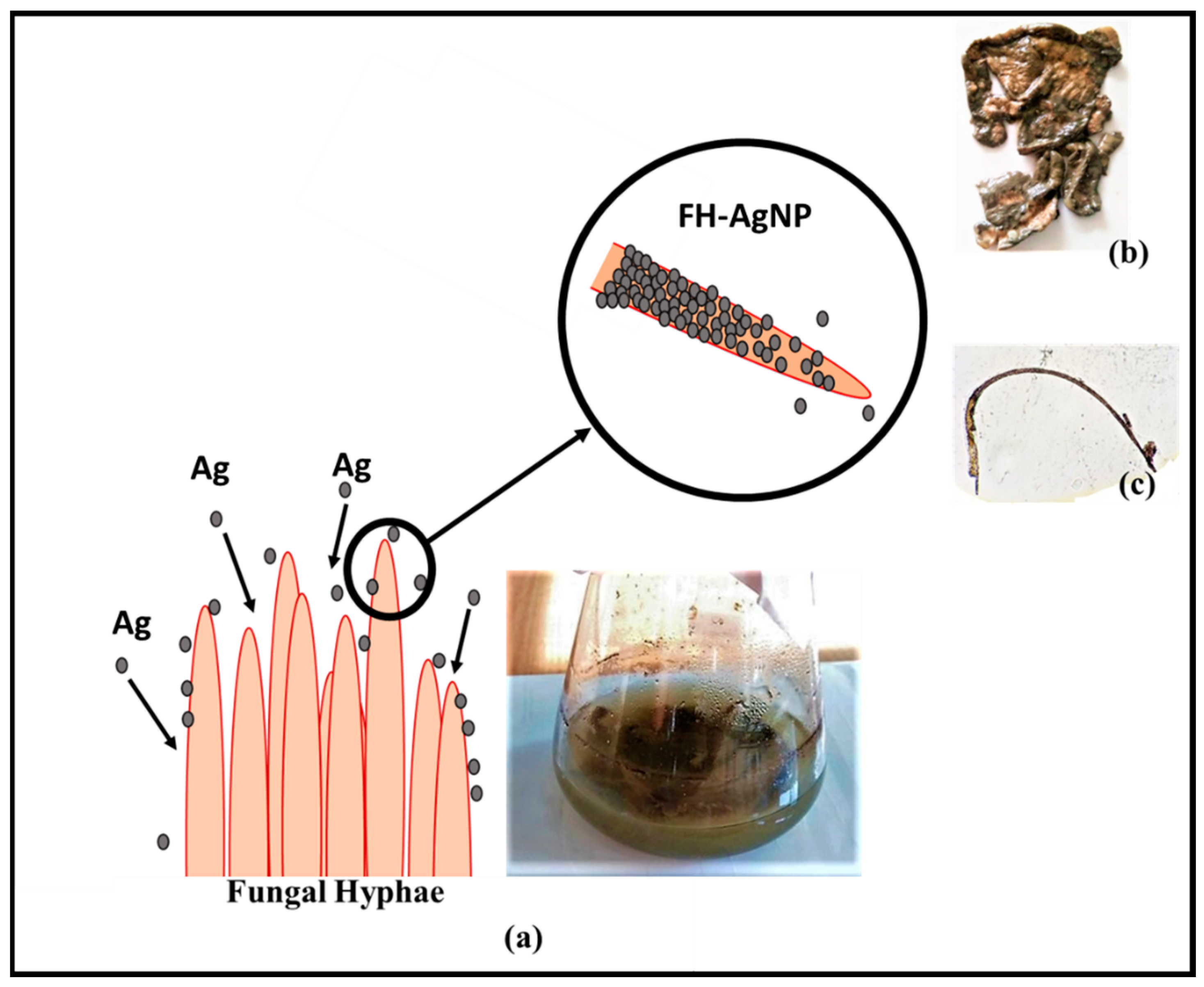
2.5. Preparation of Fungal Hyphae–Silver Nanoparticle (FH-AgNP) Composite

The fungus (Sclerotium rolfsii) was grown initially in agar medium maintained at room temperature. The fungal biomass was taken out from the medium before sporulation for templating. Later, the biomass was added to the AgNP suspension and was kept for incubation with stirring for four days. The colour of the biomass changed from white to black, indicating the complete deposition of AgNPs on the fungal hyphae. The biomass was then taken out and washed with deionized water 5 times to remove the loosely bound particles. The silver deposited biomass (FH-AgNPs) was then dried in a hot air oven at 50 °C for 5 h (Figure 1). The dried biomass was then used for characterization.

2.6. Degradation Studies

The catalytic activity of the FH-AgNPs was assessed by degradation of methylene blue (MB) at room temperature. The degradation studies were carried out in a quartz tube using a visible photoreactor. A tungsten halogen bulb of 364 nm and 250 W was used as the light source. In order to gain the absorption–desorption equilibrium of the dye, the dye sample (20 ppm) was bubbled with the catalyst (30 mg) for 30 min in the dark before irradiation. Approximately 3 mL of the solution was taken out at definite time intervals during the reaction and the catalyst particles were removed by centrifugation. A UV-Visible spectrometer was used to find out the percentage degradation of the dye with respect to time.
The catalytic efficiency was calculated using the formula
%   o f   d e g r a d a t i o n = C 0 C t C 0 × 100
where C 0 is the initial concentration and   C t is the concentration at a particular time interval.

2.7. Antibacterial Studies

Agar well diffusion studies were used to analyse the antibacterial properties of the developed composite against Gram negative Escherichia coli (E. coli) and Gram positive Staphylococcus aureus (S. aureus) bacteria. To perform antimicrobial studies, Muller–Hinton (MH) agar media plates were prepared. E. coli and S. aureus were cultured in Luria Bertani broth and its growth was monitored at 600 nm. The spread plate method was used to inoculate on MH media. A gel puncture was used to make wells of 10 mm in each petri plate. To assess the antibacterial property, various concentrations (50–400 μg) of FH-AgNPs were used. The compound for investigation was prepared in water (2 mg/mL) and using a sterile micropipette it was added into different wells as 50, 100, 200, and 400 μg, respectively. The plates were then incubated at room temperature for 24 h and after incubation the zone of inhibition was measured.

3. Results and Discussion

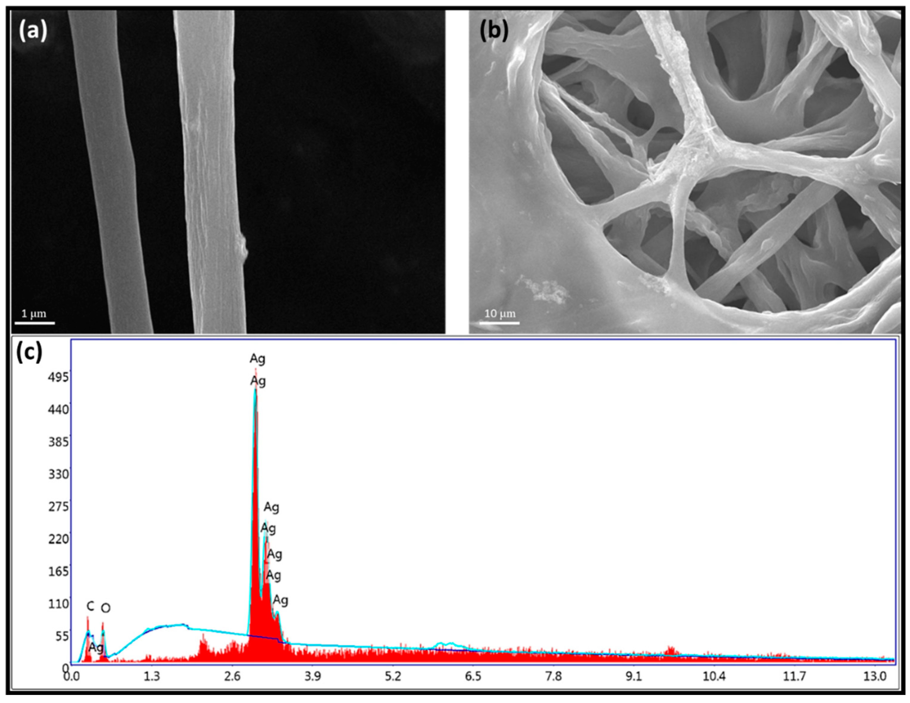
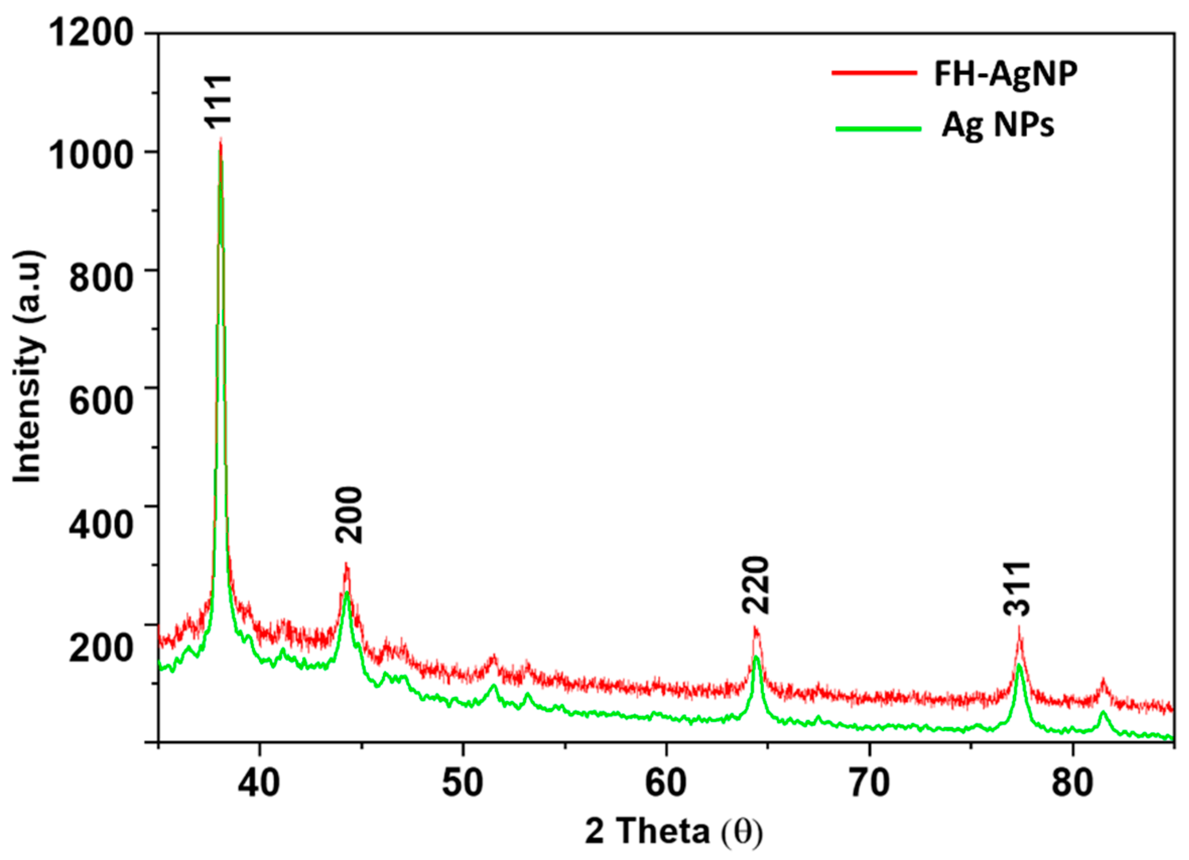
3.1. Morphological Characterization

Biogenic AgNPs were prepared economically using a plant extract rich in secondary metabolites that facilitates the stable synthesis. A colloidal suspension of AgNPs was used to synthesize FH-AgNPs. The hybrid stable structure of FH-AgNPs was produced due to the accumulation of NPs on the surface of fungal hyphae. The thicknesses of these microstructure depend on the incubation period, wherein the NPs assemble as multilayers on the biomass. Studies emphasize that heavy metals such as silver can interfere with fungal growth and metabolism, and hence spores were germinated to adequate biomass in nutritional media devoid of AgNPs. The harvested biomass was suspended in AgNPs that serve as a framework for the self-assembling of NPs to form stable heterogeneous microtubules. Fungal cells, especially the chitin and chitosan, are associated with amino groups that encourage effective assembling of AgNPs on hyphae due to the electrostatic attraction between the fungal cell wall and negatively charged AgNPs. Furthermore, fungi secrete adhesive proteins and project glycoproteins on the surface of the cell wall that can also improve the self-assembling of nanoparticles [22,23].
In order to investigate the functional properties of the synthesized composite, the morphology and the associated parameters must be studied thoroughly. The morphology of the synthesized FH-AgNPs was determined using SEM and EDAX analysis with elemental mapping. The SEM images suggest that the fungal mycelium is retained after bio-templating and the EDS analysis indicates the presence of Ag in the microstructures. It was found that during bio-templating, the fungal hyphae collapse to form a sheet-like structure, and the SEM images (Figure 2a,b) were obtained by tearing off the upper layer which exposed the fungal hyphae. When the fungus is subjected to environmental stress, it tends to produce numerous secondary metabolites such as hydrophobic proteins, glycoproteins, and lipids that are hydrophobic in nature. The hydrophobic interactions between the nanoparticles and the metabolites produced by the fugal hyphae result in binding of the nanoparticles. The EDS analysis shows that the atomic weight percentage of Ag is 83.31%, suggesting a uniform distribution of AgNPs over the fungal hyphae (Figure 2c). Upon analysis of the results obtained from SEM and EDS, it can be clearly seen that the mycelium remains intact after integrating AgNPs onto it, thereby forming tubular structures which can be utilized for various applications. The fungal mycelium acts as a solid support for the AgNPs, wherein it efficiently adheres to the mycelium via various non-covalent interactions.
Figure 3 depicts the XRD patterns of AgNPs and FH-AgNPs. The diffraction patterns obtained for FH-AgNPs are quite like the diffraction patterns procured for AgNPs. For FH-AgNPs, the characteristic diffraction peaks (111, 200, 320, and 311) for Ag appear at 38.04°, 44.21°, 64.4°, and 77.3°, respectively [24]. The comparative XRD analysis of AgNPs and FH-AgNPs confirms the formation of the silver nanoparticle fungi composite, wherein the AgNPs are efficiently doped on the filamentous fungi. The adopted methodology for the generation of the FH-AgNP composite proves to be an efficient macroscale assembly of nanoparticles, wherein the unique properties of individual nanoparticles are translated into macroscopic materials.

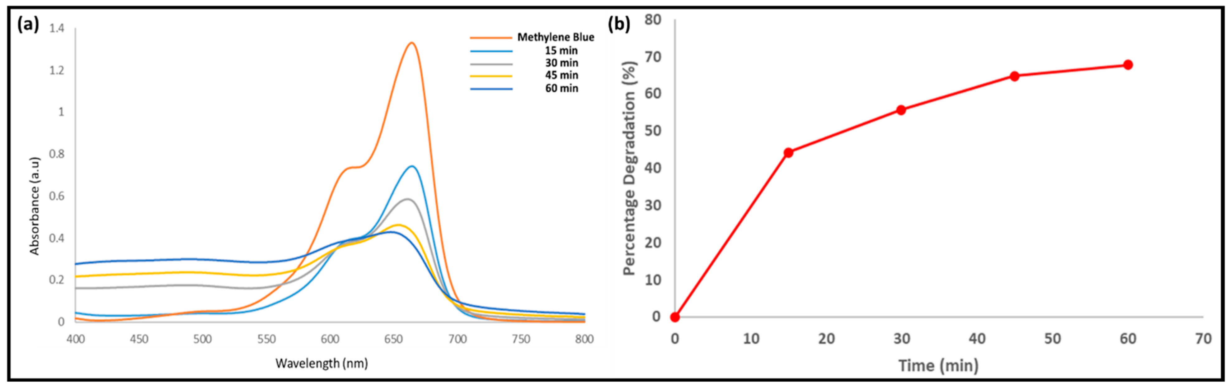
3.2. Photocatalytic Degradation of Methylene Blue

Several industries such as textile, plastic, and paper are known to generate streams of waste effluents containing substantial amounts of organic dyes which are eventually released into water bodies. These industrial effluents, when released into water bodies without any prior treatment, cause severe damage to both aquatic plants and animals. Recently, it is seen that conventional water treatment methods are replaced by advanced oxidation processes such as the Fenton method, photolysis, ozonolysis, and photocatalysis. In photocatalysis, using a suitable catalyst, degradation of organic pollutants present in wastewater is carried out in the presence of UV radiation associated with sunlight. Methylene blue (MB) is an aromatic heterocyclic dye which is commonly found in industrial wastewater that causes calamitous effects on the environment [25]. It is a carcinogenic thiazine pollutant which upon ingestion poses serious threats to human health, and it is known to cause severe damage to the nervous system. Nausea, breathing difficulties, and gastric infections are the other ailments associated with the ingestion of methylene blue.
Methylene blue shows an intense absorbance at 655 nm. The FH-AgNPs prove to be effective in the degradation of methylene blue in the presence of visible light. A catalyst load of 30 mg was used for the degradation studies. The fungal biomass is known to degrade dye by a mechanism known as biosorption. Biosorption includes various physico-chemical interactions such as adsorption, deposition, electrostatic interactions, and ion exchange that trigger the dye degradation [26]. The functional groups present in the fungal cell wall serve as the binding sites for the dye molecules. The amino, carboxylic acid, phosphate groups and lipids are prominent binding sites. Pre-treatment of the fungal biomass is found to increase the adsorption capacity of the fungi. Bio-templating AgNPs on the fungal hyphae increases the adsorption capacity and the catalytic activity. A percentage degradation of 67.86% was obtained within 60 min (Figure 4b), which highlights the potent catalytic activity of FH-AgNPs in dye degradation. A UV-Visible spectrophotometer was used to monitor the extent of degradation of MB. There was a significant reduction in the absorption band of MB with an increase in time (Figure 4a) that indicates degradation of MB catalysed by FH-AgNPs The FH-AgNPs stand out as potent catalysts due to the combined effect of the fungi and the AgNPs.
The possible mechanism for the photocatalytic degradation of methylene blue by the FH-AgNPs composite occurs via a series of photochemical reactions. The synthesized composite material possesses combined properties of both the fungi and the nanoparticle, thereby exhibiting an enhanced catalytic activity when compared to activity exhibited when they are present individually. The enhanced surface area provided by the fungi and the unique surface plasmon resonance phenomenon [27] of AgNPs are believed to be the reasons for the enhanced catalytic activity exhibited by the composite. When the surface of the nanoparticles is irradiated with high-energy photons, a collective oscillation of electrons occurs from the outermost band to a higher energy state. Later, molecular oxygen present in water undergoes plasmonic excitation of the surface electrons and becomes converted to the free radical species O2*. This is followed by an electron capture process by the holes generated in the nanoparticles, wherein electrons are captured from methylene blue adsorbed on the surface of the nanoparticles. This results in the oxidation of methylene blue, and the free radical (O2*) generated reacts with H+ and forms other active radicals such as OH* and OH2*. These free radicals are responsible for the degradation of methylene blue into various azo dye intermediates. The OH radicals are known to attack the C-S*=C functional group of MB, which results in the opening of the central aromatic ring containing both the heteroatoms. A control experiment was effectuated to study comparatively the degradation efficiency of AgNPs and FH-AgNPs. For AgNPs, a percentage degradation of 40.2% was obtained, which was found to be less when compared to FH-AgNPs wherein a percentage degradation of 67.86% was obtained in 60 min. This in turn proves the enhanced catalytic activity of FH-AgNPs over AgNPs.

3.3. Antibacterial Studies

AgNPs are known to possess a wide spectrum of antibacterial, antifungal, and antiviral properties. AgNPs can penetrate the bacterial cell wall, thereby changing the structural properties of the cell membrane, which results in cell death. The pronounced antibacterial effects of AgNPs can be attributed to the particle size and large surface-to-volume ratio. AgNPs efficiently increase the cell membrane’s permeability, interrupt cell replication by hindering the replication of deoxyribonucleic acid, and produce reactive oxygen species. AgNPs, when integrated into a solid support, exhibit enhanced antibacterial activity due to the slow and steady release of silver ions that results in cell death. The silver ions released adhere to the cytoplasmic membrane and cell wall, which eventually increases cell permeability and thereby leads to disruption of the bacterial envelope. The uptake of silver ions deactivates the respiratory enzymes, thereby generating reactive oxygen species. Reactive oxygen species efficiently disrupt the cell membrane and manifest the denaturation of deoxyribonucleic acid (DNA). Silver ions specifically interact with the sulphur and phosphorous present in DNA, leading to problems in DNA replication, reproduction of cells, or termination of microorganisms. Additionally, this interrupts the synthesis of proteins, leading to the denaturation of ribosomes in the cytoplasm of the cell (Figure 5) [17].
The antibacterial property of FH-AgNPs was studied against Escherichia coli and Staphylococcus aureus using the agar gel diffusion technique [28]. The FH-AgNPs’ activity was compared with the standard antibiotic ampicillin. The antibacterial activity of the composite with silver nanoparticles was close to that of ampicillin. In general, AgNP-based composites are found to possess higher antibacterial activity due to the combined effect of the counterpart and the slow silver release rate. Ag ions are gradually released from FH-AgNPs, which can increase the exposure time and result in better bacteriolysis. Ag ions can bind crucial biomolecules such as DNA, RNA, and proteins, inhibiting their function. Furthermore, the composite can induce oxidative stress in bacterial cells. As a result, these composites render an oligodynamic effect, wherein they exhibit a bacteriostatic or even a bactericidal impact. The compound was prepared in water (2 mg/mL) and the zone of inhibition was measured after 48 h. An amount of 200 μg of the composite exhibited a maximum antibacterial property against E. coli. The results are tabulated in Table 1. In presence of AgNPs in the composite induce oxidative stress releasing hydroxyl radicals that attack the bacterial cell wall, thereby disintegrating the same and leading to bacteriostatic/bactericidal effects.

4. Conclusions

To summarize, a multifunctional fungi silver nanoparticle composite was synthesized and characterized. The previously synthesized AgNPs were efficiently coated on the fungal mycelium to generate microtubules coated with AgNPs. The FH-AgNP composite was found to efficiently catalyse the degradation of an industrial pollutant dye, methylene blue. A percentage degradation of 67.86% was obtained within 60 min, highlighting the potent catalytic activity of the synthesized composite. Moreover, the composite displayed a potent antibacterial property against E. coli and S. aureus. The synthesis and applications rendered by FH-AgNPs open a new era of the utilization of organic–inorganic hybrid materials for various multifunctional applications. The developed strategy offers varying versatility wherein the methodology can be extended to other microorganisms, thereby providing tantalizing possibilities for the hierarchical assembly of macroscale structures with controllable electronic, magnetic, and optical properties which can be efficiently utilized for applications involving sensing, catalysis, energy storage, and biomedical fields.

Author Contributions

Methodology, F.J.; Formal analysis, F.J., J.D. and V.V.L.; Investigation, F.J.; Data curation, J.D., A.N. and V.V.L.; Writing—original draft, F.J., J.D. and A.N.; Writing—review & editing, A.N. and S.B.N.K.; Visualization, F.J.; Supervision, A.N.; Project administration, A.N. All authors have read and agreed to the published version of the manuscript.

Funding

This research received no external funding.

Acknowledgments

The authors are thankful to CHRIST (Deemed to be University) and the Centre for Research, CHRIST (Deemed to be University), for the research support. Aatika would like to acknowledge the support from CHRIST (Deemed to be University) for funding this research (MRP # MRPDSC-1722). KSBN is thankful to Durban University of Technology for the awarding of the research fellowship.

Conflicts of Interest

The authors declare no conflict of interest.

References

  1. Ozden, S.; MacWan, I.G.; Owuor, P.S.; Kosolwattana, S.; Autreto, P.A.S.; Silwal, S.; Vajtai, R.; Tiwary, C.S.; Mohite, A.D.; Patra, P.K.; et al. Bacteria as Bio-Template for 3D Carbon Nanotube Architectures. Sci. Rep. 2017, 7, 1–8. [Google Scholar] [CrossRef]
  2. Baig, N.; Kammakakam, I.; Falath, W.; Kammakakam, I. Nanomaterials: A Review of Synthesis Methods, Properties, Recent Progress, and Challenges. Mater. Adv. 2021, 2, 1821–1871. [Google Scholar] [CrossRef]
  3. Herrera-Beurnio, M.C.; Hidalgo-Carrillo, J.; López-Tenllado, F.J.; Martin-Gómez, J.; Estévez, R.C.; Urbano, F.J.; Marinas, A. Bio-Templating: An Emerging Synthetic Technique for Catalysts. A Review. Catalysts 2021, 11, 1364. [Google Scholar] [CrossRef]
  4. Zheng, X.T.; Xu, H.V.; Tan, Y.N. Bioinspired Design and Engineering of Functional Nanostructured Materials for Biomedical Applications. ACS Symp. Ser. 2017, 1253, 123–152. [Google Scholar] [CrossRef]
  5. Selvakumar, R.; Seethalakshmi, N.; Thavamani, P.; Naidu, R.; Megharaj, M. Recent Advances in the Synthesis of Inorganic Nano/Microstructures Using Microbial Biotemplates and Their Applications. RSC Adv. 2014, 4, 52156–52169. [Google Scholar] [CrossRef]
  6. Kubo, A.M.; Gorup, L.F.; Toffano, L.; Amaral, L.S.; Rodrigues-Filho, E.; Mohan, H.; Aroca, R.; Camargo, E.R. Nanostructured Assemblies of Gold and Silver Nanoparticles for Plasmon Enhanced Spectroscopy Using Living Biotemplates. Colloids Interfaces 2017, 1, 1–11. [Google Scholar] [CrossRef]
  7. Kubo, A.M.; Gorup, L.F.; Amaral, L.S.; Filho, E.R.; Camargo, E.R. Kinetic Control of Microtubule Morphology Obtained by Assembling Gold Nanoparticles on Living Fungal Biotemplates. Bioconjug. Chem. 2016, 27, 2337–2345. [Google Scholar] [CrossRef]
  8. Kubo, A.M.; Gorup, L.F.; Amaral, L.S.; Rodrigues-Filho, E.; de Camargo, E.R. Heterogeneous Microtubules of Self-Assembled Silver and Gold Nanoparticles Using Alive Biotemplates. Mater. Res. 2018, 21, 1–7. [Google Scholar] [CrossRef]
  9. Rehman, A.; Majeed, M.I.; Ihsan, A.; Hussain, S.Z.; Saif-Ur-Rehman; Ghauri, M.A.; Khalid, Z.M.; Hussain, I. Living Fungal Hyphae-Templated Porous Gold Microwires Using Nanoparticles as Building Blocks. J. Nanopart. Res. 2011, 13, 6747–6754. [Google Scholar] [CrossRef]
  10. Zhu, W.K.; Cong, H.P.; Guan, Q.F.; Yao, W.T.; Liang, H.W.; Wang, W.; Yu, S.H. Coupling Microbial Growth with Nanoparticles: A Universal Strategy to Produce Functional Fungal Hyphae Macrospheres. ACS Appl. Mater. Interfaces 2016, 8, 12693–12701. [Google Scholar] [CrossRef]
  11. Sabah, A.; Kumar, P.; Mohammed, W.S.; Dutta, J. Visible-Light-Induced Directed Gold Microwires by Self-Organization of Nanoparticles on Aspergillus Niger. Part. Part. Syst. Charact. 2013, 30, 473–480. [Google Scholar] [CrossRef]
  12. Ullah, M.W.; Manan, S.; Khattak, W.A.; Shahzad, A.; Ul-Islam, M.; Yang, G. Biotemplate-Mediated Green Synthesis and Applications of Nanomaterials. Curr. Pharm. Des. 2020, 26, 5819–5836. [Google Scholar] [CrossRef] [PubMed]
  13. Gao, J.F.; Zhang, Q.; Wang, J.H.; Wu, X.L.; Wang, S.Y.; Peng, Y.Z. Contributions of Functional Groups and Extracellular Polymeric Substances on the Biosorption of Dyes by Aerobic Granules. Bioresour. Technol. 2011, 102, 805–813. [Google Scholar] [CrossRef] [PubMed]
  14. Abdi, O.; Kazemi, M. A Review Study of Biosorption of Heavy Metals and Comparison between Different Biosorbents. J. Mater. Environ. Sci. 2015, 6, 1386–1399. [Google Scholar]
  15. Kitching, M.; Ramani, M.; Marsili, E. Fungal Biosynthesis of Gold Nanoparticles: Mechanism and Scale Up. Microb. Biotechnol. 2015, 8, 904–917. [Google Scholar] [CrossRef]
  16. Li, Z.; Chung, S.-W.; Nam, J.-M.; Ginger, D.S.; Mirkin, C.A. Living Templates for the Hierarchical Assembly of Gold Nanoparticles. Angew. Chem. 2003, 115, 2408–2411. [Google Scholar] [CrossRef]
  17. Yin, I.X.; Zhang, J.; Zhao, I.S.; Mei, M.L.; Li, Q.; Chu, C.H. The Antibacterial Mechanism of Silver Nanoparticles and Its Application in Dentistry. Int. J. Nanomed. 2020, 15, 2555. [Google Scholar] [CrossRef]
  18. Xia, S.; Zhang, L.; Pan, G.; Qian, P.; Ni, Z. Photocatalytic Degradation of Methylene Blue with a Nanocomposite System: Synthesis, Photocatalysis and Degradation Pathways. Phys. Chem. Chem. Phys. 2015, 17, 5345–5351. [Google Scholar] [CrossRef] [PubMed]
  19. Samari, F.; Salehipoor, H.; Eftekhar, E.; Yousefinejad, S. Low-Temperature Biosynthesis of Silver Nanoparticles Using Mango Leaf Extract: Catalytic Effect, Antioxidant Properties, Anticancer Activity and Application for Colorimetric Sensing. New J. Chem. 2018, 42, 15905–15916. [Google Scholar] [CrossRef]
  20. Res, J.M.B.; Sarsar, V.; Selwal, K.K.; Selwal, M.K. Green Synthesis of Silver Nanoparticles Using Leaf Extract of Mangifera Indica and Evaluation of Their Antimicrobial Activity. J. Microbiol. Biotech. Res. 2013, 3, 27–32. [Google Scholar]
  21. Jones, M.; Bhat, T.; Kandare, E.; Thomas, A.; Joseph, P.; Dekiwadia, C.; Yuen, R.; John, S.; Ma, J.; Wang, C.H. Thermal Degradation and Fire Properties of Fungal Mycelium and Mycelium—Biomass Composite Materials. Sci. Rep. 2018, 8, 1–10. [Google Scholar] [CrossRef]
  22. Pal, U.; Castillo López, D.N.; Carcaño-Montiel, M.G.; López-Reyes, L.; Díaz-Nuñez, P.; Peña-Rodríguez, O. Nanoparticle-Assembled Gold Microtubes Built on Fungi Templates for SERS-Based Molecular Sensing. ACS Appl. Nano Mater. 2019, 2, 2533–2541. [Google Scholar] [CrossRef]
  23. Feofilova, E.P. The Fungal Cell Wall: Modern Concepts of Its Composition and Biological Function. Microbiology 2010, 79, 711–720. [Google Scholar] [CrossRef]
  24. Zhang, H.; Wang, X.; Li, Y.; Guo, C.; Zhang, C. Preparation and Characterization of Silver-Doped Graphene-Reinforced Silver Matrix Bulk Composite as a Novel Electrical Contact Material. Appl. Phys. A Mater. Sci. Process. 2019, 125, 1–9. [Google Scholar] [CrossRef]
  25. Houas, A.; Lachheb, H.; Ksibi, M.; Elaloui, E.; Guillard, C.; Herrmann, J.M. Photocatalytic Degradation Pathway of Methylene Blue in Water. Appl. Catal. B Environ. 2001, 31, 145–157. [Google Scholar] [CrossRef]
  26. Islam, A.; Teo, S.H.; Taufiq-Yap, Y.H.; Ng, C.H.; Vo, D.V.N.; Ibrahim, M.L.; Hasan, M.M.; Khan, M.A.R.; Nur, A.S.M.; Awual, M.R. Step towards the Sustainable Toxic Dyes Removal and Recycling from Aqueous Solution- A Comprehensive Review. Resour. Conserv. Recycl. 2021, 175, 105849. [Google Scholar] [CrossRef]
  27. Singh, J.; Dhaliwal, A.S. Plasmon-Induced Photocatalytic Degradation of Methylene Blue Dye Using Biosynthesized Silver Nanoparticles as Photocatalyst. Environ. Technol. 2018, 41, 1520–1534. [Google Scholar] [CrossRef]
  28. Jose, A.; Sunaja Devi, K.R.; Pinheiro, D.; Lakshmi Narayana, S. Electrochemical Synthesis, Photodegradation and Antibacterial Properties of PEG Capped Zinc Oxide Nanoparticles. J. Photochem. Photobiol. B Biol. 2018, 187, 25–34. [Google Scholar] [CrossRef] [PubMed]
Figure 1. Schematic representation of the synthesis of FH-AgNPs. (a) Fungal biomass incubated with AgNPs. (b) Fungi-templated AgNPs. (c) Optical microscopic image of fungi filament templated with AgNPs.
Applsci 13 02158 g001
Figure 2. SEM image of FH-AgNP composite: (a) 1 μm magnification; (b) 10 μm magnification. (c) Elemental analysis of FH-AgNP composite.
Applsci 13 02158 g002
Figure 3. XRD analysis of AgNPs and FH-AgNPs.
Applsci 13 02158 g003
Figure 4. (a) Degradation studies using UV-visible spectroscopy. (b) Plot of percentage decomposition vs. time.
Applsci 13 02158 g004
Figure 5. Mechanism of antibacterial action of AgNPs. (1) Ag+ ions disrupt cell wall. AgNPs release Ag+ ions which adhere to the bacterial cell wall and thereby pass through the cell wall and cytoplasmic membrane. (2) Denaturation of cell wall by AgNPs. AgNPs accumulate on the cell wall and cytoplasmic membrane and cause membrane denaturation. (3) Disruption of cell membrane by reactive oxygen species. The reactive oxygen species produced by AgNPs lead to disruption of cell membrane. (4) Denaturation of ribosomes. Silver ions, after moving into the cell cytoplasm, adhere to the ribosome and thereby hinder the synthesis of proteins. (5) Interruption in DNA replication. The process of DNA replication is hindered when AgNPs bind to various heteroatoms such as nitrogen and sulphur present in DNA.
Applsci 13 02158 g005
Table 1. Antimicrobial studies of FH-AgNPs against E coli and S. aureus.
Concentration (μg)Zone of Inhibition—E. coli (mm)Zone of Inhibition—S. aureus (mm)
FH-AgNPs5055.5
1005.85.7
20066.5
40066.5
Ampicillin (Standard)506.66.2
10077.5
2007.47.5
40088.7
Disclaimer/Publisher’s Note: The statements, opinions and data contained in all publications are solely those of the individual author(s) and contributor(s) and not of MDPI and/or the editor(s). MDPI and/or the editor(s) disclaim responsibility for any injury to people or property resulting from any ideas, methods, instructions or products referred to in the content.

