Article

Impact of Preceded Tumor Therapeutic Irradiation on the Microtensile Bond Strength of Universal Adhesives Applied in Self-Etch Mode to Human Dentin In Vitro

by
Sina Broscheit
1,*,
Dirk Vordermark
2,
Reinhard Gerlach
2 and
Christian Ralf Gernhardt
1
1
University Outpatient Clinic for Conservative Dentistry and Periodontology, Medical Faculty, Martin-Luther-University Halle-Wittenberg, 06112 Halle, Germany
2
Department of Radiotherapy, University Clinic, Martin-Luther-University Halle-Wittenberg, Ernst-Grube-Strasse 40, 06120 Halle, Germany
*
Author to whom correspondence should be addressed.
Appl. Sci. 2023, 13(13), 7873; https://doi.org/10.3390/app13137873
Submission received: 9 June 2023 / Revised: 27 June 2023 / Accepted: 3 July 2023 / Published: 4 July 2023

Abstract

:
The aim of this study was to investigate the effect of preceded tumor therapeutic irradiation on the tensile bond strength of three modern universal adhesives applied in self-etch mode on dentin. Specimens prepared from 135 extracted human third molars were divided into three superior groups. These received either no radiation, 5 Gy, or a total dose of 60 Gy in vitro irradiation, fractionally applied. The samples of each group were further randomly assigned to six subgroups to test three adhesives (Futurabond® U, Voco; AdheSE® Universal Ivoclar Vivadent; Xeno® Select, Dentsply Sirona) in the self-etch application mode (n = 15). Tensile bond strength was determined using a universal testing machine (1.0 mm/min). Data were analyzed with ANOVA (p < 0.01) and Tukey’s test (p < 0.05). The influence of irradiation on the microtensile bond strength of the used dentin adhesives proved to be significant. For each material, a decrease in adhesion value was registered after irradiation. However, only for the material Xeno® Select were significantly reduced adhesion values determined after irradiation with 60 Gy compared to 0 Gy. Within the limitations of an in vitro study, some effects of tumor therapeutic irradiation of human dentin on the tensile bond strength of universal adhesives used in self-etch mode could be observed. Those decreases were only partly significant, depending on the material and the radiation dose. Whether the tensile bond strength on irradiated dentin depends on the particular application mode (etch-and-rinse vs. self-etch) of the universal adhesives remains to be investigated.

1. Introduction

Worldwide, tumors of the oral cavity account for approximately 2% of all malignant tumors [1]. According to the German Centre for Cancer Registry Data (ZfKD) of the Robert Koch Institute, for example, 14,310 new patients with tumors of the oral cavity and throat were detected in Germany in 2018 [2], and the global incidence of lip and oral cavity cancer in 2020 accounted for 377,713 cases [1].
Radiation therapy is one important therapeutic principal for tumors of the head and neck region, and has been applied regularly [3,4,5,6]. High-energy radiation from X-rays, gamma rays, neutrons, protons and other sources is used to eliminate cancer cells and to shrink tumors [7]. The ionizing radiation causes damage to the DNA of the tumor cell, both directly, through the radiation itself, and indirectly via free radicals, which are released due to the interaction of radiation with water. Having suffered irreversible damage to the DNA, the cell loses its ability to maintain cell division. If all cells of a tumor lose their proliferative potential, the tumor can be eradicated completely [8].
Despite its anticancer effects, irradiation has some injurious impacts on healthy tissue located in the radiation field [5,6]. This results in clinical consequences such as mucositis, hyposalivation, osteoradionecrosis and radiation caries [5,6,9,10,11]. Apart from these, previous studies have shown a direct influence on the mechanical properties of the dental tissue, inter alia, the alteration of the collagenous structure [12,13,14]. Collagenous fibers are essential for the adhesive bond between the dentin surface and adhesive material for mounting restorative resin materials [15,16,17].
Restorative dental treatment is more often required in patients who have undergone radiotherapy in the head and neck area, since the risk of developing radiation caries is increased throughout life and not only during or immediately after treatment [6,18]. Adhesively bonded composite restorations are a well-established treatment option to restore caries-affected teeth [19,20]. Therefore, some studies recommend treatment with adhesively bonded composite fillings in these cases [21,22].
The two approaches to adhesive systems, etch-and-rinse and the self-etch technique, establish bonding to dentin in different ways and use these collagen fibers in different manners. While the etch-and-rinse technique completely removes the smear layer through the prior use of an acid etchant and thus exposes and infiltrates the collagen fibers, the self-etch technique only demineralizes the surface of the smear layer, leaving residual hydroxyapatite crystals still attached to the collagen fibers, which are then incorporated into the bonding layer [23].
One of the recent inventions in adhesive dentistry is universal adhesives. These adhesives, also called “multi-mode” or “multi-purpose”, combine the application methods of the classic etch-and-rinse and self-etch adhesives, i.e., they can be used with or without prior etching of the dentin or even with selective enamel etching [24,25]. This flexibility of the application method is achieved by functional monomers, which are the core components of universal adhesives [26,27,28]. Functional monomers are acidic molecules that can fulfill various functions, including the partial dissolution of the smear layer, the demineralization of hydroxyapatite, the improvement of monomer penetration into the tooth structure and the ability to form chemical bonds with tooth substrates [27,29,30].
The bonding performance of various universal adhesives on human dentin In etch-and-rinse mode compared to self-etch mode based on the assessment of the microtensile bond strength (µTBS) has already been investigated in numerous studies [24,31,32,33]. Wagner et al. [33], for example, investigated how previous etching improves the dentine penetration pattern, but without affecting the µTBS.
The effects of radiotherapy on the µTBS of various adhesives to dentine have also been published several times [34,35,36,37].
However, relatively sparse information is currently available in the dental literature about the behavior of universal adhesives on irradiated human dentin. Hence, the aim of this study was to examine the influence of preceded tumor therapeutic irradiation on the tensile bond strength of three different universal adhesives used in self-etch application mode on human dentin. The null hypothesis to be tested was that there is no influence on the microtensile bond strength in non- and severely irradiated human dentin.

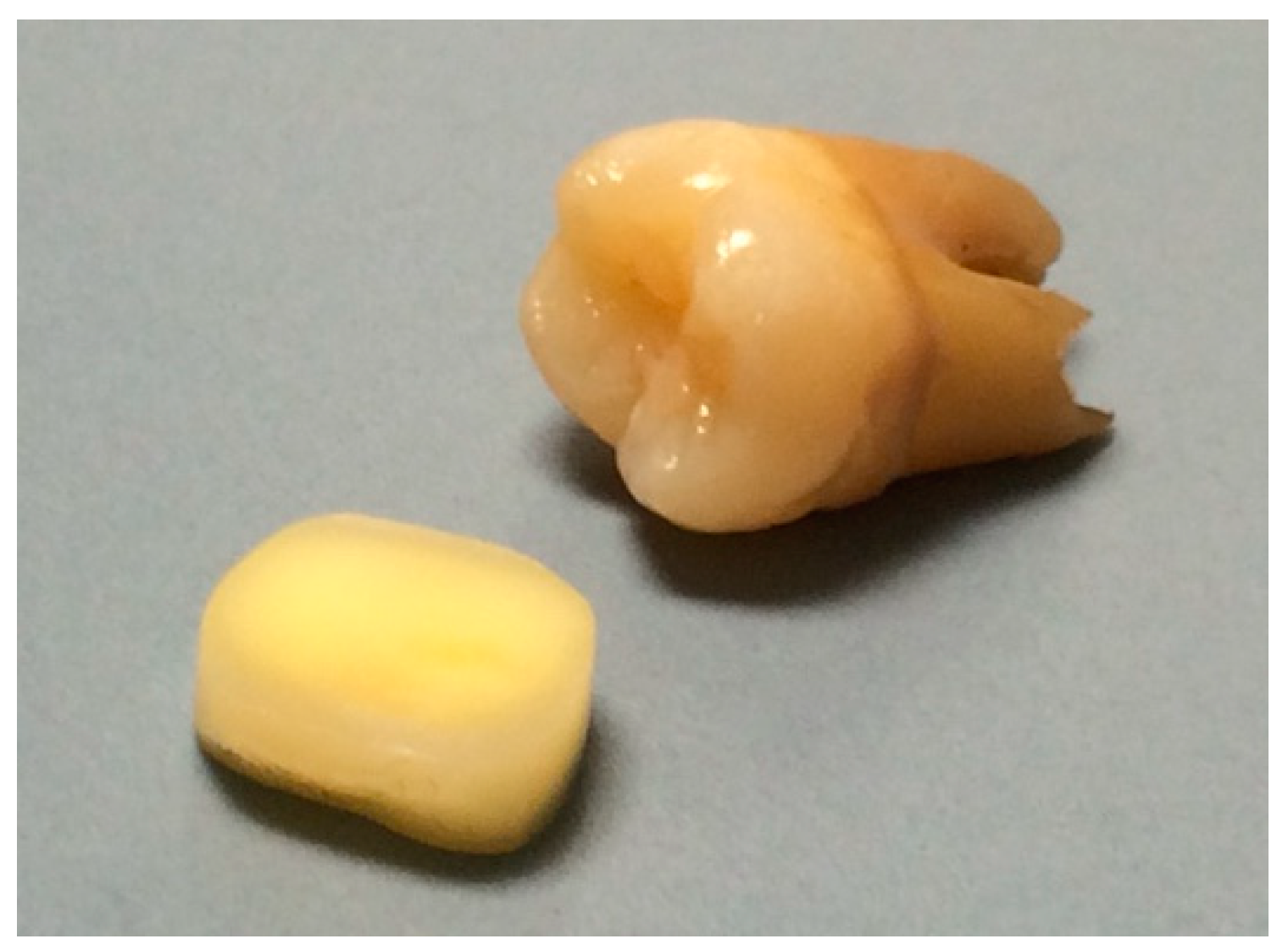
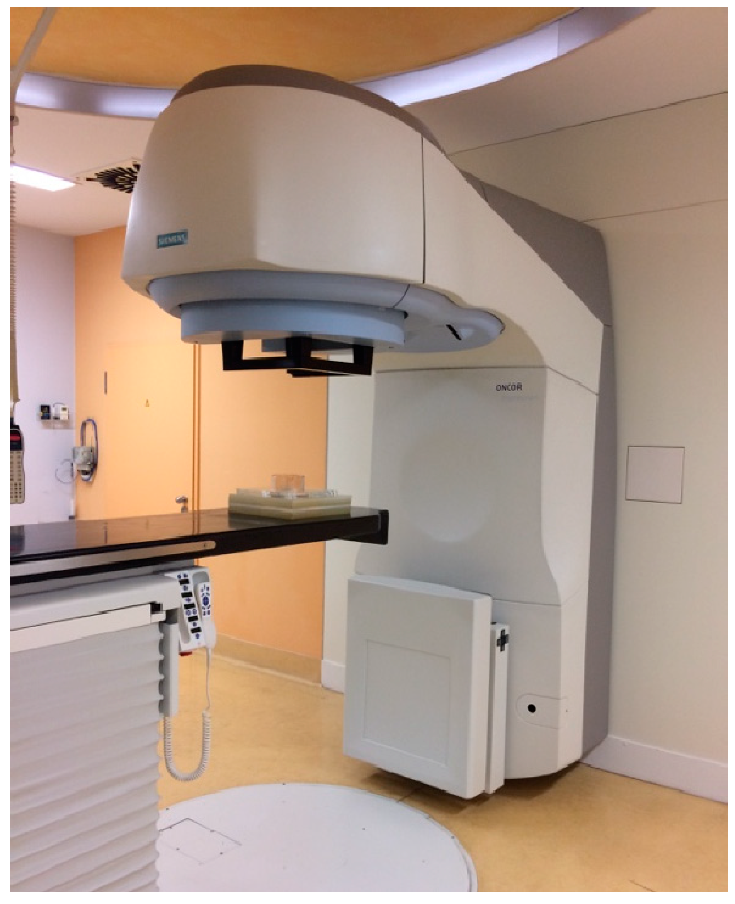
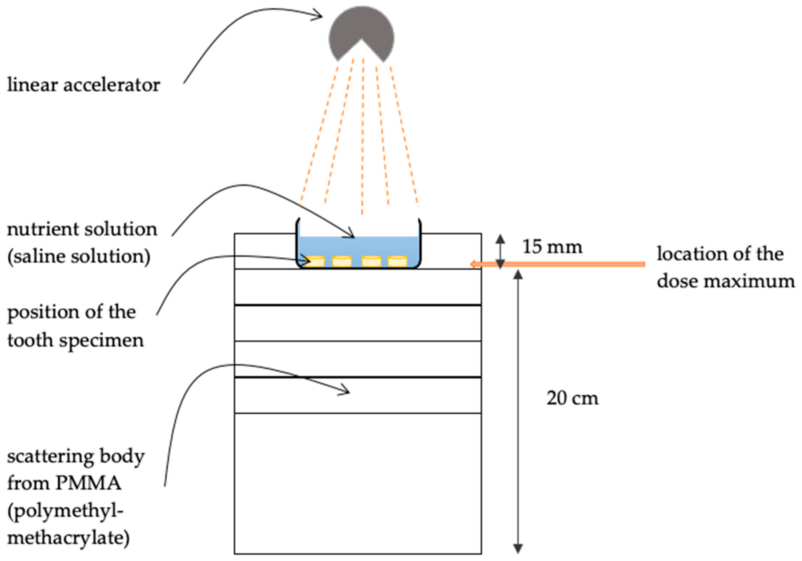
2. Materials and Methods
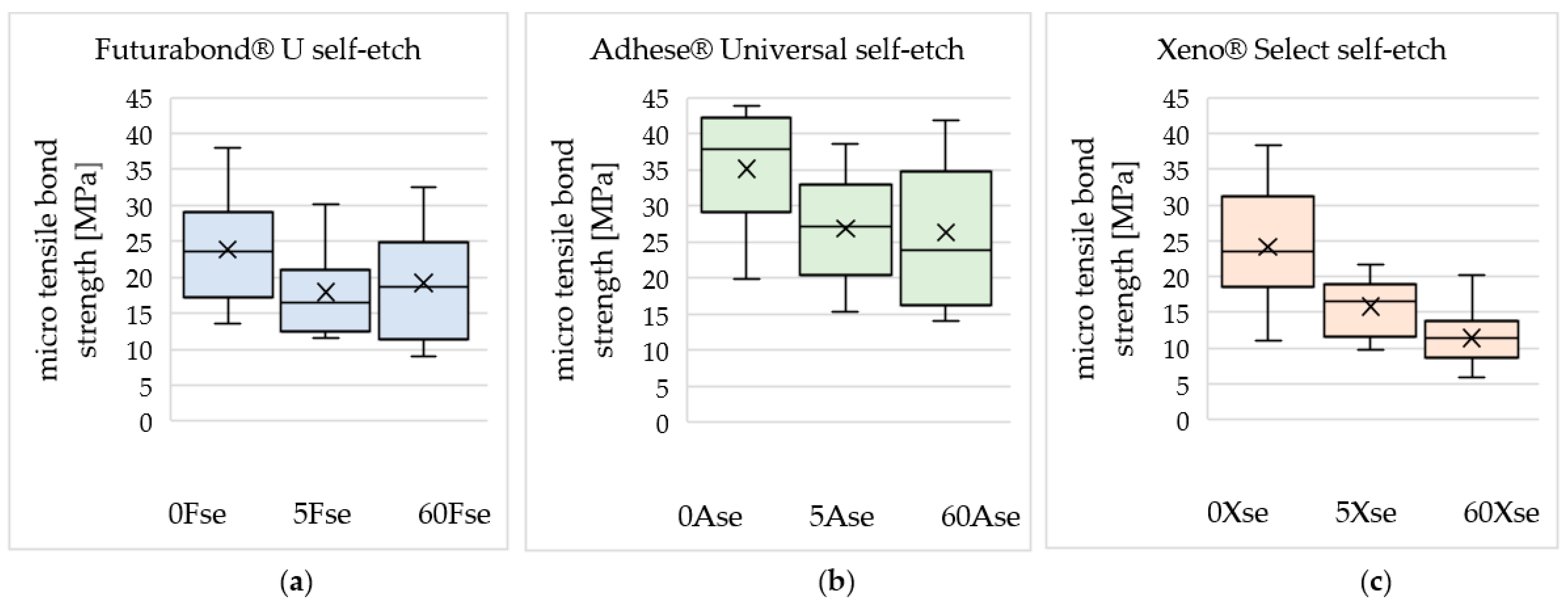
In this study, one hundred and thirty-five freshly extracted caries-free human third molars were included. Before the experimental procedure, the teeth were stored in saline solution (0.9% NaCl, B. Braun Meisungen AG, Meisungen, Germany) at room temperature. The preparation of the specimens of the teeth occurred in a manner Kobler et al. described in 2008 [38]. This technique allows dentin perfusion based on the simulation of intra-pulpal pressure. Under constant water cooling, the roots were ground down using a grinding machine (Rotopol-35, Struers GmbH, Willich, Germany) until the coronal pulp chamber was disclosed. After the removal of the pulpal tissue, the specimens were reduced from the occlusal part until the distance between the occlusal plateau and the pulp chamber was adjusted to 2.0 mm (±0.2 mm). Afterwards, the apical area needed to be parallelized to the occlusal plateau and reduced to a total sample thickness of 4.0 mm (±0.2 mm). These prepared samples (Figure 1) were stored in saline solution (0.9% NaCl) once again and randomly divided into three main groups. The first group received no further treatment; the specimens of the other two groups were irradiated in cooperation with a radiation physicist from the Department of Radiotherapy using a linear accelerator (ONCOR Impression IMRT Plus, Siemens AG, Munich, Germany) (Figure 2). Half of the irradiated samples underwent one single dose of 5 Gy (Gray), and for the other half irradiation was applied in a fractionated schedule with single doses of 2 Gy per day. For those samples it resulted in a total dose of 60 Gy, since they were fractionally irradiated 5 days per week over a period of 6 weeks, according to conventional radiotherapy of head and neck cancer [13,39,40]. Figure 3 shows a schematic illustration of the set-up of the test specimens during irradiation: For the purpose of homogeneity, the specimens were stored in daily renewed saline [40], arranged on one plane level in the zone of the dose maximum during the radiation treatment and placed on a PMMA (polymethyl-methacrylate) scattering body to minimize the scattered radiation.
The forty-five samples in each group were randomly divided into three subgroups for the reason that three different universal bonding agents were tested in self-etch mode. Finally, nine subgroups emerged, each consisting of fifteen samples (Table 1).
For the purpose of the experimental procedure, the samples were fastened to a special experimental apparatus in a universal testing machine (Zwick Z005, Zwick GmbH & Co., Ltd., KG, Ulm, Germany). Pressure of 30 cm H2O was generated via saline solution, thereby simulating intrapulpal pressure and thus dentin perfusion throughout the entire duration of the experiment. The universal bonding agents were applied according to the manufacturers’ recommendations in self-etch application mode, which did not differ for the three materials tested. One layer adhesive was applied with a brush-tip and scrubbed into the dentin surface for 20 s. Subsequently, oil-free compressed air was used in order to disperse the adhesive plus remove any solvent, and the adhesive was light-cured for 10 s. The following three universal adhesives were used:
F: Futurabond® U (Voco GmbH, Cuxhaven, Germany);
A: AdheSE® Universal (Ivoclar Vivadent AG, Schaan, Liechtenstein);
X: Xeno® Select (Dentsply Sirona Inc., Charlotte, NC, USA).
Table 2 lists the main components of the adhesive materials used according to the safety data sheets provided by the corresponding manufacturers.
For the comparability of the results, the same composite resin was used in all test groups: Grandio®SO color A2 (VOCO GmbH, Cuxhaven, Germany). The application of the composite filling material occurred through an application sleeve. This sleeve featured a continuous cylindrical slot of 1 mm diameter to accommodate the composite material, which ensured a standardized area of 0.79 mm2 as a bonding surface.
Tensile bond strength was determined 15 min after the light polymerization. The samples were loaded at a speed of 1 mm/min. The maximum force was recorded and divided by the standardized dimension of the bonding surface (diameter 1 mm = 0.79 mm2) to obtain microtensile bond strength. To avoid any impact and bias of different researchers, all experimental steps were performed by one unblinded dentist.
The sample size was calculated upon the assumption that the main endpoint (bond strength) would be 16 in the irradiated group and 25 in the non-irradiated control group, with a standard deviation of 9 in both arms. This relevant difference could be detected with 80% power by a t-test to the 5%-level if 14 specimen per group were included. Therefore, 15 specimens per group were included. Statistical analysis was performed with SPSS® 25.0 (IBM®, Ehningen, Germany). Mean microtensile bond strength and standard deviation were calculated for each experimental group. To assess differences between the results of all groups, two-way ANOVA was used at a 1% significance level. The Tukey’s test was used to calculate the differences between the various groups at a 5% significance level.

3. Results

The effect of irradiation on the tensile bond strength varied among the materials used, but a reduction was observed in all experimental groups.
The results, respectively, mean tensile bond strength and standard deviation of each group, are presented in Table 3 and Figure 4. Figure 5 illustrates the spread of the resulting data.
Considering the results for the material Futurabond® U, the value of the mean tensile bond strength without irradiation was 23.87 MPa (±7.49 MPa). Low dose irradiation (5 Gy) led to a decrease in the tensile bond strength of Futurabond® U to 17.95 MPa (±5.95 MPa). After irradiation with 60 Gy, the tensile bond strength of Futurabond® U did not decrease further (19.21 MPa ± 7.34 MPa).
Likewise, for AdheSE® Universal, the tensile bond strength of this material without irradiation was higher overall, with 35.10 MPa (±8.41 MPa). After irradiation with a low dose (5 Gy), there was a decrease in the tensile bond strength of AdheSE® Universal to 26.86 MPa (±7.41 MPa), but the dose increase to 60 Gy had no further effect; the tensile bond strength remained at 26.30 MPa (±10.07 MPa).
Regarding material Xeno® Select, a gradual decrease in the tensile bond strength according to the increase in the irradiation dose was observed. The reduction from 24.17 MPa (±8.36 MPa) at 0 Gy via 15.78 MPa (±3.89 MPa) after 5 Gy to 11.42 MPa (±3.86 MPa) after 60 Gy irradiation dose, which occurred in self-etch-mode, represented the largest loss of tensile bond strength overall.
The two-Way ANOVA revealed that the influence of irradiation and the used adhesive system on the microtensile bond strength of the used dentin adhesive systems proved to be significantly different (p < 0.01).
In all experimental groups, a decrease in tensile bond strength between non-irradiated and irradiated groups was registered, at least after irradiation with 60 Gy. The percentage decrease within one material after a radiation dose of 60 Gy versus no radiation is listed in Table 3. Nevertheless, pairwise comparisons of the different groups revealed that the effect of irradiation on tensile bond strength was not significant for Futurabond® U and AdheSE® Universal (Tukey’s test, p > 0.05). Only in the test series of the material Xeno® Select were the values of tensile bond strength in self-etch-mode after irradiation with 60 Gy significantly reduced compared to no irradiation (p < 0.05, Tukey’s test).

4. Discussion

The present study investigated the tensile bond strength of three different widely used and differently composed universal adhesives on irradiated dentin compared to non-irradiated dentin in the self-etch application mode in vitro. All experimental steps, including specimen preparation and the application of all materials, followed by bond strength testing, were performed by one researcher (dentist) to avoid any impact of different persons. This dentist was trained in advance to become familiar with the study protocol.
In some previous investigations, it was shown that the new universal bonding agents led to higher bond strength values when they were used after a preceding etching step [41,42]. Our study set-up was to evaluate solely the self-etch mode rather than the etch-and-rinse mode, and those research works investigated the effect on the enamel surface, whereas in the present study dentin tissue was tested.
According to Elkaffas et al. [43], the evaluation of numerous studies on the adhesion values of universal adhesives on dentin did not reveal any significant overall difference between etch-and-rinse and self-etch utilization. This analysis considered study results on numerous different materials, which differed in their composition, including the functional monomers contained therein, and thus in their chemical and mechanical properties. The remarkable one of these functional monomers was 10-methacryloyloxydecyl dihydrogen phosphate (10-MDP or MDP), which was able to bond to the hydroxyapatite crystals contained in the dentin [29]. By linking calcium (Ca), which is released from the hydroxyapatite (HAp) by demineralization, MDP-Ca salts were formed, which were self-assembled into the so-called “nanolayering” [44,45]. These primary chemical/ionic interactions of specific functional monomers with hydroxyapatite followed the “adhesion-decalcification” concept [45], which states that carboxylic acids either adhere to hydroxyapatite or decalcify it in accordance with the dissolution rate of the particular calcium salt in the acid solution [46]. The three universal adhesives tested contain different functional monomers that are intended to create the bond to the tooth structure. AdheSE® Universal and Futurabond® U contain MDP [47,48], which, according to Yoshida et al. [29], creates a very stable bond to the hydroxyapatite, which was validated by the low dissolution rate of its calcium salt in water. Carrilho et al. [49] affirmed the beneficial properties of MDP by evaluating a range of studies.
Long-term bonding effectiveness has not been investigated in this current study, but the results of Zhang et al. [28] indicated that the bond of universal adhesives in general is not capable of resisting ageing, although it was observed that bonds obtained by the etch-and-rinse mode are more resistant to water-aging than those achieved by the self-etch mode.
The bases of fractioned irradiation in conventional radiotherapy are the five Rs of radiobiology: repair, reassortment, repopulation, reoxygenation and radiosensitivity. These factors influence the response of tumors and normal tissue to radiation, and thus the therapy efficiency [50,51]. In this study, we applied in cooperation with one specialized radiation physicist from the Department of Radiotherapy of our University a total dose of 60 Gy, in 30 fractions of 2 Gy per day, treating 5 days per week over a period of 6 weeks, according to conventional radiotherapy [13,39,40]. The cooperation with only one radiation physicist might help to avoid the impact of different involved researchers. Today, there are several different radiotherapy schedules, such as hyperfractionation or accelerated fractionation, which are being evaluated for advantages over conventional fractionation radiotherapy, regarding patient outcome [52,53,54], which may therefore be common practice. To perform the in vitro test, the dentin specimens were irradiated directly and homogeneously. This differs from the clinical situation of radiotherapy, where the teeth do not necessarily remain in the radiation field, depending on the location of the tumor. Today, a technique called intensity-modulated radiation therapy (IMRT) is established; in this method, the direction of radiation changes continuously, with the result that the main target keeps receiving the entire amount of radiation required for the therapy, but the surrounding tissue exposed to the radiation varies, thereby limiting the radiation dose in this area [55,56]. As a result, it is assumed that the effects of irradiation on the bond with dentin found in this study appear to be even milder in the clinical situation, especially when treated with IMRT.
In this study, we solely simulated the clinical situation in which resin fillings were applied post radiotherapy. No consideration was given to the scenario where a patient might have fillings in some teeth before radiotherapy starts. If an existing dental restoration was irradiated, this could possibly have different effects on the bond strength, as Arid et al. [57] have ascertained in a previous study, or on the long-term durability of the restoration. This should therefore be investigated further, particularly in regard to the recommendation to rehabilitate patients’ dentition before radiotherapy [6].
Various studies have been published in the dental literature describing direct irradiation impacts on human tissue, and dentin in particular. The results of these studies include decreased microhardness [13,40] and micro-morphological damage, such as obliterated dentinal tubules [13,57,58,59], collagen network impairment and ruptures in the dentin structure [13,59]. These effects may explain a deterioration of the adhesive bond strength to the dentin, which was partly determined in our present study but also in a recently published study by Arid et al. [57].
These findings, in combination with the observations that irradiation causes the dentinoenamel junction (DEJ) to appear unstable [58,59,60,61] and affects enamel damage [14,59,62,63], are possibly some of several factors that all together promote the development of atypical radiation-related caries. Other effective aspects include hyposalivation, poor oral hygiene and altered microbial oral flora [64,65].
Some of the points mentioned concerning damage to the dental hard tissue caused by irradiation are still being discussed controversially, as there are also studies that refute certain aspects of them [62,66,67]. Still, it cannot be denied that patients have an increased risk of caries post radiotherapy [6,68,69] and thus there is a major necessity for dental restorations. Nevertheless, there is no consensus on which is the preferred material for conservative caries therapy in patients having undergone head and neck radiation therapy. The Oral Care Study Group of the Multinational Association of Supportive Care in Cancer and the International Society of Oral Oncology (MASCC/ISOO) published the clinical guideline ‘Evidence-Based Management Strategies for Oral Complication from Cancer Treatment’ in 2011 [70], recommending the use of resin-modified glass ionomer cement (GIC), composite resin or amalgam for restorations in post head and neck radiation patients, and so do Hong et al. [69] for patients who use fluoride. Conventional GIC restorations are recommended for patients who do not use fluoride; although they need to be replaced more frequently due to their failure rate, they might be more effective in preventing secondary caries than composite resin restorations [21,23,69], due to the fluoride-releasing [71,72] and cariostatic properties [73,74,75].
However, in those patients who follow the general recommendations for fluoride application after head and neck irradiation [6,69,76], composite resin restorations may be advantageous because they have a longer survival rate compared to GIC [77] and superior durability and erosion resistance in patients with fluoride compliance [22].
There are only a few studies that have investigated the effects of tumor therapeutic irradiation on the bond strength and clinical outcomes of an adhesively attached composite restoration. Their results vary widely and are partly even contradictory; some concluded that irradiation does not impair the adhesive bond [35,36,78], while other studies showed reduced adhesive bond strength values [34,37,57,79]. Galetti et al. [35] investigated in vivo irradiated teeth of tumor patients, i.e., those that had been exposed to the intraoral conditions during and after irradiation; which might be hyposalivation, reduced saliva pH, and changed diet and oral hygiene. This is consistent with the fact that radiation caries has a multifactorial origin [6]. On the other hand, however, the study design allowed the analysis of only a few samples from a small number of patients for whom the clinical circumstances of radiation therapy were inherently individual: The samples were exposed to different radiation doses (60–70 Gy total dose), and the study did not indicate how each individual sample was affected by the direct radiation field [35]. In the study accomplished by da Cunha et al. 2016 [78], the testing procedures were carried out under in vitro conditions, but the specimens were irradiated with a single dose, which is not consistent with the clinical procedure [13,39,40]. Another study that examined universal adhesives on irradiated dentin, as well, was the study of Ugurlu in 2020 [79]. In this publication, he observed a significantly decreased bond strength of three different universal adhesives when applied in a single layer. The universal adhesives were only used in self-etch mode, but not in etch-and-rinse mode, just as in our study. This corresponds to our findings for the material Xeno® Select, which revealed a significantly reduced bond strength in self-etch mode after irradiation. Overall, it strengthens our thesis that these impacts appear to be material-dependent [79].

5. Conclusions

Within the limitation of this in vitro study, some effects of preceded tumor therapeutic irradiation on the tensile bond strength of universal adhesives applied in self-etch mode to dentin substrate could be observed. Apparently, higher radiation does seem to decrease the tensile bond strength of the universal adhesive to the dentin. Depending on the material used, these effects turned out to be partly significant.
The question of whether universal adhesives can be recommended for conservative caries therapy in patients who have undergone radiotherapy as part of the tumor therapy for head and neck cancer cannot be conclusively clarified, as the data from the various studies are contradictory. Therefore, further investigations such as applications of in situ trial models or even long-term clinical test series are desirable, since it is worthwhile to establish a clinical guideline for the dental management of patients post radiotherapy in the near future in order to ensure these patients receive the best possible dental treatment.

Author Contributions

Conceptualization, C.R.G.; methodology, S.B. and C.R.G.; validation, S.B. and C.R.G.; formal analysis, C.R.G.; investigation, S.B.; resources, C.R.G., D.V. and R.G.; data curation, C.R.G.; writing—original draft preparation, S.B. and C.R.G.; writing—review and editing, S.B. and C.R.G.; visualization, S.B.; supervision, C.R.G.; project administration, C.R.G. and D.V.; material acquisition, C.R.G. All authors have read and agreed to the published version of the manuscript.

Funding

This research received no external funding.

Informed Consent Statement

Not applicable.

Data Availability Statement

The data are available upon request from the corresponding author.

Acknowledgments

The used dentin adhesive systems were generously provided by the manufacturers. The manufacturers providing the materials had no role in the design of the study; in the collection, analyses, or interpretation of data; in the writing of the manuscript; or in the decision to publish the results.

Conflicts of Interest

The authors declare no conflict of interest.

References

  1. Global Cancer Observatory. Cancer Today. Cancer Fact Sheets. Available online: https://gco.iarc.fr/today/fact-sheets-cancers (accessed on 28 March 2023).
  2. Robert Koch Institute; Association of Population-based Cancer Registries in Germany. Cancer in Germany 2017/2018, 13th ed.; Robert Koch Institute: Berlin, Germany, 2022; pp. 32–33. [Google Scholar] [CrossRef]
  3. Argiris, A.; Karamouzis, M.V.; Raben, D.; Ferris, R.L. Head and neck cancer. Lancet 2008, 371, 1695–1709. [Google Scholar] [CrossRef]
  4. Bourhis, J.; Etessami, A.; Lusinchi, A. New trends in radiotherapy for head and neck cancer. Ann. Oncol. 2005, 16 (Suppl. S2), ii255–ii257. [Google Scholar] [CrossRef] [PubMed]
  5. Sciubba, J.J.; Goldenberg, D. Oral complications of radiotherapy. Lancet Oncol. 2006, 7, 175–183. [Google Scholar] [CrossRef] [PubMed]
  6. Kielbassa, A.M.; Hinkelbein, W.; Hellwig, E.; Meyer-Lückel, H. Radiation-related damage to dentition. Lancet Oncol. 2006, 7, 326–335. [Google Scholar] [CrossRef] [PubMed]
  7. Mesia, R.; Iglesias, L.; Lambea, J.; Martinez-Trufero, J.; Soria, A.; Taberna, M.; Trigo, J.; Chaves, M.; Garcia-Castano, A.; Cruz, J. SEOM clinical guidelines for the treatment of head and neck cancer (2020). Clin. Transl. Oncol. 2021, 23, 913–921. [Google Scholar] [CrossRef]
  8. Ray-Chaudhuri, A.; Shah, K.; Porter, R.J. The oral management of patients who have received radiotherapy to the head and neck region. Br. Dent. J. 2013, 214, 387–393. [Google Scholar] [CrossRef] [PubMed] [Green Version]
  9. Gernhardt, C.R.; Koravu, T.; Gerlach, R.; Schaller, H.G. The influence of dentin adhesives on the demineralization of irradiated and non-irradiated human root dentin. Oper. Dent. 2004, 29, 454–461. [Google Scholar]
  10. Hey, J.; Seidel, J.; Schweyen, R.; Paelecke-Habermann, Y.; Vordermark, D.; Gernhardt, C.; Kuhnt, T. The influence of parotid gland sparing on radiation damages of dental hard tissues. Clin. Oral Investig. 2013, 17, 1619–1625. [Google Scholar] [CrossRef]
  11. Kuhnt, T.; Jirsak, N.; Muller, A.C.; Pelz, T.; Gernhardt, C.; Schaller, H.G.; Janich, M.; Gerlach, R.; Dunst, J. Quantitative and qualitative investigations of salivary gland function in dependence on irradiation dose and volume for reduction of xerostomia in patients with head-and-neck cancer. Strahlenther. Onkol. 2005, 181, 520–528. [Google Scholar] [CrossRef]
  12. Fränzel, W.; Gerlach, R.; Hein, H.J.; Schaller, H.G. Effect of tumor therapeutic irradiation on the mechanical properties of teeth tissue. Z. Med. Phys. 2006, 16, 148–154. [Google Scholar] [CrossRef]
  13. Gonçalves, L.M.; Palma-Dibb, R.G.; Paula-Silva, F.W.; Oliveira, H.F.; Nelson-Filho, P.; Silva, L.A.; Queiroz, A.M. Radiation therapy alters microhardness and microstructure of enamel and dentin of permanent human teeth. J. Dent. 2014, 42, 986–992. [Google Scholar] [CrossRef] [PubMed] [Green Version]
  14. Reed, R.; Xu, C.; Liu, Y.; Gorski, J.P.; Wang, Y.; Walker, M.P. Radiotherapy effect on nano-mechanical properties and chemical composition of enamel and dentine. Arch. Oral Biol. 2015, 60, 690–697. [Google Scholar] [CrossRef] [PubMed] [Green Version]
  15. Nakabayashi, N.; Kojima, K.; Masuhara, E. The promotion of adhesion by the infiltration of monomers into tooth substrates. J. Biomed. Mater. Res. 1982, 16, 265–273. [Google Scholar] [CrossRef] [PubMed]
  16. Hardan, L.; Daood, U.; Bourgi, R.; Cuevas-Suarez, C.E.; Devoto, W.; Zarow, M.; Jakubowicz, N.; Zamarripa-Calderon, J.E.; Radwanski, M.; Orsini, G.; et al. Effect of Collagen Crosslinkers on Dentin Bond Strength of Adhesive Systems: A Systematic Review and Meta-Analysis. Cells 2022, 11, 2417. [Google Scholar] [CrossRef] [PubMed]
  17. Eusufzai, S.Z.; Barman, A.; Jamayet, N.B.; Ahmad, W.; Mahdi, S.S.; Sheikh, Z.; Daood, U. Effects of Riboflavin Collagen Crosslinker on Dentin Adhesive Bonding Efficiency: A Systematic Review and Meta-Analysis. Materials 2023, 16, 1701. [Google Scholar] [CrossRef]
  18. Yadav, S.; Yadav, H. Ionizing irradiation affects the microtensile resin dentin bond strength under simulated clinical conditions. J. Conserv. Dent. 2013, 16, 148–151. [Google Scholar] [CrossRef]
  19. Gernhardt, C.R.; Nguyen, A.D.; Michaelis, M.; Puetz, N. Clinical Outcome of Class I and II Restorations with and without an Intermediary Layer of a Flowable Composite after 24 Months: A Prospective, Randomized, Split-Mouth-Designed, Controlled and Single-Blinded Clinical Trial. Appl. Sci. 2023, 13, 4224. [Google Scholar] [CrossRef]
  20. Badr, C.; Spagnuolo, G.; Amenta, F.; Khairallah, C.; Mahdi, S.S.; Daher, E.; Battineni, G.; Baba, N.Z.; Zogheib, T.; Qasim, S.S.B.; et al. A Two-Year Comparative Evaluation of Clinical Performance of a Nanohybrid Composite Resin to a Flowable Composite Resin. J. Funct. Biomater. 2021, 12, 51. [Google Scholar] [CrossRef]
  21. De Moor, R.J.G.; Stassen, I.G.; van ’t Veldt, Y.; Torbeyns, D.; Hommez, G.M.G. Two-year clinical performance of glass ionomer and resin composite restorations in xerostomic head- and neck-irradiated cancer patients. Clin. Oral Investig. 2011, 15, 31–38. [Google Scholar] [CrossRef]
  22. McComb, D.; Erickson, R.L.; Maxymiw, W.G.; Wood, R.E. A clinical comparison of glass ionomer, resin-modified glass ionomer and resin composite restorations in the treatment of cervical caries in xerostomic head and neck radiation patients. Oper. Dent. 2002, 27, 430–437. [Google Scholar]
  23. Van Meerbeek, B.; De Munck, J.; Yoshida, Y.; Inoue, S.; Vargas, M.; Vijay, P.; Van Landuyt, K.; Lambrechts, P.; Vanherle, G. Buonocore memorial lecture. Adhesion to enamel and dentin: Current status and future challenges. Oper. Dent. 2003, 28, 215–235. [Google Scholar] [PubMed]
  24. Hanabusa, M.; Mine, A.; Kuboki, T.; Momoi, Y.; Van Ende, A.; Van Meerbeek, B.; De Munck, J. Bonding effectiveness of a new ‘multi-mode’ adhesive to enamel and dentine. J. Dent. 2012, 40, 475–484. [Google Scholar] [CrossRef] [PubMed]
  25. Perdigão, J.; Sezinando, A.; Monteiro, P.C. Laboratory bonding ability of a multi-purpose dentin adhesive. Am. J. Dent. 2012, 25, 153–158. [Google Scholar] [PubMed]
  26. Nagarkar, S.; Theis-Mahon, N.; Perdigão, J. Universal dental adhesives: Current status, laboratory testing, and clinical performance. J. Biomed. Mater. Res. B. Appl. Biomater. 2019, 107, 2121–2131. [Google Scholar] [CrossRef]
  27. Van Landuyt, K.L.; Yoshida, Y.; Hirata, I.; Snauwaert, J.; De Munck, J.; Okazaki, M.; Suzuki, K.; Lambrechts, P.; Van Meerbeek, B. Influence of the chemical structure of functional monomers on their adhesive performance. J. Dent. Res. 2008, 87, 757–761. [Google Scholar] [CrossRef]
  28. Zhang, Z.Y.; Tian, F.C.; Niu, L.N.; Ochala, K.; Chen, C.; Fu, B.P.; Wang, X.Y.; Pashley, D.H.; Tay, F.R. Defying ageing: An expectation for dentine bonding with universal adhesives? J. Dent. 2016, 45, 43–52. [Google Scholar] [CrossRef]
  29. Yoshida, Y.; Nagakane, K.; Fukuda, R.; Nakayama, Y.; Okazaki, M.; Shintani, H.; Inoue, S.; Tagawa, Y.; Suzuki, K.; De Munck, J.; et al. Comparative study on adhesive performance of functional monomers. J. Dent. Res. 2004, 83, 454–458. [Google Scholar] [CrossRef]
  30. Yoshihara, K.; Nagaoka, N.; Okihara, T.; Kuroboshi, M.; Hayakawa, S.; Maruo, Y.; Nishigawa, G.; De Munck, J.; Yoshida, Y.; Van Meerbeek, B. Functional monomer impurity affects adhesive performance. Dent. Mater. 2015, 31, 1493–1501. [Google Scholar] [CrossRef]
  31. Chen, C.; Niu, L.N.; Xie, H.; Zhang, Z.Y.; Zhou, L.Q.; Jiao, K.; Chen, J.H.; Pashley, D.H.; Tay, F.R. Bonding of universal adhesives to dentine--Old wine in new bottles? J. Dent. 2015, 43, 525–536. [Google Scholar] [CrossRef] [Green Version]
  32. Muñoz, M.A.; Luque, I.; Hass, V.; Reis, A.; Loguercio, A.D.; Bombarda, N.H. Immediate bonding properties of universal adhesives to dentine. J. Dent. 2013, 41, 404–411. [Google Scholar] [CrossRef] [Green Version]
  33. Wagner, A.; Wendler, M.; Petschelt, A.; Belli, R.; Lohbauer, U. Bonding performance of universal adhesives in different etching modes. J. Dent. 2014, 42, 800–807. [Google Scholar] [CrossRef] [PubMed]
  34. Bernard, C.; Villat, C.; Abouelleil, H.; Gustin, M.P.; Grosgogeat, B. Tensile Bond Strengths of Two Adhesives on Irradiated and Nonirradiated Human Dentin. Biomed. Res. Int. 2015, 2015, 798972. [Google Scholar] [CrossRef] [PubMed] [Green Version]
  35. Galetti, R.; Santos-Silva, A.R.; Antunes, A.N.; Alves Fde, A.; Lopes, M.A.; de Goes, M.F. Radiotherapy does not impair dentin adhesive properties in head and neck cancer patients. Clin. Oral Investig. 2014, 18, 1771–1778. [Google Scholar] [CrossRef] [PubMed]
  36. Gernhardt, C.R.; Kielbassa, A.M.; Hahn, P.; Schaller, H.G. Tensile bond strengths of four different dentin adhesives on irradiated and non-irradiated human dentin in vitro. J. Oral Rehabil. 2001, 28, 814–820. [Google Scholar] [CrossRef]
  37. Rodrigues, R.B.; Soares, C.J.; Junior, P.C.S.; Lara, V.C.; Arana-Chavez, V.E.; Novais, V.R. Influence of radiotherapy on the dentin properties and bond strength. Clin. Oral Investig. 2018, 22, 875–883. [Google Scholar] [CrossRef] [PubMed]
  38. Kobler, A.; Schaller, H.G.; Gernhardt, C.R. Effects of the desensitizing agents Gluma and Hyposen on the tensile bond strength of dentin adhesives. Am. J. Dent. 2008, 21, 388–392. [Google Scholar]
  39. Awwad, H.K.; Lotayef, M.; Shouman, T.; Begg, A.C.; Wilson, G.; Bentzen, S.M.; Abd El-Moneim, H.; Eissa, S. Accelerated hyperfractionation (AHF) compared to conventional fractionation (CF) in the postoperative radiotherapy of locally advanced head and neck cancer: Influence of proliferation. Br. J. Cancer 2002, 86, 517–523. [Google Scholar] [CrossRef]
  40. Kielbassa, A.M.; Beetz, I.; Schendera, A.; Hellwig, E. Irradiation effects on microhardness of fluoridated and non-fluoridated bovine dentin. Eur. J. Oral Sci. 1997, 105, 444–447. [Google Scholar] [CrossRef]
  41. McLean, D.E.; Meyers, E.J.; Guillory, V.L.; Vandewalle, K.S. Enamel Bond Strength of New Universal Adhesive Bonding Agents. Oper. Dent. 2015, 40, 410–417. [Google Scholar] [CrossRef]
  42. Pouyanfar, H.; Tabaii, E.S.; Aghazadeh, S.; Nobari, S.; Imani, M.M. Microtensile Bond Strength of Composite to Enamel Using Universal Adhesive with/without Acid Etching Compared to Etch and Rinse and Self-Etch Bonding Agents. Open. Access. Maced. J. Med. Sci. 2018, 6, 2186–2192. [Google Scholar] [CrossRef] [Green Version]
  43. Elkaffas, A.A.; Hamama, H.H.H.; Mahmoud, S.H. Do universal adhesives promote bonding to dentin? A systematic review and meta-analysis. Restor. Dent. Endod. 2018, 43, e29. [Google Scholar] [CrossRef]
  44. Yoshida, Y.; Yoshihara, K.; Nagaoka, N.; Hayakawa, S.; Torii, Y.; Ogawa, T.; Osaka, A.; Meerbeek, B.V. Self-assembled Nano-layering at the Adhesive interface. J. Dent. Res. 2012, 91, 376–381. [Google Scholar] [CrossRef]
  45. Yoshihara, K.; Yoshida, Y.; Hayakawa, S.; Nagaoka, N.; Irie, M.; Ogawa, T.; Van Landuyt, K.L.; Osaka, A.; Suzuki, K.; Minagi, S.; et al. Nanolayering of phosphoric acid ester monomer on enamel and dentin. Acta Biomater. 2011, 7, 3187–3195. [Google Scholar] [CrossRef]
  46. Yoshida, Y.; Van Meerbeek, B.; Nakayama, Y.; Yoshioka, M.; Snauwaert, J.; Abe, Y.; Lambrechts, P.; Vanherle, G.; Okazaki, M. Adhesion to and decalcification of hydroxyapatite by carboxylic acids. J. Dent. Res. 2001, 80, 1565–1569. [Google Scholar] [CrossRef] [PubMed]
  47. Tian, F.; Zhou, L.; Zhang, Z.; Niu, L.; Zhang, L.; Chen, C.; Zhou, J.; Yang, H.; Wang, X.; Fu, B.; et al. Paucity of Nanolayering in Resin-Dentin Interfaces of MDP-based Adhesives. J. Dent. Res. 2016, 95, 380–387. [Google Scholar] [CrossRef] [PubMed]
  48. Torres, C.R.; Zanatta, R.F.; Silva, T.J.; Huhtala, M.F.; Borges, A.B. Influence of previous acid etching on bond strength of universal adhesives to enamel and dentin. Gen. Dent. 2017, 65, e17–e21. [Google Scholar]
  49. Carrilho, E.; Cardoso, M.; Marques Ferreira, M.; Marto, C.M.; Paula, A.; Coelho, A.S. 10-MDP Based Dental Adhesives: Adhesive Interface Characterization and Adhesive Stability-A Systematic Review. Materials 2019, 12, 790. [Google Scholar] [CrossRef] [Green Version]
  50. Steel, G.G.; McMillan, T.J.; Peacock, J.H. The 5Rs of radiobiology. Int. J. Radiat. Biol. 1989, 56, 1045–1048. [Google Scholar] [CrossRef] [PubMed] [Green Version]
  51. Withers, H. The four R’s of radiotherapy. Adv. Radiat. Biol. 1975, 5, 261. [Google Scholar]
  52. Bourhis, J.; Overgaard, J.; Audry, H.; Ang, K.K.; Saunders, M.; Bernier, J.; Horiot, J.C.; Le Maître, A.; Pajak, T.F.; Poulsen, M.G.; et al. Hyperfractionated or accelerated radiotherapy in head and neck cancer: A meta-analysis. Lancet 2006, 368, 843–854. [Google Scholar] [CrossRef]
  53. Fu, K.K.; Pajak, T.F.; Trotti, A.; Jones, C.U.; Spencer, S.A.; Phillips, T.L.; Garden, A.S.; Ridge, J.A.; Cooper, J.S.; Ang, K.K. A Radiation Therapy Oncology Group (RTOG) phase III randomized study to compare hyperfractionation and two variants of accelerated fractionation to standard fractionation radiotherapy for head and neck squamous cell carcinomas: First report of RTOG 9003. Int. J. Radiat. Oncol. Biol. Phys. 2000, 48, 7–16. [Google Scholar] [CrossRef] [PubMed]
  54. Mallick, S.; Benson, R.; Julka, P.K.; Rath, G.K. Altered fractionation radiotherapy in head and neck squamous cell carcinoma. J. Egypt. Natl. Cancer Inst. 2016, 28, 73–80. [Google Scholar] [CrossRef] [PubMed] [Green Version]
  55. Bortfeld, T. IMRT: A review and preview. Phys. Med. Biol. 2006, 51, R363–R379. [Google Scholar] [CrossRef]
  56. Nutting, C.M.; Morden, J.P.; Harrington, K.J.; Urbano, T.G.; Bhide, S.A.; Clark, C.; Miles, E.A.; Miah, A.B.; Newbold, K.; Tanay, M.; et al. Parotid-sparing intensity modulated versus conventional radiotherapy in head and neck cancer (PARSPORT): A phase 3 multicentre randomised controlled trial. Lancet Oncol. 2011, 12, 127–136. [Google Scholar] [CrossRef] [PubMed] [Green Version]
  57. Arid, J.; Palma-Dibb, R.G.; de Oliveira, H.F.; Nelson-Filho, P.; de Carvalho, F.K.; da Silva, L.A.B.; de Siqueira Mellara, T.; da Silva, R.A.B.; Faraoni, J.J.; de Queiroz, A.M. Radiotherapy impairs adhesive bonding in permanent teeth. Support. Care Cancer 2020, 28, 239–247. [Google Scholar] [CrossRef] [PubMed]
  58. Grötz, K.; Duschner, H.; Kutzner, J.; Thelen, M.; Wagner, W. Neue Erkenntnisse zur Ätiologie der sogenannten Strahlenkaries. Nachweis direkter radiogener Veränderungen an der Schmelz-Dentin-Grenze. Strahlenther. Onkol. 1997, 173, 668–676. (In German) [Google Scholar] [CrossRef]
  59. Lu, H.; Zhao, Q.; Guo, J.; Zeng, B.; Yu, X.; Yu, D.; Zhao, W. Direct radiation-induced effects on dental hard tissue. Radiat. Oncol. 2019, 14, 5. [Google Scholar] [CrossRef] [Green Version]
  60. Fonseca, J.M.; Troconis, C.C.; Palmier, N.R.; Gomes-Silva, W.; Paglioni, M.D.; Araújo, A.L.; Arboleda, L.P.; Filho, A.J.; González-Arriagada, W.A.; Goes, M.F.; et al. The impact of head and neck radiotherapy on the dentine-enamel junction: A systematic review. Med. Oral Patol. Oral Cir. Bucal 2020, 25, e96–e105. [Google Scholar] [CrossRef]
  61. Pioch, T.; Golfels, D.; Staehle, H.J. An experimental study of the stability of irradiated teeth in the region of the dentinoenamel junction. Endod. Dent. Traumatol. 1992, 8, 241–244. [Google Scholar] [CrossRef]
  62. al-Nawas, B.; Grötz, K.A.; Rose, E.; Duschner, H.; Kann, P.; Wagner, W. Using ultrasound transmission velocity to analyse the mechanical properties of teeth after in vitro, in situ, and in vivo irradiation. Clin. Oral Investig. 2000, 4, 168–172. [Google Scholar] [CrossRef]
  63. Seyedmahmoud, R.; Wang, Y.; Thiagarajan, G.; Gorski, J.P.; Reed Edwards, R.; McGuire, J.D.; Walker, M.P. Oral cancer radiotherapy affects enamel microhardness and associated indentation pattern morphology. Clin. Oral Investig. 2018, 22, 1795–1803. [Google Scholar] [CrossRef] [PubMed] [Green Version]
  64. Lieshout, H.F.; Bots, C.P. The effect of radiotherapy on dental hard tissue--a systematic review. Clin. Oral Investig. 2014, 18, 17–24. [Google Scholar] [CrossRef] [PubMed]
  65. Schweyen, R.; Hey, J.; Fränzel, W.; Vordermark, D.; Hildebrandt, G.; Kuhnt, T. Radiation-related caries: Etiology and possible preventive strategies. What should the radiotherapist know? Strahlenther. Onkol. 2012, 188, 21–28. [Google Scholar] [CrossRef] [PubMed]
  66. Kielbassa, A.M.; Wrbas, K.T.; Schulte-Mönting, J.; Hellwig, E. Correlation of transversal microradiography and microhardness on in situ-induced demineralization in irradiated and nonirradiated human dental enamel. Arch. Oral Biol. 1999, 44, 243–251. [Google Scholar] [CrossRef]
  67. Springer, I.N.; Niehoff, P.; Warnke, P.H.; Böcek, G.; Kovács, G.; Suhr, M.; Wiltfang, J.; Açil, Y. Radiation caries--radiogenic destruction of dental collagen. Oral Oncol. 2005, 41, 723–728. [Google Scholar] [CrossRef]
  68. Frank, R.M.; Herdly, J.; Philippe, E. Acquired dental defects and salivary gland lesions after irradiation for carcinoma. J. Am. Dent. Assoc. 1965, 70, 868–883. [Google Scholar] [CrossRef]
  69. Hong, C.H.L.; Hu, S.; Haverman, T.; Stokman, M.; Napeñas, J.J.; Braber, J.B.; Gerber, E.; Geuke, M.; Vardas, E.; Waltimo, T.; et al. A systematic review of dental disease management in cancer patients. Support. Care Cancer 2018, 26, 155–174. [Google Scholar] [CrossRef]
  70. Clinical Guideline “Evidence-Based Management Strategies for Oral Complication from Cancer Treatment”. Available online: http://www.isoo.world/continuing-education.html#guidelines (accessed on 22 September 2020).
  71. Haveman, C.W.; Summitt, J.B.; Burgess, J.O.; Carlson, K. Three restorative materials and topical fluoride gel used in xerostomic patients: A clinical comparison. J. Am. Dent. Assoc. 2003, 134, 177–184. [Google Scholar] [CrossRef] [Green Version]
  72. Thornton, J.B.; Retief, D.H.; Bradley, E.L. Fluoride release from and tensile bond strength of Ketac-Fil and Ketac-Silver to enamel and dentin. Dent. Mater. 1986, 2, 241–245. [Google Scholar] [CrossRef]
  73. Hicks, J.; Garcia-Godoy, F.; Donly, K.; Flaitz, C. Fluoride-releasing restorative materials and secondary caries. Dent. Clin. N. Am. 2002, 46, 247–276. [Google Scholar] [CrossRef]
  74. Wiegand, A.; Buchalla, W.; Attin, T. Review on fluoride-releasing restorative materials—Fluoride release and uptake characteristics, antibacterial activity and influence on caries formation. Dent. Mater. 2007, 23, 343–362. [Google Scholar] [CrossRef] [PubMed]
  75. Krämer, N.; Schmidt, M.; Lücker, S.; Domann, E.; Frankenberger, R. Glass ionomer cement inhibits secondary caries in an in vitro biofilm model. Clin. Oral Investig. 2018, 22, 1019–1031. [Google Scholar] [CrossRef] [PubMed]
  76. Horiot, J.C.; Schraub, S.; Bone, M.C.; Bain, Y.; Ramadier, J.; Chaplain, G.; Nabid, N.; Thevenot, B.; Bransfield, D. Dental preservation in patients irradiated for head and neck tumours: A 10-year experience with topical fluoride and a randomized trial between two fluoridation methods. Radiother. Oncol. 1983, 1, 77–82. [Google Scholar] [CrossRef] [PubMed]
  77. Mjör, I.A. The reasons for replacement and the age of failed restorations in general dental practice. Acta Odontol. Scand. 1997, 55, 58–63. [Google Scholar] [CrossRef]
  78. da Cunha, S.R.; Ramos, P.A.; Haddad, C.M.; da Silva, J.L.; Fregnani, E.R.; Aranha, A.C. Effects of Different Radiation Doses on the Bond Strengths of Two Different Adhesive Systems to Enamel and Dentin. J. Adhes. Dent. 2016, 18, 151–156. [Google Scholar] [CrossRef]
  79. Ugurlu, M. Effect of the double application of universal adhesives on the dentine bond strength after radiotherapy. Aust. Dent. J. 2020, 65, 181–188. [Google Scholar] [CrossRef]
Figure 1. Photograph of a ready-to-use specimen from a freshly extracted caries-free human third molar.
Applsci 13 07873 g001
Figure 2. Photograph of the linear accelerator: ONCOR Impression IMRT Plus (Siemens AG, Munich, Germany) in the Department of Radiotherapy, University Clinic, Martin-Luther-University Halle-Wittenberg, Halle, Germany.
Applsci 13 07873 g002
Figure 3. Schematic illustration of the set-up during irradiation.
Applsci 13 07873 g003
Figure 4. Mean microtensile bond strength and standard deviation of the experimental groups.
Applsci 13 07873 g004
Figure 5. Boxplot of the microtensile bond strengths (a) of the material Futurabond® U; (b) of the material AdheSE® Universal; and (c) of the material Xeno® Select. X = mean values in MPa.
Applsci 13 07873 g005
Table 1. Classification of the experimental groups. Each group consisted of n = 15 samples.
0 Gy 5 Gy60 Gy
Futurabond® U0 Fse 15 Fse60 Fse
AdheSE® Universal0 Ase5 Ase60 Ase
Xeno® Select0 Xse5 Xse60 Xse
1 se for self-etch.
Table 2. List of materials used.
AdhesiveBatch NumberpHMain Components *
FFuturabond® U, VocoLOT 15271162.3BIS-GMA 1
HEMA 2
HEDMA 3
Acidic adhesive monomer
Urethanedimethacrylate
Catalyst
AAdheSE® Universal, Ivoclar VivadentLOT U540132.5–3HEMA 2
BIS-GMA 1
Ethanol
D3MA 4
MDP 5
MCAP 6
Camphorquinone
XXeno® Select, Dentsply SironaLOT 1602000694<2Bifunctional acrylates
Tert-butyl alcohol
Functionalized phosphoric acid ester (ethyl 2-[5-dihydrogen phosphoryl-5,2-dioxapentyl]acrylate)
Acidic acrylates
4-dimetylaminobenzonitril
* According to the material safety data sheets provided by the manufacturers. 1 BIS-GMA: bisphenol A-gycidyl methacrylate; 2 HEMA: 2-hydroxyethyl methacrylate; 3 HEDMA: 1,6-hexanediylbismethacrylate; 4 D3MA: 1,10-decandiol dimethacrylate; 5 MDP: methacryloyloxydecyl dihydrogen phosphate; 6 MCAP: methacrylated cyboxylic acid polymer.
Table 3. Testing results: mean microtensile bond strength in MPa (megapascal) and standard deviation.
Futurabond® UAdheSE® UniversalXeno® Select
0 Gy23.8735.1024.17
±7.49±8.41±8.36
5 Gy17.9526.8615.78
±5.95±7.41±3.89
60 Gy19.2126.3011.42
±7.34±10.07±3.86
Decrease in mean microtensile bond strength after irradiation with 60 Gy compared to 0 Gy−19.5%−25.1%−52.8%
Disclaimer/Publisher’s Note: The statements, opinions and data contained in all publications are solely those of the individual author(s) and contributor(s) and not of MDPI and/or the editor(s). MDPI and/or the editor(s) disclaim responsibility for any injury to people or property resulting from any ideas, methods, instructions or products referred to in the content.

