Article

Does Anterior Cruciate Ligament Reconstruction with a Hamstring Tendon Autograft Predispose to a Knee Valgus Alignment on Initial Contact during Landing? A Drop Vertical Jump Movement Analysis

1
Faculty of Medicine, University of Porto, 4200-319 Porto, Portugal
2
Biomechanics Laboratory of Porto (LABIOMEP), Faculty of Sport, University of Porto, 4200-450 Porto, Portugal
3
Centre of Research, Education, Innovation and Intervention in Sport (CIFI2D), Faculty of Sport, University of Porto, 4200-450 Porto, Portugal
*
Authors to whom correspondence should be addressed.
Appl. Sci. 2023, 13(13), 7363; https://doi.org/10.3390/app13137363
Submission received: 1 May 2023 / Revised: 6 June 2023 / Accepted: 15 June 2023 / Published: 21 June 2023
(This article belongs to the Special Issue Advances in Sport Injury Prevention)

Abstract

:
The mechanism most correlated with anterior cruciate ligament (ACL) tears is the simultaneous valgus and external rotation of the knee. This study investigated if ACL reconstruction with a hamstring tendon autograft predisposes to “knee-in & toe-out” compared to ACL reconstruction with a patellar tendon autograft and to healthy individuals during a drop vertical jump. A three-dimensional markerless motion capture was used to conduct a case control study, collecting data from 11 healthy participants and 14 participants who underwent ACL reconstruction, 8 with a hamstring tendon autograft and 6 with a patellar tendon autograft, while performing a bilateral drop vertical jump. Joint kinematic variables such as angular positions, moments and velocities were obtained by processing video recordings with the Theia Markerless system and Visual3D. Differences between groups were calculated using the independent Sample T-test and One-Way ANOVA with Bonferroni post hoc adjustments. No significant differences were found at the peak knee valgus for the maximum valgus (mean difference (md): −2.14 ± 1.57 deg, t (23): 0.171, p = 0.187, d = 0.548), rotation (md: 1.04 ± 1.97°, t (23): 0.001, p = 0.601, d = 0.214) and flexion (md: −10.29 ± 11.82°, t (23): 0.917, p = 0.393, d = 0.351) of the knee, when comparing healthy participants with those who underwent ACL reconstruction. Vertical ground reaction forces were significantly higher in the healthy group when compared to the ACL reconstruction group (md: 20.11 ± 6.29 N/kg, t (23): 1.264, p = 0.049, d = 0.836). The knee extension angular moment and angular velocity were significantly higher for the healthy participants, when compared to participants who underwent ACL reconstruction with a patellar tendon autograft (md: 0.65 ± 0.18 Nm/kg, F (2.22): 7.090, p = 0.004, d = 0.804; md: −111.51 ± 38.31°/s, F (2.22): 4.431, p = 0.024, d = 1.000, respectively). ACL reconstruction with a hamstring tendon autograft does not increase the risk of a valgus knee alignment movement on initial contact during landing. Differences found in various parameters may justify the evaluation of the quality movement with a motion capture system while performing the drop vertical jump for the creation of specialized rehabilitation programs.

1. Introduction

The anterior cruciate ligament (ACL) is the central stabilizer of the knee, stabilizing the tibia against increased anterior translation and internal rotation [1]. It is estimated that approximately 250,000 ACL tears occur annually in the United States [2], resulting in approximately 120,000 ACL reconstructions (ACLR) each year [3]. The incidence increased drastically in the last two decades, both in the general population and in individuals who play sports [4]. Nowadays, the treatment of choice for ACL tears has been surgical intervention in the detriment of non-operative management, which is associated with a poor functional outcome [5], preventing the return to pre-injury activities for most patients and causing an increased incidence in secondary ACL reconstructions [6]. As a result, surgery has become the first-line treatment for ACL-deficient knees in active patients [7].
The two most used autografts for ACLR are the bone–patellar tendon–bone (PT) and the four-strand hamstring tendon (HT) [8]. ACLR with a PT autograft has shown some advantages, with faster graft incorporation [9], a higher proportion of patients returning to pre-injury activity levels [10] and lower graft failure rates when compared with HT autografts [11]. However, HT autografts have other advantages, with fewer rates of anterior knee pain, extensor strength deficits and osteoarthritis, as well as a lower rate of motion problems requiring surgical intervention, compared to ACLR with a PT autograft [12]. ACLR with a PT autograft appear to result in a loss of extension and preservation of flexion strength. On the other hand, HT reconstructions demonstrate greater flexion strength deficits [11]. After ACLR with a HT autograft, some patients had valgus knee alignment on initial contact during single leg hop landing [13]. Patients who had two HTs harvested (gracilis and semitendinosus) presented flexion and internal rotation strength deficits up to 1 year postoperative [14].
It is important to determine which treatment results in greater benefits or smaller deficits. Because of this, traditional methods have been discontinued since these methods rely on patient self-reports and on visual observations and assessments of different evaluators, which can lead to subjective measurements [15]. Other methods have been used to assess different movements: the use of force platforms has been conducted by authors to collect data from the procedure [16]; in other cases, a kinematic analysis was performed via a two-dimensional (2D) frontal plane projection angle of knee alignment measured during the DVJ [17]. Current motion capture methods, based either on anatomical marker placement or on the new markerless approaches [18,19,20], have been used to capture movements and process videos to identify limbs’ position and orientation. These methods provide a three-dimensional representation of the body segments, enabling a more precise extraction of the joint centers and angles [21] and the modeling of a six degrees-of-freedom model of the individual’s movement, which could not be achieved with other methods. However, there are significant differences between markerless and marker placement approaches. Despite marker-based motion capture still being the gold standard, it is difficult to quantify how reliable the marker position is. First, it is affected by the researcher’s ability to identify the anatomical landmarks, which is dependent on experience and the morphology of the individual. Secondly, it is affected by movement/skin artefacts and thirdly, it is affected by the ability of the system to detect the marker, and the position residual [22]. Markerless motion caption, on the other hand, is a more objective method, relying on the system algorithms. The absence of markers also increases the compliance of each participant by not posing a constraint to each participant’s movement [23]. However, while validation studies are increasing, they do not cover all movements, such as the drop jump. The current validation studies show a good agreement between marker-based and markerless (Theia) in the sagittal and frontal planes during the walking condition [24]. Theia has also provided a summary of the DVJ, demonstrating reliable and encouraging results [25].
The evaluation of the functionality of the knee after ACLR has been conducted by analyzing different movements. Hewett et al. [26] used the drop vertical jump (DVJ) for the first time to screen the risk of ACL injury, being a good screening tool since it allows the observation of the lower limbs’ reaction at the position of the initial contact with the ground. Considering the mechanism most correlated to ACL tears, known as the “knee-in & toe-out”, which translates to the valgus and external rotation of the knee [27], it is important to understand if ACLR with a HT autograft predispose to a knee valgus alignment during the DVJ. Therefore, this study aimed to determine, using a markerless motion capture system, if there is a difference between the use of a HT autograft and a PT autograft in the valgus movement of the initial contact of the DVJ.

2. Materials and Methods

2.1. Study Design and Participants

In this case-control and retrospective study, 25 participants were recruited, of which 14 were submitted to ACLR after an ACL tear. Another 11 participants were recruited as a control sample. Each case participant underwent ACLR at Centro Hospitalar de São João, E.P.E.—Hospital de São João (Portugal) in the last 6 years. To be included in this study, they had to fulfil the following inclusion criteria: being submitted to ACLR with HT reconstruction or PT reconstruction, the ACLR must have happened 6 months to 6 years prior to the moment of evaluation, not having any musculoskeletal injury in the last 6 months (including ACLR) and being able to walk without walking aids. Participants who underwent ACLR with a quadriceps tendon autograft were excluded from the study. Case participants with concomitant lesions during an ACL tear, including meniscal lesions, were included in this study. The control sample comprised healthy volunteers aged between 18 and 45 years and who had no history of lower limb surgery, knee pain, lower limb neuromuscular dysfunction or visible knee effusion. This study was conducted in accordance with the Declaration of Helsinki and received an ethical approval from the São João Hospital Center Review Board (Protocol code 291/22, approved on 21 December 2022). The participant’s characteristics are described in Table 1.

2.2. Experimental Setup

Motion capture data were recorded using eight Miqus Video cameras (Qualisys AB, Göteborg, Sweden), operating at 720p resolution and 100 Hz sampling rate, with a calibration error under 0.50 mm. Ground reaction forces were collected with a pair of 60 × 40 cm force platforms (Bertec Inc., Columbus, OH, USA) fixed, according to the manufacturer guidelines, to a concrete slab, while operating at 1000 Hz and in synchrony with the motion capture cameras. Medical records of each case participant were accessed to determine which type of ACL reconstruction they were submitted to.

2.3. Experimental Procedure

Prior to each testing session, participants received an explanation and demonstration on how to perform the DVJ, and anthropometric measurements (including height and weight) were collected. Each participant was asked to step on a platform, and its height corresponded to 20% of the participant’s stature. Then, the participant was asked to fall/drop from the step onto the force platforms (not jumping) and, when hitting the ground with both feet, immediately jump as high as possible. Only the first contact with the force platforms was included for analysis, with each force platform recording the contact of one lower limb. This task was performed by each participant three times, while barefoot. All participants were evaluated by the same examiner. The experimental procedure is represented in Figure 1.

2.4. Data Processing

Data processing was enabled with the Qualisys Functional Assessment v.2.4.0 framework (Qualisys AB, Sweden). This automatically performed the video exporting to Theia Markerless v2022. 1.0.2309 patch 20 (Theia Markerless, Kingston, ON, Canada), where a 6 degrees-of-freedom model of the participant’s movement was modeled from the video recordings. Next, this kinematic model was merged with the kinetic information provided by the force platforms and imported into Visual3D v2021.11.3 (C-Motion, Washington, DC, USA). The last step of the framework was the application of the Qualisys Functional Assessment proprietary code that provided a detailed analysis of the DVJ, from which most results of this study were extracted. Additional code was produced to obtain joint angular velocity and metrics during the maximum valgus of both knees.
Regarding signal convention of the angular position, it was established that positive values would represent flexion, dorsiflexion, adduction and internal rotation. Positive values were also used for the knee’s extension, adduction and internal rotation moments of force. Ground reaction forces and joint moments of force were both normalized to the participant’s mass.

2.5. Statistical Analysis

All statistical analyses were performed using SPSS version 27.0 (IBM, New York, NY, USA). For the statistical analysis, data were retrieved from the operated limb in the case sample, while from the control sample, a limb was chosen randomly.
To assess the normality of distribution of data, a Shapiro–Wilk test was performed [28]. Since normality tests presented a parametric distribution, all descriptive statistics are shown as mean ± standard deviation. An independent Sample t-test was used to compare the control group with the case group (including participants submitted to ACLR with a HT autograft or PT autograft). A one-way analysis of variance (ANOVA) with Bonferroni post hoc adjustments for all pair-wise comparisons was used to test statistically significant differences in the variables between the three different groups (HT autograft, PT autograft and controls). The power of analysis was performed using Cohen’s d for the independent t-test, and interpreted as small (>0.2), moderate (>0.50) or large (>0.80). Cohen’s f was calculated for the ANOVA results using the G*Power 3.1.7 software (University of Kiel, Kiel, Germany), and interpreted as small (0.1), medium (0.25) or large (0.4) [29]. The p-value was set to 0.05 to determine significant differences in all statistical procedures.

3. Results

The following DVJ biomechanical parameters were obtained, comparing the control group with the case group (Table 2) and comparing the control group to participants submitted to ACLR with a HT autograft and participants submitted to ACLR with a PT autograft (Table 3).
1. Kinematics. No significant differences were found between control and case participants for torque at the deepest position and for maximum hip, knee and ankle flexion, nor when making comparisons between the three groups. At the position of the peak knee valgus, no significant differences were found for the maximum valgus (mean difference (md): −2.14 ± 1.57°, t (23): 0.171, p = 0.187, d = 0.548), flexion (md: −10.29 ± 11.82°, t (23): 0.917, p = 0.393, d = 0.351) or rotation (md: 1.04 ± 1.97°, t (23): 0.001, p = 0.601, d = 0.214) of the knee, comparing healthy participants to participants submitted to ACLR.
2. Vertical ground reaction forces (vGRF) and jump height. vGRF was significantly higher in the control group when compared to the case group (md: 20.11 ± 6.29 N/Kg, t (23): 1.264, p = 0.049, d = 0.836), but this difference did not translate to a significant difference in the jump height. However, there were no significant differences between the three groups for vGRF and for jump height.
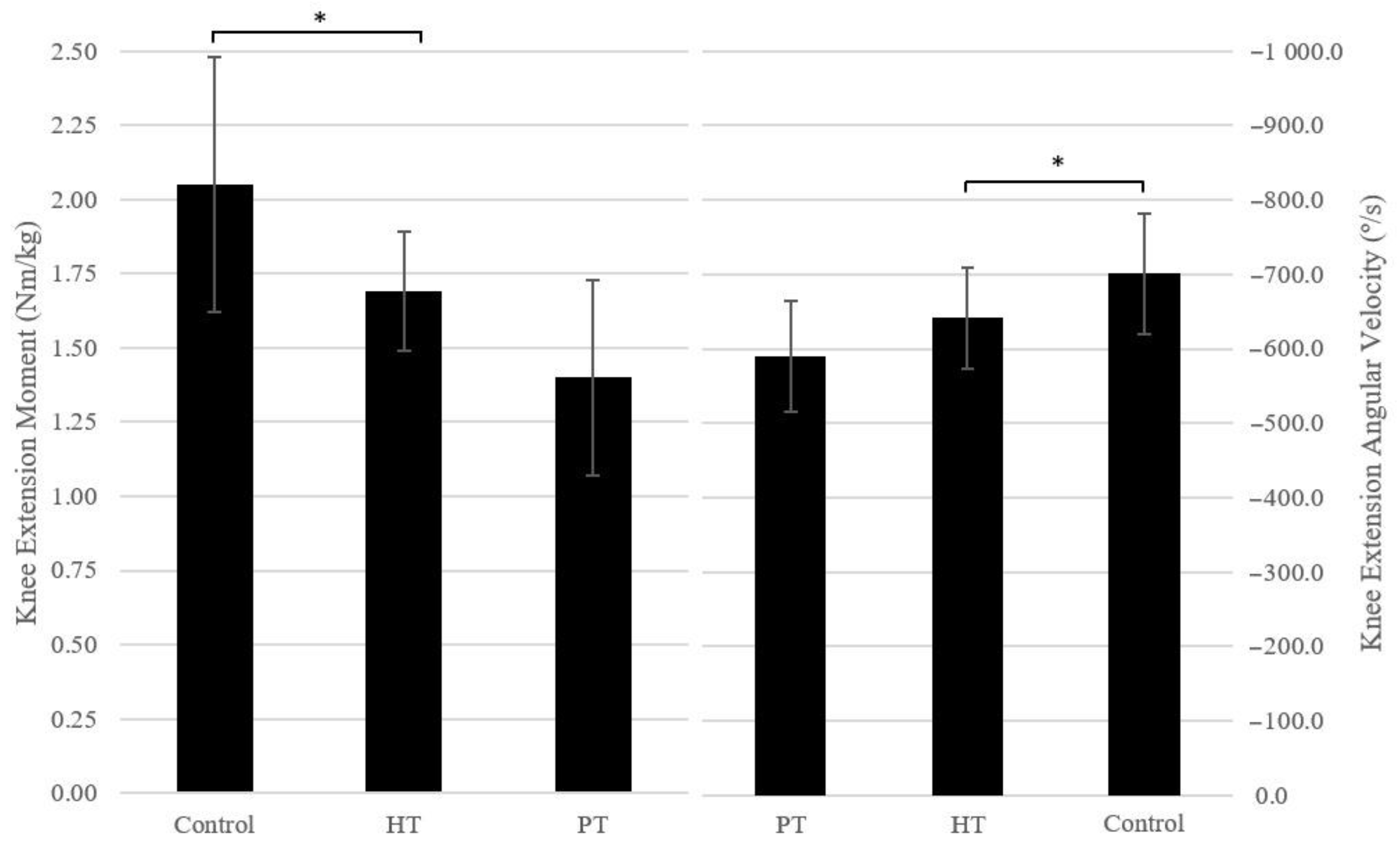
3. Angular moments and velocities. The knee extension moment and angular velocity were significantly higher for the control participants when compared to case participants (md: 0.48 ± 0.14 Nm/kg, t (23): 1.866, p = 0.003, d = 1.344; md: −81.84 ± 30.82°/s, t (23): 0.657, p = 0.014, d = 1.070, respectively). When comparing the three groups, this difference translated to significantly higher values for the control participants when compared to participants submitted to ACLR with a PT autograft (md: 0.65 ± 0.18 Nm/kg, F (2.22): 7.090, p = 0.004, d = 0.804; md: −111.51 ± 38.31°/s, F (2.22): 4.431, p = 0.024, d = 1.000, respectively). No other differences were found while comparing knee joint moments and angular velocities. A comparison of the statistically significant differences between the three groups is represented in Figure 2.

4. Discussion

The present study set forth to determine if there is a difference between the use of a HT autograft and a PT autograft in the valgus movement of the initial contact of the DVJ. There are three major findings from the current study: (1) a slight tendency for an increased valgus alignment in participants who underwent ACLR with a HT autograft; (2) a decrease in vertical ground reaction forces (vGRF) in participants who underwent ACLR; and (3) a decrease in the knee extension angular position and velocity for participants who underwent ACLR with a PT autograft.

4.1. Peak Knee Valgus

Despite Asaeda et al. [13] showing a valgus knee alignment on initial contact during DVJ landing in people who underwent ACLR with a HT autograft, no significant differences were found comparing the control group with the case group or comparing the control group with participants with a HT autograft. However, although not reaching a level of statistical significance, the group of ACLR with a HT autograft demonstrated a tendency of an increased peak knee valgus angle compared to the control group (md: 2.85 ± 1.83°, F (2.22): 1.216, p = 0.399, d = 0.899). According to other authors, semitendinosus and gracilis muscles are essential for dynamic stability of the medial knee, and they stabilize valgus moments applied to the knee when the knee is near extension [30], which might explain our findings for a tendency of an increased peak knee valgus angle in ACLR with HT. Given that an increased knee valgus moment immediately after initial contact might be a risk factor for ACL injury [31], it is essential to decrease this susceptibility to ACL tears by producing a kinetic change that decreases the first peak of the knee valgus moment after initial contact during landing. Sasaki et al. [32] showed that simple core muscle training improved neuromuscular control of the trunk and lower limbs, producing a kinetic change, with decreased lateral trunk motion or increased trunk, hip and knee flexion angles, improving the feedforward mechanism in the jump landing maneuver. These changes in the present study, including thorax lean and hip and knee flexion, were not significant in the case group, when compared to the control participants. However, since no information regarding the rehabilitation program carried out by each participant was taken into account for this study, it is not possible to corroborate or reject such relation.

4.2. Peak vGRF

In this study, participants who underwent ACLR had significantly lower vGRF compared to healthy participants. Baumgart et al. [33] corroborate these findings, demonstrating that the bilateral DVJ revealed vGRF differences between the operated and non-operated (healthy) leg of patients with low subjective knee function. vGRF differences were also present within the operated and non-operated legs between patients with low and high subjective knee function. vGRF has been used by other authors as an indirect measurement to identify knee kinetic asymmetries in ACL-reconstructed patients [34]. Baumgart et al. [35] has shown that patients who underwent ACLR with low subjective knee function reduce the load of the operated leg during eccentric quadricep movements. Other studies support these claims, showing that ski racers had greater asymmetries in the concentric phase after their return to sports, compared with healthy controls [36]. Given this information, despite not finding significant differences between the case and control groups for kinematic variables (including joint angles and joint torque), further studies could be conducted comparing the operated limb to the unoperated limb, hypothesizing asymmetries between the two.

4.3. Knee Extension Angular Moment and Angular Velocity

The case group showed a decrease in the knee extension angular moment compared to the control group. This difference is due to a significant decrease in this parameter in participants who underwent ACLR with a PT autograft compared to the control group. Significant differences were also found comparing knee extension angular velocity between the same groups.
Joint angular velocity is the speed at which a joint is moved under various loading conditions, thus being related to the dynamics of the muscle activation and force generation [37]. In this case, a decrease in the knee extension angular velocity represents a decrease in the power production during takeoff in the injured limb from the deepest position. This is supported by other studies, which concluded that people submitted to ACLR showed a decrease in the peak angular velocity when ascending from squatting [38], hypothesizing that a change in the ascending velocity might originate from altered somatosensory input due to greater tibial–femoral separation. However, Orishimo et al. [39] found that this decrease in power production during take-off was primarily compensated by higher power produced at the hip joint, whereas greater power absorption at the ankle joint was acting as a super-compensatory mechanism for a decreased power absorption at the knee joint during landing maneuvers.
Furthermore, findings of Aune et al. [40] corroborate the same results of the present study, showing that people who underwent ACLR with a HT autograft had better isokinetic knee extension strength and endurance after 6 months following surgery compared with people who underwent ACLR with a PT autograft. It is known that quadriceps muscle weakness is correlated to ACLR with a PT autograft [41]. It is suggested that this deficit in knee extension is related to patellar tendon donor-site morbidity.

4.4. Clinical Implications

There is no standard criteria to clear athletes for a safe return to sport (RTS) after ACLR [42]. This decision is mostly based on measuring variables, which include time since surgery, isokinetic tests, strength and ligamentous laxity [43]. In fact, most test batteries for decision making regarding RTS following ACLR include one- and two-legged postural stability tests, isokinetic tests (which evaluate muscle strength by determining angular velocities) and jump tasks, especially the hop test, which have become the gold-standard of performance tests prior to returning the athlete to sport [44]. These tests are, for the most part, quantitative in nature (e.g., jump height), whereas the quality of movements is not assessed in most programs. Movement-based tests, including the DVJ, with the assessment and analysis of a three-dimensional motion capture system, may complement the evaluation of an athlete for RTS, identifying biomechanical risk factors by capturing movements used to identify the limb’s position and orientation, enabling the extraction of joint centers and their respective angles and rotational forces.
Rehabilitation programs following ACLR play an important role, achieving a successful clinical outcome, as well as encouraging early motion, strength recovery and a return of function. The assessment of the quality of movement for RTS, by incorporating movement evaluations, including the DVP with a motion capture system, may identify, with a higher sensibility, deficits after ACLR. Information, such as the movement strategy, depth of movement, segment alignment, symmetry and control, may enable the creation of specific rehabilitation programs that promote a faster recovery and a lower risk of secondary complications after ACLR.

4.5. Limitations

Due to the reduced sample size, separation between male and female participants was not performed during the data analysis, despite biomechanical evidence of differences between the sexes [45].
This study utilized the bilateral DVJ as the experimental procedure, showing few differences between case and control groups. These differences may be amplified if the same protocol is applied while landing on a single foot, despite a bigger strain and stress to the knee (which is the reason this movement was not performed). Also, there may have been significant differences between participants when taking into account their daily physical activities, being a possible bias of this study. Questionnaires like Lysholm and Tegner Activity Scales [46] could have been applied to evaluate the level of activity pre- and post-surgery for the case group and evaluate the level of activity of the control group.
Participants with concomitant lesions during an ACL tear, including meniscal lesions, were included in this study, being a possible bias for a misleading cause of the participant’s deficits. As mentioned earlier, another limitation of this study is the limited sample size, which reduces the statistical power of our findings and its generalization for the overall population.

5. Conclusions

This study showed that ALCR with a HT autograft do not increase the risk of a valgus knee alignment movement on initial contact during landing compared to ACLR with a PT autograft (md: 2.85 ± 1.83°, F (2.22): 1.216, p = 0.399, d = 0.899). No differences were found for a significant predisposition of the “knee-in & toe-out” position, comparing participants who underwent ACLR with a HT autograft to participants who underwent ACLR with a PT autograft and to healthy participants. ACLR with a PT autograft showed a decrease in the knee extension angular moment (md: 0.65 ± 0.18 Nm/kg, F (2.22): 7.090, p = 0.004, d = 0.804) and angular velocity (md: −111.51 ± 38.31°/s, F (2.22): 4.431, p = 0.024, d = 1.000) compared to the healthy group. Given the differences in various parameters in participants who underwent ACLR, the evaluation of the quality movement of the DVJ with a motion capture system should be considered for the creation of focused rehabilitation programs on deficits that might be found, while enabling a more informed RTS decision.

Author Contributions

Conceptualization, D.A., P.F., F.S. and M.G.; methodology, D.A., P.F., F.S. and M.G.; software, P.F.; validation, D.A., P.F., F.S. and M.G.; formal analysis, D.A. and F.S.; investigation, D.A., P.F. and F.S.; writing—original draft preparation, D.A.; writing—review and editing, P.F., F.S. and M.G.; supervision, P.F., F.S. and M.G.; project administration, M.G. All authors have read and agreed to the published version of the manuscript.

Funding

This research received no external funding.

Institutional Review Board Statement

This study was conducted in accordance with the Declaration of Helsinki and approved by the São João University Hospital Center Review Board (protocol code 291/22 on 21 December 2022).

Informed Consent Statement

Informed consent was obtained from all subjects involved in this study.

Data Availability Statement

The data presented in this study are available within the article.

Conflicts of Interest

The authors declare no conflict of interest.

References

  1. Kohn, L.; Rembeck, E.; Rauch, A. [Anterior cruciate ligament injury in adults: Diagnostics and treatment]. Orthopade 2020, 49, 1013–1028. [Google Scholar] [CrossRef]
  2. Sepúlveda, F.; Sánchez, L.; Amy, E.; Micheo, W. Anterior Cruciate Ligament Injury: Return to Play, Function and Long-Term Considerations. Curr. Sports Med. Rep. 2017, 16, 172–178. [Google Scholar] [CrossRef]
  3. Mall, N.A.; Chalmers, P.N.; Moric, M.; Tanaka, M.J.; Cole, B.J.; Bach, B.R.; Paletta, G.A. Incidence and Trends of Anterior Cruciate Ligament Reconstruction in the United States. Am. J. Sports Med. 2014, 42, 2363–2370. [Google Scholar] [CrossRef]
  4. Siegel, L.; Vandenakker-Albanese, C.; Siegel, D. Anterior cruciate ligament injuries: Anatomy, physiology, biomechanics, and management. Clin. J. Sport Med. 2012, 22, 349–355. [Google Scholar] [CrossRef] [Green Version]
  5. Mizuta, H.; Kubota, K.; Shiraishi, M.; Otsuka, Y.; Nagamoto, N.; Takagi, K. The conservative treatment of complete tears of the anterior cruciate ligament in skeletally immature patients. J. Bone Jt. Surg. 1995, 77, 890–894. [Google Scholar] [CrossRef] [Green Version]
  6. Stone, A.V.; Marx, S.; Conley, C.W. Management of Partial Tears of the Anterior Cruciate Ligament: A Review of the Anatomy, Diagnosis, and Treatment. J. Am. Acad. Orthop. Surg. 2021, 29, 60–70. [Google Scholar] [CrossRef]
  7. Paschos, N.K.; Howell, S.M. Anterior cruciate ligament reconstruction: Principles of treatment. EFORT Open Rev. 2016, 1, 398–408. [Google Scholar] [CrossRef]
  8. Tibor, L.; Chan, P.H.; Funahashi, T.T.; Wyatt, R.; Maletis, G.B.; Inacio, M.C. Surgical Technique Trends in Primary ACL Reconstruction from 2007 to 2014. J. Bone Jt. Surg. 2016, 98, 1079–1089. [Google Scholar] [CrossRef] [PubMed]
  9. Rahr-Wagner, L.; Thillemann, T.M.; Pederson, A.B.; Lind, M. Comparison of hamstring tendon and patellar tendon grafts in anterior cruciate ligament reconstruction in a nationwide population-based cohort study: Results from the Danish registry of knee ligament reconstruction. Am. J. Sport Med. 2014, 42, 278–284. [Google Scholar] [CrossRef] [PubMed]
  10. Xie, X.; Liu, X.; Chen, Z.; Yu, Y.; Peng, S.; Li, Q. A meta-analysis of bone–patellar tendon–bone autograft versus four-strand hamstring tendon autograft for anterior cruciate ligament reconstruction. Knee 2015, 22, 100–110. [Google Scholar] [CrossRef] [PubMed]
  11. Mohtadi, N.G.; Chan, D.S.; Dainty, K.N.; Whelan, D.B. Patellar tendon versus hamstring tendon autograft for anterior cruciate ligament rupture in adults. Cochrane Database Syst. Rev. 2011, 2011, CD005960. [Google Scholar] [CrossRef]
  12. Samuelsen, B.T.; Webster, K.E.; Johnson, N.R.; Hewett, T.E.; Krych, A.J. Hamstring Autograft versus Patellar Tendon Autograft for ACL Reconstruction: Is There a Difference in Graft Failure Rate? A Meta-analysis of 47,613 Patients. Clin. Orthop. Relat. Res. 2017, 475, 2459–2468. [Google Scholar] [CrossRef]
  13. Asaeda, M.; Nakamae, A.; Hirata, K.; Kono, Y.; Uenishi, H.; Adachi, N. Factors associated with dynamic knee valgus angle during single-leg forward landing in patients after anterior cruciate ligament reconstruction. Asia-Pac. J. Sport Med. Arthrosc. Rehabil. Technol. 2020, 22, 56–61. [Google Scholar] [CrossRef]
  14. Hardy, A.; Casabianca, L.; Andrieu, K.; Baverel, L.; Noailles, T.; Junior French Arthroscopy Society. Complications following harvesting of patellar tendon or hamstring tendon grafts for anterior cruciate ligament reconstruction: Systematic review of literature. Orthop. Traumatol. Surg. Res. 2017, 103, S245–S248. [Google Scholar] [PubMed]
  15. Muro-De-La-Herran, A.; Garcia-Zapirain, B.; Mendez-Zorrilla, A. Gait Analysis Methods: An Overview of Wearable and Non-Wearable Systems, Highlighting Clinical Applications. Sensors 2014, 14, 3362–3394. [Google Scholar] [CrossRef] [PubMed] [Green Version]
  16. Kollias, I.; Panoutsakopoulos, V.; Papaiakovou, G. Comparing Jumping Ability among Athletes of Various Sports: Vertical Drop Jumping from 60 Centimeters. J. Strength Cond. Res. 2004, 18, 546–550. [Google Scholar]
  17. Paz, G.A.; de Freitas Maia, M.; Farias, D.; Santana, H.; Miranda, H.; Lima, V.; Herrington, L. Kinematic Analysis Of Knee Valgus During Drop Vertical Jump And Forward Step-Up In Young Basketball Players. Int. J. Sports Phys. Ther. 2016, 11, 212–219. [Google Scholar] [PubMed]
  18. Castro, M.; Fonseca, P.; Paiotti, F.; Pocinho, M.; Carvalho, D.; Vinha, E.; Vilas-Boas, J. Gait characteristics in people with diabetes classified in risk categories 0 and 2 of the IWGDF—International Working Group on the Diabetic Foot. Rev. Port. Endocrinol. Diabetes Metab. 2022, 16, 104–109. [Google Scholar] [CrossRef]
  19. Sousa, M.V.; Sebastião, R.; Fonseca, P.; Morais, S.; Soares, D.; de Sousa, I.; Machado, L.; Sousa, F.; Vaz, M.; Vilas-Boas, J.P. Can increased load carriage affect lower limbs kinematics during military gait? Ergonomics 2022, 65, 1194–1201. [Google Scholar] [CrossRef]
  20. Vilas-Boas, M.D.C.; Fonseca, P.F.P.; Sousa, I.M.; Cardoso, M.N.; Cunha, J.P.S.; Coelho, T. Gait Characterization and Analysis of Hereditary Amyloidosis Associated with Transthyretin Patients: A Case Series. J. Clin. Med. 2022, 11, 3967. [Google Scholar] [CrossRef]
  21. Kessler, S.E.; Rainbow, M.J.; Lichtwark, G.A.; Cresswell, A.G.; D’Andrea, S.E.; Konow, N.; Kelly, L.A. A Direct Comparison of Biplanar Videoradiography and Optical Motion Capture for Foot and Ankle Kinematics. Front. Bioeng. Biotechnol. 2019, 7, 199. [Google Scholar] [CrossRef] [PubMed] [Green Version]
  22. Kanko, R.M.; Laende, E.; Selbie, W.S.; Deluzio, K.J. Inter-session repeatability of markerless motion capture gait kinematics. J. Biomech. 2021, 121, 110422. [Google Scholar] [CrossRef] [PubMed]
  23. Kanko, R.M.; Laende, E.K.; Davis, E.M.; Selbie, W.S.; Deluzio, K.J. Concurrent assessment of gait kinematics using marker-based and markerless motion capture. J. Biomech. 2021, 127, 110665. [Google Scholar] [CrossRef] [PubMed]
  24. Kanko, R.M.; Laende, E.K.; Strutzenberger, G.; Brown, M.; Selbie, W.S.; DePaul, V.; Scott, S.H.; Deluzio, K.J. Assessment of spatiotemporal gait parameters using a deep learning algorithm-based markerless motion capture system. J. Biomech. 2021, 122, 110414. [Google Scholar] [CrossRef]
  25. Drazan, J.F.; Phillips, W.T.; Seethapathi, N.; Hullfish, T.J.; Baxter, J.R. Moving outside the lab: Markerless motion capture accurately quantifies sagittal plane kinematics during the vertical jump. J. Biomech. 2021, 125, 110547. [Google Scholar] [CrossRef]
  26. Hewett, T.E.; Myer, G.D.; Ford, K.R.; Heidt, R.S., Jr.; Colosimo, A.J.; McLean, S.G.; van Den Bogert, A.J.; Paterno, M.V.; Succop, P. Biomechanical Measures of Neuromuscular Control and Valgus Loading of the Knee Predict Anterior Cruciate Ligament Injury Risk in Female Athletes: A Prospective Study. Am. J. Sports Med. 2005, 33, 492–501. [Google Scholar] [CrossRef] [Green Version]
  27. Kobayashi, H.; Kanamura, T.; Koshida, S.; Miyashita, K.; Okado, T.; Shimizu, T.; Yokoe, K. Mechanisms of the anterior cruciate ligament injury in sports activities: A twenty-year clinical research of 1700 athletes. J. Sports Sci. Med. 2010, 9, 669–675. [Google Scholar]
  28. Shapiro, S.S.; Wilk, M.B. An Analysis of Variance Test for Normality (Complete Samples). Biometrika 1965, 52, 591–611. [Google Scholar] [CrossRef]
  29. Cohen, J. Statistical Power Analysis for the Behavioral Sciences, 2nd ed.; Lawrence Erlbaum Associates: Hillsdale, NJ, USA, 1988. [Google Scholar]
  30. Herbort, M.; Michel, P.; Raschke, M.J.; Vogel, N.; Schulze, M.; Zoll, A.; Fink, C.; Peterson, W.; Domnick, C. Should the Ipsilateral Hamstrings Be Used for Anterior Cruciate Ligament Reconstruction in the Case of Medial Collateral Ligament Insufficiency? Biomechanical Investigation Regarding Dynamic Stabilization of the Medial Compartment by the Hamstring Muscles. Am. J. Sports Med. 2017, 45, 819–825. [Google Scholar]
  31. Kimura, Y.; Ishibashi, Y.; Tsuda, E.; Yamamoto, Y.; Hayashi, Y.; Sato, S. Increased knee valgus alignment and moment during single-leg landing after overhead stroke as a potential risk factor of anterior cruciate ligament injury in badminton. Br. J. Sports Med. 2012, 46, 207–213. [Google Scholar] [CrossRef]
  32. Sasaki, S.; Tsuda, E.; Yamamoto, Y.; Maeda, S.; Kimura, Y.; Fujita, Y.; Ishibashi, Y. Core-Muscle Training and Neuromuscular Control of the Lower Limb and Trunk. J. Athl. Train. 2019, 54, 959–969. [Google Scholar] [CrossRef] [PubMed] [Green Version]
  33. Baumgart, C.; Hoppe, M.W.; Freiwald, J. Phase-Specific Ground Reaction Force Analyses of Bilateral and Unilateral Jumps in Patients With ACL Reconstruction. Orthop. J. Sports Med. 2017, 5, 2325967117710912. [Google Scholar] [CrossRef] [PubMed] [Green Version]
  34. Gokeler, A.; Hof, A.; Arnold, M.P.; Dijkstra, P.U.; Postema, K.; Otten, E. Abnormal landing strategies after ACL reconstruction. Scand. J. Med. Sci. Sports 2010, 20, e12–e19. [Google Scholar] [CrossRef]
  35. Baumgart, C.; Schubert, M.; Hoppe, M.W.; Gokeler, A.; Freiwald, J. Do ground reaction forces during unilateral and bilateral movements exhibit compensation strategies following ACL reconstruction? Knee Surg. Sports Traumatol. Arthrosc. 2017, 25, 1385–1394. [Google Scholar] [CrossRef]
  36. Jordan, M.J.; Aagaard, P.; Herzog, W. Lower limb asymmetry in mechanical muscle function: A comparison between ski racers with and without ACL reconstruction. Scand. J. Med. Sci. Sports 2015, 25, e301–e309. [Google Scholar] [CrossRef]
  37. Hahn, D.; Herzog, W.; Schwirtz, A. Interdependence of torque, joint angle, angular velocity and muscle action during human multi-joint leg extension. Eur. J. Appl. Physiol. 2014, 114, 1691–1702. [Google Scholar] [CrossRef]
  38. Stone, W.J.; Arnett, S.W.; Hoover, D.L. Lower Extremity Kinematics of Acl-Repaired and Non-Injured Females When Using Knee Savers®. Int. J. Sports Phys. Ther. 2017, 12, 737–746. [Google Scholar] [CrossRef] [Green Version]
  39. Orishimo, K.F.; Kremenic, I.J.; Mullaney, M.J.; McHugh, M.P.; Nicholas, S.J. Adaptations in single-leg hop biomechanics following anterior cruciate ligament reconstruction. Knee Surg. Sports Traumatol. Arthrosc. 2010, 18, 1587–1593. [Google Scholar] [CrossRef]
  40. Aune, A.K.; Holm, I.; Risberg, M.A.; Jensen, H.K.; Steen, H. Four-Strand Hamstring Tendon Autograft Compared with Patellar Tendon-Bone Autograft for Anterior Cruciate Ligament Reconstruction. Am. J. Sports Med. 2001, 29, 722–728. [Google Scholar] [CrossRef] [PubMed]
  41. Heijne, A.; Werner, S. A 2-year follow-up of rehabilitation after ACL reconstruction using patellar tendon or hamstring tendon grafts: A prospective randomised outcome study. Knee Surgery, Sports Traumatol. Arthrosc. 2009, 18, 805–813. [Google Scholar] [CrossRef]
  42. Ardern, C.L.; Bizzini, M.; Bahr, R. It is time for consensus on return to play after injury: Five key questions. Br. J. Sports Med. 2015, 50, 506–508. [Google Scholar] [CrossRef] [PubMed]
  43. Barber-Westin, S.D.; Noyes, F.R. Factors used to determine return to unrestricted sports activities after anterior cruciate ligament reconstruction. Arthroscopy 2011, 27, 1697–1705. [Google Scholar] [CrossRef] [PubMed]
  44. Hildebrandt, C.; Müller, L.; Zisch, B.; Huber, R.; Fink, C.; Raschner, C. Functional assessments for decision-making regarding return to sports following ACL reconstruction. Part I: Development of a new test battery. Knee Surg. Sports Traumatol. Arthrosc. 2015, 23, 1273–1281. [Google Scholar] [PubMed] [Green Version]
  45. Ryan, J.; Magnussen, R.A.; Cox, C.; Hurbanek, J.G.; Flanigan, D.C.; Kaeding, C.C. ACL reconstruction: Do outcomes differ by sex?: A systematic review. JBJS 2014, 96, 507–512. [Google Scholar] [CrossRef] [PubMed] [Green Version]
  46. Eshuis, R.; Lentjes, G.W.; Tegner, Y.; Wolterbeek, N.; Veen, M.R. Dutch Translation and Cross-cultural Adaptation of the Lysholm Score and Tegner Activity Scale for Patients With Anterior Cruciate Ligament Injuries. J. Orthop. Sports Phys. Ther. 2016, 46, 976–983. [Google Scholar] [CrossRef]
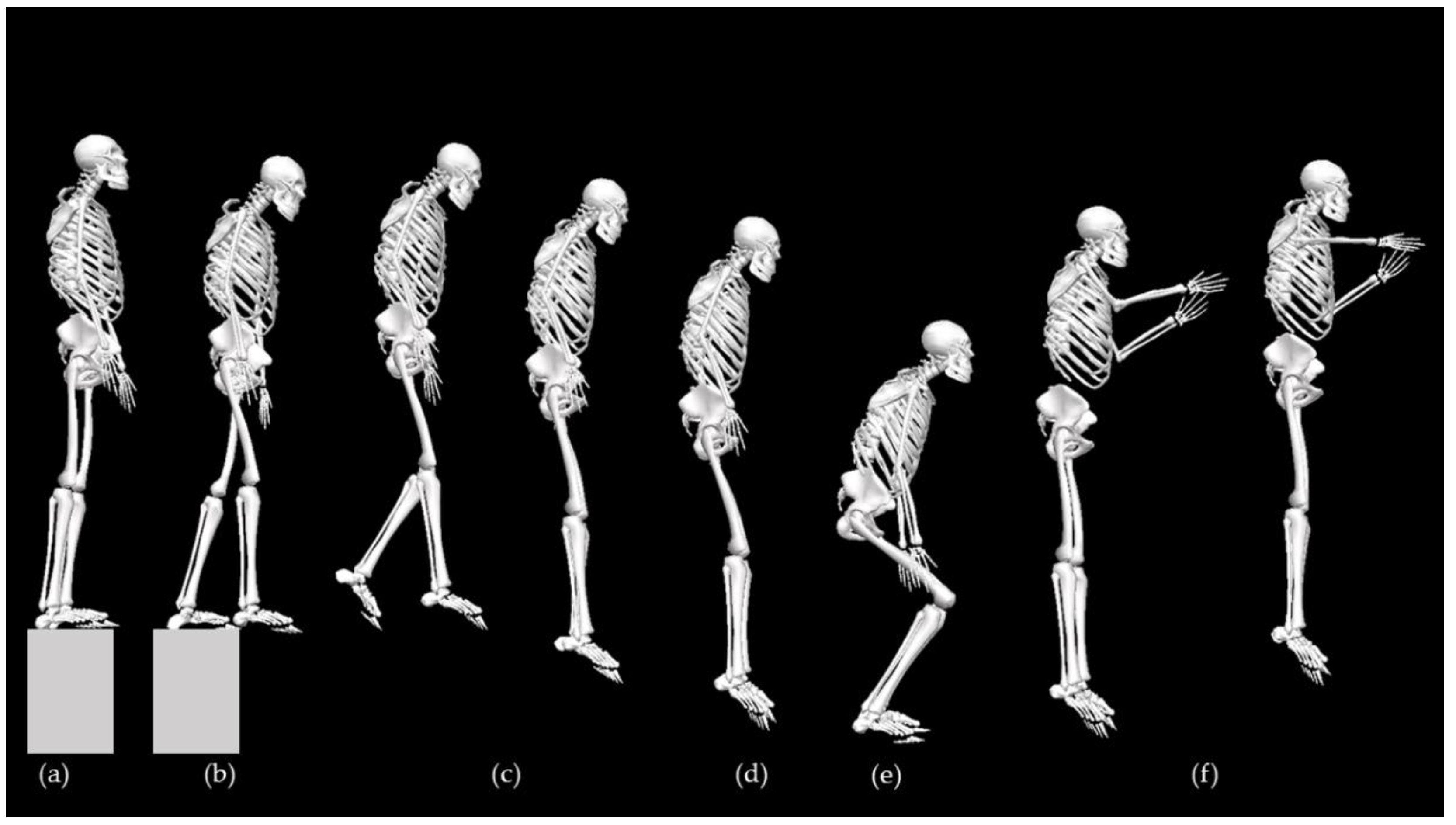
Figure 1. Sequential representation of the drop vertical jump from (a) elevation corresponding to 20% of the participant’s height; (b) the dropping instant; (c) drop flight period; (d) initial contact with the ground; (e) maximum depth after touching the ground; and (f) post-drop vertical jump flight period.
Applsci 13 07363 g001
Figure 2. Comparison of the statistically significant differences between the control, bone–patellar tendon–bone (PT) and four-strand hamstring tendon (HT) autographs in terms of the knee extension moment and angular velocity during the drop vertical jump. Significant differences (p < 0.05) are identified with an asterisk (*).
Applsci 13 07363 g002
Table 1. Participants’ information.
GroupControl vs.
Case Group
AutograftControl vs.
HT Autograft
Control vs. PT AutograftHT vs. PT Autograft
ParameterControl
(n = 11)
Case
(n = 14)
pMD ± SD HT
(n = 8)
PT
(n = 6)
pM ± SDpMD ± SD)pMD ± SD
Age (years)29.40 ± 7.3736.93 ± 8.190.031 *−7.53 ± 3.2639.75 ± 6.3433.17 ± 9.410.027 *−10.35 ± 3.601.000−3.77 ± 3.920.3716.58 ± 4.10
Height (cm)169.50 ± 6.12175.07 ± 7.380.056−5.57 ± 2.77176.25 ± 3.65173.50 ± 10. 860.144−6.75 ± 3.220.804−4.00 ± 3.521.0002.75 ± 3.74
Mass
(kg)
69.78 ± 9.6683.16 ± 16.540.026 *−13.38 ± 5.6384.73 ± 10.0781.08 ± 23.670.102−14.94 ± 6.600.394−11.30 ± 7.211.0003.64 ± 7.68
BMI (kg/m2)24.25 ± 2.7926.92 ± 3.420.047 *−2.67 ± 1.2727.28 ± 3.1726.44 ± 3.990.165−3.03 ± 1.490.577−2.20 ± 1.631.0000.84 ± 1.74
Time since surgery (years)-4.14 ± 1.66--4.13 ± 1.894.17 ± 1.47----0.965−0.04 ± 0.93
Values other than number of participants are expressed as mean ± standard deviation. Significant differences (p < 0.05) are identified with an asterisk (*). BMI, body mass index. HT, hamstring tendon. MD, mean difference. p, p-value. SD, standard deviation. PT, patellar tendon.
Table 2. Comparison between control group and case group during bilateral drop vertical jump.
UnitsControl
Group
Case
Group
pMD ± SD
Touch Down
Thorax Flexiondeg19.91 ± 9.5324.04 ± 8.070.253−4.13 ± 3.52
Hip Flexiondeg16.94 ± 16.6512.04 ± 13.020.3544.90 ± 5.18
Knee Flexiondeg25.73 ± 12.9923.56 ± 19.930.7582.17 ± 6.95
Deepest Position
Max Hip Extension TorqueNm/kg1.12 ± 0.531.13 ± 0.360.954−0.01 ± 0.18
Max Knee Extension TorqueNm/kg1.49 ± 0.831.39 ± 0.380.6970.10 ± 0.25
Max Ankle Extension TorqueNm/kg1.20 ± 0.710.86 ± 0.330.1240.34 ± 0.21
Max Knee Valgus TorqueNm/kg−0.06 ± 0.27−0.79 ± 2.830.4020.74 ± 0.86
General
Jump Heightcm22.28 ± 6.4722.29 ± 3.810.995−0.01 ± 2.07
Depth of countermovementcm24.82 ± 6.7424.93 ± 5.840.964−0.11 ± 2.42
Max Hip Flexiondeg54.10 ± 25.0266.92 ± 13.980.117−12.82 ± 7.88
Max Knee Flexiondeg80.01 ± 25.0392.04 ± 13.380.136−12.03 ± 7.79
Max Ankle Flexiondeg27.85 ± 7.3029.28 ± 4.350.550−1.42 ± 2.34
Peak vGRFN/kg20.11 ± 6.2915.44 ± 4.970.049 *4.67 ± 2.25
Hip Rotation at ICdeg−4.54 ± 3.79−5.94 ± 3.110.3211.40 ± 1.38
Knee Flexion at ICdeg25.73 ± 13.9923.56 ± 19.930.7582.17 ± 6.95
Knee Valgus at ICdeg−0.69 ± 3.981.36 ± 3.870.198−2.05 ± 1.54
Peak Foot/Pelvis Angledeg−7.97 ± 12.63−9.17 ± 4.720.7451.20 ± 3.65
At Peak Knee Valgus
Max Knee Valgusdeg1.86 ± 3.974.00 ± 3.840.187−2.14 ± 1.57
Max Knee Flexiondeg42.31 ± 32.8052.60 ± 26.380.393−10.29 ± 11.82
Max Knee Rotationdeg−11.11 ± 4.79−12.15 ± 4.950.6011.04 ± 1.97
Angular Moments
Knee ExtensionNm/kg2.05 ± 0.431.57 ± 0.300.003 *0.48 ± 0.14
Knee FlexionNm/kg−0.26 ± 0.19−0.14 ± 0.120.067−0.12 ± 0.06
Knee ValgusNm/kg0.29 ± 0.220.24 ± 0.120.4130.06 ± 0.07
Knee VarusNm/kg−0.23 ± 0.20−0.19 ± 0.080.459−0.04 ± 0.06
Angular Velocities
Knee Extensiondeg/s−701.06 ± 80.94−619.22 ± 72.910.014 *−81.84 ± 30.82
Knee Flexiondeg/s517.84 ± 78.64498.83 ± 96.790.60119.01 ± 36.00
Data are presented for the reconstructed leg of case participants along with one (randomly selected) leg of the control participants as mean ± standard deviation. The between-group comparisons show p-values, non-standardized mean differences and standard deviations. Significant differences (p < 0.05) are identified with an asterisk (*). IC, initial contact with ground. Max, maximum. MD, mean difference. p, p-value. SD, standard deviation. vGRF, vertical ground reaction forces.
Table 3. Comparison between control group, group of ACLR with HT autograft and group of ACLR with PT autograft during bilateral drop vertical jump.
Control vs. HT AutograftControl vs. PT AutograftHT vs. PT Autograft
UnitsControl GroupHT AutograftPT AutograftpMD ± SD pMD ± SD pMD ± SD
Touch Down
Thorax Flexiondeg19.91 ± 9.5327.24 ± 4.8719.77 ± 9.900.224−7.33 ± 3.921.0000.14 ± 4.230.3457.47 ± 4.55
Hip Flexiondeg16.94 ± 12.6511.25 ± 14.4513.08 ± 12.081.0005.69 ± 6.101.0003.85 ± 6.661.000−1.83 ± 7.09
Knee Flexiondeg25.73 ± 12.9925.71 ± 24.4920.68 ± 13.281.0000.01 ± 8.151.0005.04 ± 8.901.0005.03 ± 9.47
Deepest Position
Max Hip Extension TorqueNm/kg1.12 ± 0.531.09 ± 0.321.18 ± 0.441.0000.03 ± 0.211.000−0.07 ± 0.231.000−0.10 ± 0.24
Max Knee Extension TorqueNm/kg1.49 ± 0.831.51 ± 0.441.23 ± 0.231.000−0.02 ± 0.291.0000.26 ± 0.321.0000.28 ± 0.34
Max Ankle Extension TorqueNm/kg1.20 ± 0.710.90 ± 0.370.82 ± 0.310.7100.31 ± 0.250.5110.39 ± 0.271.0000.08 ± 0.29
Max Knee Valgus TorqueNm/kg−0.06 ± 0.27−1.33 ± 3.76−0.08 ± 0.110.6381.27 ± 1.081.0000.02 ± 1.080.868−1.25 ± 1.15
General
Jump Heightcm22.28 ± 6.4722.50 ± 4.1122.00 ± 3.741.000−0.23 ± 2.441.0000.27 ± 2.661.0000.50 ± 2.83
Depth of countermovementcm24.82 ± 6.7423.88 ± 6.3826.33 ± 3.781.0000.94 ± 2.821.000−1.52 ± 3.081.000−2.46 ± 3.28
Max Hip Flexiondeg54.10 ± 25.0262.96 ± 14.5772.20 ± 12.361.000−8.86 ± 9.140.250−18.10 ± 9.981.000−9.24 ± 10.62
Max Knee Flexiondeg80.01 ± 25.0389.76 ± 15.0895.08 ± 11.300.891−9.75 ± 9.130.434−15.07 ± 9.971.000−5.32 ± 10.61
Max Ankle Flexiondeg27.85 ± 7.3028.85 ± 5.7029.85 ± 1.751.000−0.99 ± 2.761.000−2.00 ± 3.011.000−1.00 ± 3.21
Peak vGRFN/kg20.11 ± 6.2916.33 ± 5.7214.26 ± 3.940.492−3.78 ± 2.620.1615.85 ± 2.871.0002.07 ± 3.05
Hip Rotation at ICdeg−4.54 ± 3.79−7.23 ± 2.98−4.22 ± 2.540.2782.69 ± 1.531.000−0.32 ± 1.670.315−3.01 ± 1.78
Knee Flexion at ICdeg25.73 ± 13.9925.71 ± 24.4920.68 ± 13.281.0000.01 ± 8.151.0005.04 ± 8.901.0005.03 ± 9.47
Knee Valgus at ICdeg−0.69 ± 3.982.28 ± 4.841.33 ± 1.740.329−2.97 ± 1.781.000−0.82 ± 1.940.9352.14 ± 2.07
Peak Foot/Pelvis Angledeg−7.97 ± 12.63−10.49 ± 5.03−7.40 ± 4.001.0002.53 ± 4.261.000−0.57 ± 4.661.000−3.09 ± 4.96
At Peak Knee Valgus
Max Knee Valgusdeg1.86 ± 3.974.71 ± 4.903.05 ± 1.650.399−2.85 ± 1.831.000−1.19 ± 2.001.0001.66 ± 2.12
Max Knee Flexiondeg42.31 ± 32.8055.55 ± 29.4448.66 ± 23.731.000−13.24 ± 13.881.000−6.35 ± 15.161.0006.90 ± 16.14
Max Knee Rotationdeg−11.11 ± 4.79−11.90 ± 4.72−12.49 ± 5.671.0000.79 ± 2.321.0001.38 ± 2.531.0000.59 ± 2.69
Angular Knee Moments
Knee ExtensionNm/kg2.05 ± 0.431.69 ± 0.201.40 ± 0.330.1130.36 ± 0.160.004 *0.65 ± 0.180.4120.29 ± 0.19
Knee FlexionNm/kg−0.26 ± 0.19−0.14 ± 0.15−0.15 ± 0.080.305−0.13 ± 0.070.526−0.11 ± 0.081.0000.01 ± 0.09
Knee ValgusNm/kg0.29 ± 0.220.27 ± 0.110.19 ± 0.121.0000.02 ± 0.080.7480.10 ± 0.091.0000.08 ± 0.09
Knee VarusNm/kg−0.23 ± 0.20−0.17 ± 0.09−0.21 ± 0.071.000−0.06 ± 0.071.000−0.02 ± 0.071.0000.04 ± 0.08
Angular Velocities
Knee Extensiondeg/s−701.06 ± 80.94−641.47 ± 68.03−589.55 ± 74.110.310−59.60 ± 35.080.024 *−111.51 ± 38.310.649−51.91 ± 40.77
Knee Flexiondeg/s517.84 ± 78.64476.06 ± 88.38529.18 ± 107.170.96941.78 ± 41.321.000−11.34 ± 45.130.842−53.12 ± 48.02
Data are presented for the reconstructed legs of participants with ACLR with HT autograft and participants with ACLR with PT autograft along with one (randomly selected) leg of the control participants as mean ± standard deviation. The between-group comparisons show p-values, non-standardized mean differences and standard deviations. Significant differences (p < 0.05) are identified with an asterisk (*). HT, hamstring tendon. IC, initial contact with ground. Max, maximum. MD, mean difference. p, p-value. PT, patellar tendon. SD, standard deviation. vGRF, vertical ground reaction forces.
Disclaimer/Publisher’s Note: The statements, opinions and data contained in all publications are solely those of the individual author(s) and contributor(s) and not of MDPI and/or the editor(s). MDPI and/or the editor(s) disclaim responsibility for any injury to people or property resulting from any ideas, methods, instructions or products referred to in the content.