Article

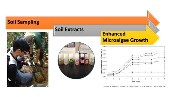
Malaysian Virgin Soil Extracts as Natural Growth Enhancer for Targeted Green Microalgae Species

by
Maegala Nallapan Maniyam
1,2,*,
Hasdianty Abdullah
1,3,
Mohd Fadzli Ahmad
1,3,
Emi Fazlina Hashim
3,
Fridelina Sjahrir
3,
Kazuhiro Komatsu
4,
Victor S. Kuwahara
5 and
Nor Suhaila Yaacob
1,2
1
Institute of Bio-IT Selangor, Universiti Selangor, Jalan Zirkon A7/A, Seksyen 7, Shah Alam 40000, Malaysia
2
Centre for Foundation and General Studies, Universiti Selangor, Jalan Timur Tambahan, Bestari Jaya 45600, Malaysia
3
Faculty of Engineering and Life Sciences, Universiti Selangor, Jalan Timur Tambahan, Bestari Jaya 45600, Malaysia
4
Faculty of Education and Graduate School of Engineering Soka University, 1-236 Tangi-Machi, Hachioji-Shi 192-8577, Japan
5
National Institute for Environmental Studies, 16-2 Onogawa, Ibaraki, Tsukuba 305-8506, Japan
*
Author to whom correspondence should be addressed.
Appl. Sci. 2022, 12(8), 4060; https://doi.org/10.3390/app12084060
Submission received: 19 February 2022 / Revised: 10 April 2022 / Accepted: 12 April 2022 / Published: 17 April 2022
(This article belongs to the Special Issue Advances in Microalgal Biomass Productions)

Abstract

:

Featured Application

The virgin soil extracts have a huge potential as growth enhancers for the mass cultivation of microalgae.

Abstract

The microalgae-based industries are trending upwards, particularly as the feed ingredient for aquaculture. Therefore, a sustainable and reasonably priced source of nutrients to support the mass cultivation of microalgae is in great demand. The present study explored the feasibility of using extracts from virgin soil as natural growth-promoting nutrients for the cultivation of Nannochloropsis oculata, Nannochloropsis oceanica, and Chlorella sorokiniana. The extracts were obtained from Bera Lake Forest using five different treatment methods. The greatest retrieval of dissolved organic carbon, total dissolved nitrogen, and total dissolved phosphorus were observed with the autoclave treatment method at 121 °C twice, yielding a respective concentration of 336.56 mg/L, 13.40 mg/L, and 0.14 mg/L, respectively. The highest growth was recorded with Nannochloropsis oculata resulting in an optical density of 0.488 ± 0.009 (×103 cell mL−1), exhibiting 43% and 44% enhanced growth in comparison to Nannochloropsis oceanica and Chlorella sorokiniana, respectively. The specific growth rate (0.114 a ± 0.007 d−1) was the highest for Nannochloropsis oculata when the 24 h-extraction method was used, whereas the utilization of the autoclave 121 °C twice treatment method contributed to the highest specific growth of Nannochloropsis ocenica (0.069 a ± 0.003 d−1) and Chlorella sorokiniana (0.080 a ± 0.001 d−1). Collectively, these findings suggested that the addition of soil extracts which is sustainable and inexpensive promoted the growth of microalgae compared to the control system. A further study investigating the optimum culture conditions for enhanced microalgae growth will be carried out for the mass production of microalgae biomass.

Graphical Abstract

1. Introduction

Population growth in addition to improved living standards and consumption levels has placed the search for new sustainable resources for the production of food, feed, and raw materials at the centre of development priorities [1]. Therefore, to meet these needs, the improvement of high-yield forms of production such as aquaculture is receiving widespread attention. Under the logic of industrial symbiosis and the circular economy, production models must be developed to minimize waste and use the waste from other processes as raw material. In this context, the cultivation of microalgae for the benefit of many industrial applications supports this agenda well [1].
Microalgae can be classified as photosynthetic microorganisms which can generate biologically active compounds, namely pigments, lipids, polyunsaturated fatty acids, carbohydrates, proteins, and vitamins, through the conversion of carbon dioxide (CO2) [2]. These bioactive compounds are applied in many industries particularly in food as feedstock and human nutrition, as pigments in cosmetics products and as antimicrobial, antiviral, antibacterial, and anticancer drugs in pharmaceuticals [3]. Moreover, due to microalgae’s high content of proteins, high-quality essential amino acids, vitamins, carotenoids, antioxidants, and other substances beneficial to animal health, these photosynthetic microorganisms contribute significantly to animal nutrition and the aquaculture industry [1]. In addition, when compared to higher plants, microalgae exhibit certain favourable characteristics such as the requirement for low water feeding, high carbon dioxide (CO2) fixation and oxygen generation, rapid growth, and simple harvest [2]. Taking into account all the exceptional characteristics of microalgae that led to their commercial importance, the strategies for mass production of microalgae at a reasonable cost must be endeavoured.
Microalgae cultivation, both prokaryotic and eukaryotic, has been studied for over 70 years and is still a hot topic of research and development. Commercialised microalgae, namely Chlorella and Spirulina, are mostly produced for human food and nutrition (nutraceuticals) [3]. In addition, Dunaliella salina and Haematococcus pluvialis were mass-produced popularly in the 1980s for microalgal pigments which were marketed mostly for human nutrition as food additives since these microalgae contain high-value carotenoids, beta-carotene, and astaxanthin, respectively [3]. In the 1990s, the commercialized production of docosahexaenoic acid (DHA) and eicosapentaenoic acid (EPA) in particular were heavily commenced for use in aquaculture feed and enrichment of nutritional products [4]. In addition, microalgae can serve as functional feed and as probiotics for aquaculture feeding which could curb the common outbreak of disease-causing enormous economic losses to the aquaculture industry. Although the production of microalgae-based products has been relatively in practice for a long time, the vast applications were directed to industries such as nutraceuticals and human consumption on a minute scale with an overall worldwide production of fewer than 15,000 tons per year and production costs limited to 10 € per kg [5]. It can then be concluded that the large-scale commercial production of microalgae is still in its infancy stage and needs substantial improvement.
There are a few challenges related to the production of microalgae, namely the necessity for a high volume of water and fertilizers usage even though arable land is not required [6]. In addition, some limitations may discourage the production of microalgae biomass. The cultivation of microalgae requires high energy consumption, incurs high costs when the biomass is converted into valuable end-products, involves technological constraints when the cultivation of microalgae carried out in photobioreactor leads to the upsurge in the formation of biofilms, and limits the light penetration and the occurrence of environmental pollution as a result from the release of the undigested nutrients [7,8]. Among these challenges, the selection of favourable culture conditions and the use of economically feasible and sustainable nutrients for microalgae cultivation should be prioritized for optimization to make commercialization more viable [6]. The formulation of microalgae cultivation medium using organic carbon such as sugar alcohols, sugars, and organic acids in the growth medium of microalgae amplifies the cost of microalgae biomass cultivation [9]. Hence, the research community has devoted immensely to the quest to search for cheaper and sustainable nutritional compositions for enhanced microalgae biomass production. The production of microalgae biomass largely depends on major nutrients, namely nitrogen, phosphorus, and carbon dioxide (CO2), whereby for the generation of 100 tons of microalgae biomass, approximately 200 tons of CO2, 5 tons of nitrogen, and 1 ton of phosphorus are required [5]. Even though sources such as fertilizers and flue gas can be employed to supply these nutrients, ultimately the utilization of these resources is limited due to the consumption of high energy and excessive emissions of carbon dioxide (CO2) [5].
Many studies have explored the use of wastewater as an alternative nutrient source which was found successful to enhance microalgae cultivation [1,5,9]. Microalgae have been attempted to bioremediate industrial, municipal, food, and animal wastewater whereby the organic compounds and nutrients are simultaneously exploited to support the growth of microalgae [10,11,12,13,14,15]. Microalgae are advantageous as biocatalysts for the treatment of wastewater due to their promising ability in removing pollutants such as carbon oxygen demand (COD), heavy metals, and emerging contaminants. Furthermore, microalgae are capable to remove and recover nutrients such as total dissolved nitrogen (TDN) and total dissolved phosphorus (TDP). The treated wastewater by microalgae additionally can be recovered for other purposes and the culture medium can be reused. The summary of the selected wastewater and the production of various microalgae biomass is presented in Table 1. However, it is necessary to explore more sustainable nutrient sources since the microalgae-based industries are blooming. Therefore, the present study focuses on investigating the potential of using extracts collected from undisturbed soils from Bera Lake Forest, Malaysia as a nutrient source for enhanced microalgae growth specifically for Nannochloropsis oculata, Nannochloropsis oceanica, and Chlorella sorokiniana. Furthermore, a high throughput method using a 96-well plate culturing was implemented in this work which facilitated time reduction for growth evaluation and contributed to cost savings.

2. Materials and Methods

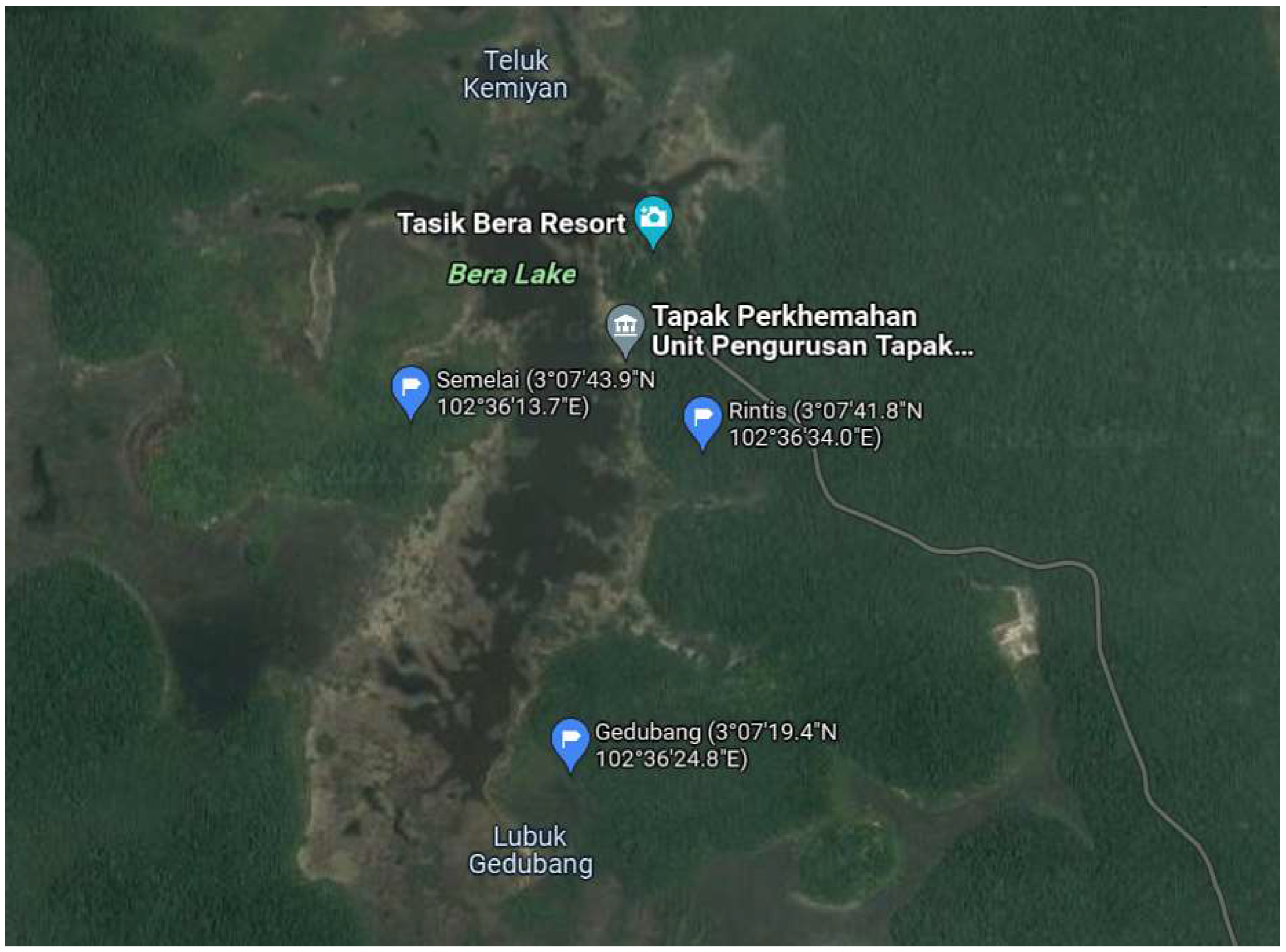
2.1. Collection of Sample and Preparation

Figure 1 shows the coordinate of Bera Lake Forest (3°07′41.8″ N 102°36′34.0″ E), located around the natural freshwater of Bera Lake in Pahang, Malaysia where the virgin soil samples were collected.
The soil samples collected from the Bera Lake Forest are categorized as silt soil containing 6.49% sand, 92.73% slit, and 0.78% clay. The soil samples which were yellowish-brown in colour exhibited acidic conditions with a pH value of 4.24 ± 0.003 and a moisture content of 1.36 ± 0.02%. The collected soil samples contained nitrogen to phosphorus to kalium ratio of 0.11: 2.38:1.05. The presence of different types of heavy metals in this soil was recorded with iron exhibiting the greatest amount of 23,884.2 mg/kg. In addition, arsenic and lead were not detected in the soil samples which is advantageous to this study.
The United States Department of Agriculture (USDA) method was employed as the random technique of soil sampling [16]. The O horizon measuring around15 cm from the soil surface was eliminated prior to sample collection. As a minimum, three sampling sites within 10 metres of each other were used to capture each sample. Five cores with uniform features were collected about 1 kg each before being combined into one composite. The samples were oven-dried at 60 °C (DKM-400 (Yamato Scientific Co., Ltd., Tokyo, Japan) for a few days until complete removal of moisture occurred, after eliminating the coarse particles such as stones, wood, and roots. The dry soil samples were crushed to powder and sieved using a 2-mm stainless steel sieve. The fine soil samples were dried and packed into Ziploc bags, which were kept in a dark environment at 24 °C until additional soil extraction processes.

2.2. Soil Extraction and Analyses

The methodology established by Anderson [17] was modified accordingly and the extraction methods as tabulated in Table 2 were carried out to collect the soil extracts.
The protocol for each extraction method was performed by transferring 200 mL ultrapure water added with 20 g of dried fine soil samples into a 400 mL centrifuge bottle (1 part of the soil to 10 parts of ultrapure water). The samples were placed in the dark at room temperature for 24 h for natural extraction. For the autoclave approach, the dried fine soil samples added to the ultrapure water were autoclaved using autoclave SX-500 (Tomy Seiko Co., Ltd., Tokyo, Japan). Beckman centrifuge Allegra X-30R (Beckman Coulter, Indianapolis, IN, USA) was used to centrifuge the samples at 2500 rpm for 15 min after the autoclaving process or the natural extraction. The resulting supernatant was carefully removed and filtered by employing Whatman Glass Microfiber Filter (GF/F) 0.7 μm via a 0.2 μm syringe filter. The filtered water samples were subsequently stored in Revco, ULT-390-10 freezers at −20 °C (Thermo Fisher Scientific, Japan) until further analyses.
Soil extracts containing non-purgeable organic carbon (NPOC) concentrations were measured as dissolved organic carbon (DOC) using Shimadzu TOC-L CSH (Shimadzu Corp., Kyoto, Japan). Each sample was acidified with 2 M of HCl and purged with carbon dioxide (CO2)-free synthetic air for the purpose of removing volatile carbon (Big Purifier with CoFree Indicator, USA). The amounts of total dissolved nitrogen (TDN) and total dissolved phosphorus (TDP) were uncovered using a portable spectrophotometer, Lovibond MD 600D (The Tintometer Limited, Amesbury, UK). To obtain the average value and standard deviation, the measurements were repeated three or four times for each sample.

2.3. Microalgae

The targeted microalgae species utilized in the present study were Nannochloropsis oceanica, Chlorella sorokiniana, and Nannochloropsis oculata which were kindly supplied by the National Institute for Environmental Studies (NIES), Japan. The preparation of Conway medium from five basic solutions followed the recipe by Khatoon [18] consisting of:
(a)
the mineral solution—100 g of NaNO3, 45 g of disodium EDTA (C6H16N2O8), 33.6 g of H3BO3, 20 g of NaH2PO4·4H2O, 1.3 g of FeCl3·6H2O, 0.36 g of MnCl2·4H2O, and 1 mL trace metal solution in 1 L of ultrapure water;
(b)
the trace metal solution—0.21 g of ZnCl2, 0.2 g of CoCl3·6H2O, 0.09 g of (NH4)6MO7O2·4H2O, and 0.2 g of CuSO4·5H2O in 0.1 L of ultrapure water;
(c)
the vitamin solution—0.2 g of thiamine (B1) and cyanocobalamin (B12) in 0.1 L of ultrapure water;
(d)
the silicate solution—2 g of Na2SiO3 in 0.1 L of ultrapure water;
(e)
the nitrate solution—2 g of KNO in 0.1 L of ultrapure water.
The medium was prepared by adding 1 mL of main mineral, silicate, and nitrate solution to the Schott bottle to prepare a 1 L volume medium. An amount of 1 mL of NH4Cl and vitamin solution was added to the cooled autoclaved medium to give a final medium concentration of 5.0 × 10⁻⁴ M. The cultures were cultivated at 25 ± 0.5 °C under a light intensity of 33.75 μmol photons m−2 s−1 on a 12 h light:12 h dark cycle.
The 96-well microplates culturing method was implemented to access the growth of Nannochloropsis oceanica, Chlorella sorokiniana, and Nannochloropsis oculata supplemented with soil extracts obtained from five different extraction methods, respectively following the methods outlined by Yaacob [19]. A maximum of 200 μL of solution can be accommodated by each well in the microplate. As an attempt to prevent evaporation during the course of experimentation, 200 μL ultrapure water was loaded into the border wells of the microplates. The remaining wells were loaded with 195 μL of an appropriate medium added with 5 μL of soil extracts obtained from the autoclave treatment method at 105 °C once in the second column (as the blank), and the third column was filled with 175 μL of an appropriate medium added with 5 μL of soil extracts obtained from autoclave treatment method at 105 °C once and 20 μL of microalgae (as the experimental groups) to document the exponential phase of microalgae.
The same experimental protocols were duplicated in the 4th to 11th columns of the microplate with autoclave treatment methods at 105 °C twice (Column 5), 121 °C once (Column 7), 121 °C twice (Column 9), and 24 h-natural extractions (Column 11), respectively. The control experiment containing the Conway medium alone was established to assess the growth of microalgae in other microplates. The air humidity in the microplate wells was preserved and the external contamination before incubation was prevented by sealing the microplates with parafilm after pipetting all the wells in the microplates.
The microplates were incubated for 9 d and the biomass or growth of microalgae was determined by optical density (OD) at 680 nm for every 24 h using the microplate reader Infinite M200 PRO (Tecan, Austria). The optical density was measured every 24 h for 9 days in each of the wells holding the controls and samples, using an 8-channel Eppendorf pipettor. The homogenization was carried out to thoroughly mix the microalgae suspended in the bottom well with the solution.

2.4. Data Analyses

Three microplate replicates for each control and sample in a column were evaluated. The net optical density was computed by subtracting the optical density from the control system with the optical density of experimental samples. The microalgae biomass was determined using optical density in the present study as the technique is straightforward, common, and rapid [20,21]. The determination of specific growth rate (μ) and the division rate of microalgae were established following the formulas below:
μ = ln ( N 2   N 1 ) t 2   t 1
k = μ ln 2
where N2 and N1 are the optical density at times t2 and t1, respectively.
The concentration of dissolved organic carbon (DOC), total dissolved nitrogen (TDN), and total dissolved phosphorus (TDP), growth of microalgae, and optical density in the respective treatment methods were examined using independent samples t-test, one-way analysis of variance (ANOVA), and Tukey post-hoc analysis. A 95% confidence interval level was secured to validate the significant difference between the various extraction parameters. All statistical analyses were achieved using SPSS (Statistical Package for the Social Sciences) statistics software version 20 (IBM SPSS, Chicago, IL, USA).

3. Results and Discussion

3.1. Nutrient Recovery from Different Extraction Methods

The selection for different extraction methods was based on autoclave methods from previous studies which used specific temperatures such as 105 °C [22] and 121 °C [23] to sterilize the sludge samples. Soil samples were used for this study due to the presence of copious amounts of humic substances. Humic substances are well recognized to aid natural plant growth as a result of the occurrence of rich bioactive organic carbon sources. Therefore, the activity of numerous antioxidant enzymes can be effectively improved to promote the physiological metabolisms of plant cells [24]. Studies carried out by Fan [24] and Zheng [25] observed that the supplementation of humic acid alleviated the growth of Euglena pisciformis AEW501 and Scenedesmus Capricornus, respectively. When the culture medium was improved with 50 mg/L humic acids, maximum biomass productivity of 35 mg/L/d and greatest high-value product (lipid content) accumulation of 60% were achieved with Euglena pisciformis AEW501 [24]. Humic substances below the concentration of 2 mgC/L had a positive effect on the growth of Scenedesmus Capricornus, which was simultaneously accompanied by increased synthesis of chlorophyll and macromolecules in the microalgae [25].
The retrieval of dissolved organic carbon (DOC), total dissolved nitrogen (TDN), and total dissolved phosphorus (TDP) is summarized in Table 3. On the whole, the recovery of TDP was substantially lower (p < 0.05) compared to that of DOC and TDN irrespective of the treatment methods used. This may be attributable to dissimilar rates of organic matter mineralization in soil [26]. Significantly lower extractable nutrients (p < 0.05) were achieved with soil extracts from the 24-h natural extraction method carried out at room temperature in comparison to autoclaved soil extracts. The use of autoclave heat shock supported the release of labile nutrients into the soil extracts with minimum alterations to the physical properties of the soil extracts [27], contributing to increased concentrations of DOC, TDN, and TDP when weighed against the natural extraction process.
It was interesting to note that the utilization of autoclave 121 °C twice treatment method yielded the greatest recovery of DOC of 336.56 mg/L, which was 474% and 108% higher than natural extraction dan autoclave 105 °C twice treatment method, respectively. The recovery of TDN and TDP observed a similar trend when the autoclave 121 °C twice treatment method was exploited to obtain the soil extracts, whereby 164% and 26% higher recovery of TDN and 75% and 40% higher recovery of TDP were recorded compared to that of natural extraction dan autoclave 105 °C twice treatment methods, respectively. Autoclave methods allowed more extraction of organic matter than natural extraction at 24 h which may be due to the disintegration of polysaccharide structures, alteration processes that influence the soil surface, and the occurrence of unknown reactions or nutrients within the soil matrix [28]. In addition, the collapse of chemical bonds could only occur when the activation energy was exceeded to disrupt the soil materials into carbon, nitrogen, and phosphorus compounds released in the soil extracts [29]. Hence, the autoclave methods assisted the supply of surplus energy which enabled the release of higher concentrations of DOC, TDN, and TDP in the soil extracts in comparison to natural extractions which occurred at a lower temperature, hampering the breakdown of any chemical bonds since the heat energy is too minimal to overcome the activation energy [19].
Although the application of different treatment methods influenced the recovery of DOC to a great extent (p < 0.05), this pattern could not be duplicated when autoclave treatment methods at 105 °C and 121 °C were used to recover TDP (p > 0.05), resulting in similar average production of 0.12 mg/L and 0.14 mg/L TDP, respectively. There was a significant difference in the recovery of TDN when autoclave treatment methods at 105 °C and 121 °C were used (p < 0.05), resulting in an average production of 10.45 mg/L and 13.40 mg/L TDP, respectively. However, the period of autoclaving did not affect the recovery of TDN between these two temperatures (p > 0.05). Similar results were observed during the recovery of TDN from soil extracts obtained from Langkawi Island and Kenyir Lake [28].
When the ratio of phosphorus was fixed to 0.1, the carbon to nitrogen ratio increased from 73.3:6.3 to 240.4:9.5 when the extraction methods were compared between natural extraction for 24 h and autoclave treatment method at 121 °C twice. Dead bacteria could be most likely responsible for the relatively low carbon to nitrogen ratio of soil extracts when natural extraction for 24 h was employed [19]. On the other hand, increased carbon to nitrogen ratio was observed when autoclave treatment methods were used, indicating the release of more carbon-rich compounds into the soil extracts [19]. Interestingly, the carbon to nitrogen ratio obtained via the autoclave 121 °C twice treatment method in the present study is quite ideal to be used for agricultural use [30]. Therefore, the findings from the present study provided a platform to further engage on the viability of using sustainable and low-cost soil extracts as an alternative source of nutrients for the growth of economically important plants.

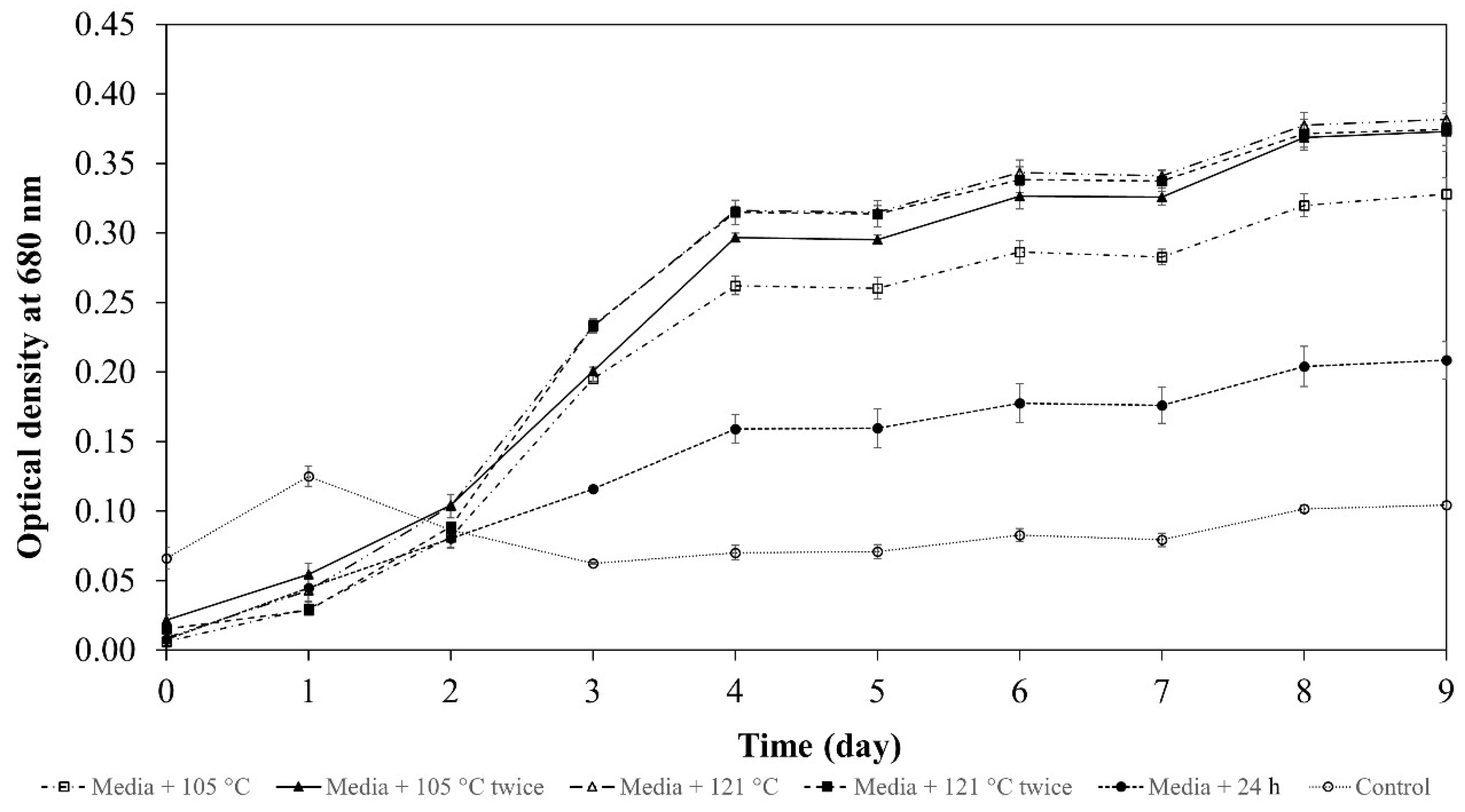
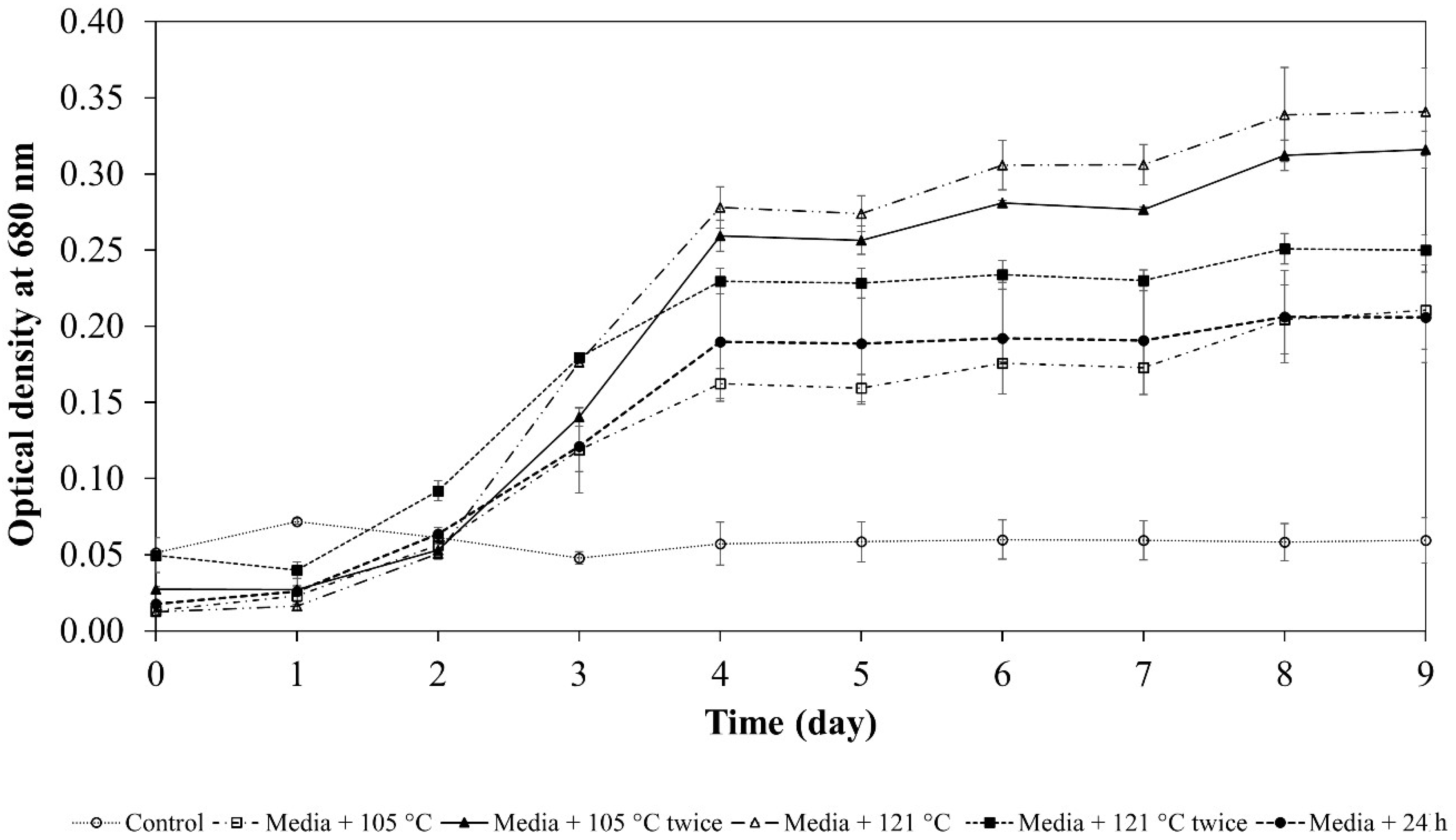
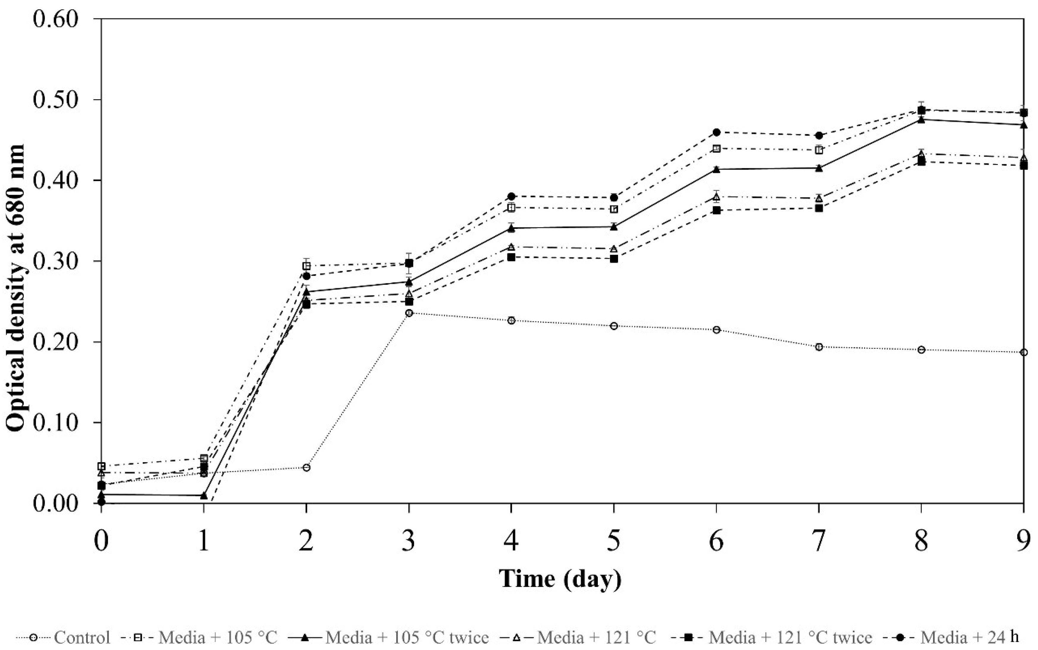
3.2. Effects of Soil Extracts on the Growth of Microalgae

Figure 2 illustrates the typical growth pattern of Nannochloropsis oceanica when cultivated with a medium amended with soil extracts obtained using five different extraction methods.
The supplementation of the soil extracts supported enhanced growth of Nannochloropsis oceanica in comparison to the control system containing the medium alone significantly (p < 0.05). The growth pattern of Nannochloropsis oceanica resembled the sigmoid growth curve for all treatment methods and the lag phase was observed for 2 d after inoculation, whereby the cell density increased gradually from an average optical density of 0.024 at day 0 to 0.063 after 2 d of the incubation period (the average value was determined as the mean of optical density between five different treatment methods).
Nannochloropsis oceanica grew more prominently into the exponential phase which began on day 2 and lasted until day 4 with an average optical density of 0.224 between the five different treatment methods, exhibiting 293% higher cell density compared to the control system containing the medium alone. The mid-log phase was achieved on day 3 with an average optical density of 0.147, indicating the rapid growth of the microalgae. The growth of Nannochloropsis oceanica was continuously monitored for 9 d and the highest optical density was observed with the autoclave treatment method at 121 °C once, yielding an optical density of 0.341 ± 0.029. When compared with natural extraction and autoclave 105 °C once treatment methods, the utilization of the autoclave treatment method at 121 °C once resulted in 64% higher cell density, depicting a significant growth rate of the microalgae when soil extracts obtained from this treatment method were used as growth enhancer (p < 0.05). In addition, the growth of Nannochloropsis oceanica was substantially influenced by the autoclave treatment methods (p < 0.05), except for natural extraction (24 h) and autoclave 105 °C once treatment methods which generated an insignificant difference (p > 0.05) in terms of growth with an optical density of 0.206 ± 0.030 and 0.210 ± 0.026, respectively, after 9 d of the incubation period.
The growth of Nannochloropsis oculata was monitored for a period of 9 d and the results are depicted in Figure 3.
The addition of soil extracts indeed supported improved growth of the microalgae with significantly increased cell density (p < 0.05) compared to that of the control system containing the medium alone. Interestingly, the control system demonstrated a lag period of 2 d, whereas the experimental groups (artificial medium containing soil extracts from five different extraction methods) showed a lag period of only 1 d. The growth pattern of the experimental groups revealed a similar trend, whereby the exponential phase began after 1 d of cultivation and the growth progressed into the early stationary phase on day 2, recording an increase in the average optical density from 0.024 on day 0 to 0.267 at day 2, presenting an increase of 1013% in the cell density. On the other hand, the optical density of the control system increased from 0.024 on day 0 to 0.236 on day 3, unveiling an increase of 883% in the cell density. However, the growth of Nannochloropsis oculata declined gradually after day 3, reaching an optical density of 0.187 on day 9 in the control system which suggested the inadequacy of using artificial medium alone in the cultivation of microalgae.
The experimental groups using the soil extracts obtained from natural extraction (24 h), autoclave 105 °C once, and autoclave 105 °C twice treatment methods established the highest growth of Nannochloropsis oculata with an average optical density of 0.484 with no significant difference (p > 0.05). However, these methods differ significantly (p < 0.05) when compared to the autoclave treatment methods at 121 °C once and 121 °C twice, which yielded an average optical density of 0.428. The utilization of soil extracts obtained from natural extraction (24 h), autoclave 105 °C once, and autoclave 105 °C twice treatment methods supported the growth of Nannochloropsis oculata 1.13-fold higher in contrast to soil extracts obtained from the autoclave treatment methods at 121 °C once and 121 °C twice.
The growth of Chlorella sorokiniana in the control system containing the medium alone showed the lowest value, recording an optical density of 0.104 after 9 d of cultivation (Figure 4).
However, there was a spike in the optical density on day 1 (0.125) indicating the occurrence of the exponential phase and from that time on, the optical density decreased to 0.063 on day 3 and became almost consistent throughout the cultivation period of 9 d, although the control system was initiated with the highest inoculation of inoculum (0.066) on day 0. This observation evidently indicated that the use of medium alone did not sustain the growth of the microalgae. Therefore, the virgin soil obtained from Bera Lake Forest was extracted using five different treatment methods and the soil extracts were used as a growth enhancer for Chlorella sorokiniana.
The growth curves of experimental groups elucidated a typical sigmoid pattern with a lag phase of 1 d, witnessing a gradual increase in the average optical density from 0.012 to 0.04, yielding an increased growth of 70%. The exponential phase was observed from day 1 to day 4 and the mid-log phase appeared around day 2.5. However, the soil extracts obtained from 24 h natural extraction exhibited a more gradual and lower growth surge compared to the rest of the experimental groups. As shown in Figure 4, it was rather apparent that by day 4 the early stationary phase was attained, resulting in an optical density of 0.159 ± 0.010 when soil extracts from natural extraction (24 h) were used. This method produced the lowest cell density by 98% (p < 0.05) when compared to that of the autoclave treatment method at 121 °C once and the autoclave treatment method at 121 °C twice, respectively. The highest cell density was observed with these autoclave treatment methods, respectively yielding an optical density of 0.316 ± 0.003 and 0.315 ± 0.009, exhibiting no discernible pattern (p > 0.05).
After the incubation period of 9 d, a substantial difference in terms of growth measured in optical density was observed between the control system containing the medium alone and the experimental groups (p < 0.05). The autoclave treatment methods at 121 °C once and at 121 °C twice emerged as the best methods for effective soil extraction which demonstrated a similar growth curve pattern with an average optical density of 0.316 achieved on day 9, documenting an insignificant difference (p > 0.05) when compared between the two cell densities. Although the autoclave treatment method at 105 °C twice exhibited slightly lower growth of Chlorella sorokiniana, during the cultivation period, the optical density on day 9 reached 0.373 ± 0.014, resembling the results obtained from autoclave treatment methods at 121 °C once and at 121 °C twice, respectively, with no significant difference (p > 0.05).
The use of soil extracts can be sustainable and cheaper support for mass cultivation of microalgae in addition to commercial medium [31]. Since the soil extracts contain the presence of important nutrients for microalgae growth such as carbon, nitrogen, phosphorus, and trace elements due to the oligotrophic nature [32], they assist the formation of protein which is vital for microalgae cultivation. The occurrence of trace elements such as phosphorus and calcium on the other hand stimulates an accelerated rate of cell division and specific growth rate of microalgae resulting in increased cell density. Furthermore, soil contains many free enzymes that play an important role in catalyzing reactions that lead to the decomposition of organic matter that stimulates the growth of microalgae [33]. A study carried out by Allaguvatova [34] reported enhanced production of Chlorella vulgaris due to the addition of soil extracts and vitamins to the culture medium as evidenced by the increase in the optical density. Similarly, Porphyridium purpureum cultivated in Kock medium in addition to soil extracts promoted the growth of the microalgae as revealed by the research established by Lu [35].
Although the soil extracts obtained from the autoclave treatment method at 121 °C twice yielded the highest ratio of carbon to nitrogen to phosphorus of 240.4:9.5:0.1, the utilization of different autoclave methods aided the growth of the tested microalgae, namely Nannochloropsis oculata, Nannochloropsis oceanica, and Chlorella sorokiniana, significantly. These findings indicated that the existence of a relatively high level of nutrients on top of distinctive extraction methods may reduce and precipitate the necessary vitamins and minerals present in the soil which enabled increased growth of microalgae [36]. In addition, the Redfeld ratio (C:N:P = 106:16:1 atomic ratio) has been set as the benchmark nitrogen to phosphorus ratio to optimally grow phytoplankton. However, different phytoplankton has different needs for nutrients to support their growth process [35]. In the present study, the ratio of nitrogen to phosphorus was quite similar between the soil extracts and the presence of a carbon ratio within the range of 162 to 164 supported the growth of Nannochloropsis spp. and Chlorella sp. effectively.

3.3. Evaluation of Specific Growth Rate (SGR, µ) and Division Rate (k) for Selected Microalgae in Modified Soil Extract Medium

The specific growth rate (μ, d−1) showed a significant difference (p < 0.05) between the modified soil extract medium when tested using three different species of microalgae, namely Nannochloropsis oculata, Nannochloropsis oceanica, and Chlorella sorokiniana (Table 4). Nannochloropsis oculata recorded the highest specific growth rate of 0.114 a ± 0.007 d−1 when a medium supplemented with soil extracts obtained from natural extraction for 24 h was used. The addition of soil extracts obtained from autoclave 121 °C once treatment method to the conventional medium was found competent to generate the highest specific growth rate of 0.069 a ± 0.003 d−1 and 0.080 a ± 0.001 d−1 for Nannochloropsis oceanica and Chlorella sorokiniana, respectively.
The relatively higher specific growth rate indicates that the addition of extraction parameters and nutrients in the modified soil extracts medium is essential to the exponential rise. The addition of soil extracts was indeed instrumental in increasing the biomass of microalgae as evidenced by the findings reported by Teo [37]. The supplementation of 7% soil extracts assisted the production of higher biomass with the specific growth rate of 0.82 d−1 of Nannochloropsis sp. after 16 d of the incubation period. Likewise, the production of Cosmarium subtumidum increased in terms of growth rate, cell number (biomass of 2629 cells per mL), and doubling time when the cultivation medium was augmented with soil extracts [38]. Accordingly, the rise in the concentration of soil extracts has affected the growth in the number of cells.
Although the soil extracts obtained from the autoclave treatment method at 121 °C twice yielded the highest nutrient recovery, the soil extracts resulting from this method of treatment did not support the highest specific growth rate of any of the microalgae species which may be due to some species dependence and variability [19]. In addition, the presence of higher microalgae populations may limit nutrient availability and this hinders the increase in total biomass concentrations over time [36]. The 96-well plate culturing method employed in the present study may contribute to certain growth inadequacies due to low volume (microwells) and eventual high concentration of the microalgae biomass on day 9 [31].
The division rate (k) displayed the same trend as the specific growth rate (μ, d−1), whereby a significant difference was seen when a different modified soil extract medium was used (p < 0.05) as shown in Table 5. The greatest division rate of Nannochloropsis oculata was achieved with a modified soil extract medium (24 h natural extraction) amounting to 0.165 a ± 0.007. Modified soil extract medium from the autoclave treatment method at 121 °C once on the other hand contributed to the highest division rate of 0.099 a ± 0.003 and 0.116 a ± 0.001 for Nannochloropsis oceanica and Chlorella sorokiniana, respectively.

4. Conclusions

Undisturbed soil from Bera Lake Forest extracted via five different methods was supplemented as a growth enhancer for the cultivation of Nannochloropsis oculata, Nannochloropsis oceanica, and Chlorella sorokiniana, respectively. The highest nutrient recovery was observed with the autoclave treatment method at 121 °C twice, recording the carbon to nitrogen to phosphorus ratio of 240.4:9.5:0.1. However, the optimal growth of Nannochloropsis oceanica and Chlorella sorokiniana was observed when soil extracts obtained from the autoclave treatment method at 121 °C once were utilized. In addition, soil extracts obtained from natural extraction for 24 h supported the maximum growth of Nannochloropsis oculata. These findings indicated different phytoplankton required different needs for nutrients to facilitate their growth process. Nonetheless, the addition of soil extracts indeed enhanced the growth of Nannochloropsis oculata, Nannochloropsis oceanica, and Chlorella sorokiniana by 478%, 158%, and 261%, respectively, in comparison to the control system containing the medium alone. As a result, soil extracts have a huge potential to be used as an alternative to expensive vitamins to enhance microalgae growth. The 96-well plate culturing method is effective as a high-throughput incubation technique to grow microalgae. However, this method provided a limited area of the microplate wells which could restrict the highest growth rate and biomass growth from cell overpopulation. Hence, further studies concentrating on optimizing the growth of microalgae using statistical modelling such as response surface methodology will be carried out to enhance this technique.

Author Contributions

Conceptualization, M.F.A. and E.F.H.; methodology, K.K.; software, H.A.; validation, M.N.M., N.S.Y. and V.S.K.; formal analysis, E.F.H. and M.N.M.; investigation, M.F.A. and K.K.; resources, F.S.; data curation, M.F.A.; writing—original draft preparation, M.N.M.; writing—review and editing, M.N.M.; visualization, N.S.Y.; supervision, V.S.K.; project administration, N.S.Y.; funding acquisition, N.S.Y. All authors have read and agreed to the published version of the manuscript.

Funding

This study was supported by the Ministry of Higher Education Malaysia (MOHE) and Japan Science and Technology Agency (JST) or Japan International Cooperation Agency (JICA), (Grant No. JPMJSA1509).

Institutional Review Board Statement

Not applicable.

Informed Consent Statement

Not applicable.

Data Availability Statement

Not applicable.

Acknowledgments

The authors are grateful to the Ministry of Higher Education Malaysia (MOHE), Japan Science and Technology Agency (JST) or Japan International Cooperation Agency (JICA), through the project for (JPMJSA1509) and the SATREPS-COSMOS Matching Fund.

Conflicts of Interest

The authors declare no conflict of interest.

References

  1. Herrera, A.; D’Imporzano, G.; Fernandez, F.G.A.; Adani, F. Sustainable production of microalgae in raceways: Nutrients and water management as key factors influencing environmental impacts. J. Clean. Prod. 2021, 287, 125005. [Google Scholar] [CrossRef]
  2. Cezare-Gomes, E.A.; del Carmen Mejia-da-Silva, L.; Pérez-Mora, L.S.; Matsudo, M.C.; Ferreira-Camargo, L.S.; Singh, A.K.; de Carvalho, J.C.M. Potential of microalgae carotenoids for industrial application. Appl. Biochem. Biotechnol. 2019, 188, 602–634. [Google Scholar] [CrossRef] [PubMed]
  3. Benemann, J.R.; Woertz, I.; Lundquist, T. Autotrophic microalgae biomass production: From niche markets to commodities. Ind. Biotechnol. 2018, 14, 3–10. [Google Scholar] [CrossRef]
  4. Camacho, F.; Macedo, A.; Malcata, F. Potential industrial applications and commercialization of microalgae in the functional food and feed industries: A short review. Mar. Drugs 2019, 17, 312. [Google Scholar] [CrossRef] [PubMed] [Green Version]
  5. Del Mar Morales-Amaral, M.; Gómez-Serrano, C.; Acién, F.G.; Fernández-Sevilla, J.M.; Molina-Grima, E. Production of microalgae using centrate from anaerobic digestion as the nutrient source. Algal Res. 2015, 9, 297–305. [Google Scholar] [CrossRef]
  6. Rizwan, M.; Mujtaba, G.; Memon, S.A.; Lee, K.; Rashid, N. Exploring the potential of microalgae for new biotechnology applications and beyond: A review. Renew. Sustain. Energy Rev. 2018, 92, 394–404. [Google Scholar] [CrossRef]
  7. Dębowski, M.; Zieliński, M.; Kazimierowicz, J.; Kujawska, N.; Talbierz, S. Microalgae cultivation technologies as an opportunity for bioenergetic system development—Advantages and limitations. Sustainability 2020, 12, 9980. [Google Scholar] [CrossRef]
  8. Mohsenpour, S.F.; Hennige, S.; Willoughby, N.; Adeloye, A.; Gutierrez, T. Integrating micro-algae into wastewater treatment: A review. Sci. Total Environ. 2021, 752, 142168. [Google Scholar] [CrossRef]
  9. Skorupskaite, V.; Makareviciene, V.; Levisauskas, D. Optimization of mixotrophic cultivation of microalgae Chlorella sp. for biofuel production using response surface methodology. Algal Res. 2015, 7, 45–50. [Google Scholar] [CrossRef]
  10. Dębowski, M.; Zieliński, M.; Kisielewska, M.; Kazimierowicz, J.; Dudek, M.; Świca, I.; Rudnicka, A. The cultivation of lipid-rich microalgae biomass as anaerobic digestate valorization technology—A pilot-scale study. Processes 2020, 8, 517. [Google Scholar] [CrossRef]
  11. Shahid, A.; Malik, S.; Zhu, H.; Xu, J.; Nawaz, M.Z.; Nawaz, S.; Alam, M.A.; Mehmood, M.A. Cultivating microalgae in wastewater for biomass production, pollutant removal, and atmospheric carbon mitigation; a review. Sci. Total Environ. 2020, 704, 135303. [Google Scholar] [CrossRef] [PubMed]
  12. Debowski, M.; Kisielewska, M.; Kazimierowicz, J.; Rudnicka, A. The effects of Microalgae Biomass Co-Substrate on Biogas Production from the Common Agricultural Biogas Plants Feedstock. Energies 2020, 13, 2186. [Google Scholar] [CrossRef]
  13. Ahmad, I.; Abdullah, N.; Koji, I.; Yuzir, A.; Mohamad, S.E. Potential of Microalgae in Bioremediation of Wastewater. Bull. Chem. React. Eng. Catal. 2021, 16, 413–429. [Google Scholar] [CrossRef]
  14. Premaratne, M.; Nishshanka, G.K.S.H.; Liyanaarachchi, V.C.; Nimarshana, P.H.V.; Ariyadasa, T.U. Bioremediation of textile dye wastewater using microalgae: Current trends and future perspectives. J. Chem. Technol. Biotechnol. 2021, 96, 3249–3258. [Google Scholar] [CrossRef]
  15. Chai, W.S.; Tan, W.G.; Munawaroh, H.S.H.; Gupta, V.K.; Ho, S.H.; Show, P.L. Multifaceted roles of microalgae in the application of wastewater biotreatment: A review. Environ. Pollut. 2021, 269, 116236. [Google Scholar] [CrossRef]
  16. Page-Dumroese, D.S. A Guide to Soil Sampling and Analysis on the National Forests of the Inland Northwest United States; (No. 326); US Department of Agriculture, Forest Service, Intermountain Research Station: Ogden, UT, USA, 1995.
  17. Anderson, B.H.; Magdoff, F.R. Autoclaving soil samples affects algal-available phosphorus. J. Environ. Qual. 2005, 34, 1958–1963. [Google Scholar] [CrossRef] [Green Version]
  18. Khatoon, H.; Banerjee, S.; Syakir Syahiran, M.; Mat Noordin, N.B.; Munafi Ambok Bolong, A.; Endut, A. Re-use of aquaculture wastewater in cultivating microalgae as live feed for aquaculture organisms. Desalination Water Treat. 2016, 57, 29295–29302. [Google Scholar] [CrossRef]
  19. Yaacob, N.S.; Ahmad, M.F.; Kawasaki, N.; Maniyam, M.N.; Abdullah, H.; Hashim, E.F.; Sjahrir, F.; Wan Mohd Zamri, W.M.I.; Komatsu, K.; Kuwahara, V.S. Kinetics Growth and Recovery of Valuable Nutrients from Selangor Peat Swamp and Pristine Forest Soils Using Different Extraction Methods as Potential Microalgae Growth Enhancers. Molecules 2021, 26, 653. [Google Scholar] [CrossRef]
  20. Sharma, A.K.; Sahoo, P.K.; Singhal, S.; Patel, A. Impact of various media and organic carbon sources on biofuel production potential from Chlorella spp. 3 Biotech 2016, 6, 116. [Google Scholar] [CrossRef] [Green Version]
  21. Bohutskyi, P.; Liu, K.; Nasr, L.K.; Byers, N.; Rosenberg, J.N.; Oyler, G.A.; Betenbaugh, M.J.; Bouwer, E.J. Bioprospecting of microalgae for integrated biomass production and phytoremediation of unsterilized wastewater and anaerobic digestion centrate. Appl. Microbiol. Biotechnol. 2015, 99, 6139–6154. [Google Scholar] [CrossRef]
  22. Mercier, A.; Michel, C.; Joulian, C.; Touzé, S.; Amalric, L.; Bataillard, P.; Morlay, C.; Battaglia-Brunet, F. Decrease of the level of extractable polychlorinated biphenyls in soil microcosms: Influence of granular activated carbon and inoculation by natural microbial consortia. Int. Biodeterior. Biodegrad. 2015, 105, 127–136. [Google Scholar] [CrossRef]
  23. Li, Y.P.; Feng, Y.L.; Chen, Y.J.; Tian, Y.H. Soil microbes alleviate allelopathy of invasive plants. Sci. Bull. 2015, 60, 1083–1091. [Google Scholar] [CrossRef] [Green Version]
  24. Fan, P.; Xu, P.; Zhu, Y.; Tu, X.; Song, G.; Zuo, Y.; Bi, Y. Addition of humic acid accelerates the growth of Euglena pisciformis AEW501 and the accumulation of lipids. J. Appl. Phycol. 2022, 34, 51–63. [Google Scholar] [CrossRef]
  25. Zheng, X.; Xu, Z.; Zhao, D.; Luo, Y.; Lai, C.; Huang, B.; Pan, X. Double-dose responses of Scenedesmus capricornus microalgae exposed to humic acid. Sci. Total Environ. 2022, 806, 150547. [Google Scholar] [CrossRef] [PubMed]
  26. Gu, S.; Gruau, G.; Dupas, R.; Petitjean, P.; Li, Q.; Pinay, G. Respective roles of Fe-oxyhydroxide dissolution, pH changes and sediment inputs in dissolved phosphorus release from wetland soils under anoxic conditions. Geoderma 2019, 338, 365–374. [Google Scholar] [CrossRef]
  27. Gebhardt, M.; Fehmi, J.S.; Rasmussen, C.; Gallery, R.E. Soil amendments alter plant biomass and soil microbial activity in a semi-desert grassland. Plant Soil 2017, 419, 53–70. [Google Scholar] [CrossRef] [Green Version]
  28. Wan Mohd Zamri, W.M.I.; Sjahrir, F.; Yaacob, N.S.; Dzulkafli, N.F.; Ahmad, M.F.; Abdullah, H.; Maniyam, M.N.; Hashim, E.F.; Kawasaki, N.; Komatsu, K.; et al. Assessment of Aqueous Extraction Methods on Extractable Organic Matter and Hydrophobic/Hydrophilic Fractions of Virgin Forest Soils. Molecules 2021, 26, 2480. [Google Scholar] [CrossRef]
  29. Steinweg, J.M.; Jagadamma, S.; Frerichs, J.; Mayes, M.A. Activation energy of extracellular enzymes in soils from different biomes. PLoS ONE 2013, 8, e59943. [Google Scholar]
  30. Kamaruddin, M.A.; Yusoff, M.S.; Ibrahim, N.; Zawawi, M.H. Resource recovery from municipal solid waste by mechanical heat treatment: An opportunity. AIP Conf. Proc. 2017, 1835, 020031. [Google Scholar]
  31. Arumugam, K.; Ahmad, M.F.; Yaacob, N.S.; Ikram, W.M.; Maniyam, M.N.; Abdullah, H.; Katayama, T.; Komatsu, K.; Kuwahara, V.S. Enhancement of targeted microalgae species growth using aquaculture sludge extracts. Heliyon 2020, 6, e04556. [Google Scholar] [CrossRef]
  32. Idris, A.; Atta, M.; Bukhari, A. Effect of soil extracts concentration on specific growth rate and lipid content of Chlorella vulgaris in bolds basal medium. Malays. J. Fundam. Appl. Sci. 2014, 10, 96–100. [Google Scholar] [CrossRef]
  33. Puglisi, I.; Barone, V.; Sidella, S.; Coppa, M.; Broccanello, C.; Gennari, M.; Baglieri, A. Biostimulant activity of humic-like substances from agro-industrial waste on Chlorella vulgaris and Scenedesmus quadricauda. Eur. J. Phycol. 2018, 53, 433–442. [Google Scholar] [CrossRef]
  34. Allaguvatova, R.; Myasina, Y.; Zakharenko, V.; Gaysina, L. A simple method for the cultivation of algae Chlorella vulgaris Bejerinck. IOP Conf. Ser. Earth Environ. Sci. 2019, 390, 012020. [Google Scholar] [CrossRef]
  35. Lu, X.; Nan, F.; Feng, J.; Lv, J.; Liu, Q.; Liu, X.; Xie, S. Effects of different environmental factors on the growth and bioactive substance accumulation of Porphyridium purpureum. Int. J. Environ. Res. Public Health 2020, 17, 2221. [Google Scholar] [CrossRef] [Green Version]
  36. Latiffi, N.A.A.; Mohamed, R.M.S.R.; Apandi, N.M.; Tajuddin, R.M. Preliminary assessment of growth rates on different concentration of microalgae Scenedesmus sp. in industrial meat food processing wastewater. MATEC Web Conf. 2017, 103, 06010. [Google Scholar] [CrossRef] [Green Version]
  37. Teo, C.L.; Idris, A.; Akhtar, J. Effect of Soil Extract in Different Culture Medium for Marine Microalgae’s Biomass and Lipid Production in Biodiesel Production. J. Life Sci. Technol. 2016, 4, 22–25. [Google Scholar] [CrossRef] [Green Version]
  38. Hammoud, N.; Mayhoub, H.; Allaan, T. The influence of illumination and soil extract concentration on growth rate and protein content of Cosmarium subtumidum microalgae. J. Entomol. Zool. Stud. 2015, 3, 27–31. [Google Scholar]
Figure 1. Sampling site of Bera Lake Forest located in Pahang, Malaysia. The coordinate of the sampling site is 3°07′41.8″ N 102°36′34.0″ E.
Applsci 12 04060 g001
Figure 2. Optical density at 680 nm of Nannochloropsis oceanica. Error bars represent the standard deviation between three replicates (n = 3).
Applsci 12 04060 g002
Figure 3. Optical density at 680 nm of Nannochloropsis oculata. Error bars represent the standard deviation between three replicates (n = 3).
Applsci 12 04060 g003
Figure 4. Optical density at 680 nm of Chlorella sorokiniana. Error bars represent the standard deviation between three replicates (n = 3).
Applsci 12 04060 g004
Table 1. Production of microalgae biomass using different types of wastewaters.
MicroalgaeWastewater SourceBiomass Production ProductivityReference
MicroalgaeDairy wastewater160 mgdm/L/d[10]
Chlorella sorokinianaCooking cocoon85.7 mg/L/d[11]
Chlorella pyrenoidosaOlive-oil mill1.25 mg/L/d[11]
Scenedesmus sp.Meat market98.5 mg/L/d[11]
Chlorella vulgarisTextile wastewater3.08 × 10−3 g per cathode electrode area[14]
Chlamydomonas sp. TRC-1Textile wastewater2.49 g/L on day 7[14]
Auxenochlorella protothecoidesConcentrated municipal wastewater0.193 g/L/d[15]
Chlamydomonas mexicanaPiggery wastewater (filter sterilized)0.028 g/L/d[15]
Table 2. Utilization of five different extraction methods for the preparation of soil extracts.
Extraction/Treatment MethodsProtocol
ApproachTemperatureExtraction Time
Natural extraction (24 h)Natural extractionRoom temperature24 h
Autoclave 105 °C onceAutoclave105 °C1 h
Autoclave 121 °C onceAutoclave121 °C1 h
Autoclave 105 °C twiceAutoclave105 °C1 h autoclave followed by 30 min cooling period and another cycle of 1 h autoclave
Autoclave 121 °C twiceAutoclave121 °C1 h autoclave followed by 30 min cooling period and another cycle of 1 h autoclave
Table 3. Effect of different extraction methods on nutrient recovery.
Treatment MethodsDOC (mg/L)TDN (mg/L)TDP (mg/L)C:N:P Ratio
Natural extraction (24 h)58.675.070.0873.3:6.3:0.1
Autoclave 105 °C once135.3010.300.12123.0:10.8:0.1
Autoclave 105 °C twice161.8010.600.12161.8:10.6:0.1
Autoclave 121 °C once229.6013.400.14164.0:9.6:0.1
Autoclave 121 °C twice336.5613.400.14240.4:9.5:0.1
Table 4. Specific growth rate, μ of Nannochloropsis ocenica, Nannochloropsis oculata, and Chlorella sorokiniana in different modified soil extract medium.
Modified Soil Extract MediumSpecific Growth Rate (SGR), μ (d−1)
Nannochloropsis ocenicaNannochloropsis oculataChlorella sorokiniana
Control medium0.032 c ± 0.0010.020 e ± 0.0010.038 e ± 0.001
Medium + 105 °C once0.038 c ± 0.0050.071 d ± 0.0010.067 c ± 0.002
Medium + 105 °C twice0.059 b ± 0.0030.105 b ± 0.0010.073 b ± 0.002
Medium + 121 °C once0.069 a ± 0.0030.069 d ± 0.0030.080 a ± 0.001
Medium + 121 °C twice0.032 c ± 0.0050.087 c ± 0.0070.076 b ± 0.004
Medium + 24 h0.037 c ± 0.0030.114 a ± 0.0070.048 d ± 0.005
Note: The values displayed are the average for replicates of three with standard deviation. a–e The overall mean of the same column with distinct superscripts changes significantly (p < 0.05) using ANOVA and Tukey post-hoc.
Table 5. The division rate, k of Nannochloropsis ocenica, Nannochloropsis oculata, and Chlorella sorokiniana in different modified soil extract medium.
Modified Soil Extract MediumThe Division Rate (k)
Nannochloropsis ocenicaNannochloropsis oculataChlorella sorokiniana
Control medium0.046 d ± 0.0010.029 e ± 0.0010.005 e ± 0.001
Medium + 105 °C once0.054 c ± 0.0050.103 d ± 0.0010.096 c ± 0.002
Medium + 105 °C twice0.085 b ± 0.0030.152 b ± 0.0010.105 b ± 0.002
Medium + 121 °C once0.099 a ± 0.0030.099 d ± 0.0030.116 a ± 0.001
Medium + 121 °C twice0.046 d ± 0.0050.126 c ± 0.0070.109 b ± 0.004
Medium + 24 h0.053 c ± 0.0030.165 a ± 0.0070.068 d ± 0.005
Note: The values displayed are the average for replicates of three with standard deviation. a–e The overall mean of the same column with distinct superscripts changes significantly (p < 0.05) using ANOVA and Tukey post-hoc.
Publisher’s Note: MDPI stays neutral with regard to jurisdictional claims in published maps and institutional affiliations.

