
Survey on Video-Based Biomechanics and Biometry Tools for Fracture and Injury Assessment in Sports

 ,
,  , ,
, ,  and
and 
Abstract
1. Introduction
1.1. Biomechanics and Biometry
1.2. Risk of Fractures and Injuries in Sports
1.3. Video-Based Biomechanics
1.4. Software
2. Discipline and Uses
2.1. Movement Studies
2.2. Injury Prevention
2.3. Sports
2.4. Ergonomy
3. Case Studies
3.1. Swimming
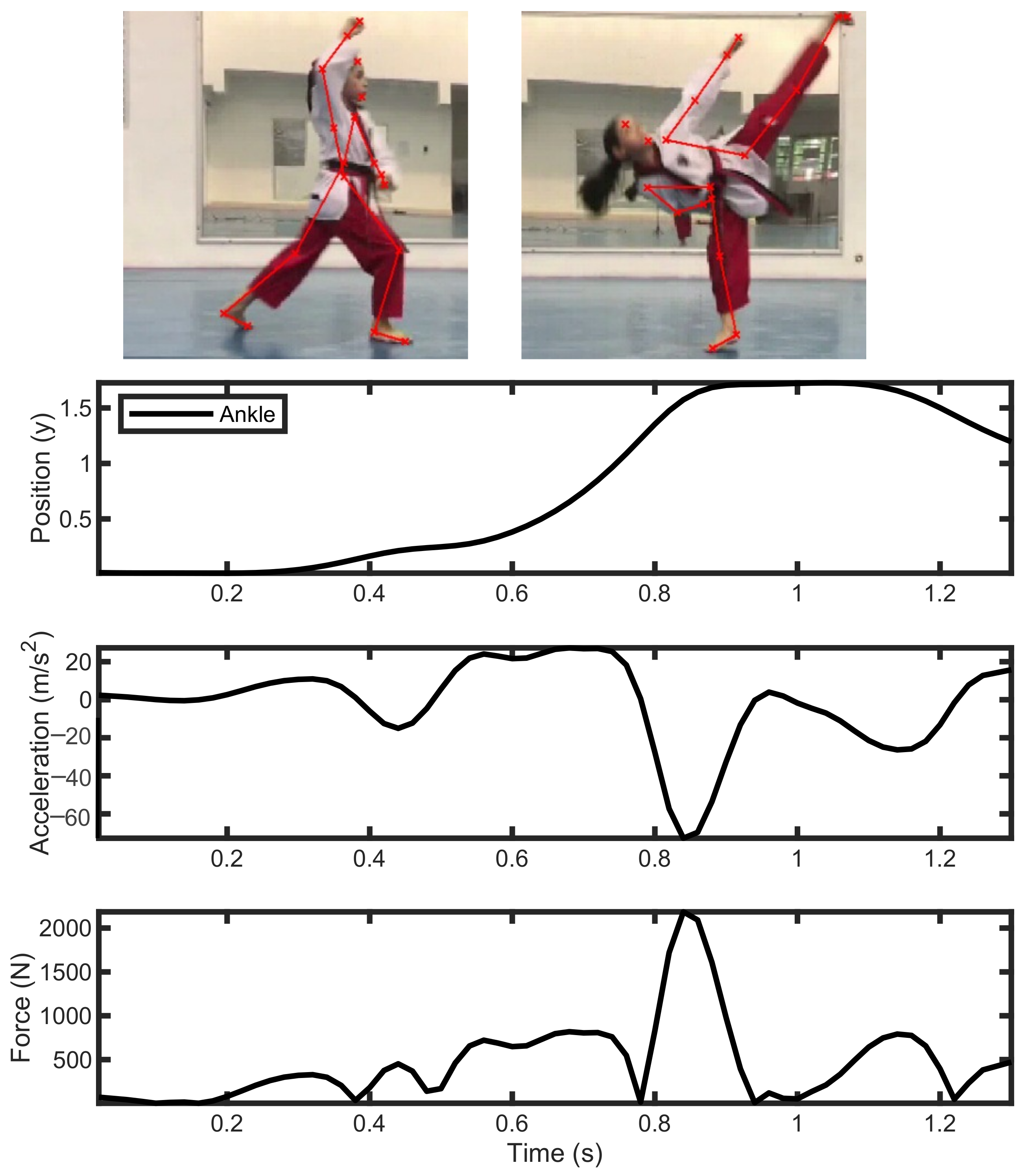
3.2. Taekwondo
3.3. Football Soccer
4. Discussion
4.1. Worlwide Roadmap in Biomechanics
4.2. Perspectives
5. Conclusions
Author Contributions
Funding
Institutional Review Board Statement
Informed Consent Statement
Data Availability Statement
Acknowledgments
Conflicts of Interest
References
- Yoshida, Y.; Nishimura, T.; Jokinen, K. Biomechanics for understanding movements in daily activities. In Proceedings of the LREC 2018 Workshop Language and Body in Real Life, Miyazaki, Japan, 9–11 May 2018. [Google Scholar]
- Singh, G.K. Biomechanics of Human Movement in Occupational Tasks with Ergonomic Considerations. Ph.D. Thesis, Deemed University, Tamilnadu, India, 2014. [Google Scholar]
- Dafoulas, G.A.; Maia, C.C.; Clarke, J.S.; Ali, A.; Augusto, J. Investigating the role of biometrics in education—The use of sensor data in collaborative learning. In Proceedings of the MCCSIS 2018—Multi Conference on Computer Science and Information Systems, Madrid, Spain, 17–19 July 2018; pp. 115–123. [Google Scholar]
- Albert, J.A.; Owolabi, V.; Gebel, A.; Brahms, C.M.; Granacher, U.; Arnrich, B. Evaluation of the pose tracking performance of the azure kinect and kinect v2 for gait analysis in comparison with a gold standard: A pilot study. Sensors 2020, 20, 5104. [Google Scholar] [CrossRef] [PubMed]
- Fernández-Baena, A.; Susín, A.; Lligadas, X. Biomechanical validation of upper-body and lower-body joint movements of kinect motion capture data for rehabilitation treatments. In Proceedings of the 2012 4th International Conference on Intelligent Networking and Collaborative Systems, Bucharest, Romania, 19–21 September 2012; pp. 656–661. [Google Scholar] [CrossRef]
- De Froda, S.F.; Thigpen, C.A.; Kriz, P.K. Two-dimensional video analysis of youth and adolescent pitching biomechanics: A tool for the common athlete. Curr. Sports Med. Rep. 2016, 15, 350–358. [Google Scholar] [CrossRef] [PubMed]
- Guten, G.N. Running Injuries; W. B. Saunders: Philadelphia, PA, USA, 1997. [Google Scholar]
- Fredericson, M.; Jennings, F.; Beaulieu, C.; Matheson, G.O. Stress fractures in athletes. Top. Magn. Reson. Imaging 2006, 17, 309–325. [Google Scholar] [CrossRef] [PubMed]
- Baggaley, M.; Esposito, M.; Xu, C.; Unnikrishnan, G.; Reifman, J.; Edwards, W.B. Effects of load carriage on biomechanical variables associated with tibial stress fractures in running. Gait Posture 2020, 77, 190–194. [Google Scholar] [CrossRef]
- Garrett, W.E. Principles and Practice of Orthopaedic Sports Medicine; Lippincott Williams & Wilkins: Philadelphia, PA, USA, 2000. [Google Scholar]
- Irani, M. Principles and Practice of Primary Care Sports Medicine; Garrett, W.E., Jr., Kirkendall, D.T., Squire, D.L., Eds.; Lippincott Williams & Wilkins: Philadelphia, PA, USA, 2001; p. 679. ISBN 0-7817-2956-4. [Google Scholar]
- Iwamoto, J.; Takeda, T. Stress fractures in athletes: Review of 196 cases. J. Orthop. Sci. 2003, 8, 273–278. [Google Scholar] [CrossRef]
- Troy, K.L.; Davis, I.S.; Tenforde, A.S. A Narrative Review of Metatarsal Bone Stress Injury in Athletic Populations: Etiology, Biomechanics, and Management. PM&R 2020, 13, 1281–1290. [Google Scholar]
- Bennell, K.; Brukner, P. Preventing and managing stress fractures in athletes. Phys. Ther. Sport 2005, 6, 171–180. [Google Scholar] [CrossRef]
- Boutefnouchet, T.; Budair, B.; Backshayesh, P.; Ali, S.A. Metatarsal fractures: A review and current concepts. Trauma 2014, 16, 147–163. [Google Scholar] [CrossRef]
- Miller, D.; Marsland, D.; Jones, M.; Calder, J. Early return to playing professional football following fixation of 5th metatarsal stress fractures may lead to delayed union but does not increase the risk of long-term non-union. Knee Surg. Sports Traumatol. Arthrosc. 2019, 27, 2796–2801. [Google Scholar] [CrossRef]
- Ekstrand, J.; van Dijk, C.N. Fifth metatarsal fractures among male professional footballers: A potential career-ending disease. Br. J. Sports Med. 2013, 47, 754–758. [Google Scholar] [CrossRef]
- Lawrence, S.J.; Botte, M.J. Jones’ fractures and related fractures of the proximal fifth metatarsal. Foot Ankle 1993, 14, 358–365. [Google Scholar] [CrossRef]
- Kuzuyama, M.; Perrier, J.; Kusaki, Y.; Sato, K.; Yamaura, I.; Tsuchiya, A. Characteristics of plantar pressure distribution in elite male soccer players with or without history of proximal fifth metatarsal fracture: A case-control study. J. Phys. Ther. Sci. 2019, 31, 530–535. [Google Scholar] [CrossRef][Green Version]
- Thomson, A.; Akenhead, R.; Whiteley, R.; D’Hooghe, P.; Van Alsenoy, K.; Bleakley, C. Fifth metatarsal stress fracture in elite male football players: An on-field analysis of plantar loading. BMJ Open Sport Exerc. Med. 2018, 4, e000377. [Google Scholar] [CrossRef]
- Sims, E.; Hardaker, W.; Queen, R. Gender differences in plantar loading during three soccer-specific tasks. Br. J. Sports Med. 2008, 42, 272–277. [Google Scholar] [CrossRef]
- Fujitaka, K.; Taniguchi, A.; Isomoto, S.; Kumai, T.; Otuki, S.; Okubo, M.; Tanaka, Y. Pathogenesis of fifth metatarsal fractures in college soccer players. Orthop. J. Sports Med. 2015, 3, 2325967115603654. [Google Scholar] [CrossRef]
- Matsuda, S.; Fukubayashi, T.; Hirose, N. Characteristics of the foot static alignment and the plantar pressure associated with fifth metatarsal stress fracture history in male soccer players: A case-control study. Sports Med.-Open 2017, 3, 1–7. [Google Scholar] [CrossRef]
- Azevedo, R.R.; da Rocha, E.S.; Franco, P.S.; Carpes, F.P. Plantar pressure asymmetry and risk of stress injuries in the foot of young soccer players. Phys. Ther. Sport 2017, 24, 39–43. [Google Scholar] [CrossRef]
- Fujitaka, K.; Tanaka, Y.; Taniguchi, A.; Ogawa, M.; Isomoto, S.; Otuki, S.; Okubo, M. Pathoanatomy of the Jones fracture in male university soccer players. Am. J. Sports Med. 2020, 48, 424–431. [Google Scholar] [CrossRef]
- Chow, J.W.; Knudson, D.V. Use of deterministic models in sports and exercise biomechanics research. Sports Biomech. 2011, 10, 219–233. [Google Scholar] [CrossRef]
- Virag, B.; Hibberd, E.E.; Oyama, S.; Padua, D.A.; Myers, J.B. Prevalence of freestyle biomechanical errors in elite competitive swimmers. Sports Health 2014, 6, 218–224. [Google Scholar] [CrossRef]
- Heinlein, S.A.; Cosgarea, A.J. Biomechanical considerations in the competitive swimmer’s shoulder. Sports Health 2010, 2, 519–525. [Google Scholar] [CrossRef]
- Tovin, B.J. Prevention and treatment of swimmer’s shoulder. N. Am. J. Sports Phys. Ther. NAJSPT 2006, 1, 166. [Google Scholar]
- De la Fuente, C.; Ramirez-Campillo, R.; Gallardo-Fuentes, F.; Alvarez, C.; Bustamante, C.; Henríquez, H.; Carpes, F.P. Pattern analysis of a complete Achilles tendon rupture suffered during high jump preparation in an official national-level athletic competition. Sports Biomech. 2019, 21, 312–322. [Google Scholar] [CrossRef]
- McIntosh, A.S.; Willmott, C.; Patton, D.A.; Mitra, B.; Brennan, J.H.; Dimech-Betancourt, B.; Howard, T.S.; Rosenfeld, J.V. An assessment of the utility and functionality of wearable head impact sensors in Australian Football. J. Sci. Med. Sport 2019, 22, 784–789. [Google Scholar] [CrossRef]
- Reyes, J.; Mitra, B.; McIntosh, A.; Clifton, P.; Makdissi, M.; Nguyen, J.V.; Harcourt, P.; Howard, T.S.; Cameron, P.A.; Rosenfeld, J.V.; et al. An Investigation of Factors Associated With Head Impact Exposure in Professional Male and Female Australian Football Players. Am. J. Sports Med. 2020, 48, 1485–1495. [Google Scholar] [CrossRef]
- Lemme, N.J.; Li, N.Y.; Kleiner, J.E.; Tan, S.; DeFroda, S.F.; Owens, B.D. Epidemiology and video analysis of Achilles tendon ruptures in the National Basketball Association. Am. J. Sports Med. 2019, 47, 2360–2366. [Google Scholar] [CrossRef]
- Clark, J.M.; Adanty, K.; Post, A.; Hoshizaki, T.B.; Clissold, J.; McGoldrick, A.; Hill, J.; Annaidh, A.N.; Gilchrist, M.D. Proposed injury thresholds for concussion in equestrian sports. J. Sci. Med. Sport 2020, 23, 222–236. [Google Scholar] [CrossRef]
- Joodaki, H.; Bailey, A.; Lessley, D.; Funk, J.; Sherwood, C.; Crandall, J. Relative motion between the helmet and the head in football impact test. J. Biomech. Eng. 2019, 141, 081006. [Google Scholar] [CrossRef] [PubMed]
- Rowson, B.; Duma, S.M. A review of on-field investigations into the biomechanics of concussion in football and translation to head injury mitigation strategies. Ann. Biomed. Eng. 2020, 48, 2734–2750. [Google Scholar] [CrossRef] [PubMed]
- Fox, D.; O’Malley, E.; Blake, C. Normative data for the Functional Movement Screen™ in male Gaelic field sports. Phys. Ther. Sport 2014, 15, 194–199. [Google Scholar] [CrossRef] [PubMed]
- Wilcox, B.J.; Machan, J.T.; Beckwith, J.G.; Greenwald, R.M.; Burmeister, E.; Crisco, J.J. Head-impact mechanisms in men’s and women’s collegiate ice hockey. J. Athl. Train. 2014, 49, 514–520. [Google Scholar] [CrossRef]
- Preatoni, E.; Cazzola, D.; Stokes, K.; England, M.; Trewartha, G. Pre-binding prior to full engagement improves loading conditions for front-row players in contested R ugby U nion scrums. Scand. J. Med. Sci. Sport. 2016, 26, 1398–1407. [Google Scholar] [CrossRef]
- Bere, T.; Flørenes, T.W.; Krosshaug, T.; Koga, H.; Nordsletten, L.; Irving, C.; Muller, E.; Reid, R.C.; Senner, V.; Bahr, R. Mechanisms of anterior cruciate ligament injury in World Cup alpine skiing: A systematic video analysis of 20 cases. Am. J. Sports Med. 2011, 39, 1421–1429. [Google Scholar] [CrossRef]
- Bakken, A.; Bere, T.; Bahr, R.; Kristianslund, E.; Nordsletten, L. Mechanisms of injuries in World Cup Snowboard Cross: A systematic video analysis of 19 cases. Br. J. Sports Med. 2011, 45, 1315–1322. [Google Scholar] [CrossRef]
- Della Villa, F.; Buckthorpe, M.; Grassi, A.; Nabiuzzi, A.; Tosarelli, F.; Zaffagnini, S.; Della Villa, S. Systematic video analysis of ACL injuries in professional male football (soccer): Injury mechanisms, situational patterns and biomechanics study on 134 consecutive cases. Br. J. Sports Med. 2020, 54, 1423–1432. [Google Scholar] [CrossRef]
- Grassi, A.; Tosarelli, F.; Agostinone, P.; Macchiarola, L.; Zaffagnini, S.; Della Villa, F. Rapid Posterior Tibial Reduction After Noncontact Anterior Cruciate Ligament Rupture: Mechanism Description From a Video Analysis. Sports Health 2020, 12, 462–469. [Google Scholar] [CrossRef]
- Schmitt, K.U.; Schlittler, M.; Boesiger, P. Biomechanical loading of the hip during side jumps by soccer goalkeepers. J. Sports Sci. 2010, 28, 53–59. [Google Scholar] [CrossRef]
- Waldén, M.; Krosshaug, T.; Bjørneboe, J.; Andersen, T.E.; Faul, O.; Hägglund, M. Three distinct mechanisms predominate in non-contact anterior cruciate ligament injuries in male professional football players: A systematic video analysis of 39 cases. Br. J. Sports Med. 2015, 49, 1452–1460. [Google Scholar] [CrossRef]
- Parsons, J.L.; Alexander, M.J. Modifying spike jump landing biomechanics in female adolescent volleyball athletes using video and verbal feedback. J. Strength Cond. Res. 2012, 26, 1076–1084. [Google Scholar] [CrossRef]
- You, D.Z.; Tomlinson, M.; Borschneck, G.; Borschneck, A.; MacDonald, M.; Deluzio, K.; Borschneck, D. The Effect of Ankle Brace Use on a 3-Step Volleyball Spike Jump Height. Arthrosc. Sports Med. Rehabil. 2020, 2, e461–e467. [Google Scholar] [CrossRef]
- Van Trigt, B.; Schallig, W.; Van der Graaff, E.; Hoozemans, M.J.; Veeger, D. Knee angle and stride length in association with ball speed in youth baseball pitchers. Sports 2018, 6, 51. [Google Scholar] [CrossRef]
- Reiser, M.; Zentgraf, K.; Kindermann, S.; Künzell, S. An approach to quantify the float effect of float serves in indoor and beach volleyball. Front. Sports Act. Living 2020, 2, 559277. [Google Scholar] [CrossRef]
- Wild, J.J.; Bezodis, I.N.; North, J.S.; Bezodis, N.E. Differences in step characteristics and linear kinematics between rugby players and sprinters during initial sprint acceleration. Eur. J. Sport Sci. 2018, 18, 1327–1337. [Google Scholar] [CrossRef]
- Linthorne, N.P.; Patel, D.S. Optimum projection angle for attaining maximum distance in a soccer punt kick. J. Sports Sci. Med. 2011, 10, 203. [Google Scholar]
- Barkwell, G.E.; Dickey, J.P. Backstroke start performance: The impact of using the Omega OBL2 backstroke ledge. Sports Biomech. 2018, 17, 429–441. [Google Scholar] [CrossRef]
- Callaway, A.J. Measuring kinematic variables in front crawl swimming using accelerometers: A validation study. Sensors 2015, 15, 11363–11386. [Google Scholar] [CrossRef]
- De Jesus, K.; De Jesus, K.; Medeiros, A.I.; Gonçalves, P.; Figueiredo, P.; Fernandes, R.J.; Vilas-Boas, J.P. Neuromuscular activity of upper and lower limbs during two backstroke swimming start variants. J. Sports Sci. Med. 2015, 14, 591. [Google Scholar]
- De Jesus, K.; Sanders, R.; de Jesus, K.; Ribeiro, J.; Figueiredo, P.; Vilas-Boas, J.P.; Fernandes, R.J. The effect of intensity on 3-dimensional kinematics and coordination in front-crawl swimming. Int. J. Sports Physiol. Perform. 2016, 11, 768–775. [Google Scholar] [CrossRef]
- Ikeda, Y.; Ichikawa, H.; Nara, R.; Baba, Y.; Shimoyama, Y.; Kubo, Y. Functional role of the front and back legs during a track start with special reference to an inverted pendulum model in college swimmers. J. Appl. Biomech. 2016, 32, 462–468. [Google Scholar] [CrossRef]
- Mooney, R.; Corley, G.; Godfrey, A.; Quinlan, L.R.; ÓLaighin, G. Inertial sensor technology for elite swimming performance analysis: A systematic review. Sensors 2016, 16, 18. [Google Scholar] [CrossRef]
- Osborough, C.; Daly, D.; Payton, C. Effect of swim speed on leg-to-arm coordination in unilateral arm amputee front crawl swimmers. J. Sports Sci. 2015, 33, 1523–1531. [Google Scholar] [CrossRef] [PubMed]
- Papic, C.; Sanders, R.H.; Naemi, R.; Elipot, M.; Andersen, J. Improving data acquisition speed and accuracy in sport using neural networks. J. Sports Sci. 2020, 39, 513–522. [Google Scholar] [CrossRef] [PubMed]
- Hamidi Rad, M.; Gremeaux, V.; Dadashi, F.; Aminian, K. A Novel Macro-Micro Approach for Swimming Analysis in Main Swimming Techniques Using IMU Sensors. Front. Bioeng. Biotechnol. 2021, 8, 1511. [Google Scholar] [CrossRef] [PubMed]
- Schnitzler, C.; Seifert, L.; Chollet, D.; Toussaint, H. Effect of aerobic training on inter-arm coordination in highly trained swimmers. Hum. Mov. Sci. 2014, 33, 43–53. [Google Scholar] [CrossRef]
- Diniz, R.; Del Vecchio, F.B.; Schaun, G.Z.; Oliveira, H.B.; Portella, E.G.; da Silva, E.S.; Formalioni, A.; Campelo, P.C.; Peyré-Tartaruga, L.A.; Pinto, S.S. Kinematic comparison of the roundhouse kick between taekwondo, karate, and muaythai. J. Strength Cond. Res. 2021, 35, 198–204. [Google Scholar] [CrossRef]
- Gutiérrez-Santiago, A.; Pereira-Rodríguez, R.; Prieto-Lage, I. Detection of the technical and tactical motion of the scorable movements in taekwondo. Physiol. Behav. 2020, 217, 112813. [Google Scholar] [CrossRef]
- Kim, J.W.; Kwon, M.S.; Yenuga, S.S.; Kwon, Y.H. The effects of target distance on pivot hip, trunk, pelvis, and kicking leg kinematics in Taekwondo roundhouse kicks. Sports Biomech. 2010, 9, 98–114. [Google Scholar] [CrossRef]
- Maloney, M.A.; Renshaw, I.; Headrick, J.; Martin, D.T.; Farrow, D. Taekwondo fighting in training does not simulate the affective and cognitive demands of competition: Implications for behavior and transfer. Front. Psychol. 2018, 9, 25. [Google Scholar] [CrossRef]
- Pradhan, A.; Kuruganti, U.; Chester, V. Biomechanical parameters and clinical assessment scores for identifying elderly fallers based on balance and dynamic tasks. IEEE Access 2020, 8, 193532–193543. [Google Scholar] [CrossRef]
- Lloyd, D. The future of in-field sports biomechanics: Wearables plus modelling compute real-time in vivo tissue loading to prevent and repair musculoskeletal injuries. Sports Biomech. 2021, 1–29. [Google Scholar] [CrossRef]
- Dingenen, B.; Malfait, B.; Nijs, S.; Peers, K.H.; Vereecken, S.; Verschueren, S.M.; Staes, F.F. Can two-dimensional video analysis during single-leg drop vertical jumps help identify non-contact knee injury risk? A one-year prospective study. Clin. Biomech. 2015, 30, 781–787. [Google Scholar] [CrossRef]
- Shere, M.; Kim, H.; Hilton, A. 3D Human Pose Estimation From Multi Person Stereo 360° Scenes. In Proceedings of the CVPR Workshops 2019, Long Beach, CA, USA, 16–20 June 2019. [Google Scholar]
- Polak, E.; Kulasa, J.; VencesBrito, A.; Castro, M.A.; Fernandes, O. Motion analysis systems as optimization training tools in combat sports and martial arts. Rev. Artes Marciales Asiáticas 2016, 10, 105. [Google Scholar] [CrossRef]
- Ceseracciu, E.; Sawacha, Z.; Fantozzi, S.; Cortesi, M.; Gatta, G.; Corazza, S.; Cobelli, C. Markerless analysis of front crawl swimming. J. Biomech. 2011, 44, 2236–2242. [Google Scholar] [CrossRef]
- Makdissi, M.; Davis, G. Using video analysis for concussion surveillance in Australian football. J. Sci. Med. Sport 2016, 19, 958–963. [Google Scholar] [CrossRef]
- Syed, S.H.; Willing, R.; Jenkyn, T.R.; Yazdani, A. Video analysis of the biomechanics of a bicycle accident resulting in significant facial fractures. J. Craniofacial Surg. 2013, 24, 2023–2029. [Google Scholar] [CrossRef]
- Hanley, B.; Tucker, C.B.; Bissas, A. Differences between motion capture and video analysis systems in calculating knee angles in elite-standard race walking. J. Sports Sci. 2018, 36, 1250–1255. [Google Scholar] [CrossRef]
- Schurr, S.A.; Marshall, A.N.; Resch, J.E.; Saliba, S.A. Two-dimensional video analysis is comparable to 3D motion capture in lower extremity movement assessment. Int. J. Sports Phys. Ther. 2017, 12, 163–172. [Google Scholar]
- Chan, C.K.; Loh, W.P.; Rahim, I.A. Human motion classification using 2D stick-model matching regression coefficients. Appl. Math. Comput. 2016, 283, 70–89. [Google Scholar] [CrossRef]
- Barre, A.; Armand, S. Biomechanical ToolKit: Open-source framework to visualize and process biomechanical data. Comput. Methods Programs Biomed. 2014, 114, 80–87. [Google Scholar] [CrossRef]
- Pfister, A.; West, A.M.; Bronner, S.; Noah, J.A. Comparative abilities of Microsoft Kinect and Vicon 3D motion capture for gait analysis. J. Med. Eng. Technol. 2014, 38, 274–280. [Google Scholar] [CrossRef]
- Valdivia, C.; Ortega, A.; Salazar, M.; Escobedo, J. Therapeutic Motion Analysis of Lower Limbs Using Kinovea. Int. J. Soft Comput. Eng. 2013, 3, 359–365. [Google Scholar]
- Ana, F.; Pedro, R. Use of Open-Source Technology to Teach Biomechanics. Ann. Univ. Oradea Fac. Phys. Educ. Sport. 2016, 26, 18–24. [Google Scholar]
- Moghaddami, A.; Gerek, Z.; Karimiasl, A.; Nozohouri, H. The Effect of Acute Dehydration and Rehydration on Biomechanical Parameters of Elite Wrestling Techniques. J. Sports Sci. 2016, 4, 93–101. [Google Scholar] [CrossRef]
- Delp, S.L.; Anderson, F.C.; Arnold, A.S.; Loan, P.; Habib, A.; John, C.T.; Guendelman, E.; Thelen, D.G. OpenSim: Open-source software to create and analyze dynamic simulations of movement. IEEE Trans. Biomed. Eng. 2007, 54, 1940–1950. [Google Scholar] [CrossRef]
- Shippen, J.; May, B. BoB—Biomechanics in MATLAB. In Proceedings of the 11th International Conference BIOMDOLE, Druskininkai, Lithuania, 20–22 October 2016; pp. 11–13. [Google Scholar] [CrossRef]
- Maas, S.A.; Ellis, B.J.; Ateshian, G.A.; Weiss, J.A. FEBio: Finite elements for biomechanics. J. Biomech. Eng. 2012, 134, 1–10. [Google Scholar] [CrossRef]
- Preuschl, E.; Hassmann, M.; Baca, A. A kinematic analysis of the jumping front-leg axe-kick in taekwondo. J. Sports Sci. Med. 2016, 15, 92–101. [Google Scholar]
- Li, X. 3-D image analysis and discussion on characteristics of taekwondo flank kick movement. RISTI—Revista Ibérica Sistemas Tecnologias Informação 2016, 2016, 170–178. [Google Scholar]
- Winiarski, S.; Rutkowska-Kucharska, A. Estimated ground reaction force in normal and pathological gait. Acta Bioeng. Biomech. 2009, 11, 53–60. [Google Scholar]
- Chen, T.L.W.; Wong, D.W.C.; Wang, Y.; Ren, S.; Yan, F.; Zhang, M. Biomechanics of fencing sport: A scoping review. PLoS ONE 2017, 12, e0171578. [Google Scholar] [CrossRef]
- Estevan, I.; Falco, C. Mechanical analysis of the roundhouse kick according to height and distance in taekwondo. Biol. Sport 2013, 30, 275–279. [Google Scholar] [CrossRef]
- Bencke, J.; Aagaard, P.; Zebis, M.K. Muscle activation during ACL injury risk movements in young female athletes: A narrative review. Front. Physiol. 2018, 9, 1–10. [Google Scholar] [CrossRef] [PubMed]
- Weiss, K.; Whatman, C. Biomechanics Associated with Patellofemoral Pain and ACL Injuries in Sports. Sports Med. 2015, 45, 1325–1337. [Google Scholar] [CrossRef] [PubMed]
- Ning, X.; Zhou, J.; Dai, B.; Jaridi, M. The assessment of material handling strategies in dealing with sudden loading: The effects of load handling position on trunk biomechanics. Appl. Ergon. 2014, 45, 1399–1405. [Google Scholar] [CrossRef] [PubMed]
- Chedi, J.M.; Mustapha, R. Biomechanics, visual and carrying discomfort: The role of ergonomics among technology education professionals on the use of laptop in no-desk settings. J. Phys. Conf. Ser. 2020, 1456, 012014. [Google Scholar] [CrossRef]
- Suri, C.; Shojaei, I.; Bazrgari, B. Effects of School Backpacks on Spine Biomechanics During Daily Activities: A Narrative Review of Literature. Hum. Factors 2020, 62, 909–918. [Google Scholar] [CrossRef]
- Karakolis, T.; Barrett, J.; Callaghan, J.P. A comparison of trunk biomechanics, musculoskeletal discomfort and productivity during simulated sit-stand office work. Ergonomics 2016, 59, 1275–1287. [Google Scholar] [CrossRef]
- Ali, S.; Mina, A.; Mehdi, S.; Iman, S.M. C3D data based on 2-dimensional images from video camera. Ann. Biomed. Sci. Eng. 2021, 5, 1–5. [Google Scholar] [CrossRef]
- Agarwal, P.; Sahu, S. Determination of hand and palm area as a ratio of body surface area in Indian population. Indian J. Plast. Surg. 2010, 43, 49–53. [Google Scholar] [CrossRef]
- Webb, A.; Banks, J.; Phillips, C.; Hudson, D.; Taunton, D.; Turnock, S. Prediction of passive and active drag in swimming. Procedia Eng. 2011, 13, 133–140. [Google Scholar] [CrossRef][Green Version]
- Hildebrand, F.; Schüler, A. Swimming propulsion and muscle force moments. Math. Comput. Model. Dyn. Syst. 2010, 16, 443–453. [Google Scholar] [CrossRef]
- O’Sullivan, D.; Chung, C.; Lee, K.; Kim, E.; Kang, S.; Kim, T.; Shin, I. Measurement and comparison of Taekwondo and Yongmudo turning kick impact force for two target heights. J. Sports Sci. Med. 2009, 8, 13–16. [Google Scholar]
- Ali, M.F.M.; Yaakub, N.; Haris, M.N.; Kamarudin, M.K.A.; Industry, F.; Sultan, U.; Abidin, Z.; Sultan, U.; Abidin, Z.; Campus, B. Modelling equation of knee force during instep kicking using dominant’s leg in football. J. Fundam. Appl. Sci. 2017, 9, 750–760. [Google Scholar] [CrossRef][Green Version]
- Barbosa, T.M.; Barbosa, A.C.; Simbaña Escobar, D.; Mullen, G.J.; Cossor, J.M.; Hodierne, R.; Arellano, R.; Mason, B.R. The role of the biomechanics analyst in swimming training and competition analysis. Sports Biomech. 2021, 1–18. [Google Scholar] [CrossRef]
- Vagenas, G.; Palaiothodorou, D.; Knudson, D. Thirty-year Trends of Study Design and Statistics in Applied Sports and Exercise Biomechanics Research. Int. J. Exerc. Sci. 2018, 11, 239–259. [Google Scholar]
- Howard, R.M.; Conway, R.; Harrison, A.J. A survey of sensor devices: Use in sports biomechanics. Sports Biomech. 2016, 15, 450–461. [Google Scholar] [CrossRef]
- Phinyomark, A.; Petri, G.; Ibáñez-Marcelo, E.; Osis, S.T.; Ferber, R. Analysis of Big Data in Gait Biomechanics: Current Trends and Future Directions. J. Med. Biol. Eng. 2018, 38, 244–260. [Google Scholar] [CrossRef]
- Hollander, K. Biomechanics of Running—Implications for Running-Related Injuries and Future Areas for Research. Sports Med. 2019, 42, 53–54. [Google Scholar] [CrossRef]
- Muñoz, M.M.; Price, S.A. The Future is Bright for Evolutionary Morphology and Biomechanics in the Era of Big Data. Integr. Comp. Biol. 2019, 59, 599–603. [Google Scholar] [CrossRef]
- Peng, X.; Tang, L. Biomechanics analysis of real-time tennis batting images using Internet of Things and deep learning. J. Supercomput. 2021, 78, 5883–5902. [Google Scholar] [CrossRef]


| Reference | Sport | Method/Software | Analysis |
|---|---|---|---|
| [30] | High-jump | Pixel segmentation | Biomechanical analysis on risks and cases of Achilles tendon injury in high-jump attempts. |
| [31] | Australian football | Head of an instrumented Hybrid III anthropomorphic test device (ATD), | Head impact exposure and concussion risk under video analysis |
| [32] | Australian football | X-Patch | Head acceleration measurements and impacts by video analysis |
| [6] | Baseball | High-speed motion analysis | Two-dimensional biomechanical analysis use in assessing proper pitching techniques |
| [33] | Basketball | Video analysis software | Risk factors and causes of Achilles tendon (AT) ruptures in NBA players. |
| [34] | Equestrian Sports | Kinovea 0.8.20; Head Impact Telemetry (HIT) System | Helmet impact mechanics; concussion thresholds. |
| [35] | American football | Optical motion capture system (Vicon, Los Angeles, CA) | Helmet head protection. Kinematics among head and helmet. |
| [36] | American football | Head impact sensors | Review on the concussion biomechanics and use of head impact sensors. |
| [37] | Gaelic field sports | Scoring Gray Cook | Normative reference values from the study of functional movement |
| [38] | Ice Hockey | Head Impact Telemetry (HIT) System | Identification of per-game frequencies of head impacts and concussions |
| [39] | Rugby | Manual digitizing Vicon Motus V9, Vicon Motion Systems, USA | Prevention for chronic injuries, towards safer engagement conditions |
| [40] | Alpine Skiing | Consensus decision by experts | Injury mechanisms descriptions of anterior cruciate ligament (ACL) injury during competition |
| [41] | Snowboard | Consensus decision by experts | Snowboard cross (SBX) descriptions of injury mechanisms |
| [42] | Football | Consensus decision by experts | Kinematics and frequency cases of ACL injuries in professional Italian football |
| [43] | Football | Kinovea software, consensus decision by experts | Approach for intra-articular lesions by late phases of ACL injury |
| [44] | Football | Force plate Type 9285, Kistler Instruments, high-speed camera | Injury risk by hip loading under diving in goalkeepers |
| [45] | Football | Consensus decision by experts | ACL injury cases and contact mechanisms |
| [46] | Volleyball | Dartfish motion analysis software | Improving landing techniques to prevent ACL injuries |
| [47] | Volleyball | Visual3D tracking system | Wearing ankle braces effect and tendency to injury development |
| [48] | Baseball | Stalker pro radar gun (Stalker Radar, Plano, TX, USA) | Biomechanics of the stride length and knee angle with high ball speeds |
| [49] | Beach volleyball | Peak Motus Software (Version 9, Vicon Motion Systems. Inc.) | Biomechanics of float serve |
| [50] | Rugby | Kinovea (v.0.8.15) | Biomechanics of sprint initial steps |
| [51] | Football soccer | Ariel Performance Analysis System (Ariel Dynamics, Trabuco Canyon, CA, USA) | Measurement of soccer goalkeeper punt kick optimum projection angle |
| [52] | Swimming | Kinovea software (Version 0.8.15) | Biomechanics on the optimal start performance in backstroke swimming |
| [53] | Swimming | Accelerometer waterproof X6-2mini (Gulf Coast Data Concepts, Waveland, MA, USA); Sensors | Multiple sensor systems for biomechanical analysis in front crawl swimming |
| [54] | Swimming | 24 anatomical markers; linear transformation algorithm | Use of differing handgrips in backstroke swimming start performance |
| [55] | Swimming | Ariel Dynamics Inc., San Diego, CA, USA | |
| [56] | Swimming | Digitizing system (FrameDIAS V, DKH, Inc., Itabashi-ku, Tokyo, Japan) | Involvement of left of mass and flight distance in a proper track start |
| [57] | Swimming | Inertial sensors | Analysis of the use of inertial sensors and detection algorithms in swimming phases |
| [58] | Swimming | SIMI Motion 7.2 software (SIMI Reality Motion Systems GmbH, Unterschleißheim, Germany) | Biomechanics of the leg to arm coordination in unilateral arm amputee swimmers |
| [59] | Swimming | “Cinalysis” software (Elipot et al., 2010) | Use of software for auto-digitization and proper analysis of glide posture |
| [60] | Swimming | Wearable measurement system including six IMUs (Physilog R IV, GaitUp, CH) | Motion phase approach for covering the analysis of full swimming training sessions |
| [61] | Swimming | Dartfish software | Effect of aerobic training towards the increasing of swimming speed |
| [62] | Taekwondo | Vicon Motion Capture System, Los Angeles, CA, USA | Kinematic characteristics for improving roundhouse kick technique performance |
| [63] | Taekwondo | LINCE v.1.2.1 software | Analysis of technical and tactical move patterns towards scoring during combat |
| [64] | Taekwondo | Kwon3D XP Motion Analysis Suite (Version 4.1, Visol, Seoul, Korea) | Target distance analysis during Taekwondo roundhouse kick movement |
| [65] | Taekwondo | Kinovea, version 0.8.25 | Analysis on the levels of anxiety and mental challenge during fighting in competition |
| Software | Ease of Access | Distinctive Features |
|---|---|---|
| Kinovea | Open source | Utilities to capture, slow down, compare, annotate and measure motion in videos. |
| Dartfish | Monthly subscription | Available for PC and cell phone. Allows to annotate, compare videos, perform angle measurements. |
| SkillSpector | Free software | Manual digitization of joints and transformation to real coordinates using DLT transform. Can handle 2D and 3D movement analysis. Advanced analysis of linear and angular kinematic data, inertia calculation, 3D movement animations, and video calibration. |
| Coach’s Eye | Paid app, available on iOS, Android and Windows | Interactive and user-friendly GUI for sports assessment. Slowmotion and playback functions. |
| Tracker | Open source, continuously updated | Automated tracking based on image recognition which can help in marker-less studies. Ample data analysis tools. |
| Biomechanical Toolkit | Open source, Python-based | Several online repositories, ease of communication with data formats from other softwares used in biomechanics research. |
| OpenSim | Open source | Development of models of musculoskeletal structures and generation of dynamic simulations. |
| Vicon | Paid | High-tech software and algorithms. Multiple solutions for different needs of the research and athletes, both marker-less and with markers. |
| Ariel Performance Analysis System | Free software | 3D motion analysis system. Automated biomechanical analysis from multiple, simultaneous video recordings. Can integrate information of EMG, force platforms and video. |
| Visual3D | Free and paid options | Users can import motion and force data and perform computations, transformations, models and analysis with reports. |
Publisher’s Note: MDPI stays neutral with regard to jurisdictional claims in published maps and institutional affiliations. |
© 2022 by the authors. Licensee MDPI, Basel, Switzerland. This article is an open access article distributed under the terms and conditions of the Creative Commons Attribution (CC BY) license (https://creativecommons.org/licenses/by/4.0/).
Share and Cite
Ortiz-Padilla, V.E.; Ramírez-Moreno, M.A.; Presbítero-Espinosa, G.; Ramírez-Mendoza, R.A.; Lozoya-Santos, J.d.J. Survey on Video-Based Biomechanics and Biometry Tools for Fracture and Injury Assessment in Sports. Appl. Sci. 2022, 12, 3981. https://doi.org/10.3390/app12083981
Ortiz-Padilla VE, Ramírez-Moreno MA, Presbítero-Espinosa G, Ramírez-Mendoza RA, Lozoya-Santos JdJ. Survey on Video-Based Biomechanics and Biometry Tools for Fracture and Injury Assessment in Sports. Applied Sciences. 2022; 12(8):3981. https://doi.org/10.3390/app12083981
Chicago/Turabian StyleOrtiz-Padilla, Vanessa E., Mauricio A. Ramírez-Moreno, Gerardo Presbítero-Espinosa, Ricardo A. Ramírez-Mendoza, and Jorge de J. Lozoya-Santos. 2022. "Survey on Video-Based Biomechanics and Biometry Tools for Fracture and Injury Assessment in Sports" Applied Sciences 12, no. 8: 3981. https://doi.org/10.3390/app12083981
APA StyleOrtiz-Padilla, V. E., Ramírez-Moreno, M. A., Presbítero-Espinosa, G., Ramírez-Mendoza, R. A., & Lozoya-Santos, J. d. J. (2022). Survey on Video-Based Biomechanics and Biometry Tools for Fracture and Injury Assessment in Sports. Applied Sciences, 12(8), 3981. https://doi.org/10.3390/app12083981