Article

Effects of Different Adhesive Systems and Orthodontic Bracket Material on Enamel Surface Discoloration: An In Vitro Study

1
Department of Preventive Dental Sciences, College of Dentistry, Prince Sattam bin Abdulaziz University, Al-Kharj 11942, Saudi Arabia
2
Department of Conservative Dentistry, Faculty of Dentistry, Alexandria University, Alexandria 5423012, Egypt
3
Department of Dentistry, King Abdullah International Medical Research Center, King Saud bin Abdulaziz University for Health Sciences, King Abdulaziz Nationals Guard Hospital, Riyadh 11481, Saudi Arabia
4
Department of Restorative and Prosthetic Dental Sciences, College of Dentistry, Dar Al Uloom University, Riyadh 13314, Saudi Arabia
5
Department of Prosthodontics, College of Dentistry, Ibb University, Ibb 70270, Yemen
*
Author to whom correspondence should be addressed.
Appl. Sci. 2022, 12(24), 12885; https://doi.org/10.3390/app122412885
Submission received: 19 November 2022 / Revised: 5 December 2022 / Accepted: 12 December 2022 / Published: 15 December 2022
(This article belongs to the Special Issue Present and Future of Orthodontics)

Abstract

:
The aim of this in vitro study was to assess the effect of the type of orthodontic brackets and adhesive systems on enamel discoloration. The baseline color of the buccal surfaces of 50 extracted human premolars was recorded using a spectrophotometer according to the CIE Lab system. The teeth were randomly divided into five groups according to the bracket type and adhesive system used: Group A: metal brackets + a Three-step etch and rinse adhesive (E&R); Group B: metal brackets + a two-step self-adhesive adhesive (SE); Group C: ceramic brackets + (E&R); Group D: ceramic brackets + (SE); Group E: no bracket bonding, serving as a control. All teeth were thermocycled, the brackets were debonded, and the enamel surfaces were finished and polished. The tooth color was then re-assessed, and the change in color (∆E*) was calculated. Pairwise comparisons revealed significant differences in favor of ceramic brackets (ΔE = 3.77 ± 3.60; p = 0.009) and (E&R) (ΔE= 3.52 ± 2.45; p = 0.008). A significant difference was found among the different groups, with group C having the least change in color (ΔE = 2.00 ± 0.89) and group B having the highest (ΔE = 8.42 ± 5.66). Both the type of orthodontic bracket and adhesive system influenced tooth color change. The teeth bonded with ceramic and E&R had the least effect on color changes, whereas teeth bonded with metal and SE showed the highest color changes after debonding.

1. Introduction

Despite its well-recognized benefits, orthodontic treatment remains associated with adverse effects, including the associated enamel surface and structural detrimental changes such as decalcification, cracks, and abrasion [1]. Moreover, orthodontic treatment is considered as a risk factor for tooth discoloration, ranging from imperceivable changes and loss of translucency, to white spot lesions, to brown color changes depicting overt carious lesions [2]. Studies have shown that different etiologic factors could directly or indirectly affect the color changes of enamel related to orthodontic treatment, including the type of adhesive system used in the bonding of the orthodontic brackets. Etch and rinse (E&R) and self-etch (SE) adhesive systems are used for the bonding procedures, each with different penetration lengths of the formed resin tags within the enamel. Since these changes occur in the enamel subsurface, they cannot be reversed after debonding of the brackets with the finishing procedures; therefore, adverse effects could be long-lasting [1,3,4,5].
The type of orthodontic bracket, metal, or ceramic is another variable that might affect enamel color changes related to orthodontic treatment [6]. The orthodontic bracket material has been reported to affect the capacity of microorganisms to adhere to and grow on the bonded tooth surface. In an in vitro study, Almosa et al. reported that enamel demineralization was higher in teeth bonded with ceramic brackets compared with those bonded with metal brackets [6]. The increased demineralization may be because of a greater bacterial accumulation around ceramic brackets [7]. However, adherence of lipopolysaccharides (LPS) from Gram-negative bacteria has been shown to be higher to steel brackets than to porcelain, plastic, and gold brackets [8].
The finishing and polishing method after bracket debonding could also affect the final tooth color change. Following the debonding of the brackets, composite resin remnants cannot be completely removed without causing structural defects to enamel. A systemic review has reported that tungsten carbide burs damage enamel surfaces less than Arkansas or green stones, diamond burs, steel burs, or lasers [9]. Post-finishing polishing has been recommended to minimize the damage and the subsequent color changes of enamel [10]. Factors, related to patient habits, such as the use of mouthwash or bleaching agents, or the consumption of chromogenic food during or after orthodontic treatment, may also affect tooth color changes related to orthodontic treatment [5,11,12,13].
Of the factors under the orthodontist’s control, controversy still exists regarding which type of orthodontic brackets and adhesive systems would cause the least amount of tooth surface discoloration. Therefore, the aim of this in vitro study was to assess the effect of the type of orthodontic bracket and adhesive system on tooth surface discoloration. The null hypotheses were that, regarding tooth surface discoloration, no difference would be found between the metal or the ceramic orthodontic brackets and that no difference would be found between the three-step and two-step adhesive systems.

2. Materials and Methods

The study was conducted after receiving ethical approval from the local research and ethics committee (approval #COD/IRB/2020/31). Fifty human premolars recently extracted for either orthodontic or periodontal reasons were collected and stored for a maximum of 1 month in 0.1% thymol solution at room temperature until use [4]. The teeth were then cleaned with a hand scaler and examined under a stereomicroscope at ×25 magnification to detect any incipient defects. Teeth with no visible caries, decalcification, discoloration, or cracks on their buccal surface were selected and then stored in distilled water at room temperature in a black container. To facilitate handling, the teeth were embedded in autopolymerizing acrylic resin to 2 mm apical to the cemento-enamel junction using custom metal molds (Figure 1). All teeth were polished with nonfluorinated and oil-free pumice, rinsed, and dried for 10 s.

2.1. Baseline Tooth Color Assessment

The baseline color of the buccal surface of all teeth was measured using a spectrophotometer (Vita Easyshade, Vita Zahnfabrik, Bad Saeckingen, Germany) against a standard white ceramic background (L* = 89. a* = −2.6, b* = 5). The spectrophotometer displayed the different color parameters (L*, a*, and b*) according to the Commission Internationale de l’Eclairage (CIE) Lab color system, where L* describes the luminance reflectance, a* describes the red–green coordinates, and b* describes the yellow–blue color coordinates.

2.2. Grouping

The teeth were randomly divided into 5 groups of 10 teeth each (n = 10) according to the assigned adhesive system and bracket type used. The resin adhesives and bracket types used in the study and their composition, application method, and manufacturer are shown in Table 1.
In groups A and B, the teeth received metal brackets (Victory Series™ Low Profile, 3M Unitek), which were bonded in group A using a 3 step etch and rinse resin cement (Transbond XT, 3M Unitek, Monrovia, CA, USA) and using 2 step self-adhesive resin cement (U-Cem Vericom) in group B. In groups C and D, the teeth received ceramic brackets (Clarity Advanced ceramic brackets, 3M Unitek), which were bonded in group C using 3 step etch and rinse resin cement (Transbond XT, 3M Unitek, Monrovia, CA, USA) and using 2 step self-adhesive resin cement (U-Ce, Vericom) in group D. In group E, 10 teeth received no bonding procedure to serve as a control group.

2.3. Bonding of the Brackets

In groups A and C, where the 3 step etch and rinse adhesive was used, the buccal surface of the teeth was etched with 37% phosphoric acid etching gel (Acido gel, Maquira, Portugal) for 30 s, rinsed, and dried with oil-free compressed air. Then, XT primer was applied and light cured for 5 s. After that, brackets were bonded to the center of the prepared buccal surfaces using Transbond XT adhesive. Excess material was then removed from around the brackets with a sharp explorer. Light curing using an LED light-curing unit (Slimax-C Plus LED Curing Light System, Beyes Dental Canada Inc., Toronto, ON, Canada) was performed for 5 s at each surface (mesial, distal, incisal and cervical surfaces) of the metal brackets (in group A) and for 5 s through the ceramic brackets (in group C).
In groups B and D, where the 2 step self-etch adhesive was used, the brackets were bonded using U-Cem adhesive and light cured for 20 s for both metal and ceramic brackets. For all groups, the intensity of the light cure was regularly checked with the built-in radiometer at >1200 mW/cm2. All materials were applied according to the manufacturers’ instructions.

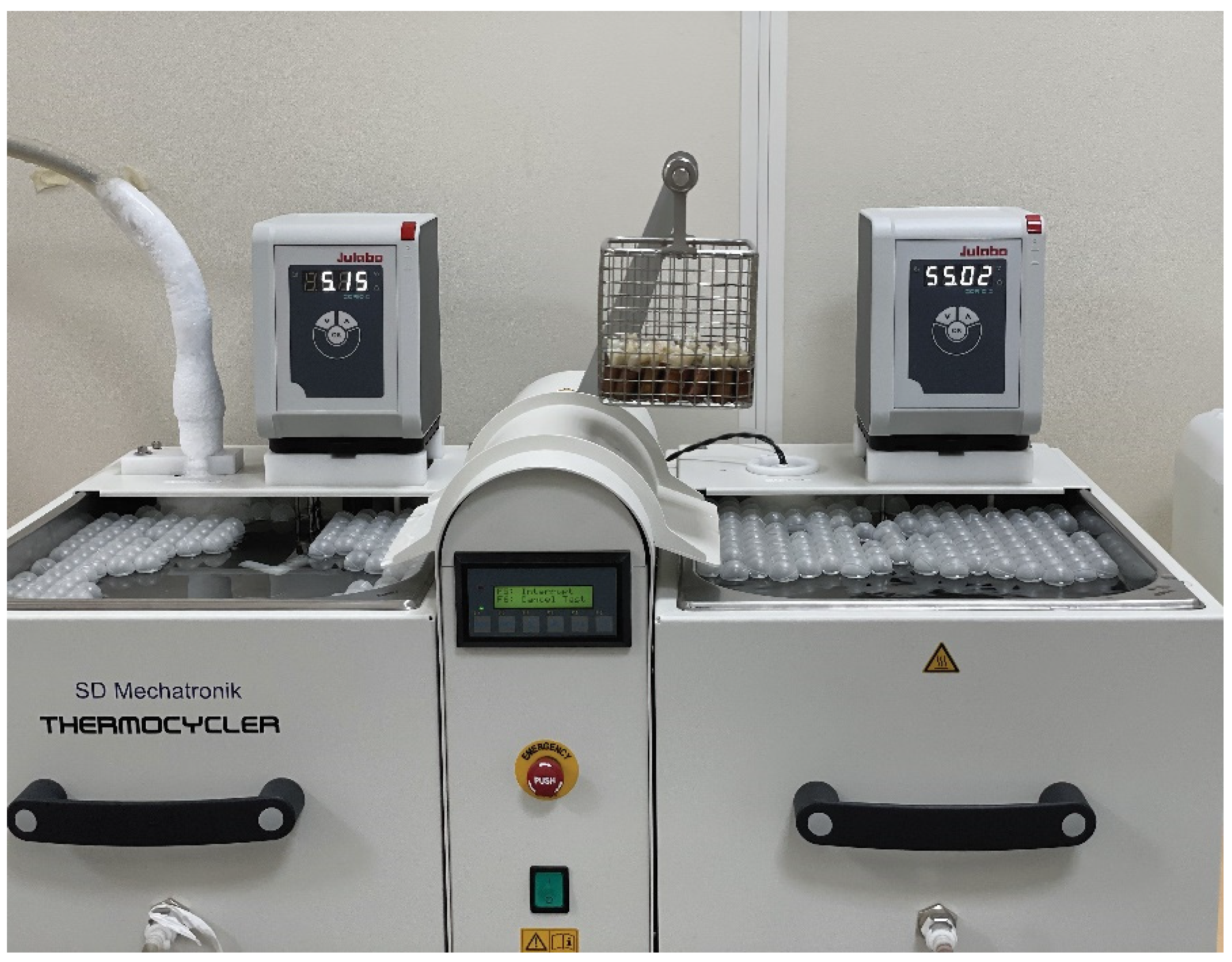
2.4. Storage and Thermocycling

To simulate temperature changes in the mouth, specimens were placed in a thermocycler (SD Mechatronic, Feldkirchen-Westerham, Germany) and exposed to 7500 thermocycles at a temperature of 5 °C/55 °C with a dwell time of 30 s and a transfer time of 15 s (Figure 2).

2.5. Debonding of the Brackets

After thermocycling procedures, the brackets were debonded with bracket removal pliers with a peeling force. The remaining adhesive was cleaned using a 12-fluted tungsten carbide bur (Komet Geber, Brasseler, Lemgo, Germany) with sufficient water cooling operated at a low speed. The teeth were polished using polishing discs (EVE Flexi-d flexible polishing and finishing discs mandrel Soflex style) until normal luster, as observed with the naked eye, was restored. The enamel surfaces were then observed under ×3 magnification to ensure the removal of any adhesive residues. All the previous steps were performed by a single examiner under a dental chair light.

2.6. Post-Debonding Tooth Color Assessment

The color of the buccal surfaces of the teeth was re-assessed after debonding of the brackets and finishing and polishing of the teeth surfaces using the Vita Easyshade spectrophotometer as described for baseline tooth color assessment. Data were recorded on a master sheet (MS Excel 2016) and coded. Change in color (∆E*) was calculated from the numerical variables L*, a*, and b* of each the specimen according to the following formula:
∆E = √([(L1 − L2)]2 + [(a1 − a2)]2 + [(b1 − b2)]2),
where L1, a1, and b1 refer to the values before bonding, and L2, a2, and b2 to the values after debonding. All measurements were repeated 3 times, and the average was calculated and recorded. The National Bureau of Standards (NBS) protocol was used to describe the ΔE using the formula: NBS units = ΔE × 0.92, as shown in Table 2 [14].

2.7. Statistical Analysis

The data were analyzed using statistical software (IBM SPSS for Windows, Version 25.0, IBM SPSS, Inc., Chicago, IL, USA) (α = 0.05). A test of normality was applied for the distribution of the data. Accordingly, nonparametric tests were used. Two-way ANOVA was used for the interaction effect of the independent variables (bracket and adhesive types) on the dependent variable (change in color). The Mann–Whitney U test was used for differences between metal and ceramic brackets and between Transbond XT and U-Cem adhesive system groups. The Kruskal–Wallis test was used for differences between the individual groups tested, and, if significant, the Dunnett test for nonparametric multiple comparison was performed.

3. Results

The interaction effect of bracket and adhesive types using a two-way ANOVA (Table 3) revealed significant differences for both the brackets and the adhesive types, separately. However, the effect of both variables together was not significant (p = 0.952). Pairwise comparison for the differences between the two types of brackets and two types of adhesives (Table 4) revealed significant differences in favor of ceramic brackets (ΔE= 3.77 ± 3.60; p= 0.009) and the Transbond XT adhesive system (ΔE = 3.52 ± 2.45; p = 0.008).
When analyzing the differences between the different groups tested, the Kruskal–Willis nonparametric test (Table 5) revealed a significant difference (p = 0.006) among the different groups, with group C having the least change in color (ΔE= 2.00 ± 0.89) and group B having the highest (ΔE = 8.42 ± 5.66). According to the NBS units, changes in group C were considered noticeable, while in all the other groups tested, changes in tooth color ranged from “appreciable” to “much” as in group B.
Further multiple comparisons revealed that a significant difference was found between group C and group B (Table 6).

4. Discussion

The aesthetic appearance of teeth after debonding of the orthodontic appliances is a primary concern for both the patient and the orthodontist. However, orthodontic treatment may present a risk factor for tooth discoloration. This study evaluated the effect of orthodontic bracket type and adhesive system on tooth color change. Metal and ceramic brackets along with etch and rinse and self-etch adhesives were investigated. Considering that the sensitivity of the human eye in detecting small color differences is both restricted and subjective, a spectrophotometer was used in the study to provide a consistent and objective assessment of the tooth color changes in response to the different procedures tested. Tooth shades were measured before the placement of the brackets and after debonding. Before debonding, the teeth were subjected to 7500 thermal cycles, simulating more than 7 months of clinical use [15]. Thermocycling simulated the temperature changes that occur in the oral cavity and is a substitute for time-consuming and costly clinical studies [16].
The study results revealed that the ceramic brackets were associated with significantly less tooth discoloration compared with metal brackets. Therefore, the first null hypothesis was rejected. To our knowledge, no previous studies have compared the effect of the bracket type, whether metal or ceramic, on tooth discoloration after debonding. Fusco et al. [17] evaluated the tooth color change of bonded metal and ceramic brackets after they were subjected to different beverages. Consistent with our results, teeth with metal brackets showed a greater color change than those with ceramic brackets. They attributed their results to the possible corrosion of the metallic orthodontic appliances when subjected to staining beverages with low pH. In our study, no staining beverages were used, but the teeth were subjected to thermocycling. In addition to the environmental conditions present in the oral cavity, thermocycling has been reported to affect the structural stability of the brackets, resulting in the release of metal ions and the decrease of their corrosion resistance [18,19]. According to Eliadis et al. [20], metal ions (mainly chromium and nickel) released by corrosion of the orthodontic brackets may diffuse through the adhesive system and even penetrate into the tooth enamel in a process known as metallosis. In cases of intense bracket oxidation, tooth staining may occur, requiring, in extreme situations, aesthetic restorations [21]. Such a color change was confirmed by the findings of our study, where tooth color change after debonding of the metal brackets was considered “appreciable” according to the NBS scale.
The other variable investigated in our study was the type of adhesive system used to bond the ceramic brackets. Less tooth discoloration was recorded with the use of the etch and rinse adhesive compared with the self-etch adhesive, which also led to the rejection of the second null hypothesis. Both in vitro and in vivo studies have reported conflicting results regarding the effects of orthodontic adhesive systems on tooth color change [22]. The separate etching step with the use of etch and rinse adhesive results in the dissolution of the superficial enamel and allows for subsequent penetration of the adhesive. In contrast, the self-etch adhesives simultaneously demineralize and penetrate the enamel’s surface [23]. When the adhesive is polymerized, the formed resin tags have been reported to be shorter with the use of self-etching primers in comparison with the use of the conventional etching technique [24,25]. The effect of the length of formed resin tags on tooth color change is unclear. Al Maaitah et al. reported that the two etching techniques (E&R and SE) had no statistically significant effect on tooth color difference and that the depth of the resin tag did not influence short-term superficial discoloration [26]. In contrast, Zaher et al. and Boncuk et al. reported a reduced color change using self-etching techniques and attributed this to the formation of shorter resin tags [3,23]. The results of our study do not support this finding and are more aligned with those of Joo et al., who reported that the use of SEPs with orthodontic adhesives showed increased tooth color change after debonding owing to their greater stain susceptibility [4]. According to Alshamsi et al., a thin residual adhesive layer of an average thickness of 31.2 to 200.2 μm remains on the enamel surface after debonding of the brackets and even after polishing [27]. This layer is susceptible to discoloration partly due to endogenous changes related to the structural properties of adhesives. The chemical composition of adhesive, the quality of polymerization, and curing time could all play a role in the color stability of the adhesive resin and hence of the color stability of the enamel surface when residual resin remains on the enamel surface after debonding [22]. In clinical situations, these factors would be heightened by exogenous factors related to the consumption of staining food and beverages [28].
When comparing the four test groups of the current study, group C, where the teeth received ceramic brackets bonded using a three step E&R adhesive system, showed the least amount of color change. In contrast, in group B, where the teeth received metal brackets bonded with self-adhesive cement, the highest color changes were found. The difference could be explained by the combined effect of the individual variables, the bracket types and adhesive systems used, as discussed earlier.
The teeth in group E, which served as control without any bonding procedure, showed a color change after thermocycling that is considered appreciable according to the NBS system. Delta E values in that group were not significantly different from those in all other test groups except group C (Table 6). Tooth color change after thermocycling may be from an increase in enamel surface roughness when subjected to cyclic thermal changes in water baths, as reported by Zhao et al. [29]. Vieira-Junior et al. demonstrated that alteration of the surface roughness of natural teeth does have an effect on the lightness of the tooth color due to alteration in the surface reflectance of the enamel surface [30].
Subjecting the control and the test groups in the current study to different post-debonding cleaning, finishing, and polishing protocols could have given an expanded view of the possible effect of the bracket type and adhesive systems on tooth color after debonding in comparison with untreated teeth; however, this was beyond the scope of the study. Further limitations of the study are that the bonded teeth were not subjected to staining solutions or demineralization challenges, as they would have encountered clinically.

Author Contributions

Concept—A.A. (Ali Alqerban), A.S.A.; Design—D.R.M.A.; Supervision—A.S.A.; Materials—D.A.; Data Collection and/or Processing—A.A. (Amjad AlShahrani); Analysis and/or Interpretation—H.A.; Literature Review—D.R.M.A.; Writing—A.S., D.A.; Critical Review—A.A. (Ali Alqerban), A.S.A., A.S. All authors have read and agreed to the published version of the manuscript.

Funding

This research was funded by Deputyship for Research & Innovation, Ministry of Education in Saudi Arabia for funding this research work through the project number (IF-PSAU-2021/03/18524).

Institutional Review Board Statement

The study was conducted in accordance with the Declaration of Helsinki, and approved by the Research Ethics Committee of College of Dentistry/Dar Aluloom University (KSA), (Approval No: COD/IRB/2020/31).

Informed Consent Statement

Informed consent was obtained from all subjects involved in the study.

Data Availability Statement

The data used to support the findings of this study are available from the corresponding author upon request.

Acknowledgments

The authors extend their appreciation to the Deputyship for Research & Innovation, Ministry of Education in Saudi Arabia for funding this research work through the project number (IF-PSAU-2021/03/18524).

Conflicts of Interest

The authors have no conflict of interest to declare.

References

  1. Sandison, R.M. Tooth surface appearance after debonding. Br. J. Orthod. 1981, 8, 199–201. [Google Scholar] [CrossRef] [PubMed]
  2. Ogaard, B. Prevalence of white spot lesions in 19-year-olds: A study on untreated and orthodontically treated persons 5 years after treatment. Am. J. Orthod. Dentofac. Orthop. 1989, 96, 423–427. [Google Scholar] [CrossRef] [PubMed]
  3. Zaher, A.R.; Abdalla, E.M.; Abdel Motie, M.A.; Rehman, N.A.; Kassem, H.; Athanasiou, A.E. Enamel colour changes after debonding using various bonding systems. J. Orthod. 2012, 39, 82–88. [Google Scholar] [CrossRef] [PubMed]
  4. Joo, H.J.; Lee, Y.K.; Lee, D.Y.; Kim, Y.J.; Lim, Y.K. Influence of orthodontic adhesives and clean-up procedures on the stain susceptibility of enamel after debonding. Angle Orthod. 2011, 81, 334–340. [Google Scholar] [CrossRef] [PubMed] [Green Version]
  5. Wu, H.M.; Ye, C.; Chen, D. Comparative study of enamel discoloration related to bonding with different orthodontic adhesives and cleaning-up with different procedures. Shanghai Kou Qiang Yi Xue 2018, 27, 257–260. [Google Scholar]
  6. Almosa, N.A.; Sibai, B.S.; Rejjal, O.A.; Alqahtani, N. Enamel demineralization around metal and ceramic brackets: An in vitro study. Clin. Cosmet. Investig. Dent. 2019, 11, 37–43. [Google Scholar] [CrossRef] [Green Version]
  7. Brusca, M.I.; Chara, O.; Sterin-Borda, L.; Rosa, A.C. Influence of different orthodontic brackets on adherence of microorganisms in vitro. Angle Orthod. 2007, 77, 331–336. [Google Scholar] [CrossRef]
  8. Knoernschild, K.L.; Rogers, H.M.; Lefebvre, C.A.; Fortson, W.M.; Schuster, G.S. Endotoxin affinity for orthodontic brackets. Am. J. Orthod. Dentofac. Orthop. 1999, 115, 634–639. [Google Scholar] [CrossRef]
  9. Janiszewska-Olszowska, J.; Szatkiewicz, T.; Tomkowski, R.; Tandecka, K.; Grocholewicz, K. Effect of orthodontic debonding and adhesive removal on the enamel-current knowledge and future perspectives—A systematic review. Med. Sci. Monit. 2014, 20, 1991–2001. [Google Scholar]
  10. Pandian, A.; Ranganathan, S.; Padmanabhan, S. Enamel color changes following orthodontic treatment. Indian J. Dent. Res. 2017, 28, 330–336. [Google Scholar] [CrossRef]
  11. Korkmaz, Y.N.; Bulut, M. Effect of mouthwashes on the discoloration of bracket-bonded tooth surfaces: An in vitro study. Clin. Oral Investig. 2020, 24, 3855–3861. [Google Scholar] [CrossRef]
  12. de Melo Oliveira, I.; Santana, T.R.; Correia, A.C.C.; Fontes, L.S.; Griza, S.; Faria, E.S.A.L. Color heterogeneity and individual color changes in dentin and enamel bleached in the presence of a metallic orthodontic bracket. J. Esthet. Restor. Dent. 2021, 33, 262–268. [Google Scholar] [CrossRef]
  13. George, J.; Mathews, T.A. Spectrophotometric Analysis of Tooth Discoloration after Bracket Debonding Induced by Coffee and Tea Grown in South India: An In vitro Study. J. Interdiscip. Dent. 2018, 8, 1. [Google Scholar] [CrossRef]
  14. Babikir, M.O.; Gilada, M.W.; Fahmy, F.; Ismail, I.A.; Alhajj, M.N.; Fadul, A.A.; Elasyouti, A. Effect of Commonly Consumed Beverages on Color Stability of Polymethyl Methacrylate Denture Base Material. Compend. Contin. Educ. Dent. 2019, 40, e1–e8. [Google Scholar]
  15. Ghavami-Lahiji, M.; Firouzmanesh, M.; Bagheri, H.; Jafarzadeh Kashi, T.S.; Razazpour, F.; Behroozibakhsh, M. The effect of thermocycling on the degree of conversion and mechanical properties of a microhybrid dental resin composite. Restor. Dent. Endod. 2018, 43, e26. [Google Scholar] [CrossRef]
  16. Valizadeh, S.; Hashemi, S.F.; Hashemikamangar, S.S.; Kharazifard, M.J. Microleakage of a Self-adhesive Composite of Class V Cavities: Effect of Surface Treatment and Thermocycling. J. Contemp. Dent. Pract. 2020, 21, 781–786. [Google Scholar]
  17. Fusco, A.; Ahmed, S.; Link, J.; Al-Talib, T.; Abubakr, N.H. Performance of Different Orthodontic Brackets after Exposure to Dietary Components: An in Vitro Pilot Study. Open J. Stomatol. 2021, 11, 341–348. [Google Scholar] [CrossRef]
  18. Gursoy, S.; Acar, A.G.; Sesen, C. Comparison of metal release from new and recycled bracket-archwire combinations. Angle Orthod. 2005, 75, 92–94. [Google Scholar]
  19. Novianti, S.; Siregar, E.; Anggani, H.S. Corrosion Resistance of Stainless Steel Brackets After Thermal Recycling by Direct Flaming. Pesqui. Bras. Em Odontopediatria E Clínica Integr. 2019, 19. [Google Scholar] [CrossRef]
  20. Eliades, T.; Athanasiou, A.E. In Vivo aging of orthodontic alloys: Implications for corrosion potential, nickel release, and biocompatibility. Angle Orthod. 2002, 72, 222–237. [Google Scholar]
  21. Hodges, S.J.; Spencer, R.J.; Watkins, S.J. Unusual indelible enamel staining following fixed appliance treatment. J. Orthod. 2000, 27, 303–306. [Google Scholar] [CrossRef]
  22. Haghighi, A.H.S.; Emami, M.; Fakhri, E.; Rezaei, Y. Effects of Orthodontic Adhesives on Dental Enamel Color Alteration Using Chemically Cured and Light-Cured Composites. Front. Dent. 2020, 17, 1. [Google Scholar]
  23. Boncuk, Y.; Cehreli, Z.C.; Polat-Ozsoy, O. Effects of different orthodontic adhesives and resin removal techniques on enamel color alteration. Angle Orthod. 2014, 84, 634–641. [Google Scholar] [CrossRef]
  24. Cal-Neto, J.P.; Miguel, J.A. Scanning electron microscopy evaluation of the bonding mechanism of a self-etching primer on enamel. Angle Orthod. 2006, 76, 132–136. [Google Scholar]
  25. Horiuch, S.; Kaneko, K.; Mori, H.; Kawakami, E.; Tsukahara, T.; Yamamoto, K.; Hamada, K.; Asaoka, K.; Tanaka, E. Enamel bonding of self-etching and phosphoric acid-etching orthodontic adhesives in simulated clinical conditions: Debonding force and enamel surface. Dent. Mater. J. 2009, 28, 419–425. [Google Scholar] [CrossRef] [Green Version]
  26. Al Maaitah, E.F.; Abu Omar, A.A.; Al-Khateeb, S.N. Effect of fixed orthodontic appliances bonded with different etching techniques on tooth color: A prospective clinical study. Am. J. Orthod. Dentofac. Orthop. 2013, 144, 43–49. [Google Scholar] [CrossRef]
  27. Al Shamsi, A.H.; Cunningham, J.L.; Lamey, P.J.; Lynch, E. Three-dimensional measurement of residual adhesive and enamel loss on teeth after debonding of orthodontic brackets: An in-vitro study. Am. J. Orthod. Dentofac. Orthop. 2007, 131, 301.e9–301.e15. [Google Scholar] [CrossRef]
  28. Corekci, B.; Irgin, C.; Malkoc, S.; Ozturk, B. Effects of staining solutions on the discoloration of orthodontic adhesives: An in-vitro study. Am. J. Orthod. Dentofac. Orthop. 2010, 138, 741–746. [Google Scholar] [CrossRef]
  29. Zhao, X.; Ren, Y.F. Surface Properties and Color Stability of Resin-Infiltrated Enamel Lesions. Oper. Dent. 2016, 41, 617–626. [Google Scholar] [CrossRef] [Green Version]
  30. Vieira-Junior, W.-F.; Vieira, I.; Ambrosano, G.-M.-B.; Aguiar, F.-H.-B.; Lima, D.-A.-N. Correlation between alteration of enamel roughness and tooth color. J. Clin. Exp. Dent. 2018, 10, e815–e820. [Google Scholar] [CrossRef]
Figure 1. Mounted samples.
Applsci 12 12885 g001
Figure 2. Thermocycling procedure.
Applsci 12 12885 g002
Table 1. Bracket types and adhesive systems used in the study.
MaterialBrand NameCompositionManufacturer
Ceramic bracketsClarity™ Advanced ceramic bracketsPolycrystalline alumina3M Unitek, Monorovia, CA, USA
Metal bracketsVictory Series™ Low ProfileStainless Steel3M Unitek, Monorovia, CA, USA
Acid etching gelAcido Gel 37%37% phosphoric acidMaquira, Setubal, Portugal
Adhesive systemTransbond XT Primer* TEGDMA, Bisphenol A diglycidyl ether dimethacrylate, Hydroquinone, Camphorquinone,
Triphenylantimony,
4-(Dimethylamino)-Benzene ethanol
3M Unitek, Monorovia, CA, USA
Transbond XTBisphenol A diglycidyl ether
dimethacrylate, Bisphenol A
Bis(2-hydroxyethyl ether) dimethacrylate, Silane treated quartz, Silane treated silica
3M Unitek, Monorovia, CA, USA
Self-adhesive resin adhesiveU-Cem™Base: methacrylate monomers containing acid groups, methacrylate monomers, silanated fillers, initiator components, stabilizer
Catalyst: methacrylate monomer, alkaline fillers, silanated fillers, initiator components
Vericom, Chuncheon-si, Korea
* TEGDMA (Triethylene glycol dimethacrylate).
Table 2. National Bureau of Standards system of expressing color difference (NBS).
NBS UnitsCritical Remarks for Color Differences
0.0–0.5Trace
0.5–1.5Slight
1.5–3.0Noticeable
3.0–6.0Appreciable
6.0–12.0Much
≥12.0Very much
Table 3. Two-way ANOVA for the interaction effect of bracket and adhesive on change in tooth color (ΔE).
SourceType III Sum of SquaresdfMean SquareFp
Corrected Model206.898368.9664.6600.007
1101.13511101.13574.3980.000
Bracket type87.232187.2325.8940.020 *
Adhesive type119.6121119.6128.0820.007 *
Bracket—Adhesive0.05410.0540.0040.952
Error532.8243614.801
Total1840.85740
Corrected Total739.72239
* Statistically significant at p value < 0.05. R2 = 0.280; Adjusted R2 = 0.220.
Table 4. Comparison of means and SDs of change in color (ΔE) between metal and ceramic brackets and between Transbond XT and U-Cem adhesive systems using Mann-Whitney nonparametric test.
Item NMeanSDMean Difference95% CI of the Differencep *
LowerUpper
Bracket TypeMetal206.724.622.950.305.610.009 *
Ceramic203.773.60
Adhesive TypeTransbond XT203.522.45−3.46−6.08−0.840.008 *
U-Cem206.985.16
* Statistically significant at p value < 0.05.
Table 5. Comparison of means and SDs of ΔE among the different groups tested using Kruskal-Willis nonparametric test.
DeltaGroup AGroup BGroup CGroup DGroup Ep *
Mean5.038.422.005.545.570.006 *
SD2.615.660.894.433.63
* Statistically significant at p value < 0.05.
Table 6. Multiple comparison between the different groups tested.
GroupsMean Difference95% CI of the Differencep *
LowerUpper
Group AGroup B−3.39−6.810.041.000
Group C3.03−0.406.450.121
Group D−0.50−3.932.921.000
Group E−0.54−3.972.891.000
Group BGroup C6.412.989.840.003
Group D2.88−0.556.311.000
Group E2.84−0.586.271.000
Group CGroup D−3.53−6.96−0.100.121
Group E−3.57−7.00−0.140.091
Group DGroup E−0.04−3.463.391.000
* Dunnett test for nonparametric multiple comparison; Significance values have been adjusted by the Bonferroni correction for multiple tests.
Publisher’s Note: MDPI stays neutral with regard to jurisdictional claims in published maps and institutional affiliations.

