Communication

Penetration Depth of Initiated and Non-Initiated Diode Lasers in Bovine Gingiva

1
Department of Periodontology, Laboratory for Periodontal-, Implant-, Phototherapy (La-PIP), School of Dental Medicine, Stony Brook University, 106 Rockland Hall, Stony Brook, NY 11794, USA
2
Department of Dermatology, School of Medicine, Stony Brook University, Stony Brook, NY 11794, USA
3
Department of Family, Population & Preventive Medicine, Stony Brook University, Stony Brook, NY 11794, USA
*
Author to whom correspondence should be addressed.
Appl. Sci. 2022, 12(24), 12771; https://doi.org/10.3390/app122412771
Submission received: 19 November 2022 / Revised: 30 November 2022 / Accepted: 8 December 2022 / Published: 13 December 2022
(This article belongs to the Section Optics and Lasers)

Abstract

:
Background: This study aimed to evaluate the penetration depth of 445 and 970 nm diode lasers using both initiated and non-initiated fibers on bovine gingiva in an ex vivo model. Methods: In this in vitro experiment, 445 nm and 970 nm diode lasers were compared in pulsed (35 Hz, duty cycle: 50%) and continuous mode with both initiated and non-initiated tips. All experimental groups had laser output settings of 2 Watts with a 320 μm thick glass fiber utilized for delivery. Two posterior teeth were randomly selected from four bovine mandibles for each group. The teeth were irradiated for 30 s buccal and 30 s lingual before the gingiva was harvested for histological evaluation. Samples were fixed in 10% formalin and stained via elastin Van Gieson. Sections were examined microscopically to evaluate collagen denaturation due to thermal effects, which were measured and compared using a mixed-effect model with Tukey adjustment. Results: The 445 nm wavelength groups displayed significantly higher penetration depths when compared to the 970 nm wavelength groups. The initiated tip groups tended to display a lower penetration depth than non-initiated groups; however, these values were similar (p > 0.05) for the 445 nm pulsed lasers (initiated vs. non-initiated). Conclusions: We can conclude that the 445 nm laser wavelength displayed greater thermal denaturation in bovine gingiva ex vivo when compared to the 970 nm wavelength. Additionally, the pulsed settings displayed less thermal penetration depth when compared to the continuous emission modes of the same power output. However, additional studies are necessary to better compare initiated and non-initiated tips using the novel blue laser light.

1. Introduction

Lasers are being incorporated into all facets of clinical dentistry, especially in periodontics [1,2]. The photothermal effects that can be achieved with laser therapy include increased coagulation [2], less post-operative pain [3], and the ablation of dark-pigmented bacteria [4]. Additionally, lasers have been shown to decrease epithelial downgrowth, which is advantageous in periodontal therapy to provide connective tissue attachment rather than long-junctional epithelial repair [5]. Therefore, periodontal phototherapy can provide advantageous effects in non-surgical therapy compared to conventional scaling and root planing [1].
Diode lasers are highly absorbed by melanin and hemoglobin (especially 445 nm blue light [6]), making diode lasers a great hemostatic agent [7]. Diode lasers are utilized in clinical settings as they have a relatively low cost and small size while still displaying great incision quality [3,7]. Additionally, diode lasers provide versatility in clinical settings as the tips can be initiated or thermo-optically powered (TOP) [8]. TOP surgery can control the risks of overheating the targeted tissue by focusing the beam for more precise thermomechanical cutting and adjusting the thermal changes [8,9]. However, without initiating the tip, diode lasers can be used in a non-contact manner as a defocused beam to coagulate the targeted site, as well as in contact with the tissues for photobiomodulation [10].
Given the flexibility of the usage of diode lasers, it is important to evaluate the laser–tissue interactions in periodontal tissues and compare initiated vs. non-initiated and pulsed vs. continuous laser beams for both visible and near-infrared wavelengths in a contact mode. This study aimed to evaluate the penetration depth and tissue denaturation of both 445 nm and 970 nm diode lasers.

2. Materials and Methods

Bovine mandibles from freshly slaughtered cows were utilized in this study. The specimens were stored at room temperature before the experiment. All treatment groups were randomly assigned to one of four mandibles, and a total of two posterior molars were utilized for each experimental group.

2.1. Experimental Groups

Four experimental groups were evaluated in this study. Two teeth were selected randomly among the four bovine jaws and were irradiated for 30 s on buccal and lingual surfaces.
Our previous study showed that the means of thermal penetration depth ranged from 0.06 to 0.24 mm for carbon dioxide, Er, Cr: YSGG, and Er: YAG lasers, with a pooled standard deviation of 0.12 mm [11]. Based on a repeated ANOVA model, the present study had 95% power to detect the differences in thermal penetration depth.
All non-surgical treatment procedures were performed by the same laser expert (G.R.). Interproximal areas were avoided so as to prevent an overlap between the treatment groups. A diode (Class IV) laser (SIROLaser Blue®, Dentsply Sirona, Charlotte, NC, USA) was utilized at 445 nm (±5 nm) and 970 nm (−10/+15 nm) wavelengths and a power setting of 2 W (as recommended by the manufacturer and the literature). The lasers were utilized in chopped-pulse (50% duty cycle, 35 Hz; pulse duration: 10 µs–0.99 s; mean power: 1 W) and continuous emission modes, with each mode evaluated utilizing initiated and non-initiated fiberoptic disposable tips (diameter: 320 µm, EasyTip 320). The tips were initiated using a dark blue articulating paper (Accufilm; Parkell) for 5 s. This allowed for a total of eight treatment groups for gingival biopsies. The glass fibers were inserted parallel to the tooth axis at a depth of 3–4 mm. The fibers were moved in the buccal surface from the interproximal area to the buccal tooth surface and then in a similar way in the lingual aspect of the posterior teeth attached to the root surface and the inner part of the sulcus. The laser irradiation was measured optically according to the changes in the tissue color during irradiation. After laser radiation, soft-tissue full-thickness specimens, including the complete gingiva to the mucogingival junction, were excised using a no.15 scalpel blade on both the buccal and lingual surfaces of each tooth and prepared for histologic and histomorphometric analysis. This led to a total of four samples per experimental group.

2.2. Histological Processing Method

The specimens were prepared for conventional histological staining with elastin Van Gieson (EVG) after fixation and paraffin embedment. Serial sections (5 μm thick) were prepared. Verhoeff’s staining procedure began with de-paraffinization and hydration to water. The specimens were then rinsed in running tap water for 3 min and stained in Verhoeff’s working solution for 15 min. Next, they were dipped 10 times in distilled water and a few times in ferric chloride 2% aqueous solution until the tissues appeared gray. After rinsing in tap water, the samples were treated in sodium thiosulfate 5% aqueous solution for 1 min before being counter-stained in Van Gieson’s solution. Slides were blotted dry before being placed in xylene and mounted with a Poly Mount. Sections were examined by conventional microscopy using an Olympus BX61 microscope; photographed with an Olympus DP71 digital camera (Olympus USA, Center Valley, PA, USA); and measured using a calibrated ocular micrometer. Histological images were evaluated at 1360 × 1024 pixels, 2X 1.05 zoom, 4.141 mm wide.
Sections were examined microscopically by two experienced examiners (S.M. and A.L.) to evaluate the penetration depth due to photothermal effects by measuring the maximum distance from the tooth side of the gingiva to the extent of collagen denaturation towards the outer epithelium.
The entire protocol for laser safety followed the Standards of the American National Standards Institute (ANSI, Z136.3 series) and the guidelines of the Laser Institute of America (LIA) for the use of laser-protecting glasses.

2.3. Statistical Analysis

The penetration depths of all experimental groups were statistically compared using a mixed-effect model with Tukey adjustment and a p-value of 5%. All statistical analyses were performed using SAS v9.4 (SAS Institute, Cary, NC, USA).

3. Results

The histological evaluation of the specimens showed differences in the penetration depth of the laser light between the continuous and pulsed emission modes at both laser wavelengths (Figure 1). The initiated tips created a more localized effect in the pocket epithelium. A histomorphometric comparison between the treatment groups demonstrating the amount of collagen fiber denaturation and the heat (energy) effects within the tissue is presented in Table 1. In the continuous mode, the initiated 445 nm laser presented a significantly different penetration depth compared to the 970 nm laser (p < 0.0001). Similar effects were found for the non-initiated lasers (p < 0.0001). In contrast, the pulsed mode lasers showed a statistically significant differences in penetration depth between initiated vs. non-initiated lasers for the 970 nm wavelength (p < 0.001) but no statistical difference for the 445 nm laser (p = 0.7939).

Evaluation of Penetration Depths among Treatment Groups

Overall, the 970 nm wavelength displayed a significantly lower penetration depth when compared to the 445 nm wavelength. In continuous non-initiated mode, the 970 nm wavelength displayed a mean penetration depth of 0.131 mm, while the 445 nm wavelength displayed a penetration depth of 0.219 mm under the same settings (p < 0.0001). Additionally, the pulsed groups displayed a lower penetration depth than the continuous groups. The initiated tip groups tended to display a lower penetration depth than the non-initiated groups. The only comparison without statistically significant differences was between the 445 nm pulsed groups, with both groups demonstrating similar values (0.136 mm for the pulsed initiated vs. 0.131 mm for the pulsed non-initiated). The 970 nm pulsed initiated group displayed an average penetration depth of 0.105 mm. This was the lowest penetration depth among all the groups, though it was statistically higher than the 970 nm pulsed non-initiated group, which measured an average penetration depth of 0.015 mm (p < 0.0001).

4. Discussion

The purpose of this investigation was to compare the penetration depth of near-infrared (970 nm) and visible (445 nm) diode lasers in both pulsed and continuous emission modes using initiated and non-initiated tips in bovine gingiva ex vivo. The results showed that, overall, the pulsed mode displayed less thermal damage than continuous mode. This was explained by the fact that the pulsed setting allows for the thermal relaxation of the targeted tissues and therefore a lower thermal penetration depth.
Additionally, the 445 nm laser wavelength displayed a higher thermal penetration depth than the 970 nm laser wavelength. This was consistent with another study displaying a higher temperature rise under a 445 nm compared to a 980 nm wavelength when uncovering implants in pigs [12]. This may have been because the 445 nm wavelength has a higher degree of absorption by both hemoglobin and melanin than the 970 nm wavelength [13]. Although this was exemplified in this study, it is still unknown how this effect translates to wound healing in vivo. In an in vitro study by Reichelt et al. using cell cultures, the 445 nm wavelength demonstrated faster wound healing [6]. Additionally, it has been shown that periodontal pathogens such as P. gingivalis can replicate in the host tissue [14,15]. Therefore, it may be beneficial to have a deeper-penetrating laser to accurately remove all the contaminated soft tissues. Additionally, removing inflamed epithelium in periodontal pockets helps to create a stronger attachment apparatus [5,16,17]. It has been demonstrated that the pulsed Nd: YAG laser can be effectively used to remove pocket epithelium in humans [18]. Most commonly applied is a trademarked technique called the “Laser-Assisted New Attachment Procedure” (LANAP), which has one of the highest thermal penetration depths among commonly used laser wavelengths [1]. Despite this deeper penetration, it is usually applied under a specific protocol for regenerative periodontal therapy with histological evidence [19,20].
Additionally, the 970 nm initiated laser group showed a significantly greater thermal penetration depth when compared to the non-initiated group. This was consistent with the study by Romanos et al., which highlighted a lower increase in temperature in the deep layers of the tissue when blue articulating paper was used to initiate the tip compared to when a non-initiated tip was used [9]. However, this phenomenon was not evident when comparing the 445 nm pulsed groups, as there was no difference in penetration depth between the initiated and non-initiated groups. This may have been due to the penetration of the 445 nm wavelength being dependent on the chromophores and, specifically, the presence of hemoglobin in the blood vessels of the connective tissue; whether or not an initiator was used seemed to have no influence in the pulsed mode. Since recent studies have shown that longer pulse widths are recommended in the case of long working distances and shorter pulse widths in the case of short working distances [21] due to changes in the fluence, there is ample opportunity to develop ideal pulse widths in periodontology by considering the thermal effects and tissue phenotype. Regardless, further research with more specific initiating protocols is necessary to fully compare the impact of initiated glass fibers in periodontal tissues using this novel blue laser light wavelength.
Recent recommendations from the World Workshop of Periodontal and Peri-implant Diseases showed that subgingival curettage has no additional benefits in non-surgical periodontal therapy, and therefore there is no need for the utilization of lasers in addition to non-surgical instrumentation [22]. However, the clinical study by Kamma et al. [23] involving aggressive periodontitis patients determined better microbiological and clinical outcomes using the 2 W power setting. This study showed the modification of the biofilm in patients with aggressive periodontal disease within a period ranging from 2 weeks to 6 months, transforming the dysbiotic microbiome into more homeostatic conditions within the periodontal tissues. Therefore, an uncontrolled destructive periodontal disease might become homeostatic due to a less-susceptible host response by the pathogenic bacteria. Similarly, a recent randomized clinical trial showed the efficacy of a low-power 980 nm diode laser in conjunction with traditional therapy and the removal of the pocket epithelium. The study showed a improvements in the clinical attachment levels and probing depths within 1 year of follow-up [5], promoting gingival de-epithelization as a method of treatment to control the migration of the long-junctional epithelium and therefore promote connective tissue regeneration.
The weaknesses of the present study included the fact that the utilized bovine mandibulae were taken from relatively young animals and therefore did not contain periodontally diseased tissues with extensively infiltrated rete pegs in the long-junctional epithelium. Additionally, the localized pigmentation in the bovine gingiva was not considered when randomizing the treatment groups. The pigmented gingiva may have affected the thermal penetration depth of the wavelengths due to the affinity of diode lasers for hemoglobin.
The use of elastin Van Gieson (EVG) staining facilitated the measurement of the thermal collagen denaturation of the histological samples by the examiners (data not shown). We believe that the use of this staining method could facilitate the evaluation of thermal damage by laser light and should be increasingly used in future studies.

5. Conclusions

We can conclude that the 445 nm wavelength displayed greater thermal denaturation in bovine gingiva when compared to the 970 nm wavelength in our ex vivo study. Additionally, the pulsed mode displayed a lower thermal penetration depth when compared to the continuous setting at the same power output. However, more studies are necessary to better compare initiated vs. non-initiated tips for the novel blue laser light in different clinical conditions.

Author Contributions

Conceptualization, G.E.R.; methodology, G.E.R. and S.M.; validation, G.E.R., N.E.E. and A.L.; formal analysis, N.E.E., A.L. and S.M.; investigation, N.E.E., A.L., S.M. and W.H.; resources, G.E.R.; writing—original draft preparation, N.E.E. and G.E.R.; writing—review and editing, G.E.R., N.E.E. and A.L.; visualization, N.E.E. and A.L.; supervision, G.E.R. and S.M.; project administration, G.E.R.; funding acquisition, G.E.R. All authors have read and agreed to the published version of the manuscript.

Funding

This research received no external funding.

Institutional Review Board Statement

Not applicable for this in vitro study.

Informed Consent Statement

Not applicable.

Conflicts of Interest

The authors declare no conflict of interest.

References

  1. Aoki, A.; Mizutani, K.; Schwarz, F.; Sculean, A.; Yukna, R.A.; Takasaki, A.A.; Romanos, G.E.; Taniguchi, Y.; Sasaki, K.M.; Zeredo, J.L.; et al. Periodontal and peri-implant wound healing following laser therapy. Periodontology 2000 2015, 68, 217–269. [Google Scholar] [CrossRef]
  2. Aoki, A.; Sasaki, K.M.; Watanabe, H.; Ishikawa, I. Lasers in nonsurgical periodontal therapy. Periodontology 2000 2004, 36, 59. [Google Scholar] [CrossRef]
  3. Romanos, G.E. Diode laser soft-tissue surgery: Advancements aimed at consistent cutting, improved clinical outcomes. Compend. Contin. Educ. Dent. 2013, 34, 752–757, quiz 758. [Google Scholar] [PubMed]
  4. Kranendonk, A.; Van der Reijden, W.; Van Winkelhoff, A.; Van der Weijden, G. The bactericidal effect of a Genius® Nd:YAG laser. Int. J. Dent. Hyg. 2010, 8, 63–67. [Google Scholar] [CrossRef] [PubMed]
  5. Nammour, S.; El Mobadder, M.; Maalouf, E.; Namour, M.; Namour, A.; Rey, G.; Matamba, P.; Matys, J.; Zeinoun, T.; Grzech-Leśniak, K. Clinical Evaluation of Diode (980 nm) Laser-Assisted Nonsurgical Periodontal Pocket Therapy: A Randomized Comparative Clinical Trial and Bacteriological Study. Photobiomodul. Photomed. Laser Surg. 2021, 39, 10–22. [Google Scholar] [CrossRef] [PubMed]
  6. Reichelt, J.; Winter, J.; Meister, J.; Frentzen, M.; Kraus, D. A novel blue light laser system for surgical applications in dentistry: Evaluation of specific laser-tissue interactions in monolayer cultures. Clin. Oral Investig. 2017, 21, 985–994. [Google Scholar] [CrossRef]
  7. Romanos, G.; Nentwig, G.H. Diode laser (980 nm) in oral and maxillofacial surgical procedures: Clinical observations based on clinical applications. J. Clin. Laser Med. Surg. 1999, 17, 193–197. [Google Scholar] [CrossRef]
  8. Romanos, G.E.; Belikov, A.V.; Skrypnik, A.V.; Feldchtein, F.I.; Smirnov, M.Z.; Altshuler, G.B. Uncovering dental implants using a new thermo-optically powered (TOP) technology with tissue air-cooling. Lasers Surg. Med. 2015, 47, 411–420. [Google Scholar] [CrossRef]
  9. Romanos, G.E.; Sacks, D.; Montanaro, N.; Delgado-Ruiz, R.; Calvo-Guirado, J.L.; Javed, F. Effect of initiators on thermal changes in soft tissues using a diode laser. Photomed. Laser Surg. 2018, 36, 386–390. [Google Scholar] [CrossRef]
  10. Romanos, G.E. Advanced Laser Surgery in Dentistry; Wiley: Hoboken, NJ, USA, 2021. [Google Scholar]
  11. Estrin, N.E.; Lesniewski, A.; McClain, S.; Hou, W.; Romanos, G.E. Thermal Penetration Depth of Pulsed Lasers in Gingival Tissues: An In-Vitro Study. Photobiomodul. Photomed. Laser Surg. 2022, 40, 410–416. [Google Scholar] [CrossRef]
  12. Matys, J.; Flieger, R.; Dominiak, M. Effect of diode lasers with a wavelength of 445 and 980 nm on a temperature rise when uncovering implants for second stage surgery: An ex-vivo study in pigs. Adv. Clin. Exp. Med. 2017, 26, 687–693. [Google Scholar] [CrossRef] [Green Version]
  13. Boulnois, J.-L. Photophysical processes in recent medical laser developments: A review. Lasers Med. Sci. 1986, 1, 47–66. [Google Scholar] [CrossRef]
  14. Lamont, R.J.; Chan, A.; Belton, C.M.; Izutsu, K.T.; Vasel, D.; Weinberg, A. Porphyromonas gingivalis invasion of gingival epithelial cells. Infect. Immun. 1995, 63, 3878–3885. [Google Scholar] [CrossRef] [Green Version]
  15. Bostanci, N.; Belibasakis, G.N. Porphyromonas gingivalis: An invasive and evasive opportunistic oral pathogen. FEMS Microbiol. Lett. 2012, 333, 1–9. [Google Scholar] [CrossRef] [Green Version]
  16. Romanos, G.E.; Henze, M.; Banihashemi, S.; Parsanejad, H.R.; Winckler, J.; Nentwig, G.-H. Removal of epithelium in periodontal pockets following diode (980 nm) laser application in the animal model: An in vitro study. Photomed. Laser Surg. 2004, 22, 177–183. [Google Scholar] [CrossRef]
  17. Goldman, H.M. A Rationale for the Treatment of the Intrabony Pocket; One Method of Treatment, Subgingival Curettage. J. Periodontol. 1949, 20, 83–91. [Google Scholar] [CrossRef]
  18. Gold, S.I.; Vilardi, M.A. Pulsed laser beam effects on the gingiva. J. Clin. Periodontol. 1994, 21, 391–396. [Google Scholar] [CrossRef]
  19. Harris, D.M.; Gregg, R.H.; McCarthy, D.K.; Colby, L.E.; Tilt, L.V. Laser-assisted new attachment procedure in private practice. Gen. Dent. 2004, 52, 396–403. [Google Scholar]
  20. Nevins, M.L.; Camelo, M.; Schupbach, P.; Kim, S.-W.; Kim, D.M.; Nevins, M. Human clinical and histologic evaluation of laser-assisted new attachment procedure. Int. J. Periodontics Restor. Dent. 2012, 32, 497–507. [Google Scholar]
  21. Nguyen, D.D.; Pang, J.Y.; Madill, C.; Novakovic, D. Effects of 445-nm Laser on Vessels of Chick Chorioallantoic Membrane with Implications to Microlaryngeal Laser Surgery. Laryngoscope 2021, 131, E1950–E1956. [Google Scholar] [CrossRef]
  22. Salvi, G.E.; Stähli, A.; Schmidt, J.C.; Ramseier, C.A.; Sculean, A.; Walter, C. Adjunctive laser or antimicrobial photodynamic therapy to non-surgical mechanical instrumentation in patients with untreated periodontitis: A systematic review and meta-analysis. J. Clin. Periodontol. 2020, 47 (Suppl. 22), 176–198. [Google Scholar] [CrossRef] [Green Version]
  23. Kamma, J.; Romanos, G.E.; Vasdekis, V. The effect of diode laser (980 nm) treatment on aggressive Periodontitis. Evaluation of microbial and clinical parameters. Photomed. Laser Surg. 2009, 27, 11–19. [Google Scholar] [CrossRef] [PubMed]
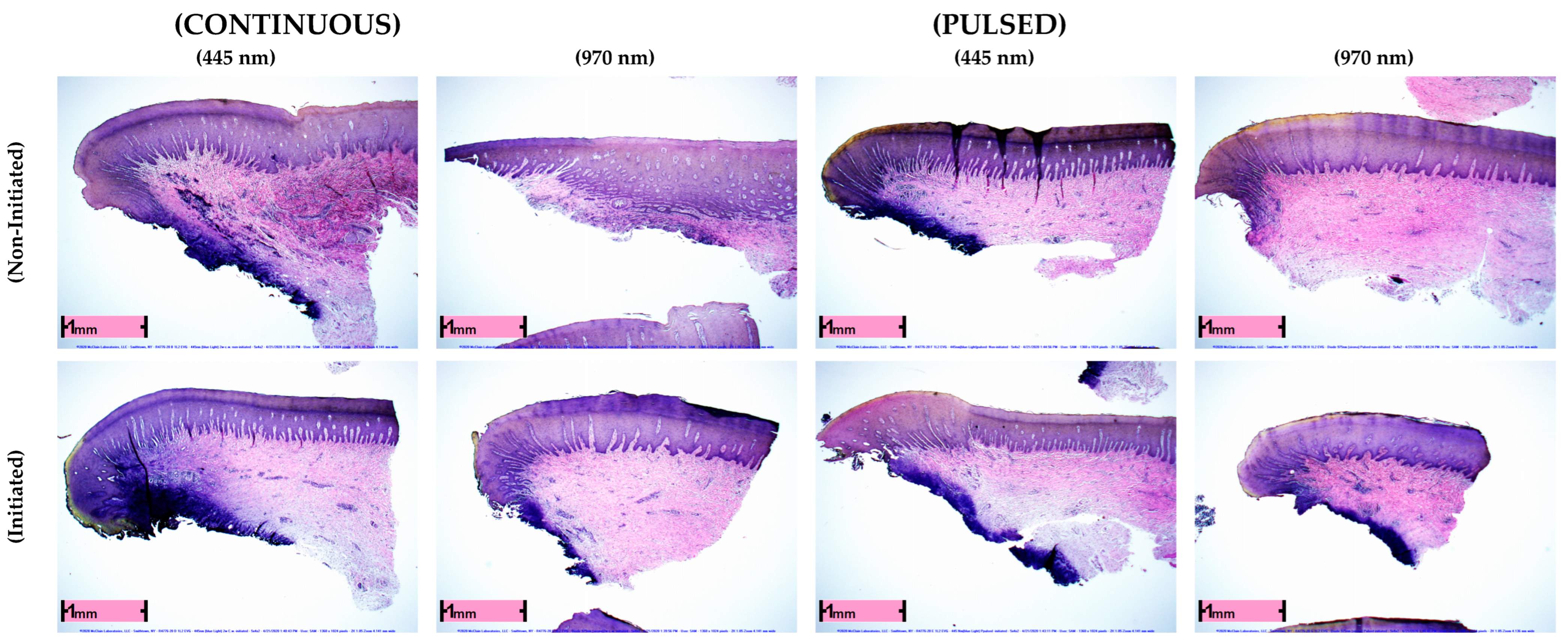
Figure 1. Histological samples of all experimental groups in continuous and pulsed modes. Coagulation of underlying tissue is visible via dark purple staining through EVG methods (1360 × 1024 pixels, 2X 1.05 zoom, 4.141 mm wide for all histological images).
Applsci 12 12771 g001
Table 1. Comparison between treatment groups when measuring the amount of collagen denaturation and thermal effect within the tissue (photothermal effects), which could be observed and measured through EVG staining (all measurements in mm; C.W.: continuous-wave mode).
Experimental GroupNMeanStd. Dev.Min.Max.p-Value
970 nm C.W. Non-Initiated960.1310.16100.46<0.001
445 nm C.W. Non-Initiated960.220.1500.6
970 nm C.W. Initiated960.200.1400.8<0.001
445 nm C.W. Initiated960.350.200.090.8
445 nm Pulsed Initiated960.130.1400.480.7939
445 nm Pulsed Non-Initiated960.130.1500.5
970 nm Pulsed Initiated960.100.1000.46<0.001
970 nm Pulsed Non-Initiated960.010.0500.25
Publisher’s Note: MDPI stays neutral with regard to jurisdictional claims in published maps and institutional affiliations.