Article

The Topical Effect of rhGDF-5 Embedded in a Collagen–Gelatin Scaffold for Accelerated Wound Healing †

1
Department of Hand, Plastic, Reconstructive and Burn Surgery, BG Klinik Tuebingen, Eberhard Karls University Tuebingen, 72076 Tuebingen, Germany
2
Department of Plastic, Hand, Reconstructive and Aesthetic Surgery, Hand Surgery, Traunstein Hospital, Ludwig-Maximilians-Universität Munic, 80539 Munich, Germany
3
Department of Plastic Surgery, Hand Surgery, Cologne-Merheim Hospital, University Witten/Herdecke, 58455 Witten, Germany
4
Department of Dermatology, University Hospital Erlangen, Friedrich-Alexander-University Erlangen-Nuernberg, 91054 Erlangen, Germany
*
Author to whom correspondence should be addressed.
The work was performed at the University of Tuebingen, Germany.
Appl. Sci. 2022, 12(2), 867; https://doi.org/10.3390/app12020867
Submission received: 20 December 2021 / Revised: 10 January 2022 / Accepted: 12 January 2022 / Published: 15 January 2022

Abstract

:
The application of exogenous growth factors such as the recombinant human growth and differentiation factor 5 (rhGDF-5) represents a major research topic with great potential for the treatment of complex wounds. In a randomized, controlled minipig study, the topical effect of rhGDF-5 on full-thickness skin defects was evaluated. A total of 60 deep dermal wounds were either treated with rhGDF-5 embedded in an innovative collagen scaffold or another commonly used collagen matrix or left untreated. Wound healing was analyzed by planimetric analysis to determine wound closure over time. After 21 days, the areas of the initial wounds were excised, and the newly formed tissue was examined histologically. In comparison to untreated wounds, all examined matrices accelerated dermal wound healing. The largest acceleration of wound healing was seen with the high-dose rhGDF-5-treated wounds, which, compared to the untreated wounds, accelerated wound healing by 2.58 days, improved the neoepidermal thickness by 32.40 µm, and increased the epidermal cell density by 44.88 cells. The innovative collagen scaffold delivered rhGDF-5 adequately, served as a template to guide proliferating and restructuring cells, and accelerated wound healing. Thus, this composite product offers a novel tool for developing effective wound dressings in regenerative medicine.

1. Introduction

Growth factors are endogenous, soluble polypeptides, which belong to the class of cytokines and are essential for the physiology of wound healing. All wound healing processes (hemostasis, inflammation, proliferation, and the remodeling phase) are regulated by a variety of endogenous growth factors, produced and secreted in response to stimuli from the wound [1,2,3,4]. Especially during the exudation phase, many growth factors such as the vascular endothelial growth factor (VEGF), epidermal growth factor (EGF), and transforming growth factor alpha (TGF-α) and beta (TGF-β), which exert chemotactic stimuli and control cell proliferation, are secreted. These growth factors exert regenerative stimuli and are important components of the re-epithelialization, angiogenesis, and formation of granulation tissue during wound healing [5,6,7].
The recombinant human growth and differentiation factor 5 (rhGDF-5) is a highly effective peptide hormone from the TGF-β superfamily [8] and influences the development of many cells and tissues during the embryonic and mature stages. It controls complex interactions between the migration and proliferation of keratinocyte progenitor cells [9], accelerates the formation of granulation tissue, and induces angiogenesis [8,10]. Furthermore, rhGDF-5 can accelerate ligament and tendon healing, collagen synthesis, and the organization of bone and cartilage tissue as well [11,12,13,14].
Through their central role and positive impact on wound healing, the application of exogenous growth factors presents a major research topic with great potential for the treatment of complex wounds. To approve rhGDF-5’s effectiveness, the results in this study were compared to a commercially available collagen–elastin matrix (1.00 mm single layer, MedSkin Solutions Dr. Suwelack AG, Billerbeck, Germany) as a representative of a commonly used product for skin replacement in dermal wounds.
In this context, the topical effect of rhGDF-5 on full-thickness skin defects was evaluated in a randomized, controlled minipig study.

2. Materials and Methods

2.1. Application of rhGDF-5

rhGDF-5 was topically applied, embedded in an innovative collagen–gelatin matrix (developed in cooperation with Freudenberg New Technologies KG, Weinheim, Germany and Biopharm GmbH, Heidelberg, Germany). The scaffold is based on a compound from animal derived collagen and gelatin produced with a novel standardized spinning procedure. The gelatin and collagen were processed into fibers with rhGDF-5 by rotation spinning. Briefly, by spinning the rotor and cooling the material, liquid jets of the precursor solution transitioned from a solution to a gel and completely solidified during flight. During this process, the liquid jet of the precursor solution became desiccated, which resulted in the formation of stable fibers with all the components homogeneously distributed. Secondly, these fibers were deposited as a composite biomaterial. This final fiber biomaterial was bioresorbable, nonwoven, non-crosslinked, and consisted of fibers with a bimodal distribution between 2 and 10 μm in diameter and pore sizes between 35 and 70 μm.
The collagen–gelatin scaffold was made with various surface densities: 30 g/m², 75 g/m², 90 g/m² and 150 g/m². rhGDF-5 was obtained via expression in Escherichia coli (strain W3110 BP) and, after isolation, was added to the collagen–gelatin scaffold at various concentrations: 100 ng/m², 500 ng/m², 1000 ng/m², and 5000 ng/m². The composite biomaterial was applied on the first day of the study for the single-application groups, only being applied once, and the multiple-application groups, which were refreshed every second day. These groups were then compared to untreated wounds and wounds treated with the commercially available collagen–elastin matrix according to the manufacturer’s instructions. It is of bovine origin and consists of a three-dimensional, native, structurally intact collagen matrix, which is a combination of collagen types I, III, and V coated with a freeze-dried, non-crosslinked α-elastin hydrolysate that has an open-pore structure.

2.2. Animal and Wound Treatment

Animals were treated according to the German Law on the Protection of Animals, and the study was performed with permission from the local animal welfare committee. On the first day of the study, female Göttingen minipigs (Ellegaard Göttingen Minipigs A/S, Dalmose, Denmark) were anesthetized and shaved, followed by the tattooing of the outline of circles and squares on their dorsum. The minipigs had an average weight of 22.6 kg (±1.4 kg) and an average age of 39 weeks (±12 days). After drawing the tattoos, sterilization of the dorsum on each minipig followed in a standardized manner, and multiple full-thickness skin defects of 2.0 cm in diameter and a mean depth of 0.6 cm were created paravertebrally by surgical excision. The standardized distance between the wounds of each minipig measured 6.0 cm to avoid cross-contamination. In total, 60 circular full-thickness skin defects were made and randomly covered according to various conditions:
  • Single application with a dressing of 30 g/m² scaffold with 100 ng rhGDF-5 (n = 6);
  • Single application with a dressing of 75 g/m² scaffold with 500 ng rhGDF-5 (n = 6);
  • Single application with a dressing of 150 g/m² scaffold with 1000 ng rhGDF-5 (n = 6);
  • Single application with a dressing of 90 g/m² scaffold with 5000 ng rhGDF-5 (n = 6);
  • Multiple applications with a dressing of 30 g/m² scaffold with 100 ng rhGDF-5 (n = 6);
  • Multiple applications with a dressing of 75 g/m² scaffold with 500 ng rhGDF-5 (n = 6);
  • Multiple applications with a dressing of 150 g/m² scaffold with 1000 ng rhGDF-5 (n = 6);
  • Multiple applications with a dressing of scaffold 90 g/m² with 5000 ng rhGDF-5 (n = 6);
  • Single application of the collagen–elastin matrix (n = 6);
  • Untreated wounds without a dressing as control (n = 6).
All wounds were then covered with an adhesive occlusion foil (Smith & Nephew Orthopaedics GmbH, Tuttlingen, Germany). To finalize the conditions, a customized minipig jacket (Ellegaard Minipig Jacket Large Full Body, Lomir Biomedical Inc., Notre Dame de L’Ille Perrot, QC, Canada) fixed with Fixomull tape (BSN medical GmbH, Hamburg, Germany) was used to protect the wounds from contamination or fluid loss.

2.3. Experimental Setup

The experimental period was set to 21 days based on previous data that showed a complete closure of the wound within this period [15]. Wound healing was evaluated by planimetric analysis to determine the wound closure per time. For planimetric evaluation, photos of the wounds were taken every second day during bandage renewal and then imported into Adobe Photoshop (Adobe Systems Inc., San Jose, CA, USA). The percentage of wound closure over time per day was then determined [16]. Wounds were considered closed if granulation tissue was no longer apparent and if the wound appeared to be covered with new epithelium tissue. At 21 days after initial treatment, the minipigs were euthanized by intravenous injection of a lethal dose of potassium chloride (14.9%) under anesthesia and necropsied for the wounds to perform histological hematoxylin and eosin (H&E) staining to determine the epidermal thickness and cell density. The neoepidermal thickness was measured at three equidistant points with an interval of 100 μm to determine the mean neoepidermal thickness. For the mean epidermal cell count, the keratinocytes in three different sections of each 100 μm obtained from the stratum basale to the stratum corneum were counted per sample using a Zeiss microscope (Axio Observer.Z1, Carl Zeiss Microscopy GmbH, Jena, Germany), a connected digital microscope camera (AxioCam ERc 5 s, Carl Zeiss Microscopy GmbH, Jena, Germany), and the ZEN blue edition (2011) microscopy software (Carl Zeiss Microscopy GmbH, Jena, Germany). Moreover, enzyme-linked immunosorbent assay (ELISA) blood screening (Biopharm GmbH, Heidelberg, Germany) was performed prior to the study, during the study, and at the end of the study to examine the rhGDF-5 serum concentration.
All obtained data were examined by using non-parametric methods. Statistical significance was set at 5% (p ≤ 0.05). All the results were confirmed by using the Wilcoxon rank-sum test (Mann–Whitney U test). Analysis was performed with SPSS software version 20.0.

3. Results

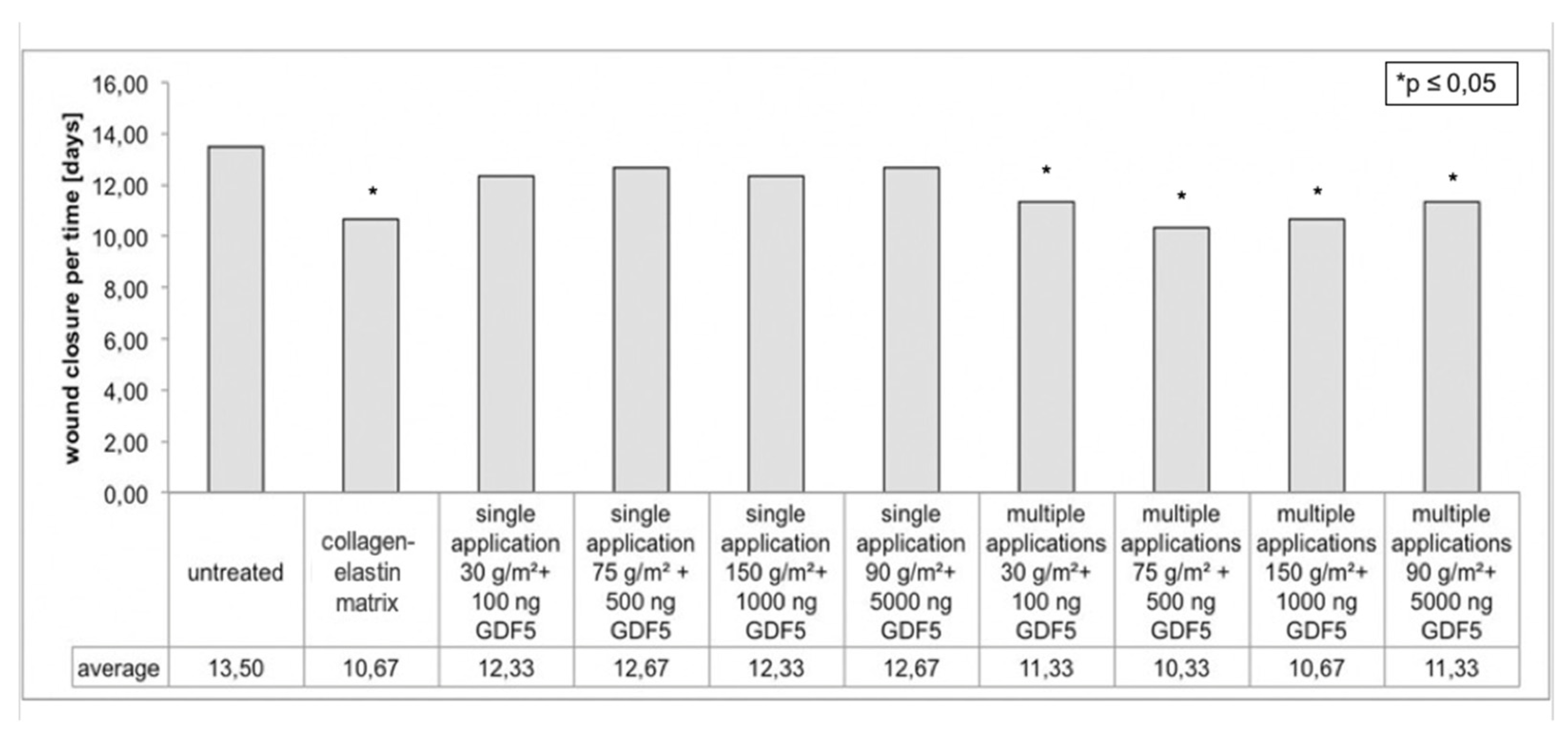
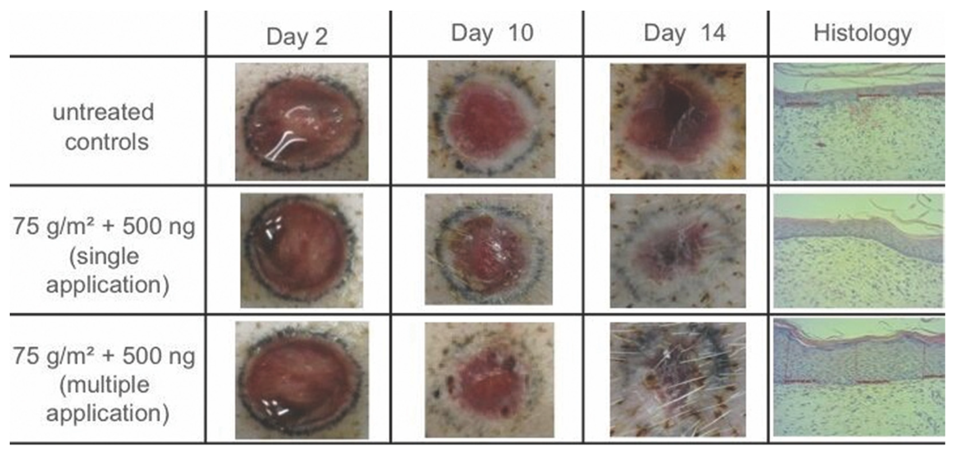
3.1. Planimetric Evaluation

Removal of both the skin substitutes and the epidermal coverage was completed for all wounds on the designated days (see Materials and Methods) to investigate wound closure. For the control wounds, complete epithelialization and closure of the wound were observed after 13.50 days (±1.19 days) on average postoperatively. For the collagen–elastin matrix-treated wounds, these occurred after 10.67 days (±0.94 days, p = 0.0002). In comparison, wounds treated with the collagen–gelatin scaffold containing rhGDF-5 were completely closed after 12.50 days (p = 0.0271) on average for the single-application groups and after 10.92 days (p < 0.0001) for the multiple-application groups. More specifically, the time required for complete wound closure, for the single-application dressings, was, on average, 12.33 days (±1.37 days, p = 0.0987) for the 30 g/m² scaffold with 100 ng rhGDF-5 condition, 12.67 days (±0.94 days, p = 0.1775) for the 75 g/m² scaffold with 500 ng rhGDF-5 condition, 12.33 days (±1.37 days, p = 0.0987) for the 150 g/m² scaffold with 1000 ng rhGDF-5 condition, and 12.67 days (±0.94 days, p = 0.1775) for the 90 g/m² scaffold with 5000 ng rhGDF-5 condition (Figure 1). For the groups with multiple applications of the dressing, the wounds were closed after 11.33 days (±0.94 days, p = 0.0015) for the 30 g/m² scaffold with 100 ng rhGDF-5, 10.33 days (±0.75 days, p = 0.0002) for the 75 g/m² scaffold with 500 ng rhGDF-5, 10.67 days (±0.94 days, p = 0.0002) for the 150 g/m² scaffold with 1000 ng rhGDF-5, and 11.33 days (±0.94 days, p = 0.0021) for the 90 g/m² scaffold with 5000 ng rhGDF-5 (Figure 1). These results show that the required time for complete wound closure decreased when treated with multiple applications of the wound dressing. For the multiple-application treatment of 75 g/m² scaffold with 500 ng rhGDF-5, this was especially evident as it showed an acceleration of 3.17 days in wound healing compared to the untreated controls (Figure 2).

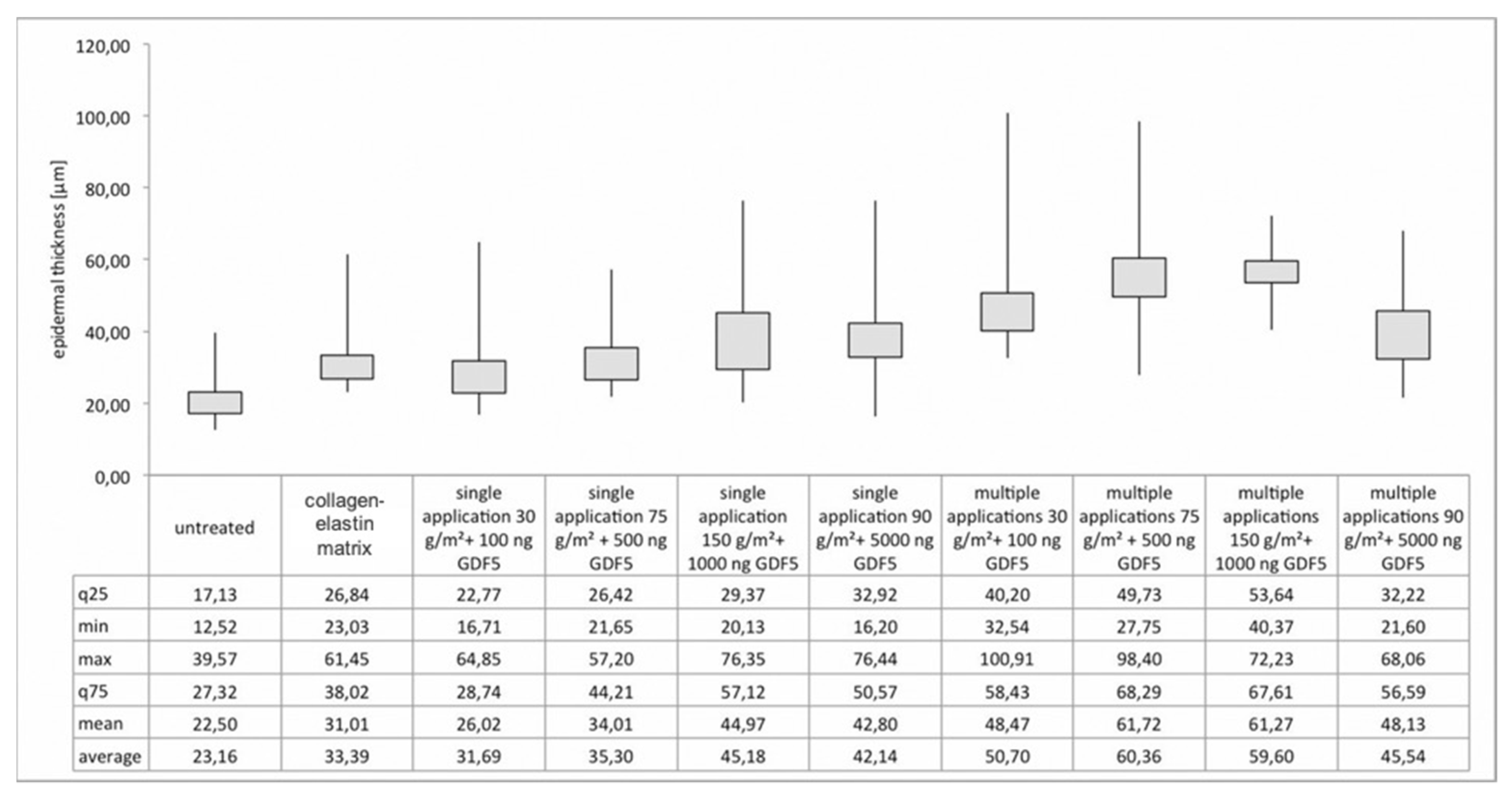
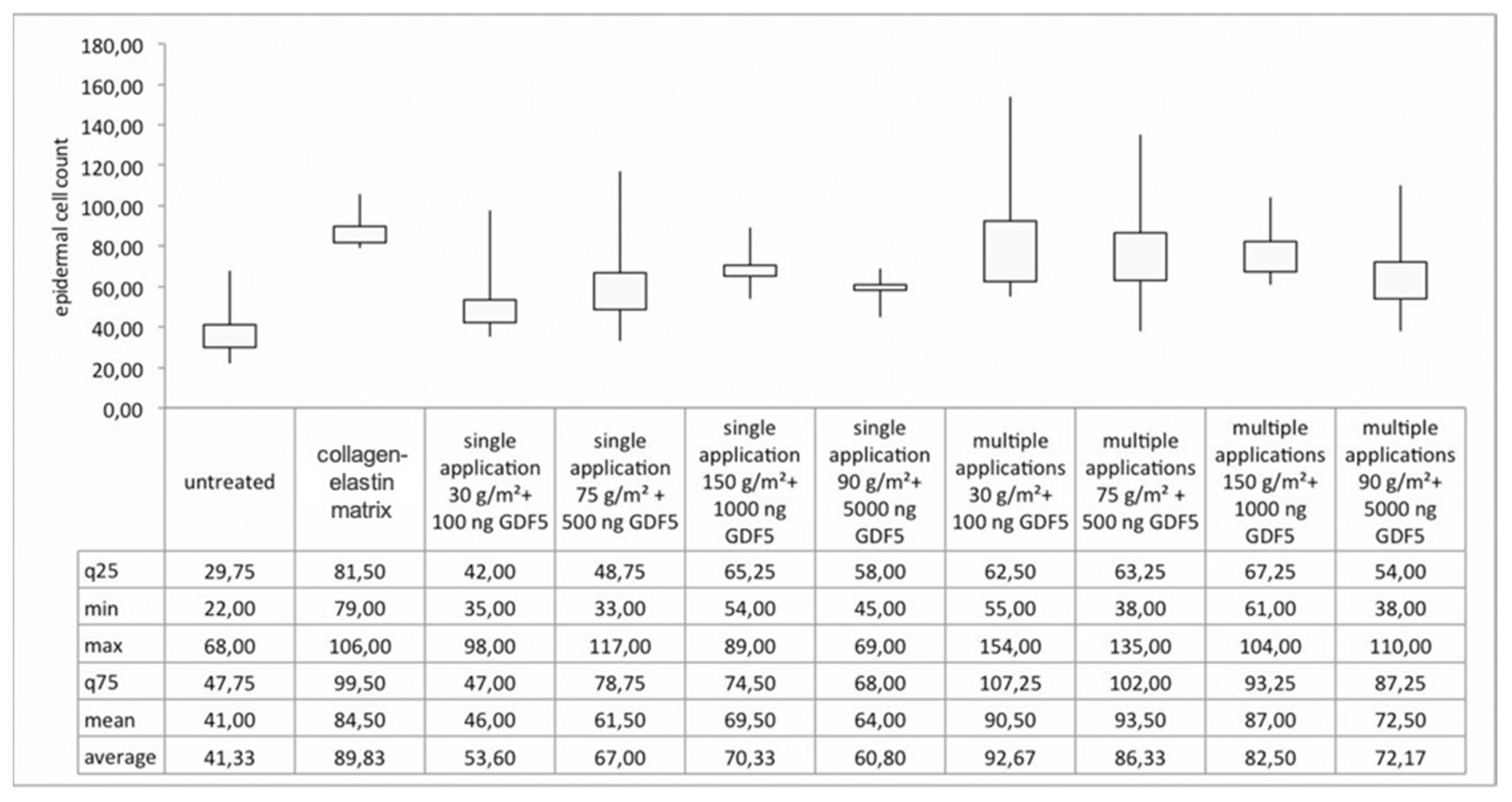
3.2. Histological Analysis

Histological analysis of the untreated control wounds showed a mean neoepidermal thickness of 22.50 µm (range of 12.52–39.57 µm). For the collagen–elastin matrix-treated group, a mean thickness of 31.01 µm (range of 23.03–61.45 µm, p = 0.0015) was found. Higher values were found for the mean neoepidermal thickness of wounds treated with the collagen–gelatin scaffold embedded with rhGDF-5. For dressings applied only once, these values were 26.02 µm (range of 16.71–64.85 µm, p = 0.0405) for the 30 g/m² scaffold with 100 ng rhGDF-5, 34.01 µm (range of 21.65–57.20 µm, p = 0.0008) for the 75 g/m² scaffold with 500 ng rhGDF-5, 44.97 µm (range of 20.13–76.35 µm, p < 0.0001) for the 150 g/m² scaffold with 1000 ng rhGDF-5, and 42.80 µm (range of 16.20–76.44 µm, p < 0.0001) for the 90 g/m² scaffold with 5000 ng rhGDF-5. For dressings applied multiple times, higher values of thickness were seen compared to the single-treated groups. These values were 48.47 µm (range of 32.54–100.91 µm, p < 0.0001) for the 30 g/m² scaffold with 100 ng rhGDF-5, 61.72 µm (range of 27.75–98.40 µm, p < 0.0001) for the 75 g/m² scaffold with 500 ng rhGDF-5, 61.27 µm (range of 40.37–72.23 µm, p < 0.0001) for the 150 g/m² scaffold with 1000 ng rhGDF-5, and 48.13 µm (range of 21.60–68.06 µm, p < 0.0001) for the 90 g/m² scaffold with 5000 ng rhGDF-5 (Figure 3).
Next, the mean epidermal cell density was determined from the neoepidermis. For untreated controls, the cell density amounted to 41.00 cells (range of 22.00–68.00 cells) (Figure 4). This value was higher for the collagen–elastin matrix-treated groups compared to the control ones and amounted to 84.50 cells (range of 79.00–106.00 cells, p = 0.0002). For the composite biomaterial groups with dressings applied only once to the skin defects, higher values of cell density were found compared to the control group, but lower than those of the collagen–elastin matrix groups. Statistically significant results were obtained, with 69.50 cells (range of 54.00–89.00 cells, p = 0.0132) for the 150 g/m² scaffold with 1000 ng rhGDF-5, and 64.00 cells (range of 45.00–69.00 cells, p = 0.0229) for the 90 g/m² scaffold with 5000 ng rhGDF-5 (Figure 4). However, for the groups with dressings applied multiple times to the skin defects, a higher cell density was seen compared to all other conditions. These values were 90.50 cells (range of 55.00–154.00 cells, p = 0.0118) for the 30 g/m² scaffold with 100 ng rhGDF-5, 93.50 cells (range of 38.00–135.00 cells, p = 0.0174) for the 75 g/m² scaffold with 500 ng rhGDF-5, 87.00 cells (range of 61.00–104.00 cells, p = 0.0019) for the 150 g/m² scaffold with 1000 ng rhGDF-5, and 72.50 cells (range of 38.00–110.00 cells, p = 0.0374) for the 90 g/m² scaffold with 5000 ng rhGDF-5 (Figure 4).
In comparison to untreated wounds, all examined wound dressing conditions functioning as skin substitutes accelerated dermal wound healing by reducing the time required to close the wound completely, to thicken the neodermal tissue, and to increase the epidermal cell density. Based on the results of the multiple-application groups, the local application of higher rhGDF-5 concentrations (100, 500, and 1000 ng) resulted in better wound healing compared to lower concentrations, indicating an rhGDF-5 dose-dependent relationship with wound healing. However, 5000 ng of rhGDF-5 did not further improve wound healing. Overall, the most optimal wound healing results were achieved with multiple rhGDF-5 treatments. This particularly included the 75 g/m² scaffold with 500 ng rhGDF-5, as it showed the fastest wound healing, the highest value for epidermal thickness, and the highest value for the epidermal cell density. The results of the ELISA blood screening showed normal values prior, during, and at the end of the study for all minipigs. rhGDF-5 could not be detected, meaning a systemic effect of rhGDF-5 was not found.

4. Discussion

Numerous acute injuries and chronic diseases often result in complex soft tissue defects. Wound healing in these cases is an advanced biochemical process in which numerous processes separated by place and time are involved. Particularly extensive burns or chronic wounds, such as diabetic pressure ulcers, often require special care and adequate wound closure to reduce long-term complications such as infection or amputation. However, current treatments do not yet offer optimal treatment for these cases [17]. Therefore, for the reasons mentioned above, there is an urgent need to support recovery treatments [18,19].
A promising approach to develop supporting treatments is the use of recombinant growth factors. Growth factors enhance processes important for tissue regeneration, including those involved in wound healing. In this study, rhGDF-5, a highly effective human peptide hormone that can promote wound healing [8,10,11,12,13,14,20], was investigated. Interestingly, all clinically approved therapies involving growth factors utilize a carrier made from a certain biomaterial for the successful delivery and targeted application of growth factors [21]. Stability and structural design are important determinants for skin tissue engineering, when constructing new dermal substitutes. In previous in vitro and in vivo studies, we already examined the innovative collagen–gelatin scaffold as a scaffold, and the results demonstrated an accelerated and improved dermal wound repair when applied as a single treatment, but even better when applied as multiple treatments [16,22,23]. Moreover, the manufacturing process of the scaffold allows the integration of bioactive substances such as antibiotics and growth factors. Therefore, we developed a composite biomaterial with the scaffold and embedded rhGDF-5 in the carrier matrix.
Previous data already showed that the open structure of the scaffold as a three-dimensional matrix enables the fast migration of different cell types (e.g., fibroblasts and keratinocytes) into the scaffold [24]. Deep dermal wounds were treated with the scaffold and rhGDF-5 at different concentrations applied multiple times during the study, which significantly accelerated wound closure, increased the epidermal cell density, and produced a thicker epidermis compared to the treatment with the scaffold itself or untreated controls [25]. When positioned in the wound, the nonwoven fibers come into contact with the wound fluid. Because of fluid absorption, the gelatin begins to swell. Additionally, when enough volume is absorbed, the gelatin dissolves. During this process, rhGDF-5 also becomes hydrated and starts to move in the matrix by diffusion. Through these diffusion processes, it travels to the cells located in the damaged tissue area of the wound. The continuous water uptake from the wound fluid progressively reduces the viscosity of the gelatin and thus also increases the mobility of rhGDF-5, resulting in a faster diffusion. The final phase is reached when the gelatin is completely dissolved and absorbed in the wound.
To further investigate improvements in wound treatment with a biomaterial composite in detail, we investigated various wound treatment conditions (applied once and multiple times) with various concentrations of rhGDF-5 and compared it to wounds treated with the collagen–elastin matrix and untreated wounds. All examined skin substitute dressings accelerated dermal wound healing, and removal of all skin substitutes was completed on day 21. The most optimal wound healing was seen for the rhGDF-5 (100, 500, 1000, and 5000 ng rhGDF-5)-treated dermal wounds when applied multiple times. Compared to the control, wound healing was accelerated by 2.58 days, neoepidermal thickness improved by 32.40 µm, and the epidermal cell density improved by 44.88 cells. Additionally, based on the results from the various treatment conditions, the enhanced wound-healing effects of rhGDF-5 were found to be dose dependent. For example, the local application of 500 ng and 1000 ng rhGDF-5 improved wound healing more than 100 ng rhGDF-5. However, 5000 ng rhGDF-5 did not further improve wound healing, possibly after having reached saturation. rhGDF-5 was not found in the analyzed blood serum. This shows that the local application of rhGDF-5 did not lead to measurable systemic doses. To ensure comparability, the values obtained from the rhGDF-5-treated groups were compared to untreated controls and the commercially available collagen–elastin matrix, which has been successfully used previously in full-thickness skin defects and showed positive long-term results [26,27,28,29,30,31]. The collagen–elastin matrix is mainly used in combination with a simultaneous application of autologous split-thickness skin grafts for full-thickness skin defect cases [26,27,28,29,30,32,33,34,35]. In our study, the collagen–elastin matrix was not used in combination with skin graft transplantation to ensure comparability with our product, which does not use skin grafts. After comparing all the conditions, the most optimal results were seen for wounds treated multiple times with 500 ng rhGDF-5 embedded in the 75 g/m² collagen–gelatin scaffold. For future investigations, additional detailed histologic evaluations at different stages of wound healing, in order to obtain more detailed information regarding the skin regeneration process between the control and treated groups, could result in stronger statistical differences for those not statistically significant.

5. Conclusions

The collagen–gelatin scaffold delivered rhGDF-5 adequately and served as a proper template to guide restructuring cells. The local application of rhGDF-5 on full-thickness skin defects was found to accelerate and improve wound healing with a faster and dose-dependent regeneration rate. Therefore, after an optimal concentration and dose analysis, this innovative composite product represents a novel tool to develop effective wound dressings in the field of regenerative medicine.

Author Contributions

Conceptualization and methodology, A.R.-S., M.H., J.S.; software, J.-O.B.; formal analysis, M.H.; validation, investigation, and data curation, W.E., M.H., A.R.-S., S.R., J.S., J.-O.B.; resources, A.R.-S., J.S.; writing—original draft preparation, W.E., J.-O.B.; writing—review and editing, W.E.; visualization, J.S.; supervision and project administration, A.R.-S., A.D.; funding acquisition, W.E. All authors have read and agreed to the published version of the manuscript.

Funding

The first and corresponding author received financial support from the research promotion program TÜFF of the local university (application number 2459-0-0). We acknowledge support by the Open Access Publishing Fund of the University of Tuebingen in Germany.

Institutional Review Board Statement

This study was conducted in accordance with the Declaration of Helsinki. Animals were treated according to the German Law on the Protection of Animals, and the study was performed with permission from the local animal welfare committee (approval code AT 1/12) of the University of Tuebingen in Germany.

Acknowledgments

There are no further contributions that do not justify authorship but need acknowledgment and no acknowledgment of technical help.

Conflicts of Interest

The authors disclose the following commercial associations that might create a conflict of interest in connection with the submitted manuscript: This research was supported partly (custom-tailored production and delivery of products) by Freudenberg Group New Technologies SE & Co. KG (Weinheim, Germany). None of the authors have a financial interest in the company. The role of the funders was to develop the analyzed material to our requirements. The funders had no further role in the design of the study; in the collection, analyses, or interpretation of data; in the writing of the manuscript, or in the decision to publish the results.

References

  1. Grose, R.; Werner, S. Wound-healing studies in transgenic and knockout mice. Mol. Biotechnol. 2004, 28, 147–166. [Google Scholar] [CrossRef]
  2. Kiritsy, C.P.; Lynch, A.B.; Lynch, S.E. Role of growth factors in cutaneous wound healing: A review. Crit. Rev. Oral Biol. Med. 1993, 4, 729–760. [Google Scholar] [CrossRef] [PubMed] [Green Version]
  3. Koveker, G.B. Growth factors in clinical practice. Int. J. Clin. Pract. 2000, 54, 590–593. [Google Scholar]
  4. Lawrence, W.T.; Diegelmann, R.F. Growth factors in wound healing. Clin. Dermatol. 1994, 12, 157–169. [Google Scholar] [CrossRef]
  5. Coerper, S.; Koveker, G.; Gottwald, T.; Becker, H.D. Cutaneous wound healing: Clinical evaluation of locally active growth factors-an overview. Zentralbl. Chir. 1996, 121, 49–51. [Google Scholar] [PubMed]
  6. Scheid, A.; Meuli, M.; Gassmann, M.; Wenger, R.H. Genetically modified mouse models in studies on cutaneous wound healing. Exp. Physiol. 2000, 85, 687–704. [Google Scholar] [CrossRef] [PubMed]
  7. Werner, S.; Grose, R. Regulation of wound healing by growth factors and cytokines. Physiol. Rev. 2003, 83, 835–870. [Google Scholar] [CrossRef]
  8. Sena, K.; Sumner, D.R.; Virdi, A.S. Modulation of VEGF expression in rat bone marrow stromal cells by GDF-5. Connect. Tissue Res. 2007, 48, 324–331. [Google Scholar] [CrossRef] [PubMed]
  9. Plichta, J.K.; Radek, K.A. Sugar-coating wound repair: A review of FGF-10 and dermatan sulfate in wound healing and their potential application in burn wounds. J. Burn Care Res. 2012, 33, 299–310. [Google Scholar] [CrossRef] [Green Version]
  10. Zeng, Q.; Li, X.; Beck, G.; Balian, G.; Shen, F.H. Growth and differentiation factor-5 (GDF-5) stimulates osteogenic differentiation and increases vascular endothelial growth factor (VEGF) levels in fat-derived stromal cells in vitro. Bone 2007, 40, 374–381. [Google Scholar] [CrossRef] [PubMed]
  11. Dines, J.S.; Cross, M.B.; Dines, D.; Pantazopoulos, C.; Kim, H.J.; Razzano, P.; Grande, D. In vitro analysis of an rhGDF-5 suture coating process and the effects of rhGDF-5 on rat tendon fibroblasts. Growth Factors 2011, 29, 1–7. [Google Scholar] [CrossRef]
  12. Forslund, C.; Rueger, D.; Aspenberg, P. A comparative dose-response study of cartilage-derived morphogenetic protein (CDMP)-1, -2 and -3 for tendon healing in rats. J. Orthop. Res. 2003, 21, 617–621. [Google Scholar] [CrossRef]
  13. Hayashi, M.; Zhao, C.; An, K.N.; Amadio, P.C. The effects of growth and differentiation factor 5 on bone marrow stromal cell transplants in an in vitro tendon healing model. J. Hand Surg. Eur. Vol. 2011, 36, 271–279. [Google Scholar] [CrossRef] [PubMed] [Green Version]
  14. James, R.; Kumbar, S.G.; Laurencin, C.T.; Balian, G.; Chhabra, A.B. Tendon tissue engineering: Adipose-derived stem cell and GDF-5 mediated regeneration using electrospun matrix systems. Biomed. Mater. 2011, 6, 025011. [Google Scholar] [CrossRef] [PubMed] [Green Version]
  15. Fu, X.; Fang, L.; Li, H.; Li, X.; Cheng, B.; Sheng, Z. Adipose tissue extract enhances skin wound healing. Wound Repair Regen. 2007, 15, 540–548. [Google Scholar] [CrossRef] [PubMed]
  16. Schiefer, J.L.; Rath, R.; Held, M.; Petersen, W.; Werner, J.O.; Schaller, H.E.; Rahmanian-Schwarz, A. Frequent Application of the New Gelatin-Collagen Nonwoven Accelerates Wound Healing. Adv. Skin Wound Care 2016, 29, 73–78. [Google Scholar] [CrossRef] [PubMed]
  17. Papini, R. Management of burn injuries of various depths. BMJ 2004, 329, 158–160. [Google Scholar] [CrossRef] [Green Version]
  18. Glowacki, J.; Mizuno, S. Collagen scaffolds for tissue engineering. Biopolymers 2008, 89, 338–344. [Google Scholar] [CrossRef] [PubMed]
  19. Kemp, P.D. Tissue engineering and cell-populated collagen matrices. Methods Mol. Biol. 2009, 522, 363–370. [Google Scholar]
  20. Yamashita, H.; Shimizu, A.; Kato, M.; Nishitoh, H.; Ichijo, H.; Hanyu, A.; Morita, I.; Kimura, M.; Makishima, F.; Miyazono, K. Growth/differentiation factor-5 induces angiogenesis in vivo. Exp. Cell Res. 1997, 235, 218–226. [Google Scholar] [CrossRef]
  21. Koria, P. Delivery of growth factors for tissue regeneration and wound healing. BioDrugs 2012, 26, 163–175. [Google Scholar] [CrossRef]
  22. Held, M.; Rahmanian-Schwarz, A.; Schiefer, J.; Rath, R.; Werner, J.O.; Rahmanian, S.; Schaller, H.E.; Petersen, W. A Novel Collagen-Gelatin Scaffold for the Treatment of Deep Dermal Wounds-An Evaluation in a Minipig Model. Dermatol. Surg. 2016, 42, 751–756. [Google Scholar] [CrossRef]
  23. Petersen, W.; Rahmanian-Schwarz, A.; Werner, J.O.; Schiefer, J.; Rothenberger, J.; Hubner, G.; Schaller, H.E.; Held, M. The use of collagen-based matrices in the treatment of full-thickness wounds. Burns 2016, 42, 1257–1264. [Google Scholar] [CrossRef] [PubMed]
  24. Shores, J.T.; Gabriel, A.; Gupta, S. Skin substitutes and alternatives: A review. Adv. Skin Wound Care. 2007, 20, 493–508; quiz 509–510. [Google Scholar] [CrossRef] [PubMed]
  25. Schiefer, J.L.; Held, M.; Fuchs, P.C.; Demir, E.; Ploger, F.; Schaller, H.E.; Rahmanian-Schwarz, A. Growth Differentiation Factor 5 Accelerates Wound Closure and Improves Skin Quality During Repair of Full-Thickness Skin Defects. Adv. Skin Wound Care. 2017, 30, 223–229. [Google Scholar] [CrossRef]
  26. Paul, S.P.; Blaikley, S.; Chinthapalli, R. Clinical update: Febrile convulsion in childhood. Community Pract. 2012, 85, 36–38. [Google Scholar] [PubMed]
  27. Graves, R.C.; Oehler, K.; Tingle, L.E. Febrile seizures: Risks, evaluation, and prognosis. Am. Fam. Phys. 2012, 85, 149–153. [Google Scholar]
  28. Auvin, S.; Desnous, B.; Bellavoine, V.; Gressens, P.; Boespflug-Tanguy, O. Febrile seizure: Underlying mechanisms, consequences and management. Arch Pediatr. 2010, 17, 686–687. [Google Scholar] [CrossRef]
  29. Fetveit, A. Assessment of febrile seizures in children. Eur. J. Pediatr. 2008, 167, 17–27. [Google Scholar] [CrossRef]
  30. Millar, J.S. Evaluation and treatment of the child with febrile seizure. Am. Fam. Phys. 2006, 73, 1761–1764. [Google Scholar]
  31. Kolokythas, P.; Aust, M.C.; Vogt, P.M.; Paulsen, F. Dermal subsitute with the collagen-elastin matrix Matriderm in burn injuries: A comprehensive review. Handchir. Mikrochir. Plast Chir. 2008, 40, 367–371. [Google Scholar] [CrossRef]
  32. Harvey, E.J.; Berger, R.A.; Osterman, A.L.; Fernandez, D.L.; Weiss, A.P. Bone-tissue-bone repairs for scapholunate dissociation. J. Hand Surg. Am. 2007, 32, 256–264. [Google Scholar] [CrossRef]
  33. Muller, M.; Reik, M.; Sauerbier, M.; Germann, G. A new bone-ligament-bone autograft from the plantar plates of the toes and its potential use in scapholunate reconstruction: An anatomical study. Ann. Plast. Surg. 2008, 61, 463–467. [Google Scholar] [CrossRef]
  34. Choi, J.Y.; Kim, S.H.; Oh, G.J.; Roh, S.G.; Lee, N.H.; Yang, K.M. Management of defects on lower extremities with the use of matriderm and skin graft. Arch. Plast. Surg. 2014, 41, 337–343. [Google Scholar] [CrossRef] [Green Version]
  35. Min, J.H.; Yun, I.S.; Lew, D.H.; Roh, T.S.; Lee, W.J. The use of matriderm and autologous skin graft in the treatment of full thickness skin defects. Arch. Plast. Surg. 2014, 41, 330–336. [Google Scholar] [CrossRef] [Green Version]
Figure 1. Planimetric analysis. The time required for complete wound closure of the untreated control group (n = 6), the collagen–elastin matrix-treated group (n = 6), and the groups treated once or multiple times with the novel composite biomaterial (n = 6 for each condition).
Applsci 12 00867 g001
Figure 2. Photographic and histological documentation. A macroscopic overview of epithelialization during wound healing in a representative untreated control, a wound treated once with the 75 g/m² collagen–gelatin scaffold and 500 ng rhGDF-5, and wounds treated multiple times with the 75 g/m² collagen–gelatin scaffold and 500 ng rhGDF-5 on days 2, 10, and 14. On day 21, microscopic images were taken from tissue stained with H&E.
Applsci 12 00867 g002
Figure 3. Histological analysis of the epidermal thickness. The epidermal thickness was histologically evaluated in micrometers by repetitive measurements from the stratum basale to the stratum corneum in three different sections per sample, with an interval of 100 µm.
Applsci 12 00867 g003
Figure 4. Histological analysis of the epidermal cell density. The epidermal cell density was histologically evaluated within a section of 100 µm width from the former sore center of the epidermis. All images per sample were taken from three different sections.
Applsci 12 00867 g004
Publisher’s Note: MDPI stays neutral with regard to jurisdictional claims in published maps and institutional affiliations.

