
An Overview of Shade Selection in Clinical Dentistry


Abstract
1. Introduction
Significance of Shade Selection in Dentistry
2. Literature Review
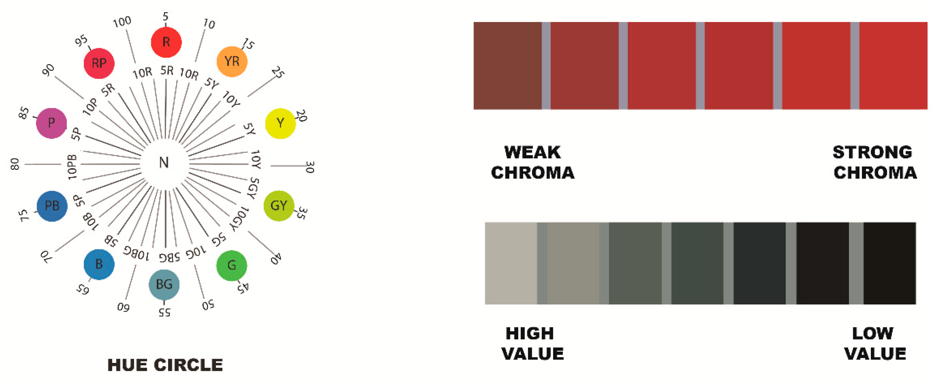
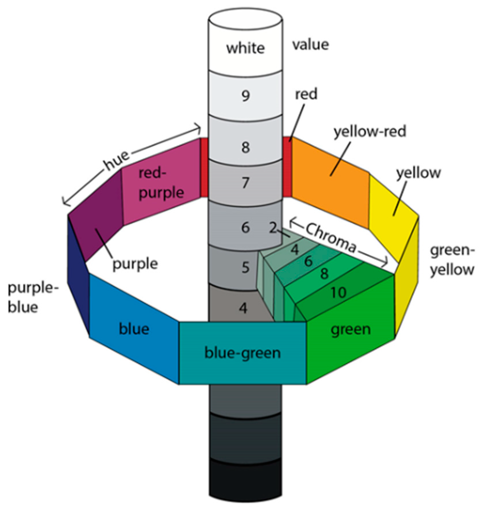
2.1. Three Color Dimensions
2.1.1. Hue
2.1.2. Value
2.1.3. Choma
2.2. Main Optical Properties of Teeth
2.2.1. Translucency
2.2.2. Fluorescence
2.2.3. Opalescence
2.2.4. Texture of Surface
2.2.5. Surface Gloss
2.2.6. Metamerism
2.3. Measurement of Colour
- Visual Technique;
- Instrumental Technique.
2.3.1. Visual Technique
Shade Guides
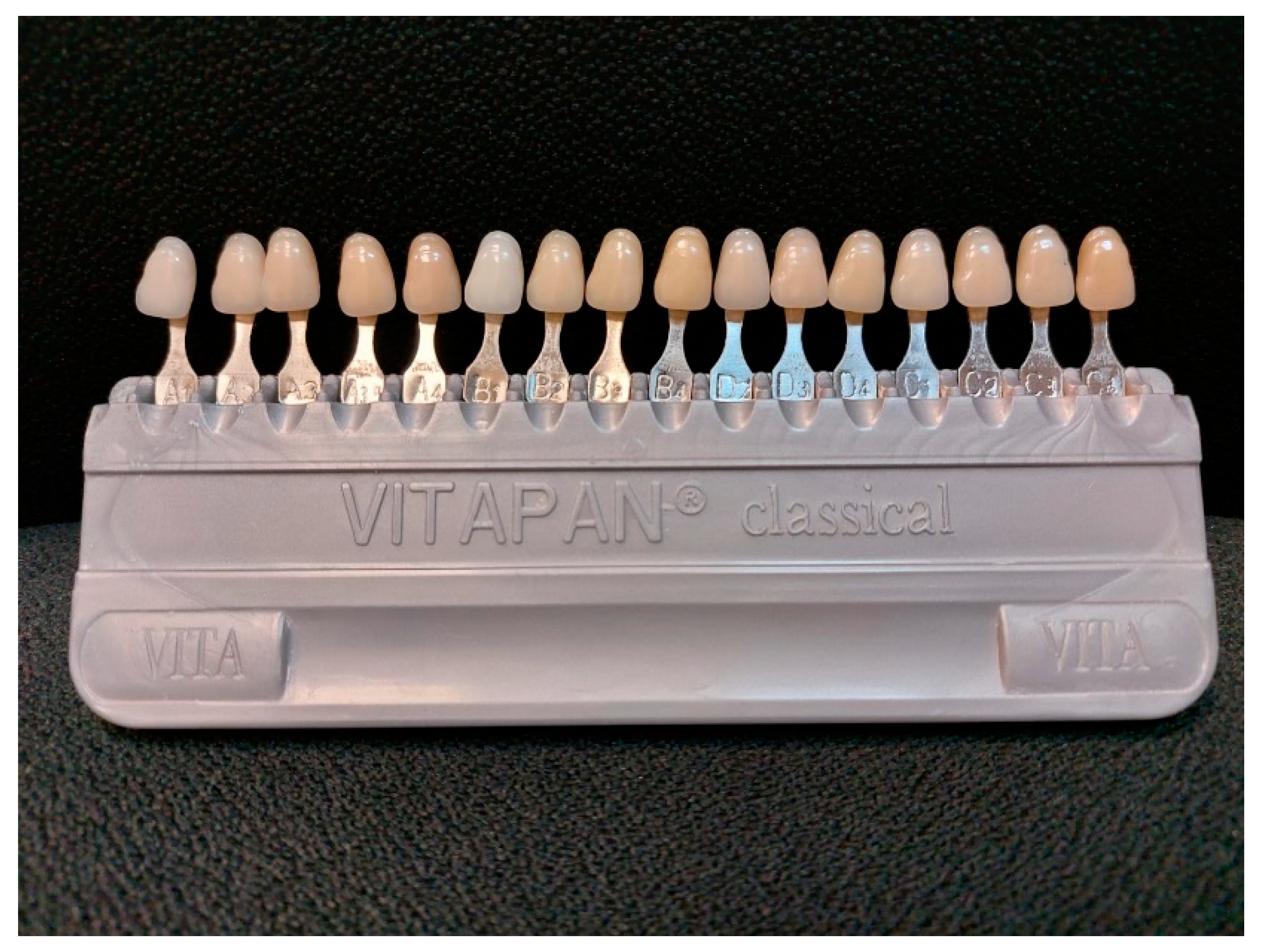
Vita Classical Shade Guide
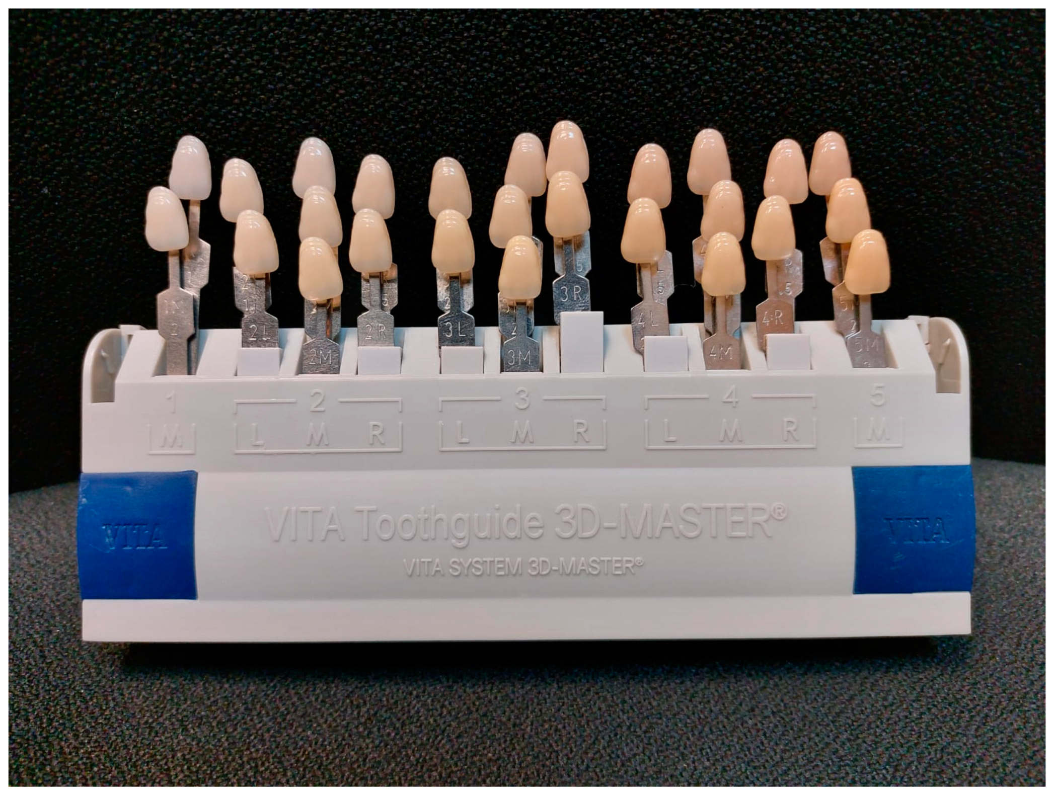
Vita 3D Master Shade Guide
- There is an extensive range of value.
- The red spectra are wide-ranging.
- The shade tabs are more correspondingly divided into the color space.
- The development in distribution of groups has improved and become more concise [32].
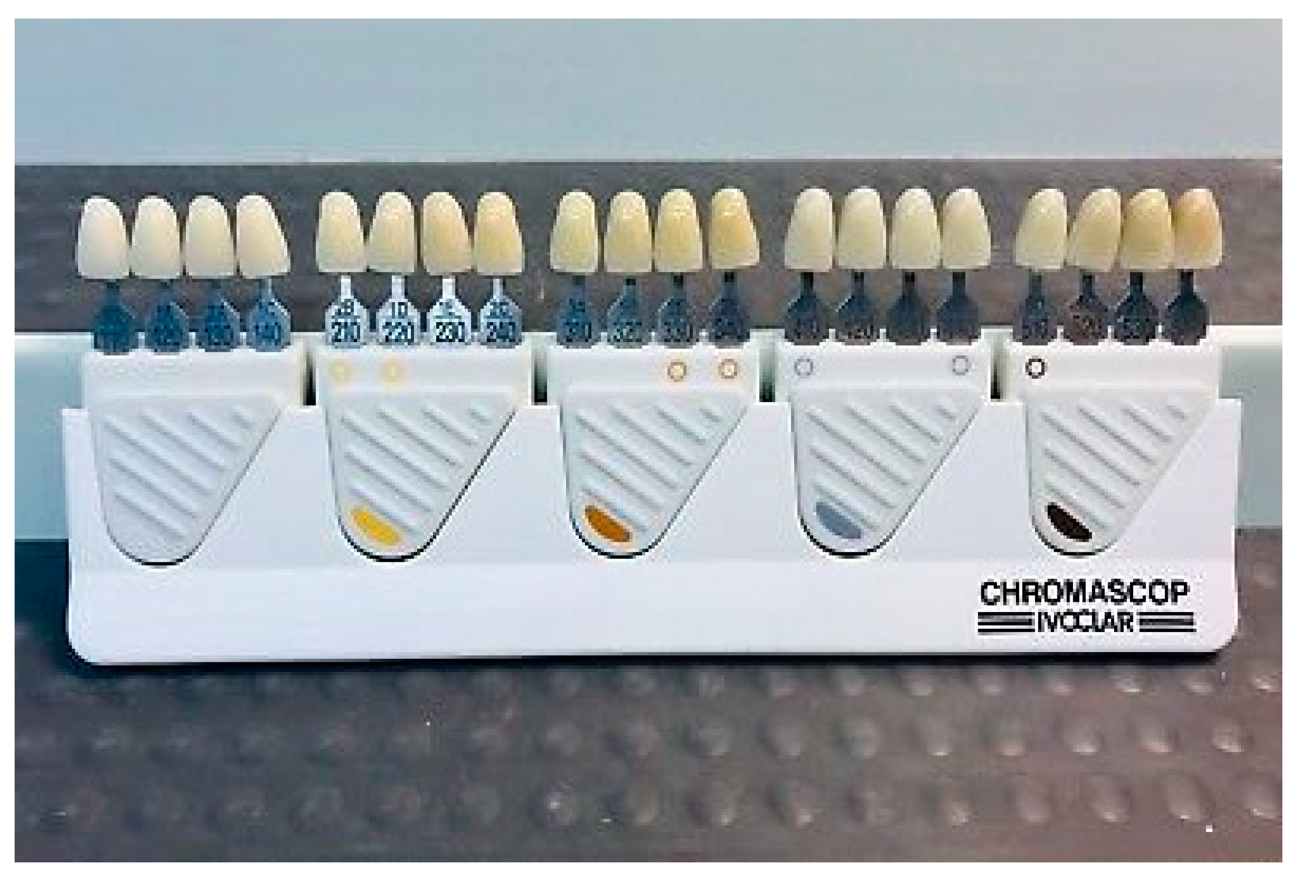
Chromascop Shade Guide
Merits of Shade Guides
- The most common method of shade selection is using a visual shade guide [6].
- It is economical and durable, i.e., does not require frequent replacement of the shade guide.
- It provides an efficient comparison with natural tooth color.
- Most frequently used by dentists, dental assistants and dental laboratory technicians to communicate the proper tooth color, brightness and translucency [9].
- Easily available.
Demerits of Shade Guides
- In shade guides, the colors may differ for each company.
- Porcelain that is used for restoration of teeth may be different with a shade guide.
- Guides are not able to direct the manufacturing of porcelain restoration.
- The shades of a tooth in a shade guide are not rationally arranged and do not cover the capacity of color space that is normally unoccupied in natural teeth.
- A normal shade tab is prepared from synthetic resin having greater density than a crown.
2.3.2. Instrumental Techniques in Tooth Shade Selection
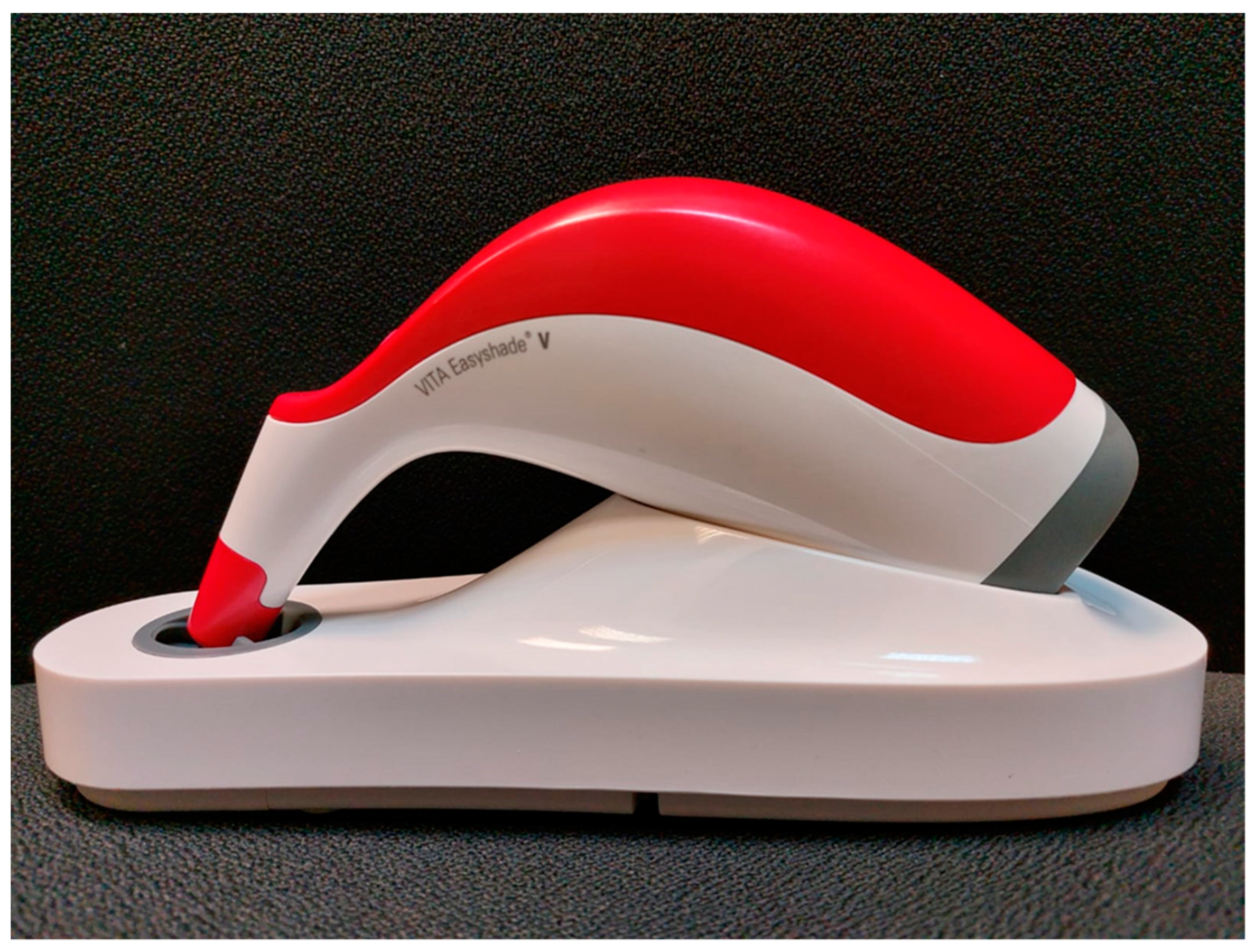
Spectrophotometers and Spectroradiometers
Colorimeters
Digital Cameras and Imaging Systems
Hybrid Devices
Restrictions of Digital Shade Guide
- The color measurement accurateness is affected due to the loss of power supply.
- For all systems, translucent recording is insufficient.
- Settlement of the probe or mouthpiece appears to be vital for the reappearance of the measurement.
- No digital shade guide is satisfactorily progressive to function in a mode of formulation.
- The research laboratory must be well equipped with the latest and most updated systems for the effective use of this process [6].
2.4. Recommended Protocols for Clinical Shade Selection
2.4.1. Working Site Lighting
2.4.2. Environment
2.4.3. Condition of Teeth
2.4.4. Distance of the Operator from the Tooth, Position of the Patient and Timing
2.5. Factors Affecting Shade Selection
2.5.1. Age
2.5.2. Gender
2.5.3. Experience
2.5.4. Eye Color
2.5.5. Color Vision Problem/Color Blindness
2.5.6. Fatigue
2.5.7. Binocular Difference
2.5.8. Environmental Influences
3. Conclusions
Author Contributions
Funding
Conflicts of Interest
References
- Pascual Moscardó, A.; Camps Alemany, I. Chromatic appreciation in the clinic and the laboratory. Med. Oral Patol. Oral Cir. Bucal. 2006, 11, E363–E368. [Google Scholar] [PubMed]
- Haralur, S.B.; Dibas, A.M.; Almelhi, N.A.; Al-Qahtani, D.A. The Tooth and Skin Colour Inter-relationship across the Different Ethnic Groups. Int. J. Dent. 2014, 2014, 146028. [Google Scholar] [CrossRef] [PubMed]
- Ten Bosch, J.J.; Coops, J.C. Tooth color and reflectance as related to light scattering and enamel hardness. J. Dent. Res. 1995, 74, 374–380. [Google Scholar] [CrossRef]
- Ciucchi, P.; Kiliaridis, S. Incisor inclination and perceived tooth colour changes. Eur. J. Orthod. 2017, 39, 554–559. [Google Scholar] [CrossRef]
- Dietschi, D.; Fahl, N., Jr. Shading concepts and layering techniques to master direct anterior composite restorations: An update. Br. Dent. J. 2016, 221, 765–771. [Google Scholar] [CrossRef]
- Borse, S.; Chaware, S.H. Tooth shade analysis and selection in prosthodontics: A systematic review and meta-analysis. J. Indian Prosthodont. Soc. 2020, 20, 131–140. [Google Scholar]
- Özat, P.B.; Tuncel, İ.; Eroğlu, E. Repeatability and reliability of human eye in visual shade selection. J. Oral Rehabil. 2013, 40, 958–964. [Google Scholar] [CrossRef]
- Terry, D.A.; Geller, W.; Tric, O.; Anderson, M.J.; Tourville, M.; Kobashigawa, A. Anatomical form defines color: Function, form, and aesthetics. Pract. Proced. Aesthetic Dent. 2002, 14, 59–68. [Google Scholar]
- Alnusayri, M.; Sghaireen, M.; Mathew, M.; Alzarea, B.; Bandela, V. Shade Selection in Esthetic Dentistry: A Review. Cureus 2022, 14, e23331. [Google Scholar]
- Fondriest, J. Shade matching in restorative dentistry: The science and strategies. Int. J. Periodontics Restor. Dent. 2003, 23, 467–479. [Google Scholar] [CrossRef]
- Rajan, N.; Rajan, A.; Singh, G.; Rani Krishna, S. Shade selection—Basic for Esthetic Dentistry: Literature Review. Int. J. Contemp. Res. Rev. 2020, 11, 20863–20868. [Google Scholar] [CrossRef]
- Lo Giudice, R.; Lipari, F.; Puleio, F.; Alibrandi, A.; Lo Giudice, F.; Tamà, C.; Sazonova, E.; Lo Giudice, G. Spectrophotometric evaluation of the variation of the enamel color using the infiltration resin treatment of the lesions of the white spots at a year of follow-up. Dent. J. 2020, 8, 35. [Google Scholar] [CrossRef] [PubMed]
- Puleio, F.; Fiorillo, L.; Gorassini, F.; Iandolo, A.; Meto, A.; D’Amico, C.; Cervino, G.; Pinizzotto, M.; Bruno, G.; Portelli, M.; et al. Systematic Review on White Spot Lesions Treatments. Eur. J. Dent. 2022, 16, 41–48. [Google Scholar] [CrossRef]
- Tashkandi, E. Consistency in color parameters of a commonly used shade guide. Saudi Dent. J. 2010, 22, 7–11. [Google Scholar] [CrossRef] [PubMed]
- Baltzer, A.; Jinoian, V.K. Determination of tooth colors. Quintessenz Zahntech. 2004, 30, 726–740. [Google Scholar]
- Gasparik, C.; Grecu, A.G.; Culic, B.; Badea, M.E.; Dudea, D. Shade-Matching Performance Using a New Light-Correcting Device. J. Esthet. Restor. Dent. 2015, 27, 285–292. [Google Scholar] [CrossRef] [PubMed]
- Öngül, D.; Şermet, B.; Balkaya, M.C. Visual and instrumental evaluation of color match ability of 2 shade guides on a ceramic system. J. Prosthet. Dent. 2012, 108, 9–14. [Google Scholar] [CrossRef]
- Hardan, L.; Bourgi, R.; Cuevas-Suárez, C.E.; Lukomska-Szymanska, M.; Monjarás-Ávila, A.J.; Zarow, M.; Jakubowicz, N.; Jorquera, G.; Ashi, T.; Mancino, D.; et al. Novel Trends in Dental Color Match Using Different Shade Selection Methods: A Systematic Review and Meta-Analysis. Materials 2022, 15, 468. [Google Scholar] [CrossRef]
- Takatsui, F.; Andrade, M.F.; Neisser, M.P.; Barros, L.A.B.; Loffredo, L.C.M. Comparison of Digital Images Obtained Photographically by Manual and Automatic Modes. Braz. Oral Res. 2012, 26, 578–583. [Google Scholar] [CrossRef]
- Sulaiman, A.O.; Adebayo, G.E. Most frequently selected shade for advance restoration delivered in a tertiary hospital facility in south western Nigeria. Ann. Ib. Postgrad. Med. 2019, 17, 157–161. [Google Scholar]
- Clary, J.A.; Ontiveros, J.C.; Cron, S.G.; Paravina, R.D. Influence of light source, polarization, education, and training on shade matching quality. J. Prosthet. Dent. 2016, 116, 91–97. [Google Scholar] [CrossRef] [PubMed]
- Aswini, K.K.; Ramanarayanan, V. The effect of gender and clinical experience on shade perception. J. Esthet. Restor. Dent. 2019, 31, 608–612. [Google Scholar] [CrossRef] [PubMed]
- Ristic, I.; Stankovic, S.; Paravina, R.D. Influence of Color Education and Training on Shade Matching Skills. J. Esthet. Restor. Dent. 2016, 28, 287–294. [Google Scholar] [CrossRef] [PubMed]
- Kröger, E.; Matz, S.; Dekiff, M.; Tran, B.L.; Figgener, L.; Dirksen, D. In vitro comparison of instrumental and visual tooth shade determination under different illuminants. J. Prosthet. Dent. 2015, 114, 848–855. [Google Scholar] [CrossRef] [PubMed]
- Clark, E.B. Tooth color selection. J. Am. Dent. Assoc. 2019, 20, 1065–1073. [Google Scholar]
- Pustina-Krasniqi, T.; Shala, K.; Staka, G.; Bicaj, T.; Ahmedi, E.; Dula, L. Lightness, chroma, and hue distributions in natural teeth measured by a spectrophotometer. Eur. J. Dent. 2017, 11, 36–40. [Google Scholar] [CrossRef]
- Mehta, S.; Nandeeshwar, D.B. A spectrophotometric analysis of extraoral aging conditions on the color stability of maxillofacial silicone. J. Indian Prosthodont. Soc. 2017, 17, 355–360. [Google Scholar] [CrossRef]
- Hyun, H.-K.; Kim, S.; Lee, C.; Shin, T.J.; Kim, Y.-J. Colorimetric distribution of human attached gingiva and alveolar mucosa. J. Prosthet. Dent. 2017, 117, 294–302. [Google Scholar] [CrossRef]
- Hammad, I.A. Intrarater repeatability of shade selections with two shade guides. J. Prosthet. Dent. 2003, 89, 50–53. [Google Scholar] [CrossRef]
- Boyce, R.A. Prosthodontic Principles in Dental Implantology. Dent. Clin. N. Am. 2021, 65, 135–165. [Google Scholar] [CrossRef]
- Paravina, R.D. Performance assessment of dental shade guides. J. Dent. 2009, 37 (Suppl. 1), e15–e20. [Google Scholar] [CrossRef] [PubMed]
- Udiljak, Z.; Illes, D.; Knezovic Zlataric, D.; Celic, R. Effect of clinical experience on the shade matching accuracy in different dental occupational groups. Acta Stomatol. Croat. 2018, 52, 132–139. [Google Scholar] [CrossRef] [PubMed]
- Brewer, J.D.; Wee, A.; Seghi, R. Advances in color matching. Dent. Clin. N. Am. 2004, 48, 341–358. [Google Scholar] [CrossRef] [PubMed]
- Basavanna, R.; Gohil, C.; Shivanna, V. Shade selection. Int. J. Oral Health Sci. 2013, 3, 26–31. [Google Scholar] [CrossRef]
- Todorov, R.; Yordanov, B.; Peev, T.; Zlatev, S. Shade guides used in the dental practice. J. IMAB 2020, 26, 3168–3173. [Google Scholar] [CrossRef]
- Joiner, A. Tooth colour: A review of the literature. J. Dent. 2004, 32, 12. [Google Scholar] [CrossRef]
- Gokce, H.S.; Piskin, B.; Ceyhan, D.; Gokce, S.M.; Arisan, V. Shade matching performance of normal and color vision-deficient dental professionals with standard daylight and tungsten illuminants. J. Prosthet. Dent. 2010, 103, 139–147. [Google Scholar] [CrossRef]
- Preethi Suganya, S.; Manimaran, P.; Saisadan, D.; Dhinesh Kumar, C.; Abirami, D.; Monnica, V. Spectrophotometric Evaluation of Shade Selection with Digital and Visual Methods. J. Pharm. Bioallied Sci. 2020, 12 (Suppl. 1), S319–S323. [Google Scholar] [CrossRef]
- Tabatabaian, F.; Beyabanaki, E.; Alirezaei, P.; Epakchi, S. Visual and digital tooth shade selection methods, related effective factors and conditions, and their accuracy and precision: A literature review. J. Esthet. Restor. Dent. 2021, 33, 1084–1104. [Google Scholar] [CrossRef]
- Sirintawat, N.; Leelaratrungruang, T.; Poovarodom, P.; Kiattavorncharoen, S.; Amornsettachai, P. The Accuracy and Reliability of Tooth Shade Selection Using Different Instrumental Techniques: An In Vitro Study. Sensors 2021, 21, 7490. [Google Scholar] [CrossRef]
- Igiel, C.; Lehmann, K.M.; Ghinea, R.; Weyhrauch, M.; Hangx, Y.; Scheller, H.; Paravina, R.D. Reliability of visual and instrumental color matching. J. Esthet. Restor. Dent. 2017, 29, 303–308. [Google Scholar] [CrossRef] [PubMed]
- Posavec, I.; Prpić, V.; Zlatarić, D.K. Influence of Light Conditions and Light Sources on Clinical Measurement of Natural Teeth Color using VITA Easyshade Advance 4,0® Spectrophotometer. Pilot Study. Acta Stomatol. Croat. 2016, 50, 337–347. [Google Scholar] [CrossRef] [PubMed]
- Chu, S.J.; Trushkowsky, R.D.; Paravina, R.D. Dental color matching instruments and systems. Review of clinical and research aspects. J. Dent. 2010, 38 (Suppl. 2), e2–e16. [Google Scholar] [CrossRef] [PubMed]
- Miyajiwala, J.S.; Kheur, M.G.; Patankar, A.H.; Lakha, T.A. Comparison of photographic and conventional methods for tooth shade selection: A clinical evaluation. J. Indian Prosthodont. Soc. 2017, 17, 273–281. [Google Scholar]
- Knezović, D.; Zlatarić, D.; Illeš, I.Z.; Alajbeg, M.; Žagar. In Vivo and in Vitro Evaluations of Repeatability and Accuracy of VITA Easyshade® Advance 4.0 Dental Shade-Matching Device. Acta Stomatol. Croat. 2015, 49, 112–118. [Google Scholar] [CrossRef]
- Paul, S.; Peter, A.; Pietrobon, N.; Hämmerle, C.H. Visual and spectrophotometric shade analysis of human teeth. J. Dent. Res. 2002, 81, 578–582. [Google Scholar] [CrossRef]
- Ballard, E.; Metz, M.J.; Harris, B.T.; Metz, C.J.; Chou, J.C.; Morton, D.; Lin, W.-S. Satisfaction of Dental Students, Faculty, and Patients with Tooth Shade-Matching Using a Spectrophotometer. J. Dent. Educ. 2017, 81, 545–553. [Google Scholar] [CrossRef]
- Blaes, J. Today’s technology improves the shade-matching problems of yesterday. J. Indiana Dent. Assoc. 2002, 81, 17–19. [Google Scholar]
- Schropp, L. Shade matching assisted by digital photography and computer software. J. Prosthodont. 2009, 18, 235–241. [Google Scholar] [CrossRef]
- Jarad, F.D.; Russell, M.D.; Moss, B.W. The use of digital imaging for colour matching and communication in restorative dentistry. Br. Dent. J. 2005, 199, 43–49; discussion 33. [Google Scholar] [CrossRef]
- Śmielecka, M.; Dorocka-Bobkowska, B. Effects of different light sources on tooth shade selection. Dent. Med. Probl. 2020, 57, 61–66. [Google Scholar] [CrossRef] [PubMed]
- Ajaj, A.M. Basics of shade selection and importance of laboratory communication restorative dentistry. J. Dent. Health Oral Disord. Ther. 2015, 2, 215–217. [Google Scholar] [CrossRef]
- Redmond, T.; Zlatkova, M.B.; Garway-Heath, D.F.; Anderson, R.S. The effect of age on the area of complete spatial summation for chromatic and achromatic stimuli. Investig. Opthalmology Vis. Sci. 2010, 51, 6533–6539. [Google Scholar] [CrossRef] [PubMed][Green Version]
- Pecho, O.E.; Ghinea, R.; Perez, M.M.; Della Bona, A. Influence of Gender on Visual Shade Matching in Dentistry. J. Esthet. Restor. Dent. 2017, 29, E15–E23. [Google Scholar] [CrossRef] [PubMed]
- Török, J.; Mauks, L.; Márton, S.; Hegedűs, C. A fogszín-meghatározást befolyásoló egyes tényezők összehasonlító vizsgálata [Comparative analysis of some factors in tooth color matching]. Fogorv. Szle. 2014, 107, 79–86. [Google Scholar]
- Pustina-Krasniqi, T.; Xhajanka, E.; Ajeti, N.; Bicaj, T.; Dula, L.; Lila, Z. The relationship between tooth color, skin and eye color. Eur. Oral Res. 2018, 52, 45–49. [Google Scholar] [CrossRef]
- Suliman, A.; Al-Abdali, T.; Taslimi, M.; Abdo, M. Prevalence of Color Vision Deficiency among Dental Practitioners and its Effect on Shade Matching Ability. Open Dent. J. 2020, 14, 539–554. [Google Scholar] [CrossRef]
- Medeiros, J.A.; Pecho, O.E.; Pérez, M.M.; Carrillo-Pérez, F.; Herrera, L.J.; Della Bona, A. Influence of background color on color perception in dentistry. J. Dent. 2021, 108, 103640. [Google Scholar] [CrossRef]
- Alomari, M.; Chadwick, R. Factors influencing the shade matching performance of dentists and dental technicians when using two different shade guides. Br. Dent. J. 2011, 211, E23. [Google Scholar] [CrossRef]


Publisher’s Note: MDPI stays neutral with regard to jurisdictional claims in published maps and institutional affiliations. |
© 2022 by the authors. Licensee MDPI, Basel, Switzerland. This article is an open access article distributed under the terms and conditions of the Creative Commons Attribution (CC BY) license (https://creativecommons.org/licenses/by/4.0/).
Share and Cite
Jouhar, R.; Ahmed, M.A.; Khurshid, Z. An Overview of Shade Selection in Clinical Dentistry. Appl. Sci. 2022, 12, 6841. https://doi.org/10.3390/app12146841
Jouhar R, Ahmed MA, Khurshid Z. An Overview of Shade Selection in Clinical Dentistry. Applied Sciences. 2022; 12(14):6841. https://doi.org/10.3390/app12146841
Chicago/Turabian StyleJouhar, Rizwan, Muhammad Adeel Ahmed, and Zohaib Khurshid. 2022. "An Overview of Shade Selection in Clinical Dentistry" Applied Sciences 12, no. 14: 6841. https://doi.org/10.3390/app12146841
APA StyleJouhar, R., Ahmed, M. A., & Khurshid, Z. (2022). An Overview of Shade Selection in Clinical Dentistry. Applied Sciences, 12(14), 6841. https://doi.org/10.3390/app12146841