Article

The Rate of Correctly Planned Size of Digital Templating in Two Planes—A Comparative Study of a Short-Stem Total Hip Implant with Primary Metaphyseal Fixation and a Conventional Stem

1
Department of Orthopaedic Surgery at Diakovere Annastift, Hannover Medical School, 30625 Hannover, Germany
2
Laboratory for Biomechanics and Biomaterials, Hannover Medical School, 30625 Hannover, Germany
3
Department of Orthopaedic Surgery, Göttingen Medical School, 37075 Göttingen, Germany
4
Go:h Gelenkchirurgie Orthopädie, 30159 Hannover, Germany
*
Author to whom correspondence should be addressed.
Appl. Sci. 2021, 11(9), 3965; https://doi.org/10.3390/app11093965
Submission received: 3 February 2021 / Revised: 25 March 2021 / Accepted: 23 April 2021 / Published: 27 April 2021

Abstract

:
(1) Background: Preoperative templating is mainly conducted on an anteroposterior pelvic overview X-ray. For short stem hip arthroplasty, the choice of the optimal size is especially crucial to avoid complications. Thus, the study aimed to determine if there is an increased rate of correctly planned sizes using two radiological planes. (2) Methods: 50 patients with a conventional stem and 100 with a short stem total hip arthroplasty were analyzed. Without knowing the implanted size, three independent orthopedic surgeons performed digital templating: once using the anteroposterior pelvic overview only and once using the lateral view in addition. (3) Results: The rate of correctly planned sizes (+/−1 size compared to the inserted size) of templating with one plane was 86.3% ± 9.5% in short stem hip arthroplasty and 88.4% ± 6.0% in conventional stem arthroplasty. By adding the lateral view, the rate of correctly planned sizes was 89.9% ± 12.0% for the short stem hip arthroplasty group and 89.4% ± 9.8% for the conventional group (p > 0.1). (4) A potential positive effect of preoperative templating using an additional lateral X-ray view for short stem implants may be suggested based on the results of this study, which did, however, not reach statistical significance.

1. Introduction

The implantation of the optimal stem size is important for the implantation of a total hip arthroplasty (THA) in order to reduce complications. Digital templating of a THA is a helpful tool for preoperative planning of surgery regarding implant type, implant size, reconstruction of the center of rotation, offset, and limb lengths [1]. This tool assists in order to receive a good outcome after a THA and to avoid complications due to anatomic variations. It helps to reduce the incidence of undersizing the femoral implant with the associated risk of increased micromotions, subsidence, and early aseptic loosening [2,3,4] and the incidence of oversizing associated with the risk of femoral fractures. Furthermore, it is helpful to avoid limb length discrepancies, instability, or excessive tension of the periarticular soft tissues by inaccurate reconstruction of the center of rotation of the hip [3,5,6,7,8,9,10].
Meanwhile, preoperative planning with plastic templates on radiographs or digital templating as a valid method to determine the correct size of the femoral stem and acetabular component belongs to the standard preoperative procedures prior to a THA [5,11,12]. Digital templating of a THA is usually performed on the anteroposterior (a.p.) pelvic overview X-ray [13]. Some software programs allow for digital templating also use the lateral view of the proximal femur for templating the stem. Although efforts have been made to further improve the accuracy of preoperative templating by using CT-based three-dimensional computerized models [14], the simple method using only the pelvic overview X-ray is still the standard procedure in clinical routine due to its high practicability on the one hand and the high radiation exposure when using CT scans on the other. The fixation of short stem THA (STHA) with predominantly metaphyseal anchorage is different from conventional THA (CTHA). While the conventional stem has a metaphyseal-diaphyseal anchorage with an intramedullary fixation within the proximal third of the femur [11], STHA with predominantly metaphyseal anchorage adapts its orientation to the femoral neck and thus to its ante torsion (Figure 1).
This study aimed to determine whether there is a difference in accuracy when templating THA stems using not only the a.p. view of the pelvis but also the lateral view of the proximal femur. Furthermore, we analyzed whether there is a difference in the rate of correctly planned sizes when templating CTHA stems in comparison to STHA implants with a predominantly metaphyseal anchorage.

2. Materials and Methods

One hundred fifty patients with a good radiological outcome after implantation of a THA were selected: 50 after implantation of a THA using a conventional femoral stem (CTHA group) and 100 after implantation of a THA using a short stem with primary metaphyseal fixation (STHA group). A good radiological outcome was judged on the postoperative X-rays by three senior surgeons according to the following criteria: under- or oversizing, early subsidence, and intra- or postoperative fractures.
Undersizing was defined as either a too low fit of the implant that could have been avoided by choosing a bigger size or by the presence of a visible distance between the cortical bone and the implant in a region of bone that is crucial for the anchorage of the implant (calcar region and lateral cortex for CTHA and cortical ring of the resected femoral neck basis as well as lateral cortex for STHA). Oversizing was defined as a too high fit of the implant that could have been avoided by choosing a smaller size, indicated by the shoulder of the implant exceeding the highest point of the greater trochanter for CTHA or the shoulder of the implant exceeding the cortical ring of the femoral neck basis by more than 10 mm for STHA. Early subsidence after full load-bearing, which was allowed for all patients, was defined as an unambiguous (>2 mm) migration of the implant within the surrounding bone in a follow-up X-ray examination 3 months after surgery. The occurrence of intra- or postoperative fractures were retrieved from the surgery reports and patients’ charts.
For these patients, who were included consecutively and underwent THA between 2010 and 2011, preoperative and postoperative digital X-rays in pairs were selected from the database of the department of orthopedic surgery of a university hospital. All these patients underwent standardized X-ray examinations, including an a.p. pelvic overview and an lateral view (Lauenstein) of the proximal femur. These X-rays existed for the preoperative and postoperative state and with a follow-up of at least 3 months. All X-rays were taken with a standardized film-focus-distance of 100 cm. An external calibration marker of 25 mm diameter was positioned between the legs of each patient for the preoperative pelvic overview.
Digital templating was performed by three senior surgeons experienced in THA. When conducting digital templating, the examiners were only able to see the preoperative, but not the postoperative X-rays.
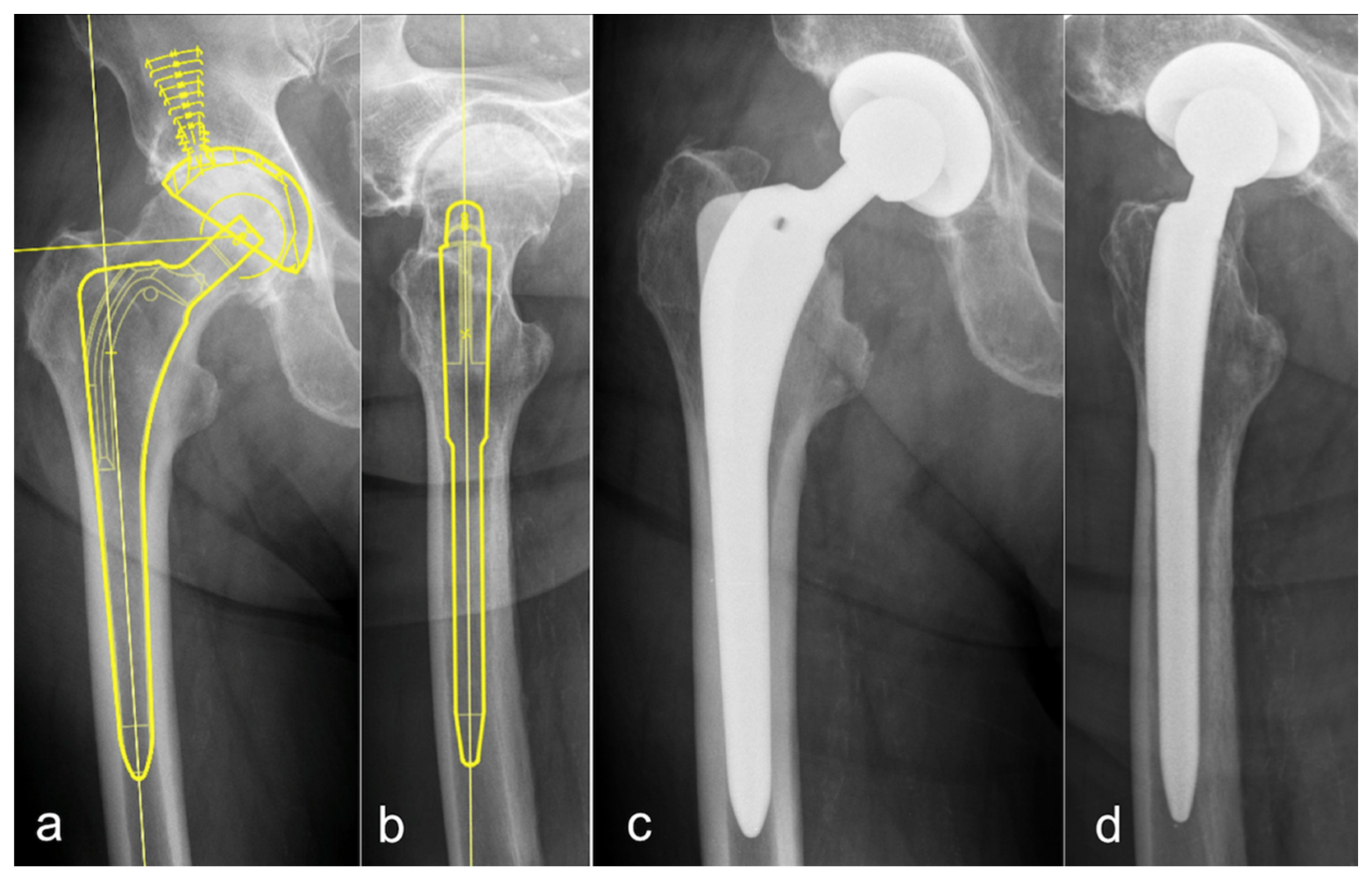
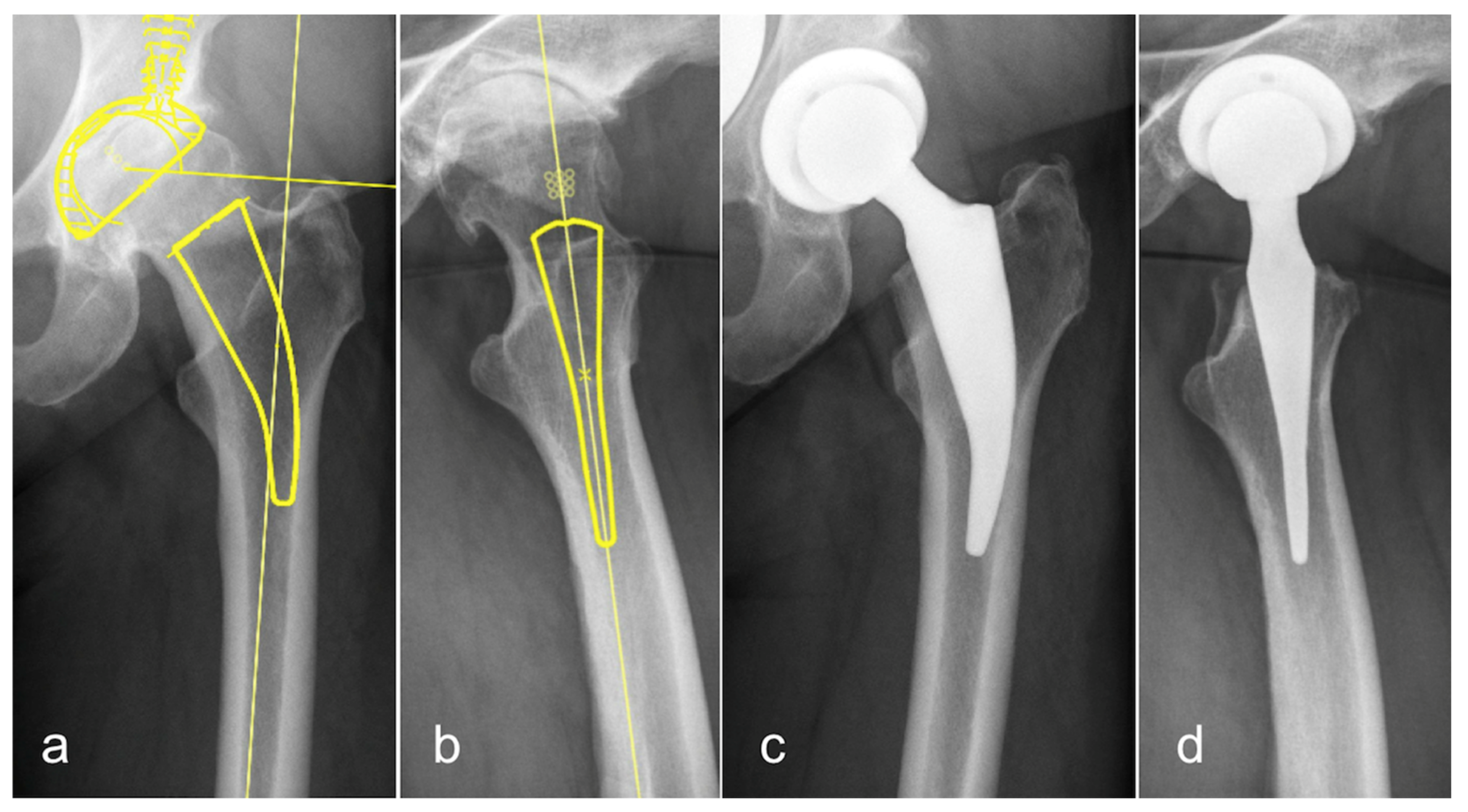
The software used for digital templating was OrthoView (OrthoView Orthopaedic Digital Planning Version 6.3, Meridian Technique Limited, Southampton, UK). For this software tool, templates for the conventional BICONTACT stem (CTHA) and the METHA short stem (STHA; both Aesculap B. Braun, Tuttlingen, Germany) were available (Figure 2 and Figure 3).
One hundred patients received a cementless METHA prosthesis with a caput-collum-diaphysis (CCD) angle of 130°, 135°, or 140°. All these stems are available in a size range of 0 to 7. The median stem size of the implanted METHA short stems was 3.
Fifty patients received a cementless conventional BICONTACT stem. These stems are monobloc-types with different offset variations: S for standard, H for High offset, and SD for dysplastic femurs. In our study, we templated and implanted only S and H types. BICONTACT stems are available in stem sizes 10–21. From the 50 truly implanted hip implants, the median stem size was 14.

Statistics

First, the results of all three raters were described with the cumulative rate of consistency between the templated and the implanted stem size. The difference was described as size derivation from the actual stem size. In addition, the planned stem sizes were presented in scatterplots. The intra-class correlation (ICC) 3,1 for consistency and 3,k for conformity for all raters was measured and quantified using Fleiss Kappa and Kendall’s coefficient of concordance. For the correlation between the templated stem size and the actual implanted stem size, a Spearman’s rank correlation coefficient was determined. Analysis was conducted in R (Version 4.0.3) [15], and figures were produced using the package ggplot2 [16].

3. Results

The study population consisted of 66 males and 84 females. The average age was 60 +/−12.8 years, ranging from 21 to 86 years. The CTHA group included 25 males and 25 females, the STHA group 41 males and 59 females. The CTHA group was significantly older than the STHA group (71 ± 14.3 years vs. 55 ± 11.5 years).

3.1. Conventional Stem Total Hip Arthroplasty (CTHA)—Conventional Templating in One Plane on the a.p. Pelvic Overview

The rate of correctly planned size of preoperative templating in one plane (a.p. view) predicted the exact CTHA stem size in 44.4% ± 6.8% cases. With a difference of +/−1 stem size, the rate of correctly planned size 88.4% ± 6.0% was detected. In 93.8% ± 5.4% of cases, the stem size was predicted within two stem sizes. In the remaining 6.2% of measurements, the templated stem size showed a deviation of +/−3 stem sizes (Table 1). The preoperative templating showed an oversizing in 27.5% and an undersizing in 28.1% of the cases.

3.2. CTHA—Templating in Two Planes—a.p. and Lateral View

Preoperative templating in two planes (a.p. view and lateral “Lauenstein” view) of CTHA showed an exact match of the implanted stem size in 40.0% ± 20.8% of cases. In 89.4% ± 9.8% an implanted size within a deviation of +/−1 stem size was found. In 99.3% ± 1.3% of our cases, we could predict the implanted stem size within a range of +/−2 sizes. The remaining 1.7% showed a deviation of +/−3 stem sizes (Table 1). In 25.5% an oversizing of the templated size was observed. In 34.5%, the templated stem size was smaller than the implanted stem size.

3.3. Short Stem Total Hip Arthroplasty (STHA)—Conventional Templating in One Plane on the a.p. Pelvic Overview

By using templating based on one radiological plane only, the rate of measurements matching the exact stem size was 36.2% ± 16.9%. By a tolerance of +/−1 stem size, the predictability raised to 86.3% ± 9.5%. A deviation of the implanted and templated stem size of up to +/−2 sizes was observed in 99.7% ± 0.6% of the cases (Table 1)—the remaining 0.3% ± 0.6% of the measurements showed a deviation of ± 3 stem sizes. An oversizing of the templated implant size was found in 31.9%, and an undersizing also in 31.9% of the cases.

3.4. STHA—Templating in Two Planes—a.p. and Lateral View

Adding a second plane (lateral view according to “Lauenstein”), the rate of planning the exact size reached 50.0% ± 27.5%. The predictability was 89.9% ± 12.0% with a tolerance of variation of +/−1 stem size. A variation of the implanted stem size lay within a range of +/−2 sizes in 98.2% ± 3.2% of the cases (Table 1). A deviation of ± 3 stem sizes was observed in the remaining measurement except for one outlier in one patient measured by one rater and showing a deviation of 4 stem sizes. In 31.8%, oversizing and 18.3%, undersizing of the templated size compared to the implanted size was measured.
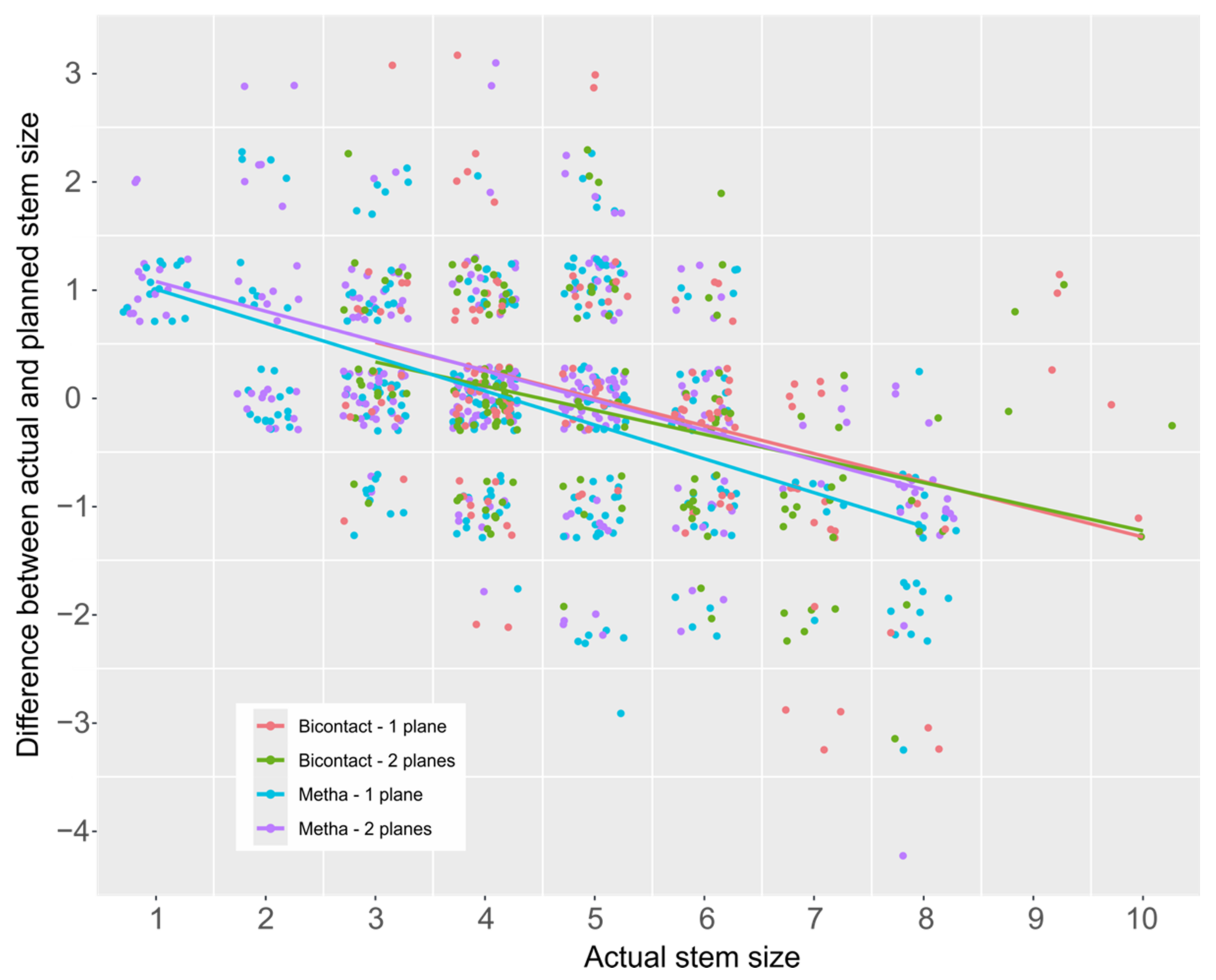
Overall, the difference between the implanted stem size and the templated size showed a lesser deviation for the STHA planned on two planes compared to one plane, whereas the deviation was rather constant for the CTHA. However, this potential effect did not reach statistical significance. In addition, there was a trend that bigger implant sizes were planned too small, while small stem sizes were planned too big (Figure 4).
An additional plane for preoperative stem templating led to an increase in the ICC for the stem size as well as for the difference between the implanted and templated stem size, except for the STHA difference values, where a decrease was observed (Table 2 and Table 3).
The conformity of the three raters was slightly fair according to Landis classification [17] and improved only for the STHA, not for the CTHA (Table 4).
The consistency of the raters showed a significant correlation for STHA templating in two planes. However, the correlation was for both stem types, and planning conditions were limited (Table 5).
The templated and the implanted stem size showed a significant correlation for both stem types. The addition of an lateral planning plane led to a stronger correlation for CTHA and STHA (Table 6).

4. Discussion

Total hip arthroplasty using short stem implants has grown up to a popular technique beside the successful conventional procedure. Short stem devices, such as the METHA stem, show promising short-term results [18,19]. Prior to implantation of the stem, digital preoperative templating is a common and routinely used tool that has not yet been determined with regards to its benefit and exactitude [11,13]. So far, nothing is known about the benefit in terms of the rate of correctly planned sizes when using additional X-rays in a second radiological plane.
This study confirms on the one hand that there is no significant difference between templating STHA in comparison to CTHA, as former studies have shown [11,13,20,21]. On the other hand, the current study indicates that the exactitude can possibly be increased by using a second radiological plane for templating. Although this effect did not reach statistical significance for neither the CTHA nor the STHA group, the tendency of the effect could be observed especially for the short stem implant.
Matching exactly the same stem size as predicted by preoperative templating occurred in 44.4% ± 6.8% with CTHA when using one plane radiographs. Other studies on conventional implants have shown an exact rate of correctly planned sizes between 31% and 58% of cases [6,11,13,22,23]. By tolerating a variation of +/− one size between templating and implantation, the data of this study showed a predictability of 88.4% ± 6.0%, again in good conformity to other studies that have reported a predictability of 71.0%–94.0% under these circumstances.
By the use of a second radiological plane, the lateral view, according to Lauenstein, in addition to the a.p. pelvic overview, a comparable rate of exact matching between templated and implanted size of 40.0% ± 20.8% was determined. With an allowed measuring tolerance of +/− one stem size, a slightly increased accordance of 89.4% ± 9.8% was reached, although the difference was not significant. A variation of more than +/−2 stem sizes between templating and implanted size occurred in 0.7% of the cases when using two radiological planes compared to 6.2% when using only one plane (p = 0.18). This reduced range of dispersion indicates a higher accuracy when using two planes, although the difference was not significant.
Results analyzing the rate of correctly planned size of preoperative templating of short stem implants are less documented in the literature. There are only two studies dealing with this question: on the one hand, for the METHA short stem by Schmidutz et al. [11] and on the other hand, for the MAYO short stem (Zimmer, Warsaw, IN, USA) by Wedemeyer et al. [24]. Both of these short stem designs aim at a metaphyseal anchorage. Wedemeyer et al. reported an exact match without size tolerance in 38% of the cases. Schmidutz et al. found for the METHA short stem an exactitude of 49% of preoperative templating. In our study, we found similar results with up to 36.2% ± 16.9%. The MAYO device showed predictability of 95%, Schmidutz et al. reported 89% for the METHA stem, and our study reports a comparable matching rate of 86.3% ± 9.5% when tolerating a difference of one size.
In analogy to the CTHA group, the second plane (lateral view) led to a smaller range of dispersion, matching the exact size in 50% ± 27.5%, and in 89.9% ± 12% with a tolerance of +/−1 stem size.
When comparing the STHA and CTHA groups, adding the second radiological plane increased the probability of matching the exact size in the STHA group (50.0% vs. 36.2%), but not in the CTHA group (40.0% vs. 44.4%). When tolerating a difference of +/− one stem size, templating with two planes revealed comparable results between the groups that were not statistically significant (89.9% for STHA vs. 89.4% for CTHA). Although none of these results reached statistical significance, these data and the graphical illustration (Figure 4) imply that using two planes for preoperative templating is more likely to have a beneficial effect with regard to a short stem than to a conventional stem.
In general, preoperative templating is important with regard to the risk of an intraoperative fracture of the femur that is a common complication in a non-cemented THA [25]. On the other hand, the undersizing of the implant may lead to early implant migration [26] that may, in turn, impair osseointegration and lead to early aseptic loosening [27].
Less experienced surgeons especially benefit from a profound preparation by means of preoperative templating that helps them to improve their orientation and to feel safer during bone preparation. The information derived from this study that the planned size tends to be too big for small implant sizes and too small for big implant sizes might be especially helpful for them.
In this study, all templating was performed by digital techniques, but there are alternative analog techniques using plastic templates. Higher exactitude for digital templating was described in studies of The et al. and Widdon et al. [12,28]. On the other hand, there are also studies that come to the conclusion that analog templating might have a higher rate of correctly planned size [23,29]. All studies showed that templating is a beneficial tool to avoid intraoperative complications.
In particular, templating in two radiological planes may also be very helpful with regard to the preoperative detection of possible contraindications for a certain implant, e.g., a retro torsion or an excessive ante torsion (Figure 5) of the femoral neck when planning the implantation of a short stem device with primary metaphyseal anchorage along the femoral neck.
This study has some limitations. Although the external calibration marker had been positioned in a standardized way for all patients, an imprecise positioning may influence the correct scaling of the X-rays and the results of THA templating [30]. A high body mass index (BMI) that was not recorded in this study may complicate the exact positioning of the calibration marker. However, the correlation between BMI and magnification has been shown to be weak so that a major bias can be ruled out [31].
Second, due to the retrospective design, the study was not able to analyze whether templating with two radiological planes would have led to a measurable effect on complication rates within this particular study population.
Furthermore, the study had an asymmetrical distribution between the CTHA and STHA groups (50 vs. 100 cases). This rather unconventional study design was chosen due to the fact that the rate of correctly planned size of digital templating of conventional stems has already been well analyzed by other studies, whereas there are very little data about short stems that were put into the focus of this study.

5. Conclusions

In conclusion, the current study indicates that preoperative templating using two radiological planes might at least have a slight, yet not significant, beneficial effect on the accuracy of the preoperative planning of short stem implants. Since contraindications for the use of a short stem can be identified more reliably and since this templating technique is easy to practice without additional expenses, its routine use can be recommended prior to surgery.

Author Contributions

Conceptualization, H.W., T.F., G.v.L.; methodology, H.W., T.F., S.B., G.v.L.; software, J.K.B., M.S., N.W., T.F., S.B.; formal analysis, M.S., N.W.; investigation, J.K.B., T.F., N.W.; data curation, J.K.B., S.B.; writing—original draft preparation, J.K.B., S.B.; writing—review and editing, M.S., N.W., S.B.; visualization, M.S., N.W., S.B.; supervision, H.W., T.F., S.B., G.v.L.; project administration, H.W., S.B.; All authors have read and agreed to the published version of the manuscript.

Funding

This research received no external funding.

Institutional Review Board Statement

Ethical review and approval were waived for this study due to the retrospective design of the study, analyzing X-rays with digital templating and comparing them to data derived from the surgery protocols.

Informed Consent Statement

Patient consent was waived due to the retrospective and anonymized design of the study.

Data Availability Statement

The data presented in this study are available on request from the corresponding author.

Conflicts of Interest

Three authors (G.v.L., H.W., and T.F.) are instructors for minimal-invasive surgery courses using the METHA stem (Aesculap).

References

  1. Efe, T.; El Zayat, B.F.; Heyse, T.J.; Timmesfeld, N.; Fuchs-Winkelmann, S.; Schmitt, J. Precision of preoperative digital templating in total hip arthroplasty. Acta Orthop. Belg. 2011, 77, 616–621. [Google Scholar] [PubMed]
  2. Haddad, F.S.; Masri, B.A.; Garbuz, D.S.; Duncan, C.P. The prevention of periprosthetic fractures in total hip and knee arthroplasty. Orthop. Clin. N. Am. 1999, 30, 191–207. [Google Scholar] [CrossRef]
  3. Haddad, F.S.; Masri, B.A.; Garbuz, D.S.; Duncan, C.P. Femoral bone loss in total hip arthroplasty: Classification and preoperative planning. Instr. Course Lect. 2000, 49, 83–96. [Google Scholar] [PubMed]
  4. Muller, M.E. Lessons of 30 years of total hip arthroplasty. Clin. Orthop. Relat. Res. 1992, 12–21. [Google Scholar]
  5. Bono, J.V. Digital templating in total hip arthroplasty. J. Bone Jt. Surg. Am. 2004, 86 (Suppl. 2), 118–122. [Google Scholar] [CrossRef]
  6. Della Valle, A.G.; Padgett, D.E.; Salvati, E.A. Preoperative planning for primary total hip arthroplasty. J. Am. Acad. Orthop. Surg. 2005, 13, 455–462. [Google Scholar] [CrossRef]
  7. Fottner, A.; Steinbruck, A.; Sadoghi, P.; Mazoochian, F.; Jansson, V. Digital comparison of planned and implanted stem position in total hip replacement using a program form migration analysis. Arch. Orthop. Trauma Surg. 2011, 131, 1013–1019. [Google Scholar] [CrossRef]
  8. Lecerf, G.; Fessy, M.H.; Philippot, R.; Massin, P.; Giraud, F.; Flecher, X.; Girard, J.; Mertl, P.; Marchetti, E.; Stindel, E. Femoral offset: Anatomical concept, definition, assessment, implications for preoperative templating and hip arthroplasty. Orthop. Traumatol. Surg. Res. 2009, 95, 210–219. [Google Scholar] [CrossRef]
  9. Kamada, S.; Naito, M.; Nakamura, Y.; Kiyama, T. Hip abductor muscle strength after total hip arthroplasty with short stems. Arch. Orthop. Trauma Surg. 2011, 131, 1723–1729. [Google Scholar] [CrossRef]
  10. Suh, K.T.; Cheon, S.J.; Kim, D.W. Comparison of preoperative templating with postoperative assessment in cementless total hip arthroplasty. Acta Orthop. Scand. 2004, 75, 40–44. [Google Scholar] [CrossRef] [Green Version]
  11. Schmidutz, F.; Steinbruck, A.; Wanke-Jellinek, L.; Pietschmann, M.; Jansson, V.; Fottner, A. The accuracy of digital templating: A comparison of short-stem total hip arthroplasty and conventional total hip arthroplasty. Int. Orthop. 2012, 36, 1767–1772. [Google Scholar] [CrossRef] [Green Version]
  12. The, B.; Verdonschot, N.; van Horn, J.R.; van Ooijen, P.M.; Diercks, R.L. Digital versus analogue preoperative planning of total hip arthroplasties: A randomized clinical trial of 210 total hip arthroplasties. J. Arthroplast. 2007, 22, 866–870. [Google Scholar] [CrossRef] [Green Version]
  13. Gamble, P.; de Beer, J.; Petruccelli, D.; Winemaker, M. The accuracy of digital templating in uncemented total hip arthroplasty. J Arthroplast. 2010, 25, 529–532. [Google Scholar] [CrossRef]
  14. Schiffner, E.; Latz, D.; Jungbluth, P.; Grassmann, J.P.; Tanner, S.; Karbowski, A.; Windolf, J.; Schneppendahl, J. Is computerised 3D templating more accurate than 2D templating to predict size of components in primary total hip arthroplasty? Hip Int. 2019, 29, 270–275. [Google Scholar] [CrossRef]
  15. R Core Team. R: A Language and Environment for Statistical Computing; R Foundation for Statistical Computing: Vienna, Austria, 2020; Available online: http://www.R-project.org (accessed on 27 April 2021).
  16. Wickham, H. ggplot2: Elegant Graphics for Data Analysis; Springer: New York, NY, USA, 2009. [Google Scholar]
  17. Landis, J.R.; Koch, G.G. The measurement of observer agreement for categorical data. Biometrics 1977, 33, 159–174. [Google Scholar] [CrossRef] [Green Version]
  18. Lerch, M.; von der Haar-Tran, A.; Windhagen, H.; Behrens, B.A.; Wefstaedt, P.; Stukenborg-Colsman, C.M. Bone remodelling around the Metha short stem in total hip arthroplasty: A prospective dual-energy X-ray absorptiometry study. Int. Orthop. 2012, 36, 533–538. [Google Scholar] [CrossRef] [Green Version]
  19. Thorey, F.; Hoefer, C.; Abdi-Tabari, N.; Lerch, M.; Budde, S.; Windhagen, H. Clinical results of the metha short hip stem: A perspective for younger patients? Orthop. Rev. 2013, 5, e34. [Google Scholar] [CrossRef] [Green Version]
  20. Iorio, R.; Siegel, J.; Specht, L.M.; Tilzey, J.F.; Hartman, A.; Healy, W.L. A comparison of acetate vs digital templating for preoperative planning of total hip arthroplasty: Is digital templating accurate and safe? J. Arthroplast. 2009, 24, 175–179. [Google Scholar] [CrossRef]
  21. Morrey, B.F.; Adams, R.A.; Kessler, M. A conservative femoral replacement for total hip arthroplasty. A prospective study. J. Bone Jt. Surg. Br. 2000, 82, 952–958. [Google Scholar] [CrossRef]
  22. Crooijmans, H.J.; Laumen, A.M.; van Pul, C.; van Mourik, J.B. A new digital preoperative planning method for total hip arthroplasties. Clin. Orthop. Relat. Res. 2009, 467, 909–916. [Google Scholar] [CrossRef] [Green Version]
  23. Hsu, A.R.; Kim, J.D.; Bhatia, S.; Levine, B.R. Effect of training level on accuracy of digital templating in primary total hip and knee arthroplasty. Orthopedics 2012, 35, e179–e183. [Google Scholar] [CrossRef] [PubMed] [Green Version]
  24. Wedemeyer, C.; Quitmann, H.; Xu, J.; Heep, H.; von Knoch, M.; Saxler, G. Digital templating in total hip arthroplasty with the Mayo stem. Arch. Orthop. Trauma Surg. 2008, 128, 1023–1029. [Google Scholar] [CrossRef] [PubMed]
  25. Jakubowitz, E.; Seeger, J.B.; Lee, C.; Heisel, C.; Kretzer, J.P.; Thomsen, M.N. Do short-stemmed-prostheses induce periprosthetic fractures earlier than standard hip stems? A biomechanical ex-vivo study of two different stem designs. Arch. Orthop. Trauma Surg. 2009, 129, 849–855. [Google Scholar] [CrossRef] [PubMed]
  26. Braun, A.; Sabah, A. Two-year results of a modular short hip stem prosthesis—A prospective study. Z. Orthop. Unfall 2009, 147, 700–706. [Google Scholar] [CrossRef]
  27. Von Lewinski, G.; Floerkemeier, T. 10-year experience with short stem total hip arthroplasty. Orthopedics 2015, 38, S51–S56. [Google Scholar] [CrossRef]
  28. Whiddon, D.R.; Bono, J.V.; Lang, J.E.; Smith, E.L.; Salyapongse, A.K. Accuracy of digital templating in total hip arthroplasty. Am. J. Orthop. 2011, 40, 395–398. [Google Scholar]
  29. Gonzalez Della Valle, A.; Comba, F.; Taveras, N.; Salvati, E.A. The utility and precision of analogue and digital preoperative planning for total hip arthroplasty. Int. Orthop. 2008, 32, 289–294. [Google Scholar] [CrossRef] [Green Version]
  30. Boese, C.K.; Wilhelm, S.; Haneder, S.; Lechler, P.; Eysel, P.; Bredow, J. Influence of calibration on digital templating of hip arthroplasty. Int. Orthop. 2019, 43, 1799–1805. [Google Scholar] [CrossRef]
  31. Boese, C.K.; Lechler, P.; Rose, L.; Dargel, J.; Oppermann, J.; Eysel, P.; Geiges, H.; Bredow, J. Calibration Markers for Digital Templating in Total Hip Arthroplasty. PLoS ONE 2015, 10, e0128529. [Google Scholar] [CrossRef]
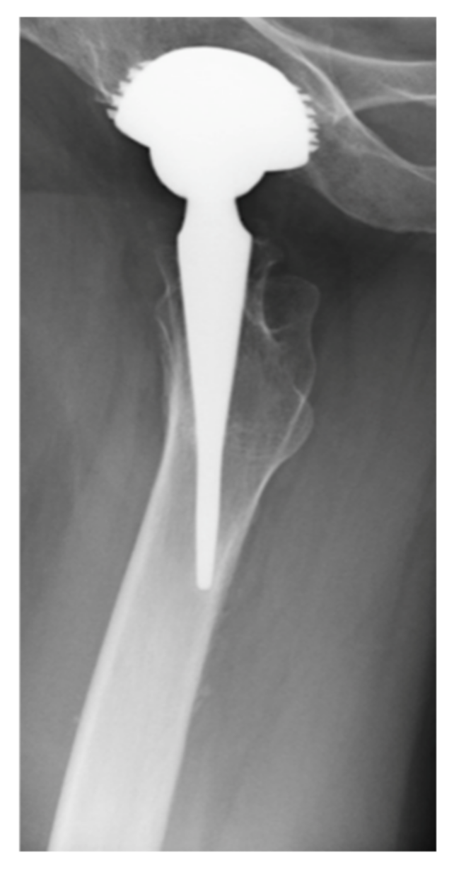
Figure 1. Lateral view of the METHA short stem total hip arthroplasty (STHA), showing the striven positioning parallel to the femoral neck and thus reconstructing the physiological ante torsion of the femoral neck.
Applsci 11 03965 g001
Figure 2. Digital templating of the BICONTACT stem (CTHA) in an anteroposterior (a) and lateral (b) view with the corresponding postoperative radiographs (c,d).
Applsci 11 03965 g002
Figure 3. Digital templating of the METHA stem (STHA) in an anteroposterior (a) and lateral (b) view with the corresponding postoperative radiographs (c,d).
Applsci 11 03965 g003
Figure 4. Scatterplots of the difference between actual and planned stem size over the actual stem size. CTHA with one plane planning is displayed in red, two plane planning is displayed in green. STHA with one plane planning is displayed in blue, two plane planning is displayed in purple. Linear regression lines are displayed for all implants and planning methods. For comparability, the implant sizes of the CTHA were substracted by 10.
Applsci 11 03965 g004
Figure 5. Digital templating of a STHA (lateral view) in a patient with excessive ante torsion of the femoral neck is an obvious contraindication for an implant with orientation parallel to the femoral neck.
Applsci 11 03965 g005
Table 1. Cumulative percentages of the three raters for the conventional (CTHA) and short stem total hip arthroplasty (STHA) groups, using one and two planes, separated in accordance with the variation between the real stem size and the measured size while templating.
Difference in Templated Size Compared to the Truly Implanted Prosthesis 0+/−1+/−2+/− > 2
CTHA (Templating a.p.)Rater 152.0%82.0%90.0%100.0%
239.189.191.3100.0
342.094.0100.0100.0
Mean ± Standard Deviation44.4 ± 6.888.4 ± 6.093.8 ± 5.4100.0 ± 0
CTHA (Templating 2 Planes)Rater 164.0%96.0%100.0%100.0%
226.178.297.8100.0
330.094.0100.0100.0
Mean ± Standard Deviation40.0 ± 20.889.4 ± 9.899.3 ± 1.3100.0 ± 0
STHA (Templating a.p.)Rater 145.0%83.0%99.0%100.0%
216.778.9100.0100.0
347.097.0100.0100.0
Mean ± Standard Deviation36.2 ± 16.986.3 ± 9.599.7 ± 0.6100.0 ± 0
STHA (Templating 2 Planes)Rater 160.0%93.0%100.0%100.0%
218.976.794.5100.0
371.0100.0100.0100.0
Mean ± Standard Deviation50.0 ± 27.589.9 ± 12.098.2 ± 3.2100.0 ± 0
Table 2. The intra-class correlation ICC (3,1) with a consistency of the values between all three raters.
Stem SizeCTHASTHA
a.p. planning0.7740.736
a.p. + lateral planning0.8380.757
Difference to Implanted SizeCTHASTHA
a.p. planning0.5860.483
a.p. + lateral planning0.6230.422
Table 3. The ICC (3,k) with conformity of the values between all three raters.
Stem SizeCTHASTHA
a.p. planning0.9110.893
a.p. + lateral planning0.940.903
Difference to Implanted SizeCTHASTHA
a.p. planning0.810.737
a.p. + lateral planning0.8320.687
Table 4. Fleiss Kappa as value for the conformity between all the three raters.
Stem SizeCTHASTHA
a.p. planning0.4110.228
a.p. + lateral planning0.270.3
Difference to Implanted SizeCTHASTHA
a.p. planning0.330.149
a.p. + lateral planning0.1780.16
Table 5. Kendall’s coefficient of concordance as a value for the consistency between the raters.
CTHASTHA
rprp
a.p. planning0.0290.2660.0230.131
a.p. + lateral planning0.0350.1960.0540.008
Table 6. Spearman’s coefficient a value for the correlation between the templated and the implanted stem size.
Stem SizeCTHASTHA
rprp
a.p. planning0.740.0010.800.001
a.p. + lateral planning0.790.0010.840.001
Publisher’s Note: MDPI stays neutral with regard to jurisdictional claims in published maps and institutional affiliations.