Article

Light-Induced Photoluminescence Quenching and Degradation in Quasi 2D Perovskites Film of (C6H5C2H4NH3)2 (CH3NH3)2[Pb3I10]

School of Electronic and Optical Engineering, Nanjing University of Science and Technology, Nanjing 210094, China
*
Author to whom correspondence should be addressed.
These authors contributed equally.
Appl. Sci. 2021, 11(6), 2683; https://doi.org/10.3390/app11062683
Submission received: 21 February 2021 / Revised: 9 March 2021 / Accepted: 15 March 2021 / Published: 17 March 2021
(This article belongs to the Section Optics and Lasers)

Abstract

:
Quasi-two-dimensional (2D) perovskites recently came into the focus because of their moisture stability. In addition to ambient air, light illumination could also cause degradation for the film of 2D perovskites; however, few studies have investigated their photostability. Here, we work on light-induced photoluminescence quenching, as well as the degradation of quasi-2D perovskites of PEA2MAn−1PbnI3n+1 (n = 3 nominally, PEA+ = C6H5(CH2)2NH3+, MA+ = CH3NH3+). Light-induced photoluminescence (PL) quenching generally happens with different speeds, depending on the wavelength and intensity of the laser as well as the film’s environment. With red light (635 nm) illumination, the film does not decompose into ambient air with an intensity below ~500 mW/cm2, although in general, a higher laser intensity and/or higher photon energy (447 nm) could render the decomposition process easier and faster. On the other hand, when the film is in a vacuum, both light-induced PL quenching and film degradation are significantly suppressed. Furthermore, we find that the multiphase of n = 1, 2, 3 in the PEA2MA2Pb3I10 film decomposes together and that the degradation processes begin with the collapses of the crystalline structures.

1. Introduction

Three-dimensional (3D) organic–inorganic metal halide perovskites have attracted attention over the past ten years because of their great promise for high-performance photovoltaics [1,2,3,4,5,6,7,8,9,10]. The recent highest certified efficiency of perovskite solar cells was about 25.5% by the National Renewable Energy Laboratory [11]. Despite the extraordinary progress in power conversion efficiencies, solar cells based on organic-lead-halide perovskites are still suffering from a number of instability mechanisms [12,13,14]. Recently, two-dimensional (2D) hybrid perovskites, especially for Ruddlesden–Poper (RP) perovskite, came into the focus. RP perovskites with a general formula of A’2An−1MnX3n+1 have a similar structure as many traditional layered 2D materials [15], where A and A are different organic cations (such as C6H5(CH2)2NH3+ (PEA+), CH3(CH2)3NH3+ (n-BA+) and CH3NH3+ (MA+)); M is a divalent metal (such as Pb2+, Sn2+); X represents halide ions (such as Cl, Br, I); and n is the number of inorganic layers [16]. Smith et al. utilized quasi-2D-layered hybrid perovskites of (PEA)2(CH3NH3)2Pb3I10 as light absorbers, which had a much higher moisture stability under an ambient environment when compared to their 3D counterparts [17]. Then, many efforts have been applied to improve the efficiency of quasi-2D hybrid perovskite solar cells by more than 15%, including a tailoring organic spacer cation, an inorganic layer thickness, etc. [18,19,20,21]. Besides that, other two-dimensional perovskites of (n-BA)2(MA)n−1PbnI3n+1 have been used in both photovoltaic and light-emitting applications, with an improvement of photostability and moisture resistance [19,22,23]. Furthermore, Grancini et al. engineered a large-area 2D/3D perovskite junction, from which solar modules delivered an efficiency of 11.2% with zero loss in performance over 10,000 h [24]. In addition to moisture and oxygen, which could be successfully excluded by an efficient encapsulation design, light-induced modification and degradation would be a more serious drawback for commercializing perovskite solar cells. Earlier work on light-induced phenomena, including long-term photostability, focused on three-dimensional (3D) perovskites with an intensely debated mechanism [25,26]. For instance, Nickel et al. demonstrated that illuminating CH3NH3PbI3 in an oxygen atmosphere resulted in swift degradation, since oxygen acted as the catalyst to deprotonate CH3NH3+ [27]. Merdasa et al. proposed that the mechanistic process of degradation occurring in a 3D MAPbI3 crystal structure collapsed smoothly to a 2D layered PbI2 structure, while the photoluminescence (PL) intensity decreased and PL spectrum blue-shifted up to 60 nm [28]. These photostability studies were all about 3D perovskites, the light-induced degradation of quasi-2D layered perovskites remaining elusive. With the development of 2D perovskites and their photovoltaic devices, research on 2D perovskites under illumination is urgently needed and may be able to contribute to an understanding of the mechanisms of light-induced degradation in 3D ones [29,30,31]. On the other hand, we noted that the degradation of encapsulated perovskite optoelectronic devices has obviously attracted more attention [32,33,34,35]. Although the degradation processes in working devices, including both solar cells [33] and light emitting diode (LED) [34,35], will be much more complicated than that in film, because of the existence of interfaces, charge transportation as well as the migration of ions under an electric field, studies on light-induced degradation in film will be helpful in understanding the degradation in devices.
In this work, we used quasi-2D perovskites of PEA2MA2Pb3I10 as a model material to investigate light-induced PL quenching and degradation. Our results clearly showed that blue light (447 nm) could generate more PL quenching and film degradation effects than red light (635 nm) when using the same illumination intensity. Additionally, when the sample was in a vacuum, light-induced degradation was significantly suppressed. Therefore, efficient encapsulation and an appropriate longpass filter could be very useful for enhancing the lifetime of devices. In addition, we found that the light degradation process might begin with the collapse of crystal structures.

2. Experimental Section

2.1. Sample Preparation

Methylammonium iodide (MAI), lead iodide (PbI2) and phenethylammonium iodide (PEAI) were purchases from Sigma Aldrich. The quasi-2D perovskite precursor solution was prepared by dissolving PEAI, MAI and PbI2 in N,N-dimethylformamide (DMF) with a molar ratio of 2:2:3. The mixed solution in DMF (35%) was stirred overnight at 60 °C inside a nitrogen-filled glove box. The glass substrates were precleaned in an ultrasonic bath with acetone, isopropanol and deionized water in sequence for 15 min, respectively, after which a nitrogen flow was used to dry the glass substrate. Finally, the glass substrate was treated with UV-ozone for 30 min. PEA2MA2Pb3I10 precursor solution was spin-cast at 4000 rpm for 30 s, and then the film was heated at 100 °C for 30 s.

2.2. Characterization of Materials and Measurement

X-ray diffraction (XRD) patterns of perovskite films were obtained by using a Bruker AXS Dimension D8 X-ray System. Photoluminescence (PL) spectra obtained using a standard continuous wave (CW) setup [36,37], the film could be placed in a dynamics vacuum of about ~0.2 Pa. A continuous wave (CW) diode laser at λ = 447 nm or 635 nm were used as the excitation source. For micro-PL measurements, the 100× objective used had a laser spot diameter of ~ 2 µm. The laser wavelength was 635 nm, and the integration time for a single PL spectrum was 0.1 s. For each time point, a 15 × 15 photoluminescence map in a 30 µm × 30 µm area was collected. The perovskite films were put on a 3-axes motorized translation stage with a minimum step of 1 µm.

3. Results and Discussion

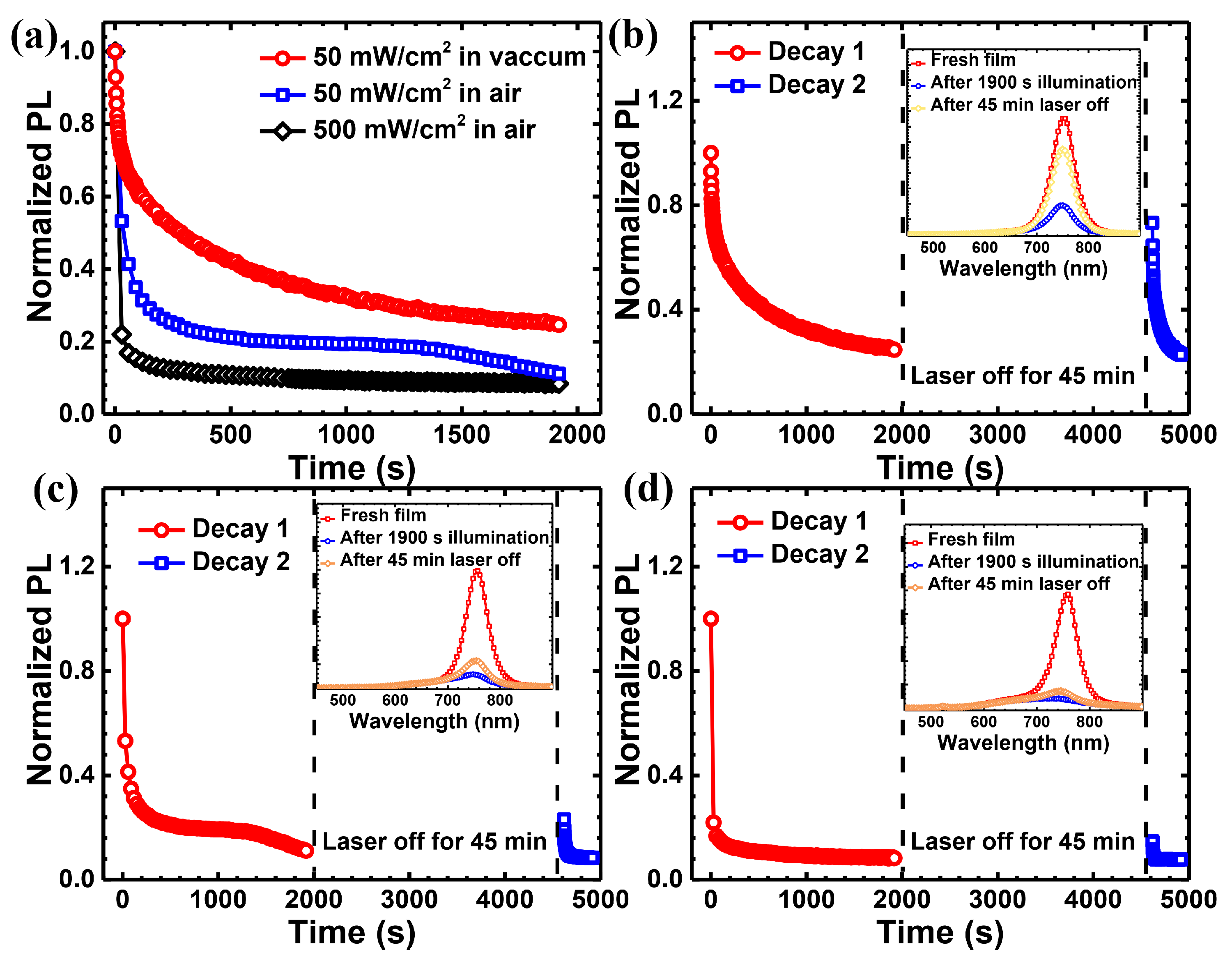
The slow dynamics of the PL intensity of PEA2MA2Pb3I10 films under continuous optical excitation using a 635 nm laser are shown in Figure 1a. We utilized three different experimental conditions to observe the PL quenching processes. When the excitation intensity is around 50 mW/cm2 and the film is in a vacuum, the PL intensity of the film slightly decreases by 20% within 1900 s, as shown in Figure 1b (red curve). Then, we blocked the laser beam and kept the film in darkness. After 45 min, after the laser beam is unblocked, the PL intensity recovers to its original level and then decreases as for the previous cycle. When a fresh film is in ambient atmosphere (25 °C, ~30% RH) with an excitation intensity of 50 mW/cm2, its PL intensity decreases by nearly 50% within 1900 s (red curve in Figure 1c), and the decay rate within the first 50 s is obviously faster than that in the vacuum. After 45 min in darkness, its PL intensity can almost recover to its original value with an identical PL spectrum (Figure 1c, blue curve and inset). However, with an excitation intensity up to 500 mW/cm2, the PL intensity of the perovskite film decreases by nearly 80% within the first 100 s and then slowly increases afterward (as shown in Figure 1d).
After a darkness treatment of 45 min, the PL spectrum does not change, although its PL intensity cannot fully recover. This suggests that the decay of the PL intensity up to 500 mW/cm2 is not due to the decomposition of perovskites, proving the stability of the quasi-2D perovskite film in air. Therefore, under a continuous 635-nm laser excitation in an ambient environment, although the ambient atmosphere accelerates the speed of PL quenching, the PEA2MA2Pb3I10 film does not decompose into other materials. Moreover, both photodegradation and photobrightening happened with a high illumination intensity, with relatively slower and weaker photobrightening effects. This is unlike the 3D perovskites, in which the photobrightening effects would dominate with a laser excitation of 632 nm [26]. Various photochemical mechanism have been introduced to describe the photobrightening effect in 3D perovskites, including temporary trap filling and the annihilation of shallow surface states, etc. [38,39]. Our results suggest that the density of naturally forming traps in 2D perovskites may not be as high as that in 3D perovskites. On the other hand, the reversible reduction of the PL intensity might result from photoinduced structural changes of the perovskites crystals [40], as well as ion migration and accumulation [41,42].
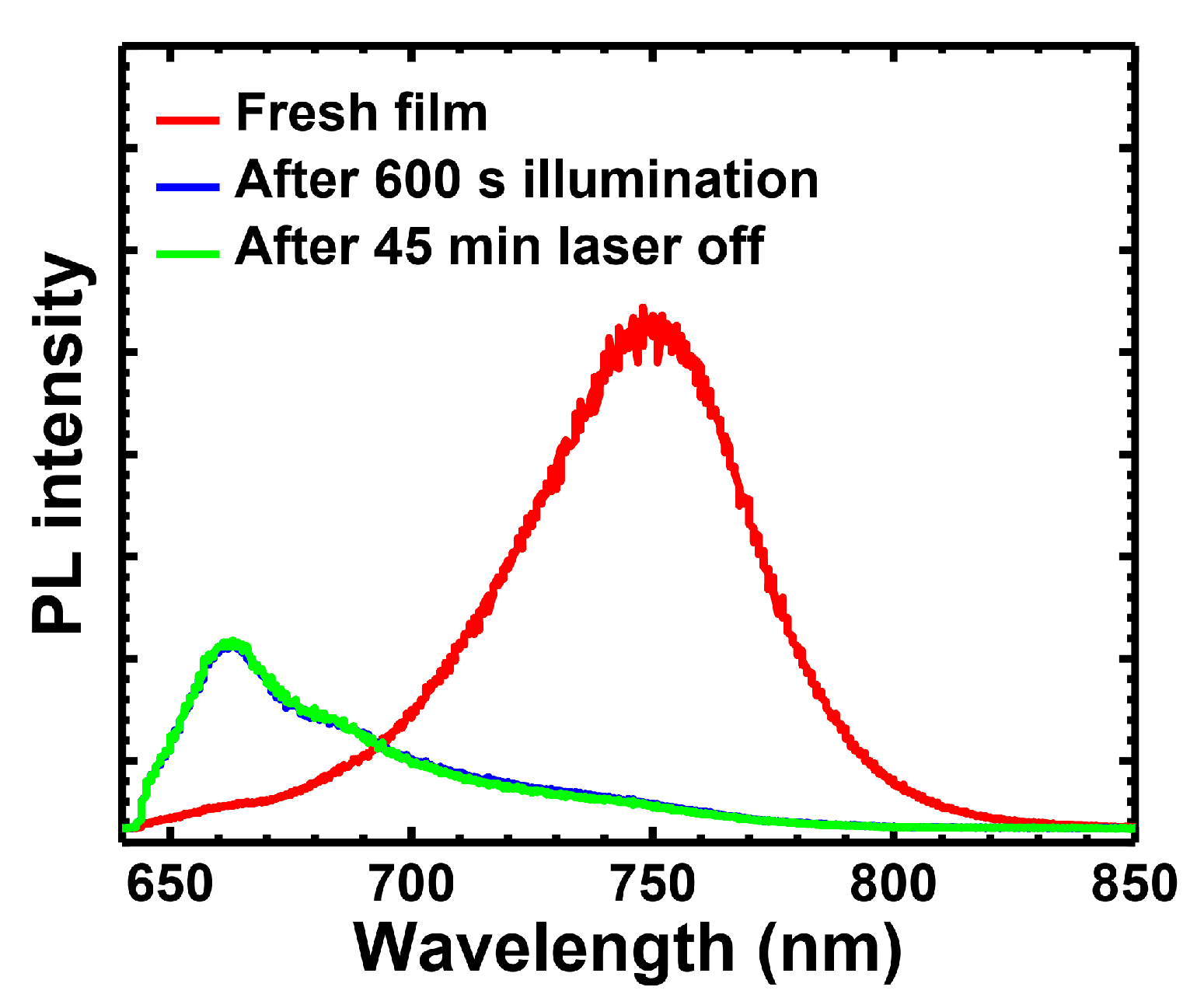
However, with a higher illumination intensity, which was achieved using a micro-PL measurement, we found that the PEA2MA2Pb3I10 film would go to unrecoverable photoinduced degradation in the same ambient environment as the previous one. As shown in Figure 2, the PL spectrum blue-shifts after a 600-s illumination with an intensity of ~1200 mW/cm2 by a 635 nm laser, and it cannot recover to the original spectrum after being kept in darkness for 45 min. Supplementary Materials Figure S1 shows that the illuminated spot of the film turns black after 600 s of illumination and is still black after 45 min of darkness treatment. Thus, here PEA2MA2Pb3I10 was subjected to an irreversible chemical decomposition.
It was known that different photon energies had different effects on the long-term photostability of 3D perovskites [12]. Here, we used a laser at 447 nm to further investigate the light-induced degradation of PEA2MA2Pb3I10. The PL quenching of PEA2MA2Pb3I10 films under different experimental conditions by a 447-nm laser is shown in Figure 3a. Compared with the laser at 635 nm, the PL quenching becomes more prominent and also faster under the same conditions when using a 447-nm laser (Figure S2). Figure 3b shows that under a continuous illumination with an excitation intensity of 50 mW/cm2, the PL intensity of the quasi-2D perovskite film in a vacuum decreases by nearly 75% within 1900 s. After 45 min in darkness, the PL intensity of the perovskite film partially recovers, while the PL spectra are almost the same before and after illumination, as shown in Figure S3a. This means that there is no obviously chemical decomposition in the perovskite film. When the film was illuminated by the same laser in an ambient environment with an excitation intensity of 50 mW/cm2, its PL intensity decreased by roughly 90% within 1900 s (Figure 3c). More importantly, Figure S3b shows that the PL spectrum blue-shifts slightly and broadens obviously. Again, after the film is kept in darkness for 45 min, its PL intensity recovers to just one-sixth of the original value with a broadened spectrum, indicating that a noticeable chemical decomposition happened in the PEA2MA2Pb3I10 film, as shown in Figure 3c. Figure 3d shows that with an excitation intensity of 500 mW/cm2, the PL intensity of the perovskite film remains only at 8% within 1900 s in an ambient environment, and we observe obvious blue-shifting with a broadened PL spectrum, while there is a new peak at around 525 nm, which is assigned to the n = 1 material of the 2D perovskites, namely PEA2PbI4 (see Figure S3c) [43]. After the same dark treatment for 45 min, it was found that only a fraction of the PL intensity could be restored. Normally, the PEA2MA2Pb3I10 film is composed of the multiphase of n = 1, 2, 3 and larger [18]; because of the effective energy transfer from a higher energy of smaller n materials to a lower energy of a larger n, the PL is single-peaked at ~750 nm. Here, the appearance of the n = 1 PL peak indicates either that an energy transfer from a smaller n to larger n is hindered or that more n = 1 material is generated from the decomposition of larger n materials. We will address this phenomenon later. Our work proved that quasi-2D perovskites of PEA2MA2Pb3I10 decomposed more easily under continuous illumination using a laser source with a shorter wavelength, in particular in ambient atmosphere. This is similar to 3D perovskites, in which the dissociation of CH3NH3+ was observed under illumination with ≥ 2.72 eV [27].
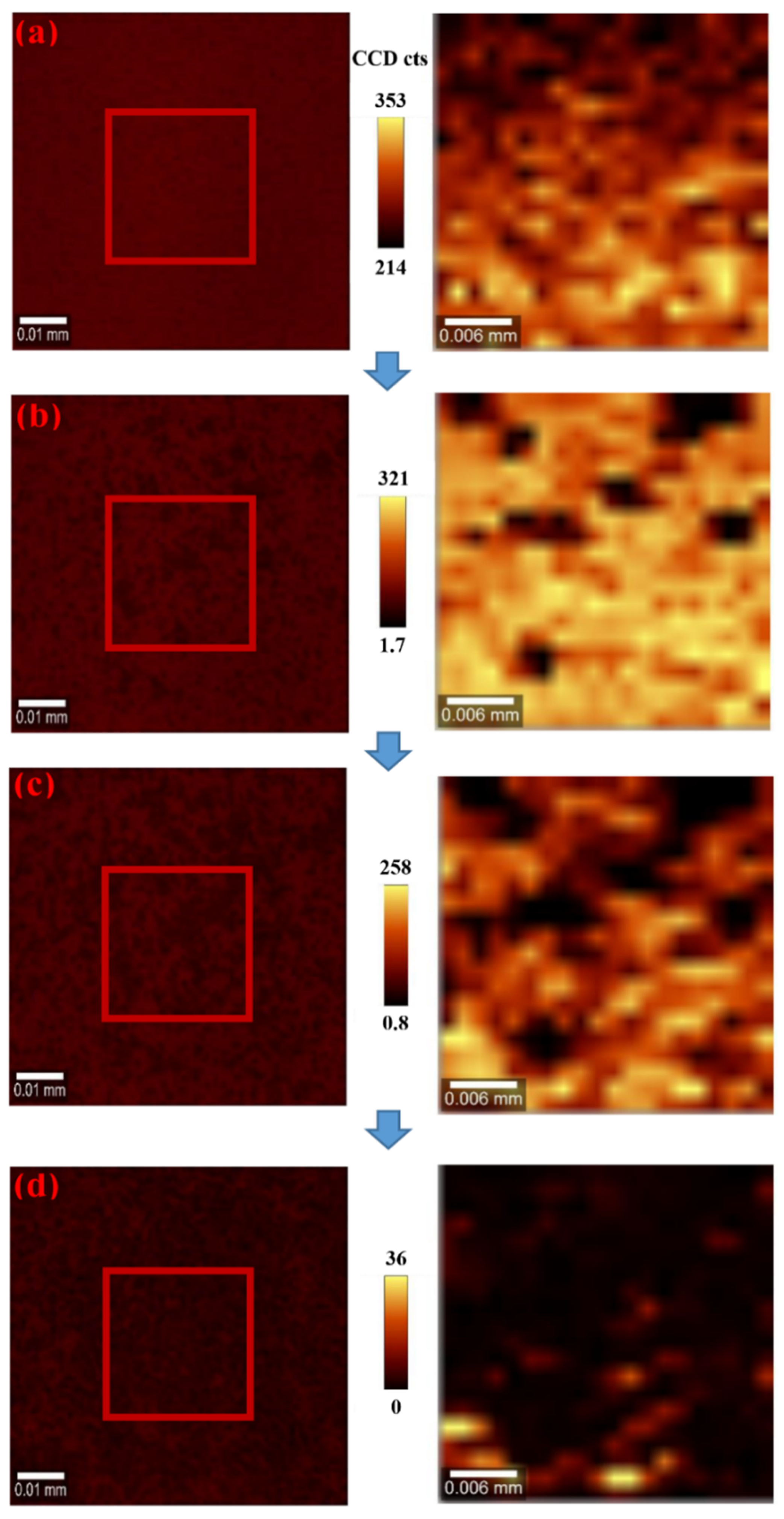
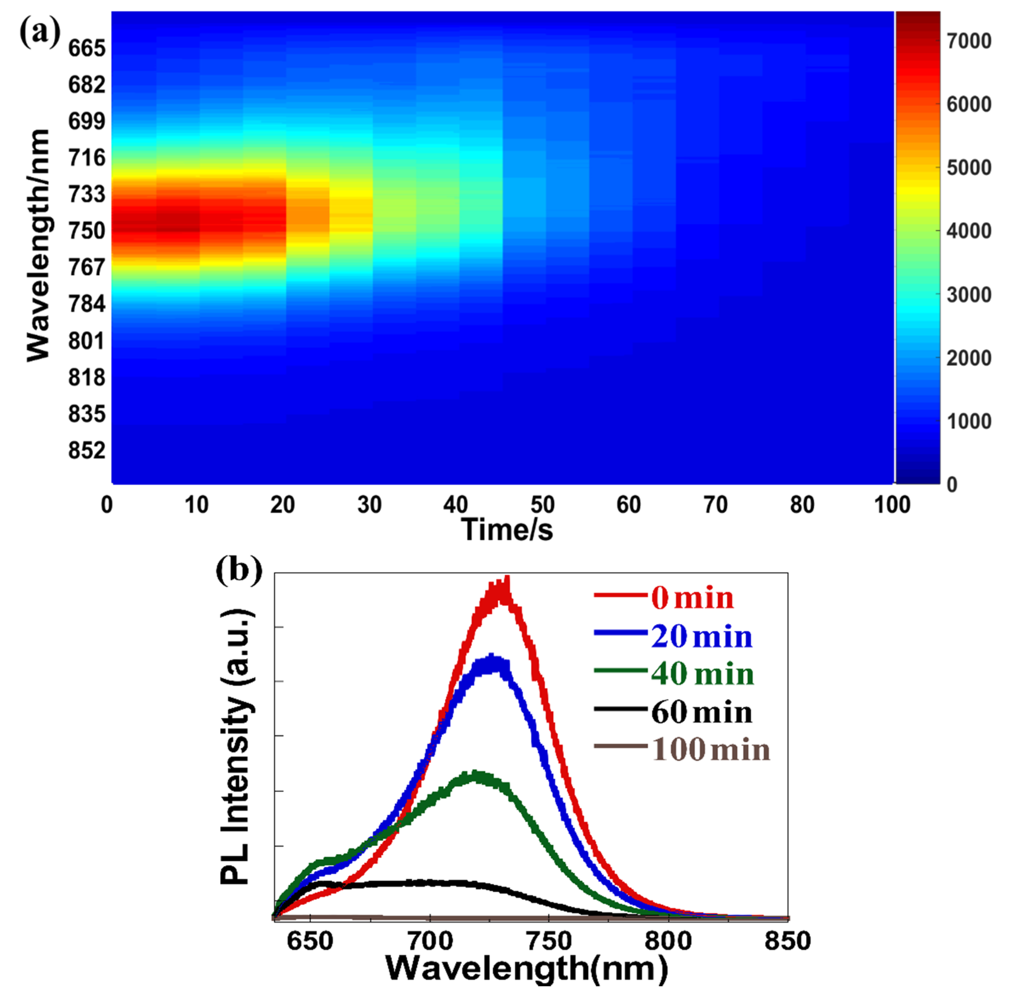
The photovoltaic devices usually work under white light illumination. To elucidate the influence of white light on quasi-2D perovskites, the specimens were characterized with confocal spectroscopy to probe spatially resolved PL spectra after continuous white light illumination by a white LED. Figure 4a–d shows microscope images and the corresponding PL intensity mapping of PEA2MA2Pb3I10 films after a continuous white light of ~1000 mW/cm2 with various times. The red square area in the microscope image was chosen as the photoluminescence intensity mapping of quasi-2D perovskites. After a given time of white light illumination, a 15 × 15 photoluminescence map in this 30 µm × 30 µm area was collected using a laser source at 635 nm with an intensity of ~50 mW/cm2 (within the red square in Figure 4). As the time of illumination increases, the whole area gradually becomes darker, as shown in the images of Figure 4. After 100 min of white light illumination, the film becomes almost completely dark. We note that the subarea for PL imaging within the square does not show a difference with other areas, indicating that the laser for PL does not generate a noticeable additional change on the film. Meanwhile, the corresponding PL intensity of each point is almost zero from the PL intensity mapping. In order to visually represent the change of the PL spectrum with time, the PL spectral evolution of the quasi-2D perovskites is shown in Figure 5a,b, in which we could observe the obvious blue-shifting and broadening of the PL spectrum and the decrease of the PL intensity along with the time. After 100 min, the PL intensity was almost zero and could not recover after the darkness treatment, proving the decomposition of the film under white light illumination in an ambient environment.
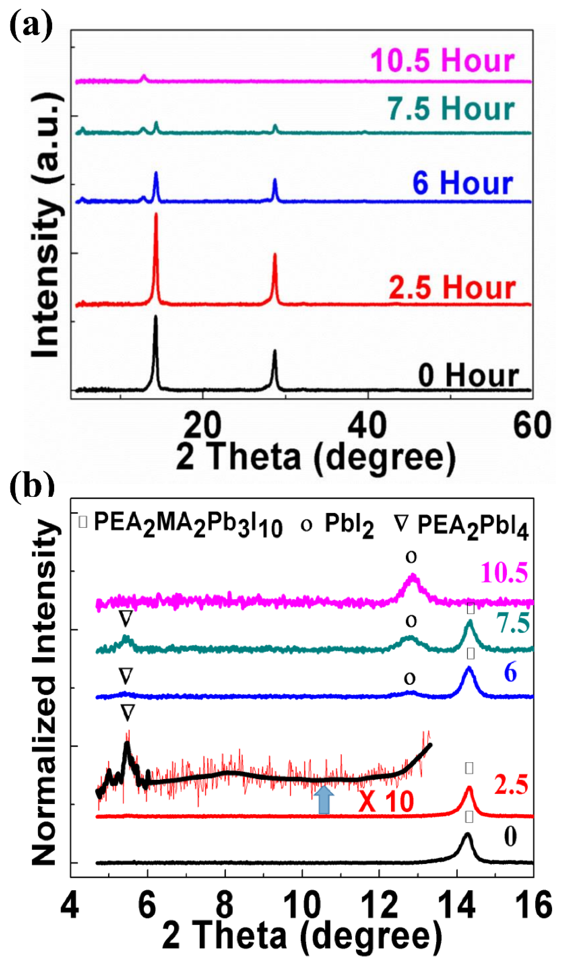
From Figure 1, Figure 2, Figure 3 and Figure 4, we conclude that the higher energy photon could cause the degradation of 2D perovskites at a modest intensity. Therefore, using an appropriate longpass filter could be useful for prolonging the lifetime of devices based on those films. On the other hand, the quasi-2D perovskites would eventually decompose into PbI2 under continuous illumination [44]. However, the entire process of degradation was still unclear for the quasi-2D PEA2MA2Pb3I10 film, which was actually composed of many phase of n = 1, 2, 3 and larger [17]. In Figure 6, the absorption of a PEA2MA2Pb3I10 film was measured under continuous illumination by a 447-nm laser source with an intensity of 350 mw/cm2 at various times. The laser-illuminated area is determined by a circular hole, which is also used for the absorption measurement. At various times of illumination, the respective peaks for n = 1, 2, 3 decrease together, indicating that they all decompose into PbI2 at roughly the same speed. This also proves that there is no n = 1 material generating from the decomposition of larger n materials. In the Figure 6 inset, after 270 min, the illuminated area changes from brown dark to yellow. At the same time, the XRD measurements, conducted in order to further observe the process of decomposition, are shown in Figure 7. Figure 7a presents the XRD patterns of the quasi-2D perovskite film at different times under a continuous illumination with white light of 300 mW/cm2. Without light illumination (0 min), the XRD patterns of the film show no sign of n = 2 and n = 1 structures, respectively. This phenomenon has been observed in multiphase PEA2MA2Pb3I10 film in the literature [45], indicating that the materials of less than n = 3 could be taken as defects within the structures of n = 3. In Figure 7b, we normalize XRD within 16 degrees of 2 theta. Under continuous white light illumination, we could firstly observe the diffraction peak at around 5.4° after 2.5 h, which was exactly same as the (002) plane peak of the PEA2PbI4 film [46]. At the same time, after being enlarged 10 times, a broad peak around 8° is also observed, which could be due to n = 2 materials with a much smaller grain size than that of the n = 1 and n = 3 materials [47,48]. After 6 h of white light illumination, the XRD peak intensity of PEA2MA2Pb3I10 decreases, the XRD peak intensity of PEA2PbI4 increases relatively, and the diffraction peak of PbI2 firstly appears. The n = 2 peak is not observable after enlargement, which may be due to the much weaker total signal (see Figure 7a). After 7.5 h of illumination, the XRD peak intensity of PEA2MA2Pb3I10 decreases consistently. At the same time, the diffraction peaks of PEA2PbI4 and PbI2 still exist. At last, after 10.5 h, the diffraction peaks of PEA2MA2Pb3I10 and PEA2PbI4 both disappear, leaving only the trace of PbI2. Although the detailed description of the decomposition process is not within the scope of the current work, we can suggest the following schematic process. Upon light illumination, the structure of PEA2MA2Pb3I10 begins to collapse, leaving the trace of PEA2PbI4, detectable by XRD (This is also consistent with the measurements of PL shown in Figure S3c, in which the PL emission from PEA2PbI4 is also observable after partial decomposition). Here, the explanation for the lacking PL of the n = 2 material is unclear. We think that the small grain size, in which the PL can easily be quenched by surface defects of the crystalline grain, could be part of the reasons. Later on, all 2D perovskites of various n will decompose into PbI2 simultaneously, as suggested by the absorption measurements shown in Figure 6. At last, only the PbI2 material will be left in the film. We noted that the XRD pattern of PEA2PbI4 emerged before that of PbI2, indicating that the crystal’s structural integrity could be important for preventing decomposition.

4. Conclusions

In summary, the light-induced PL quenching and degradation of the quasi-2D perovskites of PEA2MA2Pb3I10 have been investigated. The experimental results of the films under ambient conditions are summarized in Table 1, below. It was obvious that blue light could generate more damage than red light. Furthermore, when the sample was in a vacuum, light-induced degradation was significantly suppressed. Therefore, although quasi-2D perovskites were more robust than their 3D counterpart, effective encapsulation and an appropriate longpass filter could be very useful for enhancing the lifetime of devices.
At the same time, we found that the multiphase of n = 1, 2, 3 in the PEA2MA2Pb3I10 film decomposed together and that the degradation process may begin with the collapse of crystal structures, suggesting that the integrity of crystal could be crucial to the increase of the stability of quasi-2D perovskites.

Supplementary Materials

The following are available online at https://www.mdpi.com/2076-3417/11/6/2683/s1, Figure S1: Optical images of quasi 2D perovskite of PEA2MA2Pb3I10 taken at various illumination times under continuous illumination with excitation intensity of 1200 mW/cm2 using 635 nm laser source in the ambient environment. Figure S2: Normalized time-dependent steady-state PL intensity of quasi 2D perovskite of PEA2MA2Pb3I10 under continuous illumination using 447 nm laser source and 635 nm laser source in different condition, (a) with laser excitations at 50 mW/cm2 in vacuum, (b) with laser excitations at 50 mW/cm2 in the ambient environment, (c) with laser excitations at 500 mW/cm2 in the ambient environment. Figure S3: (a) Normalized PL spectrum of quasi 2D perovskite of PEA2MA2Pb3I10 under continuous illumination using 447 nm laser source in different condition at various time (a) laser excitations at 50 mW/cm2 in vacuum, (c) laser excitations at 50 mW/cm2 in the ambient environment, (d) laser excitations at 500 mW/cm2 in the ambient environment, stopping at 1900s for dark treatment of 45 min before resumption.

Author Contributions

Conceptualization, C.S., S.H., X.Y.; data curation, S.H., X.Y., Y.Z., B.Y.; formal analysis, S.H., X.Y., Y.Z., H.L.; software, S.H.; funding acquisition, C.S.; investigation, S.H., X.Y., Y.Z., B.Y.; methodology, H.L., C.S.; project administration, C.S.; resources, C.S.; supervision, C.S.; validation, S.H., X.Y., C.S.; visualization, S.H., X.Y., C.S.; writing—original draft, S.H., X.Y., C.S.; writing—review & editing, C.S. All authors have read and agreed to the published version of the manuscript.

Funding

This research was funded by National Natural Science Foundation of China, grant number 62074079, 61627802, and the APC was funded by National Natural Science Foundation of china (Nos. 61574078 and 61874056).

Institutional Review Board Statement

Not applicable.

Informed Consent Statement

Not applicable.

Data Availability Statement

Not applicable.

Acknowledgments

This work was supported by National Natural Science Foundation of China (Nos. 62074079, 61627802, 61874056, 61574078).

Conflicts of Interest

The authors declare no conflict of interests.

References

  1. Xiao, Z.; Bi, C.; Shao, Y.; Dong, Q.; Wang, Q.; Yuan, Y.; Wang, C.; Gao, Y.; Huang, J. Efficient, high yield perovskite photovoltaic devices grown by interdiffusion of solution-processed precursor stacking layers. Energy Environ. Sci. 2014, 7, 2619–2623. [Google Scholar] [CrossRef]
  2. Wojciechowski, K.; Saliba, M.; Leijtens, T.; Abate, A.; Snaith, H.J. Sub-150 °C processed meso-superstructured perovskite solar cells with enhanced efficiency. Energy Environ. Sci. 2014, 7, 1142–1147. [Google Scholar] [CrossRef]
  3. Wang, Q.; Shao, Y.; Dong, Q.; Xiao, Z.; Yuan, Y.; Huang, J. Large fill-factor bilayer iodine perovskite solar cells fabricated by a low-temperature solution-process. Energy Environ. Sci. 2014, 7, 2359–2365. [Google Scholar] [CrossRef]
  4. Jeon, N.J.; Lee, H.G.; Kim, Y.C.; Seo, J.; Noh, J.H.; Lee, J.; Seok, S. o-Methoxy substituents in spiro-OMeTAD for efficient inorganic–organic hybrid perovskite solar cells. J. Am. Chem. Soc. 2014, 136, 7837–7840. [Google Scholar] [CrossRef] [PubMed]
  5. Ryu, S.; Noh, J.H.; Jeon, N.J.; Kim, Y.C.; Yang, W.S.; Seo, J.; Seok, S.I. Voltage output of efficient perovskite solar cells with high open-circuit voltage and fill factor. Energy Environ. Sci. 2014, 7, 2614–2618. [Google Scholar] [CrossRef]
  6. Liu, M.; Johnston, M.B.; Snaith, H.J. Efficient planar heterojunction perovskite solar cells by vapour deposition. Nat. Cell Biol. 2013, 501, 395–398. [Google Scholar] [CrossRef] [PubMed]
  7. Burschka, J.; Pellet, N.; Moon, S.-J.; Humphry-Baker, R.; Gao, P.; Nazeeruddin, M.K.; Grätzel, M. Sequential deposition as a route to high-performance perovskite-sensitized solar cells. Nat. Cell Biol. 2013, 499, 316–319. [Google Scholar] [CrossRef] [PubMed]
  8. Jeon, N.J.; Noh, J.H.; Kim, Y.C.; Yang, W.S.; Ryu, S.; Seok, S.I. Solvent engineering for high-performance inorganic–organic hybrid perovskite solar cells. Nat. Mater. 2014, 13, 897–903. [Google Scholar] [CrossRef]
  9. Zhou, H.; Chen, Q.; Li, G.; Luo, S.; Song, T.-B.; Duan, H.-S.; Hong, Z.; You, J.; Liu, Y.; Yang, Y. Interface engineering of highly efficient perovskite solar cells. Science 2014, 345, 542–546. [Google Scholar] [CrossRef]
  10. Yang, W.S.; Noh, J.H.; Jeon, N.J.; Kim, Y.C.; Ryu, S.; Seo, J.; Seok, S.I. High-performance photovoltaic perovskite layers fabricated through intramolecular exchange. Science 2015, 348, 1234–1237. [Google Scholar] [CrossRef]
  11. Best Research-Cell Effciencies. Available online: https://www.nrel.gov/pv/assets/pdfs/cell-pv-eff-emergingpv.20200919.pdf (accessed on 19 September 2020).
  12. Yang, J.; Siempelkamp, B.D.; Liu, D.; Kelly, T.L. Investigation of CH3NH3PbI3 degradation rates and mechanisms in controlled humidity environments using in situ techniques. ACS Nano 2015, 9, 1955–1963. [Google Scholar] [CrossRef] [PubMed]
  13. Divitini, G.; Cacovich, S.; Matteocci, F.; Cinà, L.; Di Carlo, A.; Ducati, C. In situ observation of heat-induced degradation of perovskite solar cells. Nat. Energy 2016, 1, 1–6. [Google Scholar] [CrossRef] [Green Version]
  14. Yang, S.; Wang, Y.; Liu, P.; Cheng, Y.-B.; Zhao, H.J.; Yang, H.G. Functionalization of perovskite thin films with moisture-tolerant molecules. Nat. Energy 2016, 1, 15016. [Google Scholar] [CrossRef]
  15. Chen, Y.; Sun, Y.; Peng, J.; Tang, J.; Zheng, K.; Liang, Z. 2D Ruddlesden-Popper Perovskites for Optoelectronics. Adv. Mater. 2018, 30, 1703487. [Google Scholar] [CrossRef] [PubMed]
  16. Li, H.; Luo, T.; Zhang, S.; Sun, Z.; He, X.; Zhang, W.; Chang, H. Two-Dimensional Metal-Halide Perovskite-based Optoelectronics: Synthesis, Structure, Properties and Applications. Energy Environ. Mater. 2021, 4, 46–64. [Google Scholar] [CrossRef]
  17. Smith, I.C.; Hoke, E.T.; Solis-Ibarra, D.; McGehee, M.D.; Karunadasa, H.I. A Layered Hybrid Perovskite Solar-Cell Absorber with Enhanced Moisture Stability. Angew. Chem. 2014, 126, 11414–11417. [Google Scholar] [CrossRef]
  18. Yan, X.; Hu, S.; Zhang, Y.; Li, H.; Sheng, C. Methylammonium acetate as an additive to improve performance and eliminate J-V hysteresis in 2D homologous organic-inorganic perovskite solar cells. Sol. Energy Mater. Sol. Cells 2019, 191, 283–289. [Google Scholar] [CrossRef]
  19. Tsai, H.; Nie, W.; Blancon, J.-C.; Stoumpos, C.C.; Asadpour, R.; Harutyunyan, B.; Neukirch, A.J.; Verduzco, R.; Crochet, J.J.; Tretiak, S.; et al. High-efficiency two-dimensional Ruddlesden–Popper perovskite solar cells. Nat. Cell Biol. 2016, 536, 312–316. [Google Scholar] [CrossRef]
  20. Lai, H.; Kan, B.; Liu, T.; Zheng, N.; Xie, Z.; Zhou, T.; Wan, X.; Zhang, X.; Liu, Y.; Chen, Y. Two-Dimensional Ruddlesden–Popper Perovskite with Nanorod-like Morphology for Solar Cells with Efficiency Exceeding 15%. J. Am. Chem. Soc. 2018, 140, 11639–11646. [Google Scholar] [CrossRef]
  21. Fu, W.; Liu, H.; Shi, X.; Zuo, L.; Li, X.; Jen, A.K. Tailoring the Functionality of Organic Spacer Cations for Efficient and Stable Quasi-2D Perovskite Solar Cells. Adv. Funct. Mater. 2019, 29, 1900221. [Google Scholar] [CrossRef]
  22. Byun, J.; Cho, H.; Wolf, C.; Jang, M.; Sadhanala, A.; Friend, R.H.; Yang, H.; Lee, T.-W. Efficient Visible Quasi-2D Perovskite Light-Emitting Diodes. Adv. Mater. 2016, 28, 7515–7520. [Google Scholar] [CrossRef]
  23. Tsai, H.; Nie, W.; Blancon, J.-C.; Stoumpos, C.C.; Soe, C.M.M.; Yoo, J.; Crochet, J.; Tretiak, S.; Even, J.; Sadhanala, A.; et al. Stable Light-Emitting Diodes Using Phase-Pure Ruddlesden-Popper Layered Perovskites. Adv. Mater. 2018, 30, 1704217. [Google Scholar] [CrossRef]
  24. Grancini, G.; Roldán-Carmona, C.; Zimmermann, I.; Mosconi, E.; Lee, X.; Martineau, D.; Narbey, S.; Oswald, F.; De Angelis, F.; Graetzel, M.; et al. One-Year stable perovskite solar cells by 2D/3D interface engineering. Nat. Commun. 2017, 8, 15684. [Google Scholar] [CrossRef]
  25. Chen, S.; Wen, X.; Huang, S.; Huang, F.; Cheng, Y.-B.; Green, M.; Ho-Baillie, A. Light Illumination Induced Photoluminescence Enhancement and Quenching in Lead Halide Perovskite. Sol. RRL 2017, 1, 1600001. [Google Scholar] [CrossRef]
  26. Quitsch, W.-A.; Dequilettes, D.W.; Pfingsten, O.; Schmitz, A.; Ognjanovic, S.; Jariwala, S.; Koch, S.; Winterer, M.; Ginger, D.S.; Bacher, G. The Role of Excitation Energy in Photobrightening and Photodegradation of Halide Perovskite Thin Films. J. Phys. Chem. Lett. 2018, 9, 2062–2069. [Google Scholar] [CrossRef] [PubMed]
  27. Nickel, N.H.; Lang, F.; Brus, V.V.; Shargaieva, O.; Rappich, J. Unraveling the Light-Induced Degradation Mechanisms of CH3NH3PbI3Perovskite Films. Adv. Electron. Mater. 2017, 3, 1700158. [Google Scholar] [CrossRef]
  28. Merdasa, A.; Bag, M.; Tian, Y.; Källman, E.; Dobrovolsky, A.; Scheblykin, I.G. Super-Resolution Luminescence Microspectroscopy Reveals the Mechanism of Photoinduced Degradation in CH3NH3PbI3 Perovskite Nanocrystals. J. Phys. Chem. C 2016, 120, 10711–10719. [Google Scholar] [CrossRef]
  29. Fan, Z.; Xiao, H.; Wang, Y.; Zhao, Z.; Lin, Z.; Cheng, H.-C.; Lee, S.-J.; Wang, G.; Feng, Z.; Goddard, W.A. Layer-by-layer degradation of methylammonium lead tri-iodide perovskite microplates. Joule 2017, 1, 548–562. [Google Scholar] [CrossRef] [Green Version]
  30. Leijtens, T.; Eperon, G.E.; Pathak, S.; Abate, A.; Lee, M.M.; Snaith, H.J. Overcoming ultraviolet light instability of sensitized TiO2 with meso-superstructured organometal tri-halide perovskite solar cells. Nat. Commun. 2013, 4, 2885. [Google Scholar] [CrossRef] [PubMed]
  31. Berhe, T.A.; Su, W.-N.; Chen, C.-H.; Pan, C.-J.; Cheng, J.-H.; Chen, H.-M.; Tsai, M.-C.; Chen, L.-Y.; Dubale, A.A.; Hwang, B.-J. Organometal halide perovskite solar cells: Degradation and stability. Energy Environ. Sci. 2016, 9, 323–356. [Google Scholar] [CrossRef]
  32. Seitz, M.; Gant, P.; Castellanos-Gomez, A.; Prins, F. Long-Term Stabilization of Two-Dimensional Perovskites by Encapsulation with Hexagonal Boron Nitride. Nanomaterials 2019, 9, 1120. [Google Scholar] [CrossRef] [Green Version]
  33. Khadka, D.B.; Shirai, Y.; Yanagida, M.; Miyano, K. Degradation of encapsulated perovskite solar cells driven by deep trap states and interfacial deterioration. J. Mater. Chem. C 2018, 6, 162–170. [Google Scholar] [CrossRef]
  34. Li, R.; Yi, C.; Ge, R.; Zou, W.; Cheng, L.; Wang, N.; Wang, J.; Huang, W. Room-temperature electroluminescence from two-dimensional lead halide perovskites. Appl. Phys. Lett. 2016, 109, 151101. [Google Scholar] [CrossRef]
  35. Khadka, D.B.; Shirai, Y.; Yanagida, M.; Ryan, J.W.; Miyano, K. Exploring the effects of interfacial carrier transport layers on device performance and optoelectronic properties of planar perovskite solar cells. J. Mater. Chem. C 2017, 5, 8819–8827. [Google Scholar] [CrossRef]
  36. Jiang, X.; Österbacka, R.; Korovyanko, O.; An, C.; Horovitz, B.; Janssen, R.; Vardeny, Z. Spectroscopic Studies of Photoexcitations in Regioregular and Regiorandom Polythiophene Films. Adv. Funct. Mater 2002, 12, 587–597. [Google Scholar] [CrossRef]
  37. Yang, X.; Wang, R.; Wang, Y.; Sheng, C.-X.; Li, H.; Hong, W.; Tang, W.; Tian, C.; Chen, Q. Long lived photoexcitation dynamics in π-conjugated polymer and fullerene blended films. Org. Electron. 2013, 14, 2058–2064. [Google Scholar] [CrossRef]
  38. Tian, Y.; Peter, M.; Unger, E.; Abdellah, M.; Zheng, K.; Pullerits, T.; Yartsev, A.; Sundström, V.; Scheblykin, I.G. Mechanistic insights into perovskite photoluminescence enhancement: Light curing with oxygen can boost yield thousandfold. Phys. Chem. Chem. Phys. 2015, 17, 24978–24987. [Google Scholar] [CrossRef] [PubMed]
  39. Galisteo-López, J.F.; Anaya, M.; Calvo, M.E.; Míguez, H. Environmental Effects on the Photophysics of Organic–Inorganic Halide Perovskites. J. Phys. Chem. Lett. 2015, 6, 2200–2205. [Google Scholar] [CrossRef] [Green Version]
  40. Gottesman, R.; Gouda, L.; Kalanoor, B.S.; Haltzi, E.; Tirosh, S.; Rosh-Hodesh, E.; Tischler, Y.R.; Zaban, A.; Quarti, C.; Mosconi, E.; et al. Photoinduced Reversible Structural Transformations in Free-Standing CH3NH3PbI3Perovskite Films. J. Phys. Chem. Lett. 2015, 6, 2332–2338. [Google Scholar] [CrossRef] [Green Version]
  41. Unger, E.L.; Hoke, E.T.; Bailie, C.D.; Nguyen, W.H.; Bowring, A.R.; Heumuller, T.; Christoforo, M.G.; McGehee, M.D. Hysteresis and transient behavior in current–voltage measurements of hybrid-perovskite absorber solar cells. Energy Environ. Sci. 2014, 7, 3690–3698. [Google Scholar] [CrossRef]
  42. Zhang, Y.; Liu, M.; Eperon, G.E.; Leijtens, T.C.; McMeekin, D.; Saliba, M.; Zhang, W.; De Bastiani, M.; Petrozza, A.; Herz, L.M.; et al. Charge selective contacts, mobile ions and anomalous hysteresis in organic–inorganic perovskite solar cells. Mater. Horizons 2015, 2, 315–322. [Google Scholar] [CrossRef]
  43. Zhang, Y.; Wang, R.; Li, Y.; Wang, Z.; Hu, S.; Yan, X.; Zhai, Y.; Zhang, C.; Sheng, C. Optical Properties of Two-Dimensional Perovskite Films of (C6H5C2H4NH3)2[PbI4] and (C6H5C2H4NH3)2 (CH3NH3)2[Pb3I10]. J. Phys. Chem. Lett. 2018, 10, 13–19. [Google Scholar] [CrossRef]
  44. Fang, H.-H.; Yang, J.; Tao, S.; Adjokatse, S.; Kamminga, M.E.; Ye, J.; Blake, G.R.; Even, J.; Loi, M.A. Unravelling Light-Induced Degradation of Layered Perovskite Crystals and Design of Efficient Encapsulation for Improved Photostability. Adv. Funct. Mater. 2018, 28, 1800305. [Google Scholar] [CrossRef]
  45. Wu, B.; Fu, K.; Yantara, N.; Xing, G.; Sun, S.; Sum, T.C.; Mathews, N. Charge Accumulation and Hysteresis in Perovskite-Based Solar Cells: An Electro-Optical Analysis. Adv. Energy Mater. 2015, 5, 1500829. [Google Scholar] [CrossRef]
  46. Yang, S.; Niu, W.; Wang, A.-L.; Fan, Z.; Chen, B.; Tan, C.; Lu, Q.; Zhang, H. Ultrathin Two-Dimensional Organic-Inorganic Hybrid Perovskite Nanosheets with Bright, Tunable Photoluminescence and High Stability. Angew. Chem. Int. Ed. 2017, 56, 4252–4255. [Google Scholar] [CrossRef] [PubMed]
  47. Gan, X.; Wang, O.; Liu, K.; Du, X.; Guo, L.; Liu, H. 2D homologous organic-inorganic hybrids as light-absorbers for planer and nanorod-based perovskite solar cells. Sol. Energy Mater. Sol. Cells 2017, 162, 93–102. [Google Scholar] [CrossRef]
  48. Peng, W.; Yin, J.; Ho, K.-T.; Ouellette, O.; De Bastiani, M.; Murali, B.; El Tall, O.; Shen, C.; Miao, X.; Pan, J.; et al. Ultralow Self-Doping in Two-dimensional Hybrid Perovskite Single Crystals. Nano Lett. 2017, 17, 4759–4767. [Google Scholar] [CrossRef]
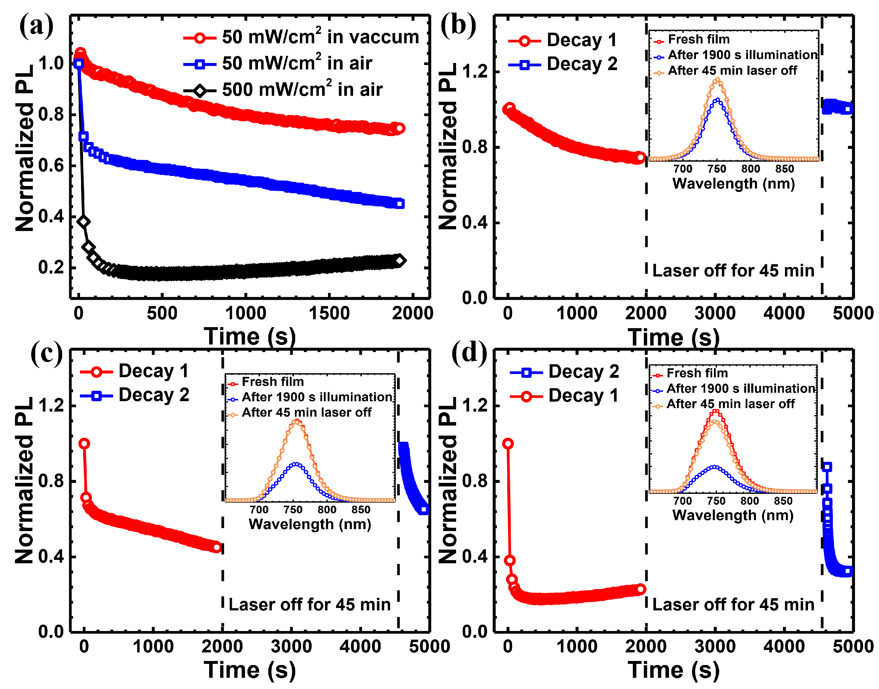
Figure 1. (a) Normalized time-dependent steady-state PL intensity of quasi-2D perovskites of PEA2MA2Pb3I10 under continuous illumination using a 635 nm laser source under different conditions. (b) The PL intensities over time of PEA2MA2Pb3I10 under laser excitations at 50 mW/cm2 in a vacuum, (c) the laser excitations at 50 mW/cm2 in an ambient environment, and (d) the laser excitations at 500 mW/cm2 in an ambient environment. The respective illumination stops at 1900 s for a dark treatment of 45 min before resumption. The corresponding PL spectra of the PEA2MA2Pb3I10 film at various time points are shown in the inset of the respective figure panel.
Applsci 11 02683 g001
Figure 2. The PL spectrum of quasi-2D perovskites of PEA2MA2Pb3I10 at various illumination times under continuous illumination with an excitation intensity of 1200 mW/cm2 using a 635-nm laser source in an ambient environment.
Applsci 11 02683 g002
Figure 3. (a) Normalized time-dependent steady-state PL intensity of quasi-2D perovskites of PEA2MA2Pb3I10 under continuous illumination using a 447-nm laser source under different conditions. (b) The PL intensities over time of PEA2MA2Pb3I10 under laser excitations at 50 mW/cm2 in a vacuum, (c) the laser excitations at 50 mW/cm2 in an ambient environment, (d) the laser excitations at 500 mW/cm2 in an ambient environment. The respective illumination stops at 1900 s for a dark treatment of 45 min before resumption. The corresponding PL spectra of the PEA2MA2Pb3I10 film at various time points are shown in the inset of the respective figure panel.
Applsci 11 02683 g003
Figure 4. Optical images and the corresponding photoluminescence intensity mapping of PEA2MA2Pb3I10 under continuous white light illumination at 1000 mW/cm2 in an ambient environment with various times: (a) 0 min, (b) 20 min, (c) 40 min and (d) 100 min. For the PL intensity mapping, a 30 µm × 30 µm area was collected. The optical excitation source was a 635-nm continuous wave (CW) laser, and the excitation intensity was approximately 50 mW/cm2.
Applsci 11 02683 g004
Figure 5. (a) PL spectral evolution and (b) spectra at various time points of PEA2MA2Pb3I10 under continuous white light illumination at 1000 mW/cm2 in an ambient environment. The optical excitation source was a 635-nm continuous wave (CW) laser, and the excitation intensity was approximately 50 mW/cm2.
Applsci 11 02683 g005
Figure 6. UV-vis spectra of the PEA2MA2Pb3I10 film at various times, under continuous illumination using a 447-nm laser source at 350 mW/cm2 in an ambient environment.
Applsci 11 02683 g006
Figure 7. (a) The X-ray diffraction (XRD) patterns of PEA2MA2Pb3I10 from 4–60 degrees, (b) Normalized XRD intensity from 4–16 degrees under continuous white light illumination at 300 mW/cm2 in an ambient environment. Part of the pattern after 2.5 h of illumination is enlarged 10-fold (the red curve is data, the black curve is for guiding the eye) in order to clearly show the peak of the n = 1 and n = 2 materials.
Applsci 11 02683 g007
Table 1. All experimental results of the quasi-2D perovskite films under ambient conditions.
Light SourceLaser at 635 nmLaser at 532 nmWhite Light
Light intensity (mW/cm2)505001200505001000
PL quenchingYesYesYesYesYesYes
PL recovery after 45 min of darkness treatmentfully90%/15%5%/
Degradation NONOYesYesYesYes
Publisher’s Note: MDPI stays neutral with regard to jurisdictional claims in published maps and institutional affiliations.