
Shear Wave Velocity Estimation Using the Real-Time Curve Tracing Method in Ultrasound Elastography


Abstract
1. Introduction
2. Method and Principle
3. Phantom Experiments
3.1. Experimental Environment
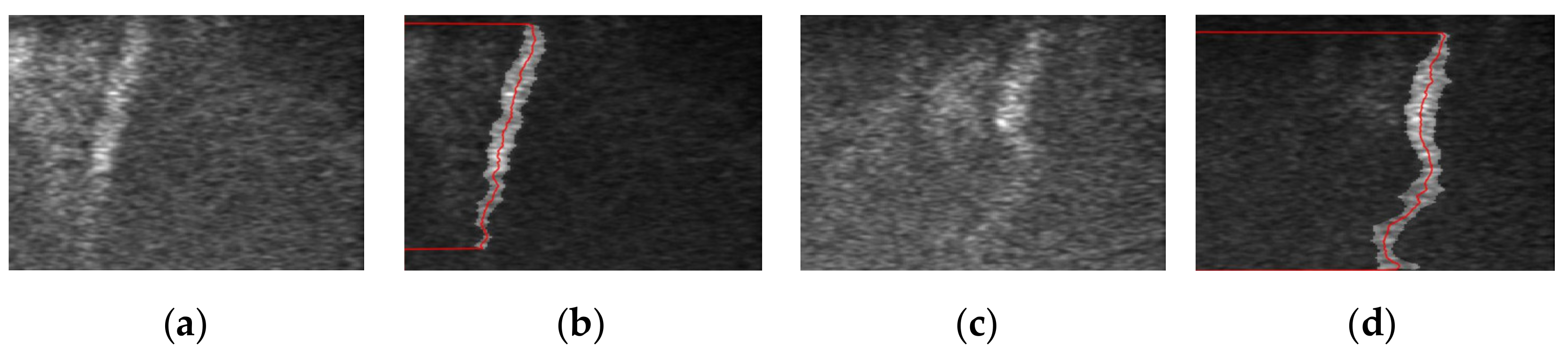
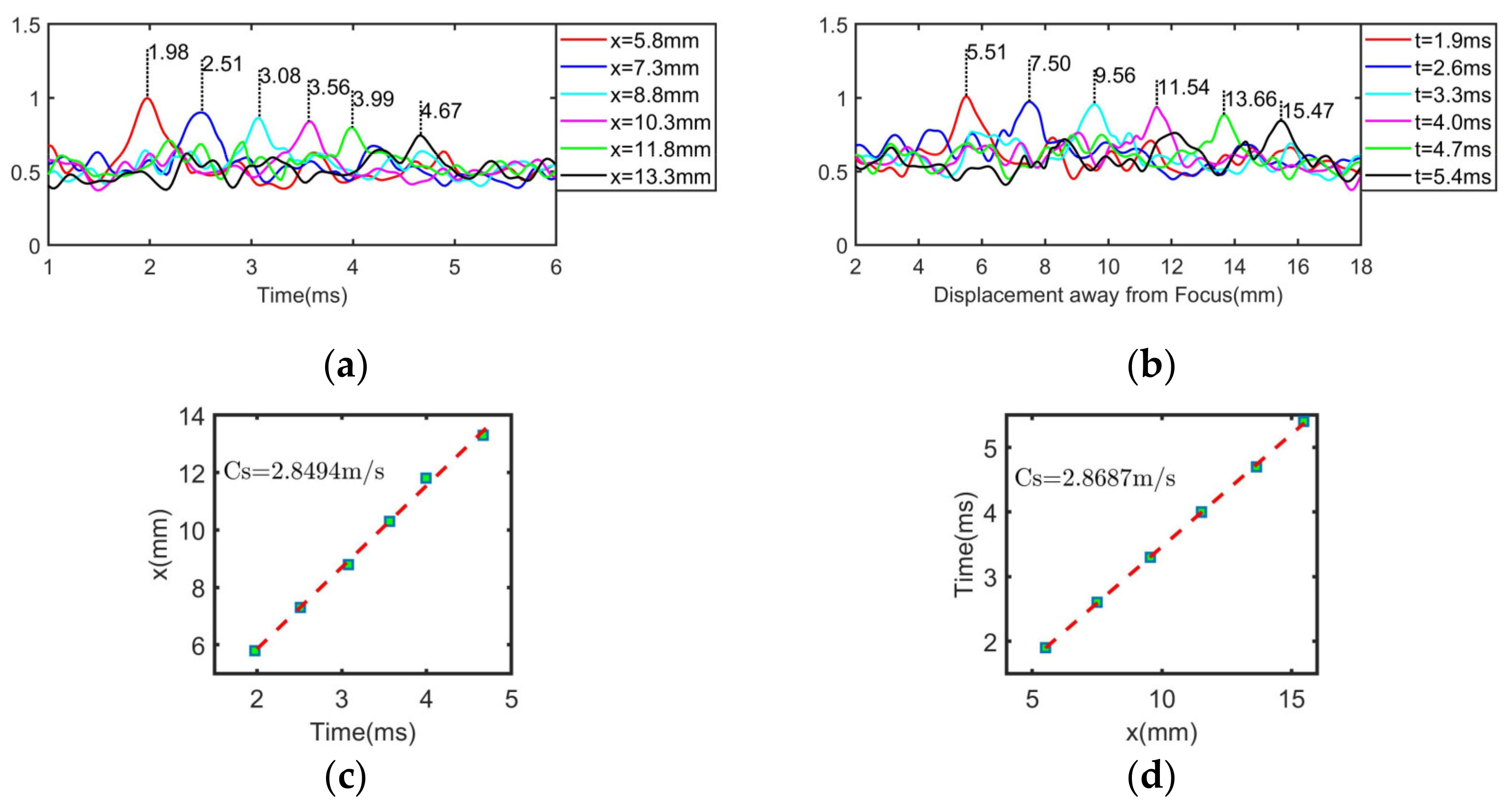
3.2. Shear Wave Velocity Estimation
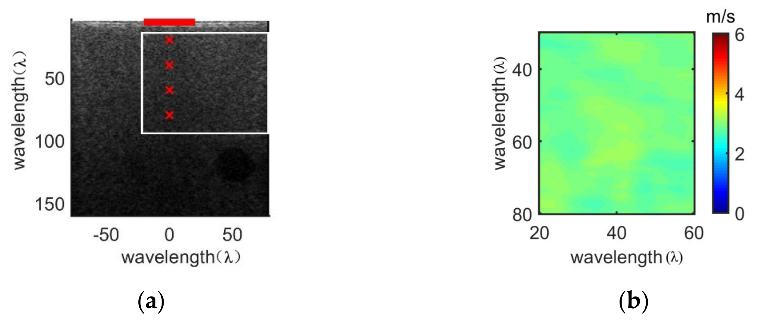
3.3. Image Reconstruction of Shear Wave Velocity
3.3.1. Homogeneous Region
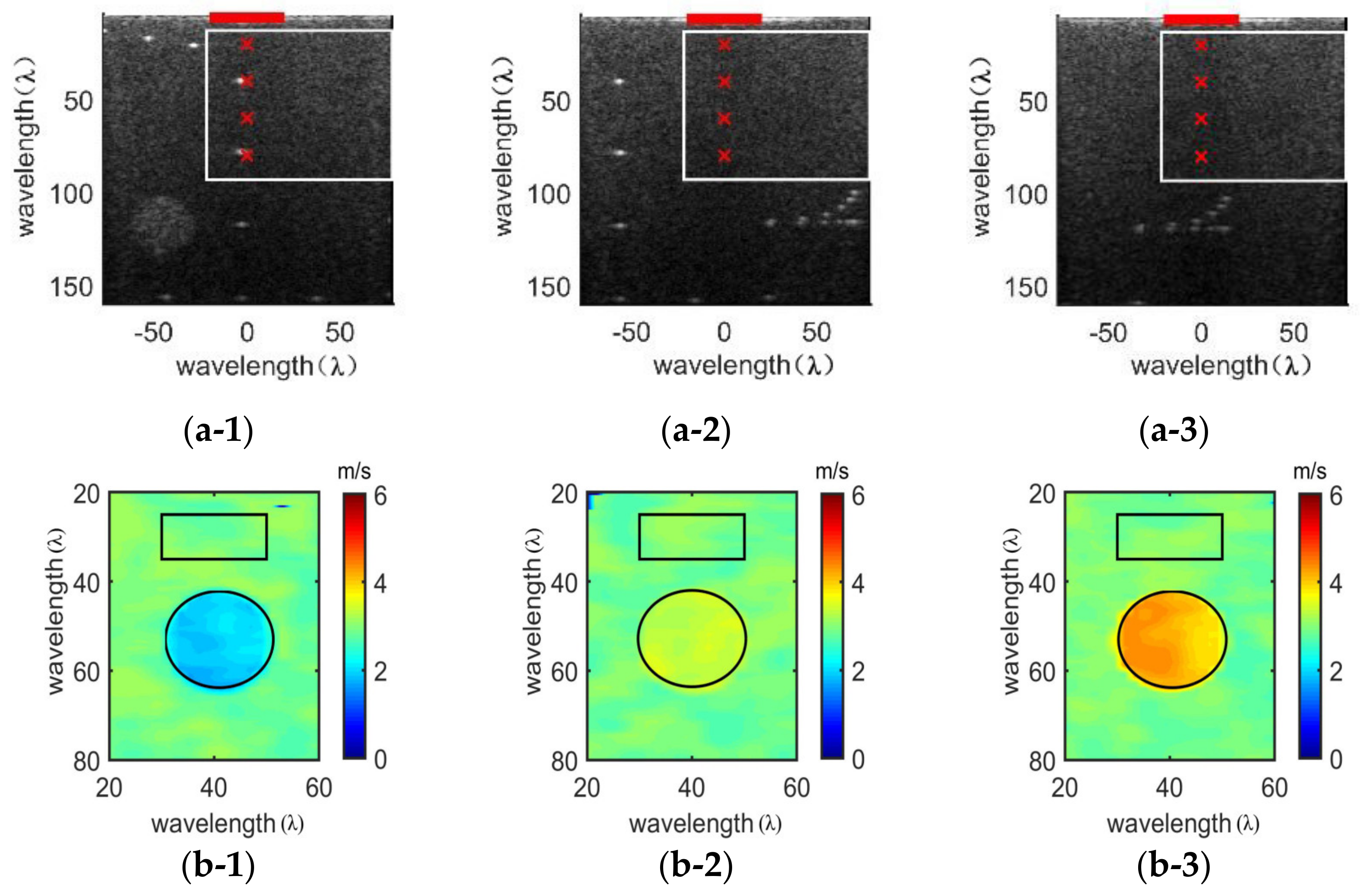
3.3.2. Stiff Spherical Inclusion Regions with Varying Elastic Values
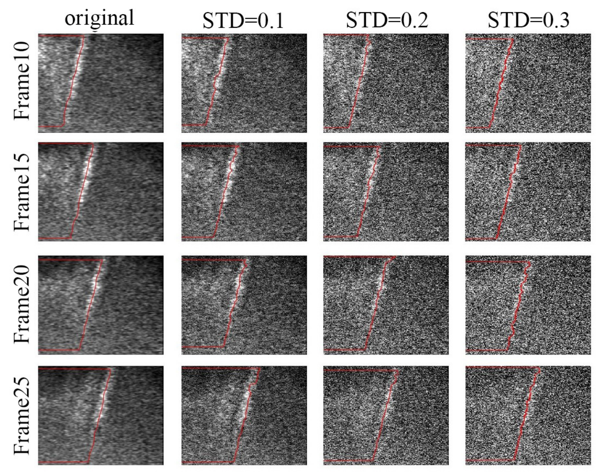
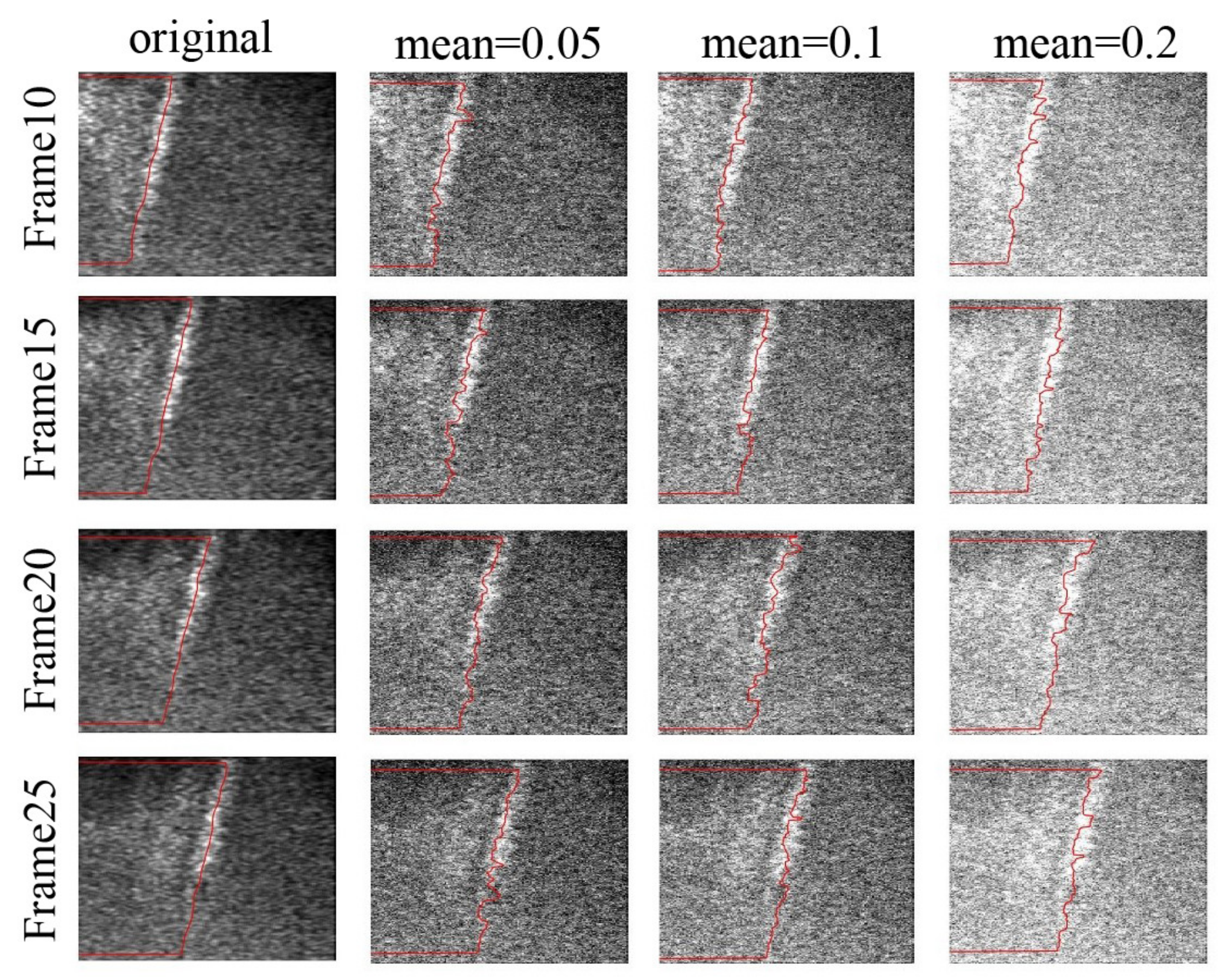
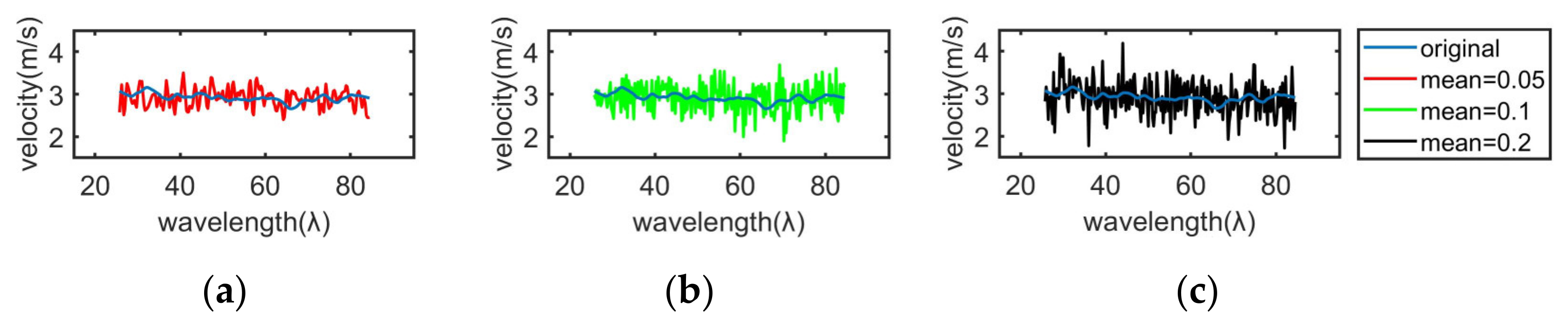
3.4. Noise Adaptability
4. Results
4.1. Evaluation of Shear Wave Velocity
4.2. Result of Velocity Reconstruction Image
4.3. Noise Adaptability Results
5. Discussion
6. Conclusions
Author Contributions
Funding
Acknowledgments
Conflicts of Interest
References
- Amzar, D.; Cotoi, L.; Sporea, I.; Timar, B.; Schiller, O.; Schiller, A.; Borlea, A.; Pop, N.G.; Stoian, D. Shear Wave Elastography in Patients with Primary and Secondary Hyperparathyroidism. J. Clin. Med. 2021, 10, 697. [Google Scholar] [CrossRef]
- Bamber, J.; Cosgrove, D.; Dietrich, C.F. EFSUMB guidelines and recommendations on the clinical use of ultrasound elastography. Part 1: Basic principles and technology. Ultraschall Med. 2013, 34, 169–184. [Google Scholar] [CrossRef]
- Pieczewska, B.; Glińska-Suchocka, K.; Niżański, W.; Dzięcioł, M. Decreased Size of Mammary Tumors Caused by Preoperative Treatment with Aglepristone in Female Domestic Dogs (Canis familiaris) Do Not Influence the Density of the Benign Neoplastic Tissue Measured Using Shear Wave Elastography Technique. Animals 2021, 11, 527. [Google Scholar] [CrossRef]
- Yamakoshi, Y.; Sato, J.; Sato, T. Ultrasonic imaging of internal vibration of soft tissue under forced vibration. IEEE Trans. Ultrason. Ferroelect. Freq. Control. 1990, 37, 45–53. [Google Scholar] [CrossRef]
- Cepeha, C.M.; Paul, C.; Borlea, A.; Fofiu, R.; Borcan, F.; Dehelean, C.A.; Ivan, V.; Stoian, D. Shear-Wave Elastography—Diagnostic Value in Children with Chronic Autoimmune Thyroiditis. Diagnostics 2021, 11, 248. [Google Scholar] [CrossRef]
- Sarvazyan, A.P.; Rudenko, O.V.; Swanson, S.D. Shear Wave Elasticity Imaging: A new ultrasonic technology of medical diagnostics. Ultrasound Med. Biol. 1998, 24, 1419–1435. [Google Scholar] [CrossRef]
- Sandrin, L.; Catheline, S.; Tanter, M.; Hennequin, X.; Fink, M. Time-resolved pulsed elastography with ultrafast ultrasonic imaging. Ultrason. Imaging 1999, 21, 259–272. [Google Scholar] [CrossRef]
- Zhang, Z.J.; Ng, G.Y.F.; Fu, S.N. Effects of habitual loading on patellar tendon mechanical and morphological properties in basketball and volleyball players. Eur. J. Appl. Physiol. 2015, 115, 2263–2269. [Google Scholar] [CrossRef]
- Kot, B.C.W.; Zhang, Z.J.; Lee, A.W.C. Elastic Modulus of Muscle and Tendon with Shear Wave Ultrasound Elastography: Variations with Different Technical Settings. PLoS ONE 2012, 7, e44348. [Google Scholar] [CrossRef]
- Maïsetti, O.; Hug, F.; Bouillard, K. Characterization of passive elastic properties of the human medial gastrocnemius muscle belly using supersonic shear imaging. J. Biomech. 2012, 45, 978–984. [Google Scholar] [CrossRef]
- Lacourpaille, L.; Hug, F.; Bouillard, K. Supersonic shear imaging provides a reliable measurement of resting muscle shear elastic modulus. Physiol. Meas. 2012, 33, N19. [Google Scholar] [CrossRef]
- Sandrin, L.; Fourquet, B.; Hasquenoph, J.M. Transient elastography: A new noninvasive method for assessment of hepatic fibrosis. Ultrasound Med. Biol. 2003, 29, 1705–1713. [Google Scholar] [CrossRef]
- Nightingale, K.; McAleavey, S.; Trahey, G. Shear-wave generation using acoustic radiation force: In vivo and ex vivo results. Ultrasound Med. Biol. 2003, 29, 1715–1723. [Google Scholar] [CrossRef]
- Nightingale, K.; Soo, M.S.; Nightingale, R. Acoustic radiation force impulse imaging: In vivo demonstration of clinical feasibility. Ultrasound Med. Biol. 2002, 28, 227–235. [Google Scholar] [CrossRef]
- Song, P.; Zhao, H.; Manduca, A. Comb-push ultrasound shear elastography (CUSE): A novel method for two-dimensional shear elasticity imaging of soft tissues. IEEE Trans. Med. Imaging 2012, 31, 1821–1832. [Google Scholar] [CrossRef]
- Song, P.; Urban, M.W.; Manduca, A. Comb-push ultrasound shear elastography (CUSE) with various ultrasound push beams. IEEE Trans. Med. Imaging 2013, 32, 1435–1447. [Google Scholar] [CrossRef] [PubMed]
- Bercoff, J.; Tanter, M.; Fink, M. Supersonic shear imaging: A new technique for soft tissue elasticity mapping. IEEE Trans. Ultrason. Ferroelect. Freq. Contr. 2004, 51, 396–409. [Google Scholar] [CrossRef]
- Tanter, M.; Bercoff, J.; Athanasiou, A. Quantitative assessment of breast lesion viscoelasticity: Initial clinical results using supersonic shear imaging. Ultrasound Med. Biol. 2008, 34, 1373–1386. [Google Scholar] [CrossRef]
- Evans, A. Breast Shear Wave Elastography in Clinical Practice. 2019 IEEE International Ultrasonics Symposium (IUS), Glasgow, UK, 6–9 October 2019; pp. 119–121. [Google Scholar]
- Bavu, E. Noninvasive in vivo liver fibrosis evaluation using supersonic shear imaging: A clinical study on 113 hepatitis C virus patients. Ultrasound Med. Biol. 2011, 37, 1361–1373. [Google Scholar] [CrossRef] [PubMed]
- Gheonea, D.I. Real-time sono-elastography in the diagnosis of diffuse liver diseases. World J. Gastroenterol. 2010, 16, 1720–1726. [Google Scholar] [CrossRef]
- Berg, W.A.; Cosgrove, D.O.; Doré, C.J. Shear-wave elastography improves the specificity of breast us: The BE1 multinational study of 939 masses. Radiology 2012, 262, 435–449. [Google Scholar] [CrossRef]
- Zhou, J.; Zhan, W.; Chang, C. Breast lesions: Evaluation with shear wave elastography, with special emphasis on the ‘stiff rim’ sign. Radiology 2014, 272, 63–72. [Google Scholar] [CrossRef]
- Palmeri, M.L.; Wang, M.H.; Dahl, J.J.; Frinkley, K.D.; Nightingale, K.R. Quantifying hepatic shear modulus in vivo using acoustic radiation force. Ultrasound Med. Biol. 2008, 34, 546–558. [Google Scholar] [CrossRef]
- McLaughlin, J.; Renzi, D. Shear wave speed recovery in transient elastography and supersonic imaging using propagating fronts. Inverse Probl. 2006, 22, 681. [Google Scholar] [CrossRef]
- Carrascal, C.A.; Chen, S.; Manduca, A.; Greenleaf, J.F.; Urban, M.W. Improved Shear Wave Group Velocity Estimation Method Based on Spatiotemporal Peak and Thresholding Motion Search. IEEE Trans. Ultrason. Ferroelect. Freq. Contr. 2017, 64, 660–668. [Google Scholar] [CrossRef]
- Oliphant, T.E.; Manduca, A.; Ehman, R.L.; Greenleaf, J.F. Complex-valued stiffness reconstruction for magnetic resonance elastography by algebraic inversion of the differential equation. Mag. Reson. Med. 2001, 45, 299–310. [Google Scholar] [CrossRef]
- Urban, M.W.; Greenleaf, J.F. Use of the radon transform for estimation of shear wave speed. J. Acoust. Soc. Am. 2012, 132, 1982. [Google Scholar] [CrossRef]
- Rouze, N.C.; Wang, M.H.; Palmeri, M.L.; Nightingale, K.R. Robust estimation of time-of-flight shear wave speed using a radon sum transformation. IEEE Trans. Ultrason. Ferroelect. Freq. Contr. 2010, 57, 2662–2670. [Google Scholar] [CrossRef]
- Chen, S.; William, W.; Callstrom, M.R. Assessment of liver viscoelasticity by using shear waves induced by ultrasound radiation force. Radiology 2013, 266, 940–947. [Google Scholar] [CrossRef]
- Braticevici, C.F.; Andronescu, D.; Usvat, R. Acoustic radiation force imaging sonoelastography for noninvasive staging of liver fibrosis. World J. Gastroenterol. 2009, 15, 5525–5532. [Google Scholar] [CrossRef]
- Wang, M.H.; Palmeri, M.L.; Rotemberg, V.M.; Rouze, N.C.; Nightingale, K.R. Improving the robustness of time-of-flight based shear wave speed reconstruction methods using RANSAC in human liver in vivo. Ultrasound Med. Biol. 2010, 36, 802–813. [Google Scholar] [CrossRef]
- Song, P.; Manduca, A.; Zhao, H. Fast Shear Compounding Using Robust Two-dimensional Shear Wave Speed Calculation and Multi-directional Filtering. Ultrasound Med. Biol. 2014, 40, 1343–1355. [Google Scholar] [CrossRef] [PubMed]
- Rifat, A.; Gerber, S.A.; Mcaleavey, S.A. Plane wave imaging improves single track location shear wave elasticity imaging. IEEE Trans. Ultrason. Ferroelect. Freq. Contr. 2018, 65, 1402–1414. [Google Scholar]
- Racedo, J.; Urban, M.W. Evaluation of Reconstruction Parameters for 2-D Comb-Push Ultrasound Shear Wave Elastography. IEEE Trans. Ultrason. Ferroelect. Freq. Contr. 2019, 66, 254–263. [Google Scholar] [CrossRef] [PubMed]
- Ahmed, R.; Doyley, M.M. Parallel Receive Beamforming Improves the Performance of Focused Transmit-Based Single-Track Location Shear Wave Elastography. IEEE Trans. Ultrason. Ferroelect. Freq. Contr. 2020, 67, 2057–2068. [Google Scholar] [CrossRef]
- Kijanka, P.; Ambrozinski, L.; Urban, M.W. Two Point Method for Robust Shear Wave Phase Velocity Dispersion Estimation of Viscoelastic Materials. Ultrasound Med. Biol. 2019, 45, 2540–2553. [Google Scholar] [CrossRef] [PubMed]
- Kijanka, P.; Matthew, W.U. Local Phase Velocity Based Imaging (LPVI): A New Technique Used for Ultrasound Shear Wave Elastography. IEEE Trans. Med. Imaging 2018, 38, 894–908. [Google Scholar] [CrossRef]
- Kijanka, P.; Qiang, B.; Song, P. Robust phase velocity dispersion estimation of viscoelastic materials used for medical applications based on the multiple signal classification method. IEEE Trans. Ultrason. Ferroelect. Freq. Contr. 2018, 65, 423–439. [Google Scholar] [CrossRef]
- Ruud, J.G. Viscoelasticity Mapping by Identification of Local Shear Wave Dynamics. IEEE Trans. Ultrason. Ferroelect. Freq. Contr. 2017, 64, 1666–1673. [Google Scholar]
- Budelli, E. A diffraction correction for storage and loss moduli imaging using radiation force based elastography. Phys. Med. Biol. 2016, 62, 91. [Google Scholar] [CrossRef]


| Acquisition Parameter | Title 3 |
|---|---|
| Wavelength (λ) | 0.2464 mm |
| Acoustic velocity (m/s) | 1540 |
| Excitation frequency (Hz) | 6.25 × 10⁶ |
| Excitation duration (cycles) | 1200 |
| Number of array elements used in acoustic radiation force generation | 32 |
| Four focal points using supersonic shear imaging technology (λ) | 20,40,60,80 |
| Transverse range of region of interest (λ) | −20~80 |
| Transverse resolution of beamforming (λ) | 0.25 |
| Longitudinal range of region of interest (λ) | 15~95 |
| Longitudinal resolution of beamforming (λ) | 0.5 |
| Excitation voltage (V) | 50 |
| Channel data sampling frequency (Hz) | 25 × 106 |
| Each frame acquisition interval (us) | 100 |
| Capture shear wave frame number | 70 |
| Background Elasticity (kPa) | Theoretical Velocity (m/s) | Estimated Method | Estimated Velocity (m/s) | Relative Error (%) |
|---|---|---|---|---|
| 30 | 3.1623 | The time-to-peak method | 2.8078 ± 0.1944 | 11.21% |
| The lateral peak method | 2.8622 ± 0.2114 | 9.49% | ||
| The real-time curve tracing method | 2.9382 ± 0.1221 | 7.09% |
| Modulus of Elasticity (kPa) | Theoretical Velocity (m/s) | Estimated Velocity (m/s) | Relative Error (%) | |
|---|---|---|---|---|
| 1 | Inclusion: 10 | 1.8257 | 1.9544 ± 0.3914 | 7.05% |
| Background: 30 | 3.1623 | 2.9179 ± 0.2236 | 7.73% | |
| 2 | Inclusion: 40 | 3.6514 | 3.3501 ± 0.5583 | 8.25% |
| Background: 30 | 3.1623 | 2.9797 ± 0.2179 | 5.77% | |
| 3 | Inclusion: 60 | 4.4721 | 4.1692 ± 0.6952 | 6.77% |
| Background: 30 | 3.1623 | 2.9754 ± 0.2328 | 5.91% | |
| Noise Level | Estimated Velocity (m/s) | Relative Error (%) | |
|---|---|---|---|
| original | 2.9193 ± 0.0930 | 7.68% | |
| mean = 0 | STD = 0.10 | 2.9185 ± 0.1021 | 7.71% |
| STD = 0.20 | 2.9167 ± 0.1119 | 7.77% | |
| STD = 0.30 | 2.9106 ± 0.1332 | 7.96% | |
| STD = 0.1 | Mean = 0.05 | 2.9111 ± 0.2110 | 7.94% |
| Mean = 0.10 | 2.9069 ± 0.3181 | 8.08% | |
| Mean = 0.20 | 2.9033 ± 0.3938 | 8.19% | |
Publisher’s Note: MDPI stays neutral with regard to jurisdictional claims in published maps and institutional affiliations. |
© 2021 by the authors. Licensee MDPI, Basel, Switzerland. This article is an open access article distributed under the terms and conditions of the Creative Commons Attribution (CC BY) license (http://creativecommons.org/licenses/by/4.0/).
Share and Cite
Li, Y.; Lv, Q.; Dai, J.; Tian, Y.; Guo, J. Shear Wave Velocity Estimation Using the Real-Time Curve Tracing Method in Ultrasound Elastography. Appl. Sci. 2021, 11, 2095. https://doi.org/10.3390/app11052095
Li Y, Lv Q, Dai J, Tian Y, Guo J. Shear Wave Velocity Estimation Using the Real-Time Curve Tracing Method in Ultrasound Elastography. Applied Sciences. 2021; 11(5):2095. https://doi.org/10.3390/app11052095
Chicago/Turabian StyleLi, Yu, Qian Lv, Jiayue Dai, Ye Tian, and Jianzhong Guo. 2021. "Shear Wave Velocity Estimation Using the Real-Time Curve Tracing Method in Ultrasound Elastography" Applied Sciences 11, no. 5: 2095. https://doi.org/10.3390/app11052095
APA StyleLi, Y., Lv, Q., Dai, J., Tian, Y., & Guo, J. (2021). Shear Wave Velocity Estimation Using the Real-Time Curve Tracing Method in Ultrasound Elastography. Applied Sciences, 11(5), 2095. https://doi.org/10.3390/app11052095