Article

Assessment of Respiratory System Resistance during High-Frequency Oscillatory Ventilation Based on In Vitro Experiment

Department of Biomedical Technology, Faculty of Biomedical Engineering, Czech Technical University in Prague, nam. Sitna 3105, 272 01 Kladno, Czech Republic
*
Author to whom correspondence should be addressed.
Appl. Sci. 2021, 11(23), 11279; https://doi.org/10.3390/app112311279
Submission received: 15 September 2021 / Revised: 19 November 2021 / Accepted: 23 November 2021 / Published: 29 November 2021

Abstract

:
High-frequency oscillatory ventilation (HFOV) is a type of mechanical ventilation with a protective potential characterized by a small tidal volume. Unfortunately, HFOV has limited monitoring of ventilation parameters and mechanical parameters of the respiratory system, which makes it difficult to adjust the continuous distension pressure (CDP) according to the individual patient’s airway status. Airway resistance Raw is one of the important parameters describing the mechanics of the respiratory system. The aim of the presented study was to verify in vitro whether the resistance of the respiratory system Rrs can be reliably determined during HFOV to evaluate Raw in pediatric and adult patients. An experiment was performed with a 3100B high-frequency oscillator, a physical model of the respiratory system, and a pressure and flow measurement system. The physical model with different combinations of resistance and compliance was ventilated during the experiment. The resistance Rrs was calculated from the impedance of the physical model, which was determined from the spectral density of the pressure at airway opening and the spectral cross-density of the gas flow and pressure at airway opening. Rrs of the model increased with an added resistor and did not change significantly with a change in compliance. The method is feasible for monitoring respiratory system resistance during HFOV and has the potential to optimize CDP settings during HFOV in clinical practice.

1. Introduction

High-frequency oscillatory ventilation (HFOV) is one of the unconventional methods of mechanical lung ventilation. It is characterized by a small tidal volume, approaching an anatomical dead space, with a protective potential [1]. Attenuation of pressure amplitude along the bronchial tree may contribute to less mechanical stress on lung tissue during HFOV compared with conventional mechanical ventilation (CMV) [2]. The patients with severe acute respiratory distress syndrome (ARDS) that do not tolerate CMV may be the target group for HFOV [3] if an alternative rescue therapy to ECMO is considered. With a number of etiologies and subtypes, ARDS is manifested by noncardiogenic pulmonary edema and hypoxia. Although new personalized pharmacological therapies for ARDS subtypes are being sought, also in the context of the COVID-19 pandemic, targeted treatment is lacking and ARDS is still the leading cause of death in critically ill patients [4,5]. Continuous distension pressure (CDP) and a set fraction of inspired oxygen determine the oxygenation of the ventilated subject in HFOV. Carbon dioxide is eliminated from the lungs by pressure oscillations that are added to CDP [6]. Recently, there have been studies that emphasize the need for an individualized approach in setting the ventilation parameters of HFOV [7,8]. It has also been shown that other monitoring and computational methods, including electrical impedance tomography (EIT) [9], optoelectronic plethysmography [10], or impedance analysis of the respiratory system [11], can lead to optimization of HFOV settings. The results of previous studies conducted with HFOV may have been influenced by settings that were not sufficiently individualized to the needs of individual patients [12,13].
Currently, there is no unified approach on how to properly set up CDP with respect to the respiratory status of individual patients. Airway resistance Raw is one of the important parameters describing the mechanics of the respiratory system. Besides tissue resistance, airway resistance Raw is a substantial part of respiratory system resistance Rrs. Elevated Raw can lead to air trapping and hyperinflation, which can result in pulmonary barotrauma [14]. Raw depends on lung volume [15,16], which is directly related to the CDP value [17]. Both ventilation at low lung volumes (CDP is too low for the patient) and ventilation at high lung volumes (CDP is too high) lead to an increase in Raw. Moreover, the increase in resistance at low lung volumes is accompanied by a significant increase in peripheral resistance, which can account for 15% of Raw. The contribution of peripheral resistance to Raw is otherwise negligible [15]. However, the possibilities for monitoring ventilation parameters are small for HFOV. The high-frequency oscillatory ventilators 3100A and 3100B (Vyaire Medical, Mettawa, IL, USA) also lack monitoring of respiratory system mechanics, such as Raw. The 3100B ventilator, designed for adult patients, was used in this study.
The forced oscillation technique (FOT) can be used to evaluate the mechanics of the respiratory system including total respiratory system resistance Rrs [18,19]. In FOT, pressure oscillations with typical frequency f = 5 Hz are applied at the airway opening and Rrs is assessed from the induced flow. Pressure oscillations at 5 Hz can penetrate the peripheral airways and detect changes in resistance in this region of the lung, allowing the assessment of Rrs [18]. In a conventional FOT configuration, an external tool with an oscillator is used to generate high-frequency oscillations. The flow caused by the external oscillations is measured at the airway opening. However, some studies have demonstrated that a high-frequency ventilator itself can be used as a generator of the pressure oscillations utilized by FOT [20,21]. The studies used small animal models whose respiratory mechanics are consistent with neonatal patients. On the contrary, we have not found a study describing the use of the method in larger physical or animal models that correspond to pediatric or adult patients.
Recently, FOT has been integrated into commercially available neonatal ventilator Fabian (Acutronic, Hirzel, Switzerland) to determine the reactance of the respiratory system of a neonatal patient. Studies described the usefulness of reactance analysis in ventilated [22] or spontaneously breathing neonatal patients [23]. In general, there is no information about the analysis of Rrs in HFOV. As the method of assessing reactance of the respiratory system by FOT becomes clinically available, we suppose that monitoring of Rrs might have similar clinical potential and could provide an early warning to elevated airway resistance.
The aim of the presented study is to verify whether it is possible, under stable and well-defined laboratory conditions, to use pressure oscillations generated by the high-frequency oscillatory ventilator to determine the resistance of the respiratory system Rrs from the measured proximal airway pressure and flow. We hypothesize that this method could be used to assess Raw at the bedside in neonatal, pediatric, and adult patients ventilated by HFOV similarly as reactance of the respiratory system. The presented method could be used also with ventilators 3100A and 3100B.

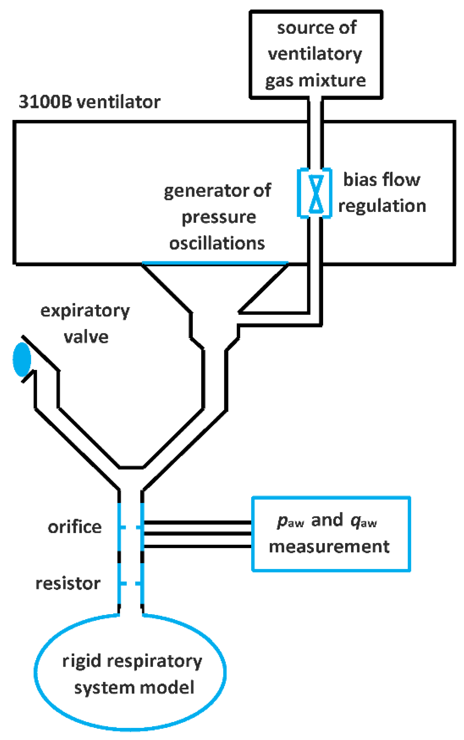
2. Materials and Methods
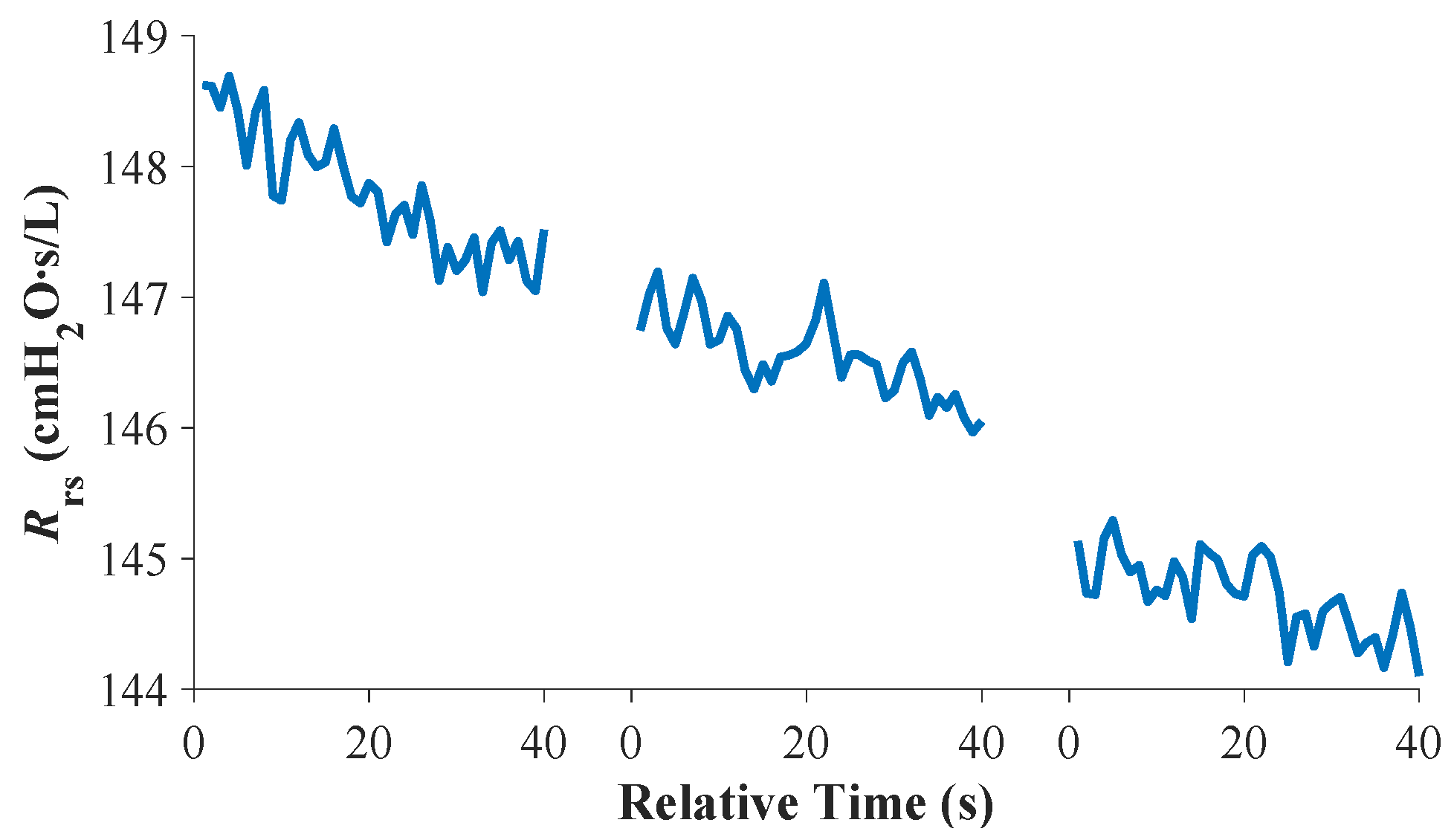
The configuration of the experiment is shown in Figure 1 [24]. The high-frequency oscillatory ventilator 3100B with standard accessories was used for the experiment. The patient circuit was connected via an endotracheal tube to a model of the respiratory system that consisted of a glass demijohn. At one phase of the experiment, an Rp5 parabolic resistor (Michigan Instruments, Grand Rapids, MI, USA) was added to the circuit. The Rp5 simulated the increased resistance of the respiratory system and the glass demijohn simulated the compliance of the lungs. Measurements performed without and with Rp5 were repeated for three glass demijohns of 54, 35, and 25 L. Values of corresponding compliances were 37, 24, and 17 mL/cmH2O, respectively [24]. The following ventilation parameters were used in the experiment: bias flow = 30 L/min, ventilatory frequency f = 5 Hz, CDP = 12 cmH2O, and pressure oscillation amplitude ΔP = 20 cmH2O. Inspiration to expiration time was set as I:E = 1:1. The ventilation parameters were set according to [25]. Pressure paw and flow qaw were recorded at the inlet of the model of the respiratory system using a measurement system specifically designed for HFOV monitoring [26]. The flow was calculated based on the pressure difference measured across an orifice. Both the signals paw and qaw were recorded at a sampling frequency f = 1000 Hz.
The respiratory system resistance Rrs measured at a pressure oscillation frequency of f = 5 Hz was calculated from the respiratory system impedance Zrs following the spectral density method described in [24]. Rrs was obtained from Zrs by converting from polar to Cartesian coordinates according to Equation (1):
R rs = Z m a g c o s Z a n g ,
where Zmag stands for the amplitude of the respiratory system impedance and Zang stands for the angle of the respiratory system impedance.

3. Results

The measurements of Rrs in our experiment are summarized in Figure 2 and Table 1. Measurements 1–3 correspond to no added resistor and measurements 4–6 correspond to the phase of the experiment with the added resistor Rp. Three demijohns representing different compliances (37, 24, and 17 mL/cmH2O) were used in both phases of the experiment. The resistance Rrs substantially increased by more than 100 cmH2O∙s/L (over 220% increase) after the addition of the resistor to the model of the respiratory system (the change between Section 3 and Section 4). The change in the compliance value did not have a substantial effect on the measured Rrs values as the mean Rrs did not differ for more than 4 cmH2O∙s/L (less than 10%) when Rp remained unchanged.
Figure 3 and Figure 4 describe in more detail the measured signal of Rrs without and with the added resistor Rp5, respectively. It can be seen in the figures that Rrs decreased over time. However, the decay of Rrs is negligible compared to the Rrs value. The decay over 40 s, estimated from the linear interpolation of Rrs signals, was 3.8% of Rrs without the resistor and 1.8% with the resistor.
It can also be seen from Figure 3 and Figure 4 in detail that change of the compliance of the respiratory system model does not affect substantially measured Rrs. For a measurement without an added resistor (Figure 3), a small increase in Rrs of 3.2 cmH2O∙s/L (7.7%) was observed with the change of compliance from 37 to 24 mL/cmH2O and a very small change in Rrs of about 0.4 cmH2O∙s/L (1.0%) was observed with the change of compliance from 24 to 17 mL/cmH2O. A decrease in Rrs about 1.2 cmH2O∙s/L and 1.8 cmH2O∙s/L (0.8% and 1.3%, respectively) was observed for measurements with the resistor.

4. Discussion

The presented results show that changes in Raw can be monitored during HFOV by measuring the resistance Rrs at an oscillation frequency of 5 Hz, when the basic mechanical properties of the respiratory system are consistent with larger animals or pediatric and adult patients. A physical model of the respiratory system was designed and an in vitro lab experiment was performed using different combinations of resistance and airway compliance values. It was shown that Rrs increases when Raw increases. The results of this in vitro study also suggest that it is possible to follow the trend of Raw under conditions of changing lung compliance.
The low standard deviations of Rrs summarized in Table 1 indicate sufficient robustness of the algorithm used in signal processing. In Figure 3 and Figure 4, small oscillations of the calculated Rrs values can be seen. The oscillations are due to the processing of the noisy pressure and flow signals. The addition of a resistor to the ventilated system increased the standard deviation of Rrs. The flow was more turbulent with the added resistor and this resulted in an increase in the noise in the flow signal [24]. However, the increased turbulence did not degrade the evaluation of Rrs.
Our in vitro study has some limitations. First, a single ventilation frequency of 5 Hz was investigated. The choice of ventilation frequency as the most appropriate was based on previous studies [18,19,24,27,28]. Second, we did not vary the CDP during the test, as this would be of little importance in a physical model with rigid walls. Animal studies [20,29,30], which mimicked immature patients and used the same FOT measurement method, reported a significant increase in Rrs during lung derecruitment because of the low CDP applied during HFOV or the low positive end-expiratory pressure (PEEP) applied during CMV. The results are in agreement with the findings presented in [15], where the decrease in airway diameter was explained by a decrease in mean airway pressure. In contrast, only small changes in Rrs are observed at CDP or PEEP values that are sufficient to maintain lung inflation. This is consistent with our simulation performed on a rigid model. Third, the results show that when the Rp5 resistor was added to the model, the measured Rrs increased by more than 100 cmH2O∙s/L on average, but the physical properties of the Rp5 resistor may have contributed to such a large increase in Rrs. Resistor Rp5 is designed as parabolic, which means that the actual resistance value depends on gas flow rate. Moreover, the resistance of Rp5 is determined by its sudden and short decrease in airway diameter, a mechanism that is not present in vivo. The choke point created by the addition of Rp5 may cause the part of pressure-flow oscillation to be reflected into the glass demijohn and not return to the measuring system, resulting in an apparently more pronounced increase in resistance. It should also be taken into account that the physical properties of the glass demijohns used differ from the actual lungs. Pressure and flow oscillations could be deflected on the wall of the glass demijohn such that the oscillations could not return to the measuring system and would instead be damped within the glass demijohn. Such deflection does not occur in the airway tree in the lungs and could explain the difference between the actual resistance and the measured Rrs. Finally, small changes in the shape of the patient circuit and endotracheal tube between measurements could also account for some of the inaccuracies in the calculation of Rrs.
The presented method of measuring Rrs during HFOV is suitable for bedside patient monitoring because only a pressure and flow orifice is added to the patient circuit. In our study, a custom-made system consisting of an orifice, sensors, digitizing hardware, and a laptop with evaluation software was used [14]. In a real clinical scenario, any monitoring device capable of measuring proximal pressure and flow during HFOV and transmitting data in real time could be used. The disadvantage of the presented method may be the increased flow resistance and dead space caused by the addition of an orifice to the patient’s circuit.
Assessment of respiratory mechanics using FOT in mechanical ventilators is now available to physicians with the Fabian neonatal ventilator [22,23]. However, the Fabian currently only determines the respiratory system reactance measured at an oscillation frequency of f = 10 Hz, which is typical for neonates. Besides the fact that in our study both parameters were measured at a frequency of f = 5 Hz, we believe that there is no significant difference between the method investigated in our study and the FOT method used by the Fabian ventilator. Therefore, no additional hardware would be required to simultaneously measure reactance and Rrs at the patient’s bedside. Based on our study, we propose that not only reactance [24] but also Rrs could be assessed during HFOV.

5. Conclusions

In this study, for the first time, the feasibility of monitoring respiratory system resistance using the FOT method during HFOV under stable well-defined laboratory conditions was verified in a physical model whose properties correspond to a large laboratory animal. The FOT method used is simple enough to be applied at the patient’s bedside in clinical practice, requires no circuit disconnection, and can be used for long-term monitoring. Ventilator operators could have information on the resistance of the respiratory system, which could facilitate an early response to an increase in resistance and thus prevent pulmonary barotrauma. As the FOT method is already used in a commercially available neonatal ventilator to determine respiratory system reactance, simultaneous measurement of resistance could be readily available in clinical practice.

Author Contributions

Conceptualization, M.R.; methodology, J.M.; software, J.M., J.R.; formal analysis, J.M.; investigation, M.R.; curation, J.M.; writing—original draft preparation, J.M.; writing—review and editing, M.R., J.R.; visualization, J.M.; supervision, M.R.; project administration, M.R.; funding acquisition, M.R. All authors have read and agreed to the published version of the manuscript.

Funding

This work was supported by the project SGS19/202/OHK4/3T/17 and SGS20/202/OHK4/3T/17.

Data Availability Statement

The data presented in this study are available on request from the corresponding author.

Conflicts of Interest

The authors declare no conflict of interest.

References

  1. Meade, M.O.; Young, D.; Hanna, S.; Zhou, Q.; Bachman, T.E.; Bollen, C.; Slutsky, A.S.; Lamb, S.E.; Adhikari, N.K.; Mentzelopoulos, S.D.; et al. Severity of Hypoxemia and Effect of High-Frequency Oscillatory Ventilation in Acute Respiratory Distress Syndrome. Am. J. Respir. Crit. Care Med. 2017, 196, 727–733. [Google Scholar] [CrossRef]
  2. van Genderingen, H.R.; van Vught, A.J.; Duval, E.L.; Markhorst, D.G.; Jansen, J.R. Attenuation of pressure swings along the endotracheal tube is indicative of optimal distending pressure during high-frequency oscillatory ventilation in a model of acute lung injury. Pediatr. Pulmonol. 2002, 33, 429–436. [Google Scholar] [CrossRef] [PubMed]
  3. Sud, S.; Sud, M.; Friedrich, J.O.; Meade, M.O.; Ferguson, N.D.; Wunsch, H.; Adhikari, N.K. High frequency oscillation in patients with acute lung injury and acute respiratory distress syndrome (ARDS): Systematic review and meta-analysis. BMJ 2010, 340, c2327. [Google Scholar] [CrossRef] [PubMed] [Green Version]
  4. Meduri, G.U.; Annane, D.; Confalonieri, M.; Chrousos, G.P.; Rochwerg, B.; Busby, A.; Ruaro, B.; Meibohm, B. Pharmacological principles guiding prolonged glucocorticoid treatment in ARDS. Intensive Care Med. 2020, 46, 2284–2296. [Google Scholar] [CrossRef] [PubMed]
  5. Williams, G.W.; Berg, N.K.; Reskallah, A.; Yuan, X.; Eltzschig, H.K. Acute Respiratory Distress Syndrome. Anesthesiology 2021, 134, 270–282. [Google Scholar] [CrossRef] [PubMed]
  6. Sklar, M.C.; Fan, E.; Goligher, E.C. High-Frequency Oscillatory Ventilation in Adults with ARDS: Past, Present, and Future. Chest 2017, 152, 1306–1317. [Google Scholar] [CrossRef]
  7. de Jager, P.; Kamp, T.; Dijkstra, S.K.; Burgerhof, J.G.; Markhorst, D.G.; Curley, M.A.; Cheifetz, I.M.; Kneyber, M.C. Feasibility of an alternative, physiologic, individualized open-lung approach to high-frequency oscillatory ventilation in children. Ann. Intensive Care 2019, 9, 9. [Google Scholar] [CrossRef]
  8. de Jager, P.; Burgerhof, J.G.; Koopman, A.A.; Markhorst, D.G.; Kneyber, M.C. Physiologic responses to a staircase lung volume optimization maneuver in pediatric high-frequency oscillatory ventilation. Ann. Intensive Care 2020, 10, 153. [Google Scholar] [CrossRef]
  9. Liu, S.; Zhao, Z.; Tan, L.; Wang, L.; Möller, K.; Frerichs, I.; Yu, T.; Huang, Y.; Pan, C.; Yang, Y.; et al. Optimal mean airway pressure during high-frequency oscillatory ventilation in an experimental model of acute respiratory distress syndrome: EIT-based method. Ann. Intensive Care 2020, 10, 31. [Google Scholar] [CrossRef] [Green Version]
  10. Lista, G.; Bresesti, I.; Cavigioli, F.; Castoldi, F.; Lupo, E.; LoMauro, A.; Aliverti, A. Efficacy of lung volume optimization maneuver monitored by optoelectronic pletismography in the management of congenital diaphragmatic hernia. Respir. Med. Case Rep. 2017, 22, 133–136. [Google Scholar] [CrossRef]
  11. Zannin, E.; Dellaca, R.L.; Dognini, G.; Marconi, L.; Perego, M.; Pillow, J.J.; Tagliabue, P.E.; Ventura, M.L. Effect of frequency on pressure cost of ventilation and gas exchange in newborns receiving high-frequency oscillatory ventilation. Pediatr. Res. 2017, 82, 994–999. [Google Scholar] [CrossRef]
  12. Kneyber, M.C.; Markhorst, D.G. Do We Really Know How to Use High-Frequency Oscillatory Ventilation in Critically Ill Children? Am. J. Respir. Crit. Care Med. 2016, 193, 1067–1068. [Google Scholar] [CrossRef]
  13. Kneyber, M.C.; Markhorst, D.G. Any trial can (almost) kill a good technique. Intensive Care Med. 2016, 42, 1092–1093. [Google Scholar] [CrossRef] [PubMed]
  14. Meyers, M.; Rodrigues, N.; Ari, A. High-frequency oscillatory ventilation: A narrative review. Can. J. Respir. Ther. 2019, 55, 40–46. [Google Scholar] [CrossRef] [PubMed]
  15. Macklem, P.T.; Mead, J. Resistance of central and peripheral airways measured by a retrograde catheter. J. Appl. Physiol. 1967, 22, 395–401. [Google Scholar] [CrossRef] [PubMed] [Green Version]
  16. Briscoe, W.A.; Dubois, A.B. The relationship between airway resistance, airway conductance and lung volume in subjects of different age and body size. J. Clin. Investig. 1958, 37, 1279–1285. [Google Scholar] [CrossRef] [PubMed] [Green Version]
  17. Miedema, M.; de Jongh, F.H.; Frerichs, I.; van Veenendaal, M.B.; van Kaam, A.H. The effect of airway pressure and oscillation amplitude on ventilation in preterm infants. Eur. Respir. J. 2012, 40, 479–484. [Google Scholar] [CrossRef] [Green Version]
  18. Bhattarai, P.; Myers, S.; Chia, C.; Weber, H.C.; Young, S.; Williams, A.D.; Sohal, S.S. Clinical Application of Forced Oscillation Technique (FOT) in Early Detection of Airway Changes in Smokers. J. Clin. Med. 2020, 9, 2778. [Google Scholar] [CrossRef]
  19. Brashier, B.; Salvi, S. Measuring lung function using sound waves: Role of the forced oscillation technique and impulse oscillometry system. Breathe 2015, 11, 57–65. [Google Scholar] [CrossRef]
  20. Dellacà, R.L.; Zannin, E.; Ventura, M.L.; Sancini, G.; Pedotti, A.; Tagliabue, P.; Miserocchi, G. Assessment of dynamic mechanical properties of the respiratory system during high-frequency oscillatory ventilation. Crit. Care Med. 2013, 41, 2502–2511. [Google Scholar] [CrossRef]
  21. Zannin, E.; Ventura, M.L.; Dellacà, R.L.; Natile, M.; Tagliabue, P.; Perkins, E.J.; Sourial, M.; Bhatia, R.; Dargaville, P.A.; Tin-gay, D.G. Optimal mean airway pressure during high-frequency oscillatory ventilation determined by measurement of respiratory system reactance. Pediatr. Res. 2014, 75, 493–499. [Google Scholar] [CrossRef] [PubMed] [Green Version]
  22. Lavizzari, A.; Veneroni, C.; Ottaviani, V.; Francesco, B.; Fumagalli, C.; Colnaghi, M.; Mosca, F.; Dellacà, R.L. Respiratory reactance (Xrs) by Forced Oscillation Technique (FOT) during the first 24h of life in non-intubated preterm infants. Eur. Respir. J. 2019, 54, PA1032. [Google Scholar] [CrossRef]
  23. Zannin, E.; Dellacà, R.L.; Neumann, R.; Schulzke, S. Assessment of lung mechanics for the prediction and evaluation of pulmonary outcome in preterm infants. Eur. Respir. J. 2018, 52, OA306. [Google Scholar] [CrossRef]
  24. Matejka, J.; Rozanek, M.; Rafl, J.; Kudrna, P.; Roubik, K. In Vitro Estimation of Relative Compliance during High-Frequency Oscillatory Ventilation. Appl. Sci. 2021, 11, 899. [Google Scholar] [CrossRef]
  25. Fessler, H.E.; Derdak, S.; Ferguson, N.D.; Hager, D.N.; Kacmarek, R.M.; Thompson, B.T.; Brower, R.G. A protocol for high-frequency oscillatory ventilation in adults: Results from a roundtable discussion. Crit. Care Med. 2007, 35, 1649–1654. [Google Scholar] [CrossRef]
  26. Roubik, K. Measuring and evaluating system designed for high-frequency oscillatory ventilation monitoring. Biomed. Tech. 2014, 59, S979. [Google Scholar] [CrossRef]
  27. Shimoda, T.; Obase, Y.; Nagasaka, Y.; Kishikawa, R.; Mukae, H.; Iwanaga, T. Peripheral bronchial obstruction evaluation in patients with asthma by lung sound analysis and impulse oscillometry. Allergol. Int. 2017, 66, 132–138. [Google Scholar] [CrossRef] [Green Version]
  28. Aronsson, D.; Hesselstrand, R.; Bozovic, G.; Wuttge, D.M.; Tufvesson, E. Airway resistance and reactance are affected in systemic sclerosis. Eur. Clin. Respir. J. 2015, 2, 28667. [Google Scholar] [CrossRef] [Green Version]
  29. Dellacà, R.L.; Zannin, E.; Kostic, P.; Olerud, M.A.; Pompilio, P.P.; Hedenstierna, G.; Pedotti, A.; Frykholm, P. Optimisation of positive end-expiratory pressure by forced oscillation technique in a lavage model of acute lung injury. Intensive Care Med. 2011, 37, 1021–1030. [Google Scholar] [CrossRef]
  30. Zannin, E.; Dellacà, R.L.; Kostic, P.; Pompilio, P.P.; Larsson, A.; Pedotti, A.; Hedenstierna, G.; Frykholm, P. Optimizing positive end-expiratory pressure by oscillatory mechanics minimizes tidal recruitment and distension: An experimental study in a lavage model of lung injury. Crit. Care 2012, 16, R217. [Google Scholar] [CrossRef] [Green Version]
Figure 1. Setup of in vitro experiment [24].
Applsci 11 11279 g001
Figure 2. The computed Rrs during ventilation of the respiratory system model without an added resistor (measurement sections 1, 2, and 3) and with added resistor Rp (measurement sections 4, 5, and 6). To investigate the effect of compliance on the measured resistance of the respiratory system, three glass demijohns with different compliance (37, 24, and 17 mL/cmH2O) were ventilated. Negligible change in Rrs signal amplitude during measurement and a small change in Rrs when compliance changed contrast with a large change in Rrs when the resistance increased.
Applsci 11 11279 g002
Figure 3. The course of computed Rrs during ventilation of the respiratory system model without an added resistor (three 40 s long measurements correspond to measurement sections 1, 2, and 3 in Figure 2). Three glass demijohns with different compliance (37, 24, and 17 mL/cmH2O) were ventilated to investigate the effect of compliance on the measured resistance of the respiratory system.
Applsci 11 11279 g003
Figure 4. The course of computed Rrs during ventilation of the respiratory system model with added resistor Rp5 (three 40 s long measurements correspond to measurement sections 4, 5, and 6 in Figure 2). Three glass demijohns with different compliance (37, 24, and 17 mL/cmH2O) were ventilated to investigate the effect of compliance on the measured resistance of the respiratory system.
Applsci 11 11279 g004
Table 1. Values of computed Rrs (cmH2O∙s/L) in both phases of the experiment (without/with the resistor) for glass demijohns of three different compliances C.
CRrs with No ResistorRrs with Resistor Rp5
(mL/cmH2O)MeanSD 1MeanSD 1
3741.50.2147.80.5
2444.70.2146.60.3
1744.30.1144.70.3
1 SD stands for standard deviation.
Publisher’s Note: MDPI stays neutral with regard to jurisdictional claims in published maps and institutional affiliations.

