Case Report

Late Developed Unusual Nasal Involvement of Postoperative Maxillary Cyst Following Maxillary Sinus Augmentation: A Case Report

1
Private Practice in Periodontics and Implant Dentistry, 75 Dolgoji-ro 27-gil, Seongbuk-gu, Seoul 02771, Korea
2
Department of Periodontology, Periodontal-Implant Clinical Research Institute, School of Dentistry, Kyungheedaero 23, Dongdaemoon-gu, Seoul 02447, Korea
*
Author to whom correspondence should be addressed.
Appl. Sci. 2021, 11(22), 10730; https://doi.org/10.3390/app112210730
Submission received: 9 October 2021 / Revised: 5 November 2021 / Accepted: 12 November 2021 / Published: 13 November 2021
(This article belongs to the Special Issue Applied Science for Oral Implantology—Fake vs. News)

Abstract

:
Postoperative maxillary cyst (POMC) is a benign expansive cystic lesion of the maxilla generally related to invasive maxillary surgeries or trauma. POMC can also develop after maxillary sinus augmentation (MSA), but many dentists are not well-aware of such complication of MSA. A 56-year-old male patient had undergone bilateral MSA. After 18 years, the patient reported painless swelling on the left palate. On the panoramic radiographs, no specific findings were found, but a large unilocular lesion was detected at the medial side of the previous augmentation of the left maxillary sinus on cone-beam computed tomographic examination. The lesion expanded medially and downward to destruct the medial wall of the maxillary sinus and palatal bone. Medial expansion of the lesion also reached the nasal septum and inferior meatus. Due to the extent and the location, the lesion was hard to manage using an intraoral surgical approach. The patient was then referred to an otolaryngologist in a university hospital. Endoscopic marsupialization was performed under general anesthesia. Previous augmentation and dental implants could be maintained during the marsupialization. The removed tissue sample revealed respiratory epithelium with inflammatory cell infiltration, confirming that the lesion was a postoperative maxillary cyst (POMC). There has been no recurrence of POMC to date. POMC is a rare postoperative complication of maxillary sinus augmentation, but clinicians should be aware of the possibility of POMC and the necessity of regular radiological monitoring.

1. Introduction

Maxillary sinus augmentation (MSA) is a well-documented treatment modality for rehabilitating pneumatized posterior maxilla [1]. Despite the reported high predictability, several complications following MSA have been reported. These include graft infection, acute/chronic sinusitis, implant failure, and implant displacement [2,3]. However, some complications, such as postoperative maxillary cyst (POMC), appear not to have wide awareness among dentists.
POMC or surgical ciliated cyst is a benign expansive cystic lesion of the maxilla. POMC was first described in a Japanese article in 1927 [4], possibly due to specific etiologic factors in a specific region. Evidence indicates that Caldwell–Luc operation, orthognathic surgery in the maxilla, and maxillofacial trauma can predispose to POMC [5,6,7]. The most commonly accepted mechanism of POMC is entrapped sinus mucosa after surgery/traumatic event and gradual growth as an epithelial lining cavity [8], potentially indicating that MSA cannot be excluded from the causality of POMC.
In 1991, Misch et al. first demonstrated POMC after MSA [9], followed by a few case studies regarding this issue [10,11,12,13]. POMC after MSA in previous literature presented with the following features: (1) sinus membrane perforation during MSA might be an etiological factor, (2) duration of development or detection largely varied between 3 months and 10 years, (3) the expansion of POMC resorbed the adjacent bone structures and augmented bone, sometimes involving a dental implant(s), and (4) intraoral surgical approach was generally chosen for complete enucleation of POMC.
This case report presents an invasive case and the relevant treatment course of POMC, which was detected 18 years after MSA and invaded the inferior meatus of the nasal cavity.

2. Case Report

A fifty-six-year-old male patient visited one of the authors’ (W-B. P) dental clinic for gradual swelling on his left palatal area in January 2019 (Figure 1). He had been a regular visitor to the clinic after dental implant surgery 18 years ago (in January 2001). However, for the past 3 years, he did not attend re-call visits due to personal reasons. He had mild hypertension and hyperlipidemia. He was a non-smoker.

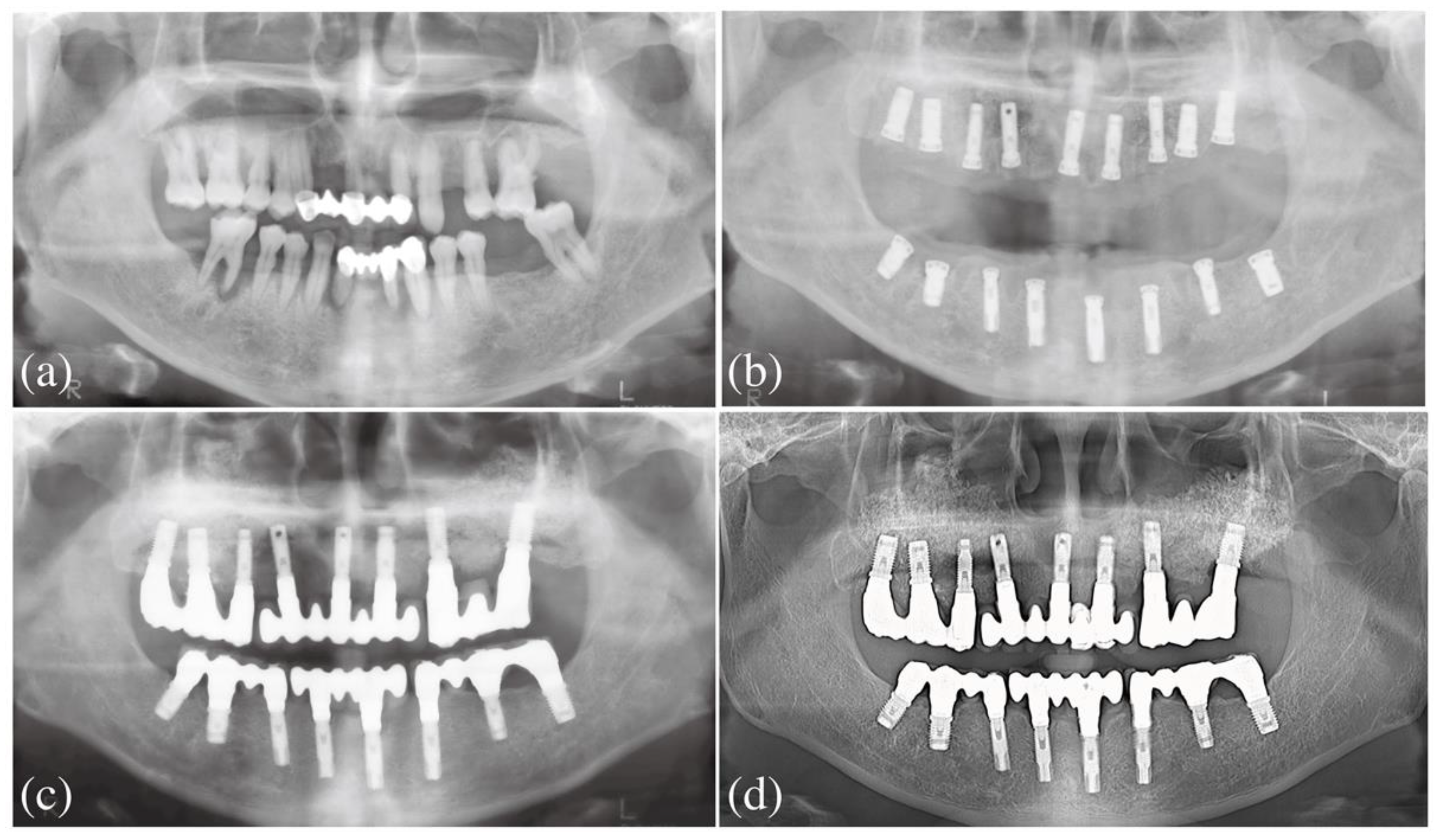
2.1. Past Dental Treatment

The patient visited the present dental clinic for hypermobile teeth, gingival bleeding, and chewing difficulty in 2001. He was diagnosed with severe periodontitis (stage III, grade B periodontitis according to new classification [14]) (Figure 2a). Due to advanced bone loss, it was decided to extract all teeth, followed by bone augmentation and implant placement. Lateral sinus augmentation and implant placement were performed at two months following the extraction for the maxilla (Figure 2b). A bony access window was made using a round bur on each lateral sinus bone wall. During the sinus membrane elevation, sinus membrane perforation occurred near the access window on both sides. After completing the elevation while taking care not to extend the perforation, the perforated sites were covered with Collatape (Zimmer Biomet, Indiana, USA) and bone substitute material (Bio-Oss, Geistlich Pharma, Wolhuson, Switzerland) was gently inserted. Subsequently, osteotomies were performed for placing implants according to the manufacturer’s guidelines. Nine HA-coated external hexed implants (Steri-Oss, Yorba Linda, CA. USA) were placed, and additional bone grafting in the sinus was performed. The access windows were covered by Collatape. Cover screws were connected to the implants for submerged healing. After 6 months, implant uncovering, and abutment connection were performed. The implant on the #14 tooth area failed to achieve proper osseointegration and was removed. Further implant placement was not performed on that site. Two months later, implant prostheses were inserted (Figure 2c). Regular re-call visits approximately one or two times a year were scheduled.

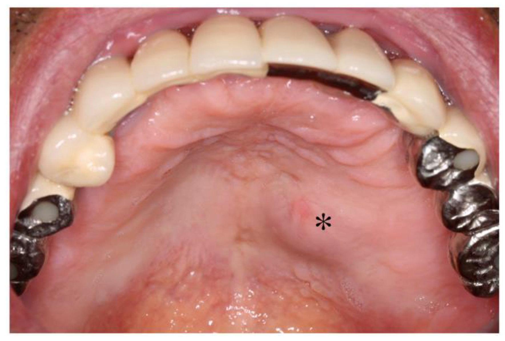
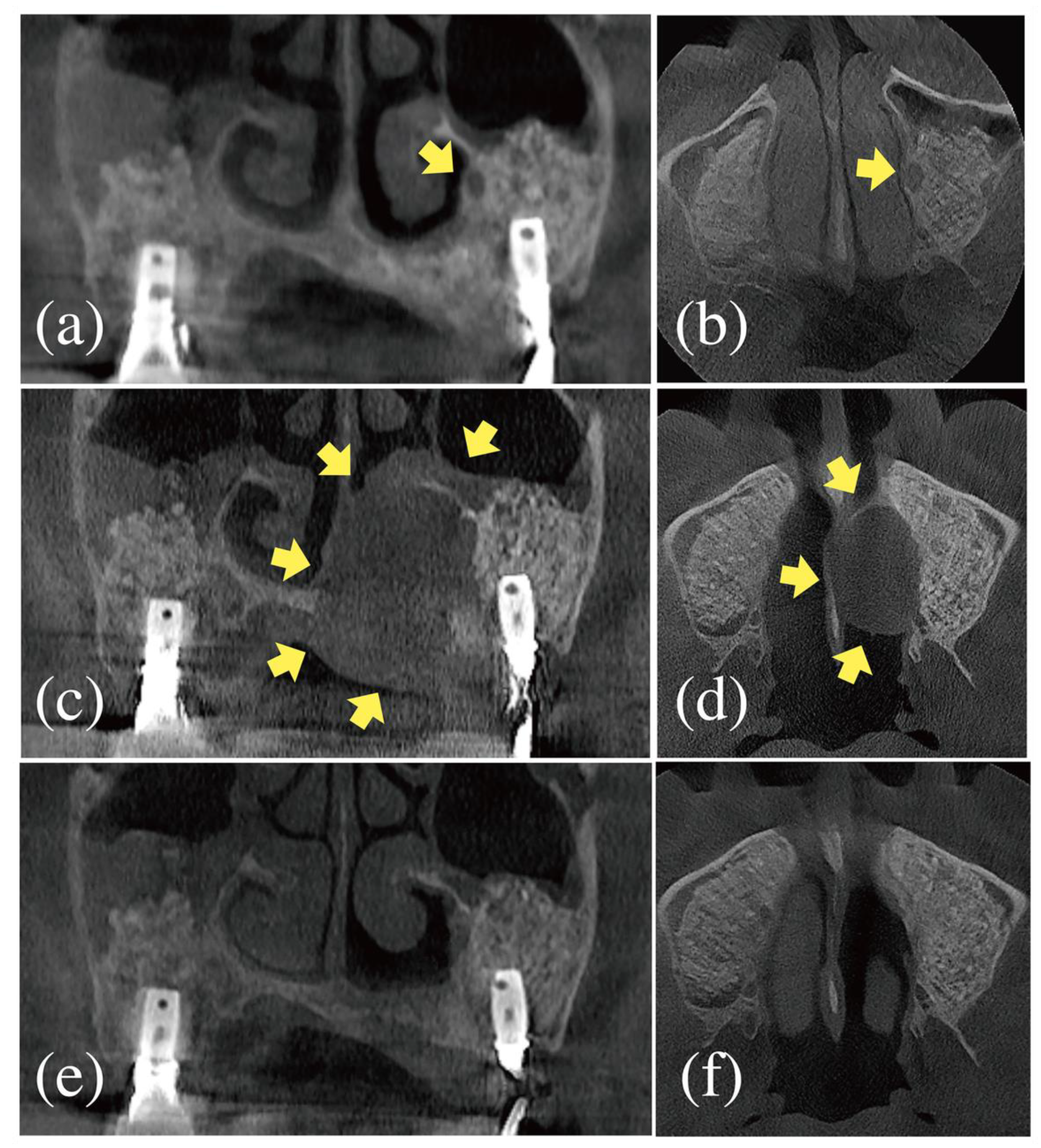
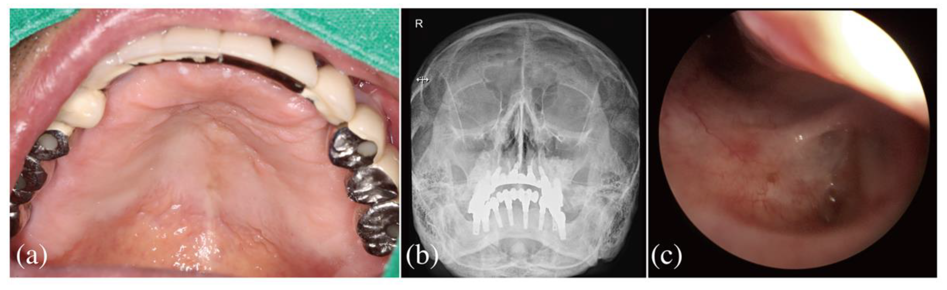
2.2. Dental Examination

After 13 years, a cone-beam computed tomography (CBCT) was taken to evaluate MSA and dental implants. On CBCT, a radiolucent lesion was found on the medial wall of the left maxillary sinus (Figure 3a,b). At this time, the attending dentist was not aware of the possibility of POMC and scheduled regular re-call visits. However, the patient did not attend the scheduled visits between 2016 and 2018.
When the patient presented to the clinic (in January 2019), there was swelling and fluctuation on the left side of the palate. The patient reported no pain around the implants and no discomfort during functioning. The implants presented bone remodeling to the level of the implant platform. Probing depth on the palatal area of the implant was roughly 5.0 mm. No specific finding related to the palatal swelling was found on the panoramic radiographs (including the past radiographs) (Figure 2d). However, CBCT revealed a large radiolucent unilateral lesion on the medial side of the left maxillary sinus (Figure 3c,d). Severe destruction on the medial wall of the sinus was observed. The lesion extended beyond the lateral aspect of the nasal cavity, reaching the nasal septum and inferior meatus. The palatal bone was also severely resorbed to the extent of the palatal suture. Slight sinus membrane thickening over the previous augmentation was observed. Ostium appeared to be patent. The lesion was not in contact with the implants.
Based on the patient’s chart review and clinical/radiographical examination, POMC was suspected. Due to the involvement of nasal structure and severe bone destruction, the patient was referred to a university hospital.

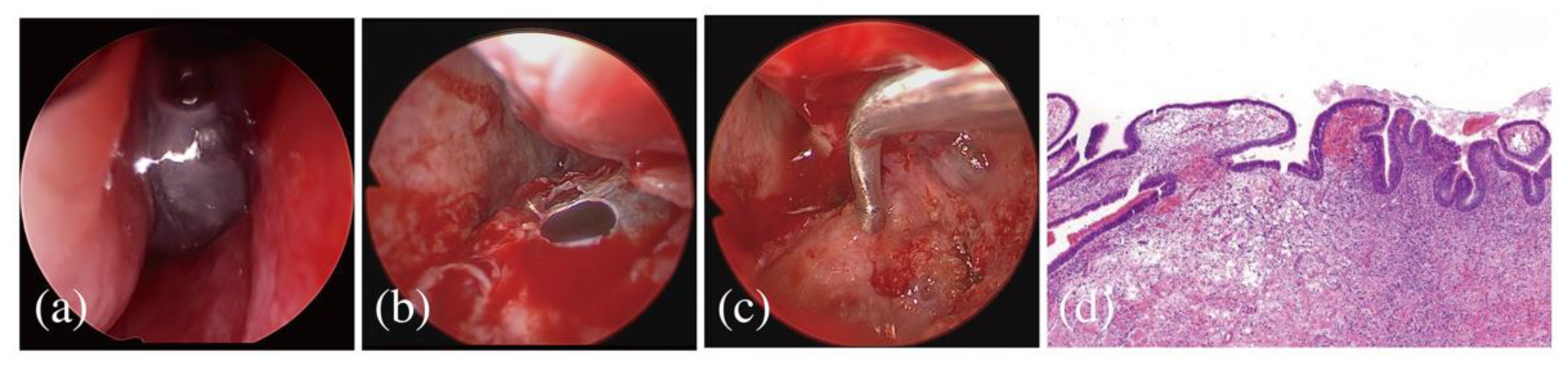
2.3. Referral to an Otolaryngological Specialist

Nasal endoscopic examination revealed a dome-shaped lesion occupying the posterior portion of the inferior meatus (Figure 4a). The base of the dome was located on the lateral part of the nasal cavity. The patient underwent nasal endoscopic marsupialization under general anesthesia. The cystic lesion was punctured and decompressed (Figure 4b). Then, the lesion extending to the inferior meatus was removed, which resulted in the partial exposure of the medial part of the previous bone augmentation (Figure 4c). This part was covered with an absorbable collagen hemostat (Novacol, Kyeron, Enschede, Netherland). The removed tissue was fixed in 10% buffered formalin for histopathologic examination.

2.4. Follow-Up

After the surgery, no adverse event was noted. Histologic specimen revealed respiratory epithelium with inflammatory cell infiltration, confirming that the lesion was POMC (Figure 4d). The swelling on the palatal area disappeared (Figure 5a). The CBCT taken at 1 year demonstrated a loss of the cystic lesion, some regeneration of the medial bone wall of the maxillary sinus, and recovery of the inferior meatus structure (Figure 3e,f). Water’s view and nasal endoscopic examination did not reveal POMC recurrence at 2 years (Figure 5b,c). Endoscopic marsupialization did not affect the survival of the augmentation and the implant. No recurrence has been found to date.

3. Discussion

The present study demonstrated unusual and invasive involvement of the nasal cavity by late developed POMC after MSA. The patient presented with clinical symptoms 18 years after MSA. Due to the location and the extent of the lesion, the intraoral surgical approach was not considered to be appropriate, and thus the patient was referred to an otolaryngologist. The POMC was successfully treated with nasal endoscopic marsupialization without implant removal. No recurrence has been found to date.
Evidence demonstrated that POMC development is related to invasive surgeries or trauma involving the maxillary sinus, such as Caldwell–Luc operation and orthognathic surgery [5,7]. During such events, the sinus mucosa might become entrapped and proliferate, eventually leading to POMC. However, a less invasive surgery was not exempted from causal factors for POMC. In the grafted bone, small fragment of the sinus mucosa might become accidentally entrapped [12]. To date, there have been nine cases of POMC related to MSA in five case studies (five case reports and one case series with a systematic review) [9,10,11,12,13].
Regarding suspected causality, for example, sinus membrane perforation, interesting findings were noted; even though hypothesized by some authors [9,11], no previous studies provide definite evidence for the perforation because the patients underwent the initial MSA in other dental clinics and perforation (or intraoperative complication) was not detected (or not documented at least) [9,10,11,12,13]. In the present study, sinus membrane perforation was found near the access window area during MSA and repaired. In the CBCT taken after 13 years, a small radiolucent cystic lesion was found near the medial wall of the nasal cavity, and this lesion became significantly larger at 18 years. The location of the cystic lesion was somewhat remote concerning the site of the perforation. This may suggest that the folding or overlapping of the sinus membrane during the membrane elevation or repair procedure may affect the development of POMC. The development of POMC might not be necessarily preconditioned by the damage to the sinus membrane (perforation and tearing) [13].
Following MSA, the primary choice for POMC was enucleation by intraoral surgery, as demonstrated in all the above case studies [9,10,11,12,13]. However, in the present study, the location of the cystic lesion was hard to manage by intraoral approach due to the invasion of the nasal cavity. Moreover, enucleation of the entire cyst might have been caused by the large scar, collateral damage to the adjacent tissues, and implant removal owing to the extent of the lesion. Thus, the patient in the present study underwent endoscopic marsupialization. Other studies also demonstrated cases similar to the present one [15,16]. In one study, POMC (due to Caldwell–Luc operation 30 years ago) invaded the orbital floor and caused diplopia, vision impairment, and limitation of eye movement. In another case, a radicular cyst expanded to fill the entire maxillary sinus cavity, thinned palatal/sinus bone walls, and invaded the inferior meatus and nasal floor. There was elevation of the nostril, protrusion of the upper lip, and septal deviation in these cases. The above cases were successfully treated with endoscopic marsupialization.
In the literature, the onset of POMC after MSA appears unclear. The range of time points of detection of POMC varied highly (between 3 months and 10 years) [9,10,11,12,13]. The detection might be missed when the patient’s symptoms, such as facial or intraoral area swelling were not apparent. For those without symptoms, POMC was found by dental radiographic examination [9,10,12]. However, the lesion may not be visible in routine peri-apical or panoramic radiography. In previous case reports, three-dimensional radiography could reveal the presence of POMC when the patient’s symptom was not absent or information about the symptoms was not given in the article [9,10,12]. In the present study, CBCT could reveal the lesion (at 13 years) despite a lack of awareness of POMC at that time point.
Another consideration is the rate of POMC enlargement. Supposing that the POMC started to develop at an early stage following MSA, the enlargement rate between 13 and 18 years became greater than before. Inflammatory cell infiltration, which was revealed in the histologic section, might be involved in this abrupt growth.
Some emphasized that POMC after MSA is being underreported [10]. The present authors partially agree with their opinion because most MSA cases are POMC-free on long-term follow-up. However, considering that the frequency of MSA surgery is increasing, clinicians should be aware that POMC is one of the postoperative complications of MSA despite its rareness.

4. Conclusions

POMC is a rare complication after MSA. However, clinicians should have knowledge of this complication because the expansion of POMC may vastly affect adjacent anatomical structures.

Author Contributions

Conceptualization, W.-B.P., H.-C.L.; methodology, W.-B.P., H.-C.L.; validation, W.-B.P., H.-C.L.; formal analysis, W.-B.P.; investigation, W.-B.P.; data curation, W.-B.P.; writing—original draft preparation, W.-B.P.; writing—review and editing, H.-C.L.; visualization, W.-B.P.; supervision, H.-C.L.; project administration, W.-B.P. All authors have read and agreed to the published version of the manuscript.

Funding

This research received no external funding.

Institutional Review Board Statement

Ethical review and approval were waived for this study, because Institutional Review Board approval is not mandatory in Korea for a case report.

Informed Consent Statement

The patients agreed to the publication.

Data Availability Statement

All data and material are presented in the manuscript.

Conflicts of Interest

The authors declare no conflict of interest.

References

  1. Pjetursson, B.E.; Tan, W.C.; Zwahlen, M.; Lang, N.P. A systematic review of the success of sinus floor elevation and survival of implants inserted in combination with sinus floor elevation. J. Clin. Periodontol. 2008, 35, 216–240. [Google Scholar] [CrossRef] [PubMed]
  2. Hamdoon, Z.; Mahmood, N.; Talaat, W.; Sattar, A.A.; Naeim, K.; Qais, A.; Kheder, W.; Jerjes, W. Evaluation of different surgical approaches to remove dental implants from the maxillary sinus. Sci. Rep. 2021, 11, 1–8. [Google Scholar] [CrossRef]
  3. Testori, T.; Weinstein, T.; Taschieri, S.; Wallace, S.S. Risk factors in lateral window sinus elevation surgery. Periodontol. 2000 2019, 81, 91–123. [Google Scholar] [CrossRef] [PubMed]
  4. Kubo, I. A buccal cyst occurred after a radical operation of the maxillary sinus. JAP J. Otolaryngol. 1927, 33, 896. [Google Scholar]
  5. An, J.; Zhang, Y. Surgical ciliated cyst of the medial canthal region after the management of a midfacial fracture: A case report. J Craniofac. Surg. 2014, 25, 70–702. [Google Scholar] [CrossRef] [PubMed]
  6. Cano, J.; Campo, J.; Alobera, M.A.; Baca, R. Surgical ciliated cyst of the maxilla. Clinical case. Medicina Oral Patología Oral y Cirugia Bucal 2009, 14, 361–364. [Google Scholar]
  7. Soares, J.C.; Villalba, N.C.; Sanromán, J.F.; Ferro, M.F.; Fernández, P.L.; Betancourt, A.L.; López, A.C. Surgical Ciliated Cysts in Orthognathic Surgery. J. Craniofacial Surg. 2021, 32, e2–e5. [Google Scholar] [CrossRef]
  8. Leung, Y.Y.; Wong, W.Y.; Cheung, L.K. Surgical Ciliated Cysts May Mimic Radicular Cysts or Residual Cysts of Maxilla: Report of 3 Cases. J. Oral Maxillofac. Surg. 2012, 70, e264–e269. [Google Scholar] [CrossRef]
  9. Misch, C.M.; Resnik, R.R.; Ismail, Y.H.; Appel, B. Post-operative maxillary cyst associated with a maxillary sinus elevation procedure: A case report. J. Oral Implant. 1991, 17, 432–437. [Google Scholar]
  10. Kahn, A.; Matalon, S.; Salem, R.; Kats, L.; Chaushu, L.; Vered, M.; Rosen, E. Sinus Floor Augmentation—Associated Surgical Ciliated Cysts: Case Series and a Systematic Review of the Literature. Appl. Sci. 2021, 11, 1903. [Google Scholar] [CrossRef]
  11. Kim, J.J.; Freire, M.; Yoon, J.-H.; Kim, H.K. Postoperative Maxillary Cyst After Maxillary Sinus Augmentation. J. Craniofacial Surg. 2013, 24, e521–e523. [Google Scholar] [CrossRef]
  12. Lockhart, R.; Ceccaldi, J.; Bertrand, J.C. Postoperative maxillary cyst following sinus bone graft: Report of a case. Int. J. Oral Maxillofac. Implant. 2000, 15, 583–586. [Google Scholar]
  13. Yamamoto, S.; Maeda, K.; Kouchi, I.; Hirai, Y.; Taniike, N.; Imai, Y.; Takenobu, T. Surgical Ciliated Cyst Following Maxillary Sinus Floor Augmentation: A Case Report. J. Oral Implant. 2017, 43, 360–364. [Google Scholar] [CrossRef]
  14. Caton, J.G.; Armitage, G.; Berglundh, T.; Chapple, I.L.C.; Jepsen, S.; Kornman, K.S.; Mealey, B.L.; Papapanou, P.N.; Sanz, M.; Tonetti, M.S. A new classification scheme for periodontal and peri-implant diseases and conditions - introduction and key changes from the 1999 classification. J Clin. Periodontol. 2018, 45 (Suppl. 20), S1–S8. [Google Scholar] [CrossRef] [PubMed]
  15. Consolo, U.; Bellini, P.; Lizio, G. Trans-nasal endoscopic marsupialization of a voluminous radicular cyst involving maxillary sinus and nasal cavity: A case report and a literature review on this surgical approach. Oral Maxillofac. Surg. Cases 2018, 4, 91–96. [Google Scholar] [CrossRef]
  16. Kim, T.H.; Kim, J.S.; Heo, S.J. Postoperative maxillary mucocele with orbital wall defect treated by transnasal endoscopic marsupialization with a penrose drain insertion: A case report. Medicine 2019, 98, e15674. [Google Scholar] [CrossRef] [PubMed]
Figure 1. Clinical situation at 18 years post-maxillary sinus augmentation. There were painless swelling and fluctuation on the left side of the palate. * The area showing swelling and fluctuation.
Applsci 11 10730 g001
Figure 2. Panoramic radiographic images of the patient. (a) Initial visit to the present dental clinic. It was planned to extract all teeth, (b) Implant placement was performed simultaneously with lateral sinus augmentation. Sinus membrane perforation occurred during sinus membrane elevation. The perforated membrane was repaired using Collatape, (c) Immediately after implant prosthesis delivery, (d) After 18 years, no specific finding was noted in the maxillary sinus.
Applsci 11 10730 g002
Figure 3. Cone-beam computed tomographic images of the patient. (a,b) Coronal and axial views after 13 years. There was a small radiolucent lesion at the medial side of the augmentation, (c,d) Coronal and axial views at 18 years. The lesion expanded in the medial and downward directions. The medial wall of the sinus, the palatal bone, and inferior meatus were invaded by the lesion, (e,f) Coronal and axial views 1 year following endoscopic marsupialization. The lesion disappeared without removal of the augmented bone and dental implants. Yellow arrows indicate POMC.
Applsci 11 10730 g003
Figure 4. Endoscopic marsupialization (ac) and histologic finding of the removed specimen (d). (a) A dome-shaped cystic lesion occupied the inferior meatus, (b) The lesion was excised, and drainage was performed, (c) After partial removal of the cystic lesion, the medial portion of the previous augmentation was exposed, (d) Respiratory epithelium and infiltration of inflammatory cells were observed in the specimen (H-E stain, x100).
Applsci 11 10730 g004
Figure 5. Clinical, radiographic, and nasal endoscopic findings after endoscopic marsupialization. (a) Swelling on the left palate disappeared, (b) PNS Water’s view, (c) Nasal endoscopic view.
Applsci 11 10730 g005
Publisher’s Note: MDPI stays neutral with regard to jurisdictional claims in published maps and institutional affiliations.

