Article

Simultaneous Measurement of Refractive Index and Flow Rate Using a Co2+-Doped Microfiber

1
School of Optics and Photonics, Beijing Institute of Technology, Beijing 100081, China
2
School of Information and Electronics, Beijing Institute of Technology, Beijing 100081, China
3
China Wuzhou Engineering Group, Beijing 100053, China
*
Authors to whom correspondence should be addressed.
Appl. Sci. 2021, 11(22), 10525; https://doi.org/10.3390/app112210525
Submission received: 19 September 2021 / Revised: 27 October 2021 / Accepted: 5 November 2021 / Published: 9 November 2021
(This article belongs to the Special Issue Advances in Fiber Optic Sensors and Their Application)

Abstract

:
This paper has proposed and experimentally demonstrated an integrated Co2+-doped microfiber Bragg grating sensor (Co-MFBGS) that can measure the surrounding liquid refractive index (LRI) and liquid flow rate (LFR) simultaneously. The Co-MFBGS provides well-defined resonant modes of core and cladding in the reflection spectrum. By monitoring the wavelength of the cladding mode, the LRI can be measured; meanwhile, by monitoring the wavelength of the core mode caused by the heat exchange, the LFR can be measured. The LRI and LFR can be distinguished by the wavelength separation between cladding mode and core mode. The experimental results show that in aqueous glycerin solution, the maximum measurement sensitivity for LRI detection is −7.85 nm/RIU (refractive index unit), and the LFR sensitivity is −1.93 nm/(μL/s) at a flow rate of 0.21 μL/s.

1. Introduction

In recent decades, on the basis of the technology developments of femtosecond laser processing [1,2], CO2 laser heating [3,4], chemical etching [5,6,7,8], and flame heating [9,10], various microfiber fabrication methods have been proposed. Microfiber-based sensors often have many important advantages, such as good flexibility, mechanical strength, compact size, quick response and high sensitivity [11,12,13], so they have many good applications, such as biological applications [8,14,15,16] and chemical analysis [12,17].
Liquid refractive index (LRI) measurement plays an important role in chemical [12], biological [14,15] and other applications. Various microfiber-based LRI sensors have been proposed, as they have high sensitivity, a robust structure, and have many applications for biosensors, chemical sensors, etc. [11]. Previous scientific research studies include fiber-optic interferometers [3,10,18], tilted fiber Bragg gratings (TFBGs) [14], long-period gratings (LPGs) [15], and many kinds of microfiber Bragg gratings (MFBGs) [1,19]. All of these previous studies bring us many useful microfiber-based sensors with specific designs.
The liquid flow rate (LFR) measurement is also a paramount index for many applications, such as medical and biological analysis [20]. In the application of optical fiber biosensors, due to the herringbone microfluidic chip’s capture efficiency being highly dependent on the flow rate, its accuracy and efficiency can be significantly improved by the measurement of flow rate [21,22].
“Hot-wire” is the most famous microfiber flowmeter [23,24,25]. For many applications, not only is the LFR needed, but also other parameters, such as LRI and temperature [19]. Sometimes simultaneous measurements may be necessary, and the measurement parameters should not be cross-impacted.
In this paper, based on the Co2+-doped microfiber Bragg grating sensor (Co-MFBGS), an integrated LRI and LFR sensing sensor has been presented and experimentally demonstrated, which can measure both parameters simultaneously. The Co-MFBGS reflection spectrum provides a well-defined core mode resonance as well as two cladding mode resonances; the wavelength shifts of two kinds of resonances have quadratic non-linear relationship to LRI and LFR. The temperature cross-impact to the LRI measurement can be eliminated by monitoring the wavelength interval change between the core mode and cladding mode. Additionally, due to the non-radiation effect of Co2+-doped fiber, the Co-MFBGS can be heated by a pump laser; by interrogating the core mode’s wavelength shift caused by the heat exchange effect, the LFR can be measured. The proposed integrated optical fiber sensor has potential value in chemical, medical and environmental applications.

2. Materials and Methods

2.1. Fabrication of the Co-MFBGS

In the proposed Co-MFBGS, the sensing device is a section of Co2+-doped optical fiber (CDOF) with 8.4/125 μm core diameter and cladding diameter, and the length is 20 mm. At a laser wavelength of 1480 nm, the section of CDOF absorption coefficient is approximately equal to 31.3 dB/m. The CDOF with a length of 20 mm was spliced to two sections of 1m length single-mode optical fibers, one at each end.
The fabrication of the proposed Co-MFBGS has two steps. First, an FBG with a length of 15 mm was written in the section of CDOF using a KrF excimer laser (248 nm) with a phase mask. Second, the CDOF with an FBG was immersed into a 20% aqueous hydrofluoric acid solution for 105 min, then removed and rinsed with ultrapure water to clean off the acid solution. Thus, a Co-MFBGS with a diameter of 16.7 μm (Figure 1a) was fabricated. With the same process, a Co-MFBGS with a diameter of 6.4 μm (Figure 1b) was also fabricated.
The reflection spectra of the Co-MFBGS with diameters of 16.7 and 6.4 μm were investigated, as shown in Figure 1c. It is clear that the Co-MFBGS with a diameter of 16.7 μm has a core mode and two cladding modes, but the Co-MFBGS with a 6.4 μm diameter has only a core mode, the wavelength range from 1525 to 1565 nm. Accordingly, the 16.7 μm Co-MFBGS was selected as the test sensor for the rest of the experiments. As shown in Figure 1c, the wavelength separation of λa (core mode) and λb (cladding mode) is 7.1 nm. The spectral characteristics of the Co-MFBGS were analyzed by using the numerical mode simulation software COMSOL, and the reflection spectrum compositions of the core mode and one cladding mode, and amplitude distributions of the transverse electric field, are as shown in the bottom of Figure 1. This indicates that on the Co-MFBGS surface, the evanescent field of the cladding mode is stronger than that of the core mode.

2.2. Principle of the Proposed Sensor

It is well known that the FBG center wavelength λB and the resonant wavelength λcl,k of the kth cladding mode can be shown as [18,26]
λ B = 2 n e f f , c o · Λ
λ c l , k = ( n e f f , c o + n e f f , c l , k ) · Λ
where neff,co and neff,cl,k are the effective index of the core mode and the kth cladding mode in the Co-MFBGS, respectively; Λ is the Bragg grating period.
As the core mode was confined in the core of the Co-MFBGS, it was insensitive to the surrounding LRI. Meanwhile, the cladding modes of the Co-MFBGS due to the evanescent field are very sensitive to the LRI. So, the Co-MFBGS can be used for LRI sensing, and from Equation (2) the wavelength drift of the cladding mode caused by LRI change is rewritten as
Δ λ c l , k ( R I ) = Δ n e f f , c l , k ( R I ) · Λ
where Δneff,cl,k(RI) is the kth cladding mode LRI change.
The wavelength interval λint is defined as
λ int = λ B λ c l , k
It is the wavelength difference between core mode and kth cladding mode. Combining Equations (3) and (4), the wavelength interval shift Δλint(RI) caused by the change of LRI can be written as
Δ λ int ( R I ) = Δ λ c l , k ( R I ) = Δ n e f f , c l , k ( R I ) · Λ
The wavelength drift of FBG caused by temperature change is well known as [27]
Δ λ B ( T ) = 2 n e f f , c o · Λ ( 1 n e f f , c o d n e f f , c o d T + 1 Λ d Λ d T ) Δ T = λ B ( α + β ) Δ T
Δ λ c l , k ( T ) = λ c l , k ( 1 n e f f , c o + n e f f , c l , k d ( n e f f , c o + n e f f , c l , k ) d T + 1 Λ d Λ d T ) Δ T = λ c l , k ( γ + β ) Δ T
where ΔλB(T) and Δλcl,k(T) are the wavelength changes of the core mode and kth cladding mode of FBG caused by temperature change, ΔT is the temperature change, α = 1 n e f f , c o d n e f f , c o d T is the thermo-optic coefficient of the optical fiber core, β = 1 Λ d Λ d T is the grating coefficient of thermal expansion, and γ = 1 n e f f , c o + n e f f , c l , k d ( n e f f , c o + n e f f , c l , k ) d T is the thermo-optic coefficient of the optical fiber cladding.
Cobalt is a kind of light absorption material and is easy to get, and the CDOF can be heated by the pump laser as a result of the non-radiation effect; meanwhile, the temperature of the Co-MFBGS increases and the wavelengths of the core mode and cladding modes have a redshift. When the Co-MFBGS was used as an LFR sensor, parts of the sensor heat can be carried away by the fluidic sample [28], which would lead to a decrease in the sensor’s temperature and a blue shift in the wavelengths of the core mode and cladding modes. So, the LFR can be measured by detecting the wavelength shift of the Co-MFBGS. Depending on the theory of hot-wire, the relationship between the LFR of the fluidic sample and heat loss is shown as [29]
Q = Δ T ( A + B ν i )
where Q is the power absorbed by the CDOF, ν is the LFR, and A, B and i are empirical coefficients. So, Equations (6) and (7) can be rewritten as
Δ λ B ( T ) = λ B ( α + β ) Q A + B ν i
Δ λ c l , k ( T ) = λ c l , k ( γ + β ) Q A + B ν i
Additionally, the wavelength interval shift Δλint(T) caused by the change of temperature can be deduced as
Δ λ int ( T ) = Δ λ B ( T ) Δ λ c l , k ( T ) = ( λ B α + λ B β λ c l , k γ λ c l , k β ) Q A + B ν i
As the difference of α and γ is very small (<0.00324 K−1) [20], Equation (11) can be rewritten as
Δ λ int ( T ) ( α + β ) ( λ B λ c l , k ) Q A + B ν i
According to Reference [20], when the LFR changes from 0 to 1 μL/s, Δλint(T) is quite small (~0.017 nm)—that is, the wavelength interval shift caused by the effect of LFR can almost be neglected. Consequently, the LRI and LFR can be measured simultaneously using the cladding mode wavelength and core mode wavelength of the Co-MFBGS, respectively, and the crosstalk can be distinguished using the wavelength interval shift.

2.3. Experimental Setup
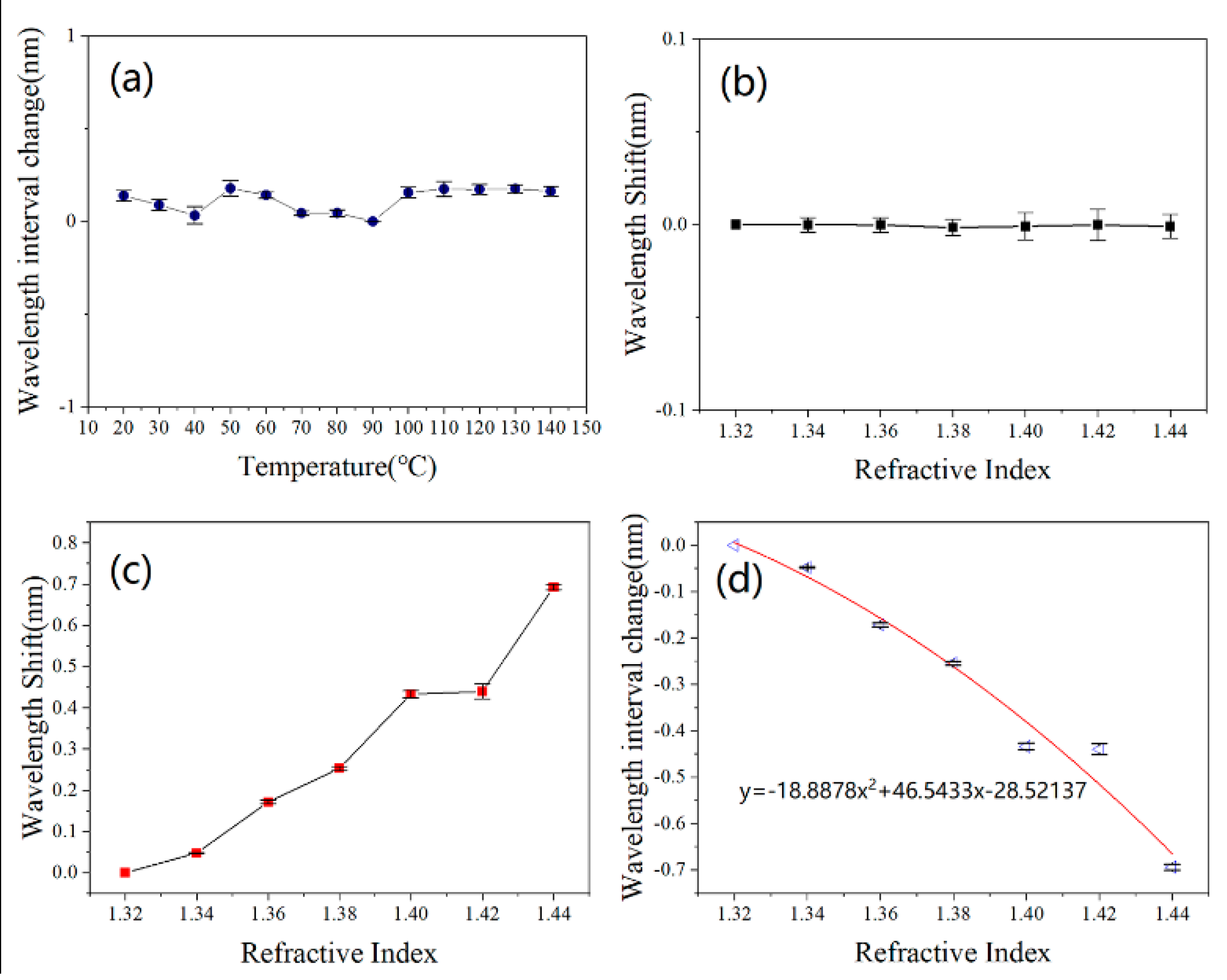
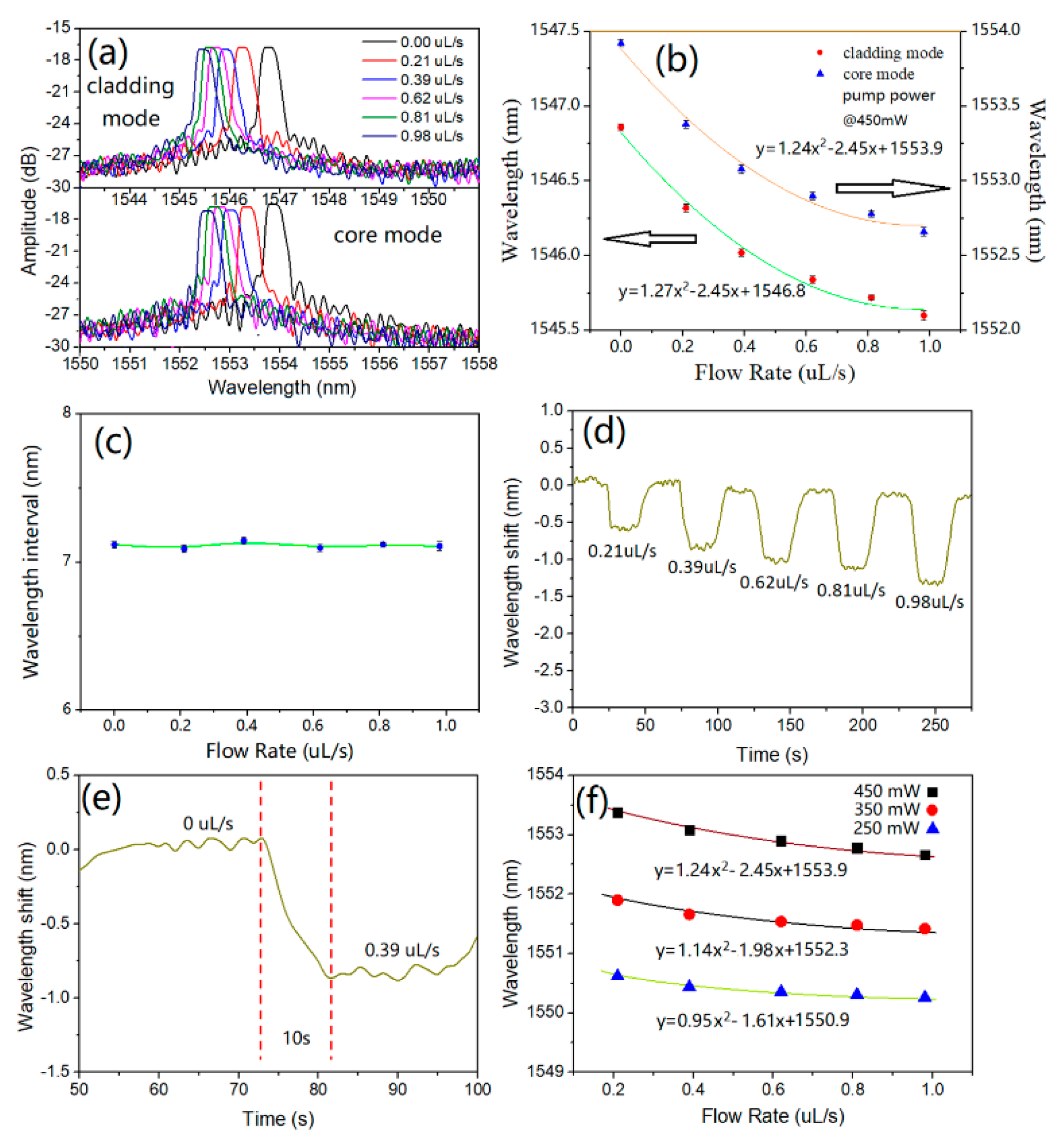
As shown in Figure 2, the experimental setup includes an amplified spontaneous emission (ASE) laser (wavelength: 1525–1565 nm, output power: 20 mW) and a tunable pump laser (1480 nm), and both the lasers are launched into the Co-MFBGS from the opposite ends. The reflective spectrum of the Co-MFBGS was interrogated by an optical spectrum analyzer (OSA, YOKOGAWA AQ6370D). At the beginning of the experiments of LRI and LFR measurement, a specially designed PDMS-based microfluidic channel was used to place the proposed Co-MFBGS, in order to impart a slight pre-stretch; then, in order to avoid the effects of other forces, a UV-sensitive adhesive was used to fix the two ends of the Co-MFBGS fiber, as shown in Figure 2. During the experiments of LRI and LFR measurement, aqueous glycerin solutions were injected into the microfluidic channel via a pressure controller whose pressure stability is as high as 10 μbar.

3. Results

The sensing characteristics of the proposed 16.7 μm diameter Co-MFBGS for temperature (20–140 °C), surrounding LRI (1.32–1.44 RIU) and LFR (0–0.98 μL/s) changes have been tested as described in the following section.
The temperature characteristics of the Co-MFBGS were tested first by putting the sensor into a chamber whose temperature could be adjusted in the range of 20–140 °C in steps of 10 °C and a resolution of 0.1 °C. The temperature experimental result of the wavelength interval (λa–b) change between λa and λb of the Co-MFBGS is shown in Figure 3a, which is clearly showing that λa–b is almost free from temperature perturbations. So, the temperature cross-sensitivity of LRI measurements can be neglected.
The LRI measurement experiment of the Co-MFBGS was conducted at room temperature (25 °C). In each measurement, the aqueous glycerin solutions, with concentrations whose RI changed from 1.32 to 1.44 RIU in steps of 0.02 RIU, were injected into the microfluidic channel. After the aqueous glycerin solution had flowed to surround the entire Co-MFBGS, the reflection spectrum was interrogated several times by the OSA until almost no change was observed, and then the spectrum was recorded, so the reliable measuring values can be obtained.
The experimental results of the proposed Co-MFBGS wavelength shift with different LRIs are shown in Figure 3b–d. Figure 3b–d shows the value change of λa, λb and λa–b with different LRIs, respectively. It is clear that the core mode wavelength of the proposed Co-MFBGS does not shift, which means it is insensitive to the LRI. Meanwhile, the cladding mode is exceedingly sensitive to the LRI; its wavelength shift decreases by 0.69 nm with the LRI changes from 1.32 to 1.44 RIU. So, the value change of λa–b is almost the same with the value change of λb, and it can be fit to a quadratic non-linear equation with LRI change (1.32–1.44 RIU). The equation indicates that the maximum LRI sensitivity of the proposed Co-MFBGS wavelength interval is about −7.85 nm/RIU, Figure 3d.
Before beginning the LFR measurement experiment, the Co-MFBGS was heated by using pumped laser powers of 250, 350 and 450 mW at a wavelength of 1480 nm, as the Co-MFBGS can be heated by pump laser due to the light absorption of cobalt. Figure 4f shows the wavelength shift of the core mode (λa) at different pumped laser powers, which clearly shows that λa has a redshift while increasing pumped laser power, and the sensitivity increases significantly with increasing heating power. That means the temperature of Co-MFBGS is increased, so, when the Co-MFBGS is used for LFR sensing, the heat of Co-MFBGS will be carried away by the flowing liquid, the temperature of Co-MFBGS will be decreased, and λa will has a blueshift.
During the LFR measurement experiment of the proposed Co-MFBGS, the LFR was varied in 0–0.98 μL/s by using a commercial electronic-controlled syringe pump to inject an aqueous glycerin solution with a constant concentration into the microfluidic channel; meanwhile, the pumped laser power was set to 450 mW. A PZT flow sensor was used to calibrate the LFR. The experimental results are shown in Figure 4.
With increasing LFR, both λa and λb experience a blueshift due to the decreasing temperature of the Co-MFBGS; reflection spectra corresponding to different flow rates are shown in Figure 4a. Figure 4b shows the value change of λb and λa with different flow rates; it can be seen that their wavelengths shift to shorter wavelengths simultaneously, but in Figure 4c it is shown that λa–b exhibited almost no change. That means the LFR and the LRI can be discriminated by wavelength interval. Figure 4b also shows that there is a quadratic non-linear relationship between the value change of λa and the LFR; a flow rate of 0.21 μL/s can lead to a sensitivity of −1.93 nm/(μL/s). From Figure 4e, it can be seen that it takes about 10 s for the reading to stabilize when the LFR increases from 0 to 0.39 μL/s; therefore, the response time of the LFR measurement of the proposed Co-MFBGS is 10 s.

4. Conclusions

This paper has presented an integrated Co-MFBGS, which can measure the LRI and LFR simultaneously. A single straight Bragg grating was written in a section of Co2+-doped fiber and then immersed into aqueous hydrofluoric acid solution to fabricate a Co-MFBGS with a diameter of 16.7 μm. The Co-MFBGS of the proposed sensor provides well-defined resonances in reflection; by detecting the wavelength of the cladding mode and core mode, the LRI and LFR can be measured, respectively, and they can be distinguished by the change of wavelength separation between cladding mode and core mode, and the temperature cross-impact of LRI measurements is also eliminated. The experimental results show that in aqueous glycerin solution the maximum measurement sensitivity for LRI detection is −7.85 nm/RIU, and the LFR sensitivity is −1.93 nm/(μL/s) at a flow rate 0.21 μL/s. The proposed Co-MFBGS can potentially be used in bioengineering, medicine, environmental protection, etc.

Author Contributions

Conceptualization, D.L., R.G. and Z.L.; Data curation, D.L. and R.G.; Formal analysis, D.L.; Funding acquisition, R.G.; Investigation, D.L. and R.G.; Methodology, D.L., R.G. and Z.L.; Project administration, R.G.; Resources, D.L., R.G. and A.Q.; Software, R.G. and A.Q.; Supervision, R.G.; Validation, R.G.; Writing—original draft, D.L.; Writing—review and editing, D.L., R.G., Z.L. and A.Q. All authors have read and agreed to the published version of the manuscript.

Funding

This research was funded in part by the National Natural Science Foundation of China (No. 62022016, 61835002, 61727817), in part by the Open Fund of IPOC (Beijing University of Posts and Telecom- munications), grant numbers IPOC2020A007 and IPOC2020A006.

Institutional Review Board Statement

Not applicable.

Informed Consent Statement

Not applicable.

Data Availability Statement

Not applicable.

Conflicts of Interest

The authors declare no conflict of interest.

References

  1. Fang, X.; Liao, C.R.; Wang, D.N. Femtosecond laser fabricated fiber Bragg grating in microfiber for refractive index sensing. Opt. Lett. 2010, 35, 1007–1009. [Google Scholar] [CrossRef] [PubMed] [Green Version]
  2. Liao, C.; Yang, K.; Wang, J.; Bai, Z.; Gan, Z.; Wang, Y. Helical Microfiber Bragg Grating Printed by Femtosecond Laser for Refractive Index Sensing. IEEE Photonics Technol. Lett. 2019, 31, 971–974. [Google Scholar] [CrossRef]
  3. Sumetsky, M.; Dulashko, Y.; Hale, A. Fabrication and study of bent and coiled free silica nanowires: Self-coupling microloop optical interferometer. Opt. Express 2004, 12, 3521–3531. [Google Scholar] [CrossRef]
  4. Kakarantzas, G.; Dimmick, T.E.; Birks, T.A.; Le Roux, R.; Russell, P.S.J. Miniature all-fiber devices based on CO(2) laser microstructuring of tapered fibers. Opt. Lett. 2001, 26, 1137–1139. [Google Scholar] [CrossRef]
  5. Zhang, E.J.; Sacher, W.D.; Poon, J.K.S. Hydrofluoric acid flow etching of low-loss subwavelength-diameter biconical fiber tapers. Opt. Express 2010, 18, 22593–22598. [Google Scholar] [CrossRef] [PubMed]
  6. Iadicicco, A.; Cusano, A.; Cutolo, A.; Bernini, R.; Giordano, M. Thinned Fiber Bragg Gratings as High Sensitivity Refractive Index Sensor. IEEE Photonics Technol. Lett. 2004, 16, 1149–1151. [Google Scholar] [CrossRef]
  7. Liang, W.; Huang, Y.; Xu, Y.; Lee, R.K.; Yariv, A. Highly sensitive fiber Bragg grating refractive index sensors. Appl. Phys. Lett. 2005, 86, 151122. [Google Scholar] [CrossRef]
  8. Bekmurzayeva, A.; Dukenbayev, K.; Shaimerdenova, M.; Bekniyazov, I.; Ayupova, T.; Sypabekova, M.; Molardi, C.; Tosi, D. Etched Fiber Bragg Grating Biosensor Functionalized with Aptamers for Detection of Thrombin. Sensors 2018, 18, 4298. [Google Scholar] [CrossRef] [Green Version]
  9. Liu, T.; Liang, L.-L.; Xiao, P.; Sun, L.-P.; Huang, Y.-Y.; Ran, Y.; Jin, L.; Guan, B.-O. A label-free cardiac biomarker immunosensor based on phase-shifted microfiber Bragg grating. Biosens. Bioelectron. 2018, 100, 155–160. [Google Scholar] [CrossRef]
  10. Ahsani, V.; Ahmed, F.; Jun, M.; Bradley, C. Tapered Fiber-Optic Mach-Zehnder Interferometer for Ultra-High Sensitivity Measurement of Refractive Index. Sensors 2019, 19, 1652. [Google Scholar] [CrossRef] [Green Version]
  11. Yan, S.-C.; Xu, F. A review on optical microfibers in fluidic applications. J. Micromech. Microeng. 2017, 27, 093001. [Google Scholar] [CrossRef]
  12. Zhang, Q.; Hu, L.; Qi, Y.; Liu, G.; Ianno, N.; Han, M. Fiber-optic refractometer based on a phase-shifted fiber Bragg grating on a side-hole fiber. Opt. Express 2015, 23, 16750–16759. [Google Scholar] [CrossRef]
  13. Miao, Y.; He, Y.; Ma, X.; Zhang, H.; Song, B.; Yang, X.; Xue, L.; Liu, B.; Yao, J. Low-Temperature Cross-Sensitivity Refractive Index Sensor Based on Single-Mode Fiber With Periodically Modulated Taper. IEEE Sens. J. 2016, 16, 2442–2446. [Google Scholar] [CrossRef]
  14. Ribaut, C.; Loyez, M.; Larrieu, J.-C.; Chevineau, S.; Lambert, P.; Remmelink, M.; Wattiez, R.; Caucheteur, C. Cancer biomarker sensing using packaged plasmonic optical fiber gratings: Towards in vivo diagnosis. Biosens. Bioelectron. 2017, 92, 449–456. [Google Scholar] [CrossRef] [PubMed]
  15. Esposito, F.; Sansone, L.; Taddei, C.; Campopiano, S.; Giordano, M.; Iadicicco, A. Ultrasensitive biosensor based on long period grating coated with polycarbonate-graphene oxide multilayer. Sens. Actuators B Chem. 2018, 274, 517–526. [Google Scholar] [CrossRef]
  16. Li, K.; Liu, G.; Wu, Y.; Hao, P.; Zhou, W.; Zhang, Z. Gold nanoparticle amplified optical microfiber evanescent wave absorption biosensor for cancer biomarker detection in serum. Talanta 2014, 120, 419–424. [Google Scholar] [CrossRef]
  17. Jaddoa, M.F.; Jasim, A.A.; Ab Razak, M.Z.; Harun, S.; Ahmad, H. Highly responsive NaCl detector based on inline microfiber Mach–Zehnder interferometer. Sens. Actuators A Phys. 2016, 237, 56–61. [Google Scholar] [CrossRef]
  18. Luo, H.; Sun, Q.; Xu, Z.; Liu, D.; Zhang, L. Simultaneous measurement of refractive index and temperature using multimode microfiber-based dual Mach-Zehnder interferometer. Opt. Lett. 2014, 39, 4049–4052. [Google Scholar] [CrossRef]
  19. Zhang, Y.; Lin, B.; Tjin, S.C.; Zhang, H.; Wang, G.; Shum, P.; Zhang, X. Refractive index sensing based on higher order mode reflection of a microfiber Bragg grating. Opt. Express 2010, 18, 26345–26350. [Google Scholar] [CrossRef]
  20. Gao, R.; Ye, J.; Xin, X. An Integrated Biological Analysis and Flow Rate Sensing for the Real-Time Detection of Carcinogen in Water Based on Co2+-Doped Optical Fibers. IEEE Sens. J. 2019, 20, 1912–1921. [Google Scholar] [CrossRef]
  21. Gong, Y.; Liu, Q.-F.; Zhang, C.-L.; Wu, Y.; Rao, Y.-J.; Peng, G.-D. Microfluidic Flow Rate Detection with a Large Dynamic Range by Optical Manipulation. IEEE Photonics Technol. Lett. 2015, 27, 2508–2511. [Google Scholar] [CrossRef]
  22. Garza-García, L.D.; García-López, E.; Camacho-León, S.; Rocha-Pizaña, M.D.R.; López-Pacheco, F.; López-Meza, J.; Araiz-Hernández, D.; Tapia-Mejía, E.J.; Santiago, G.T.-D.; Rodríguez-González, C.A.; et al. Continuous flow micro-bioreactors for the production of biopharmaceuticals: The effect of geometry, surface texture, and flow rate. Lab Chip 2014, 14, 1320–1329. [Google Scholar] [CrossRef]
  23. Gong, Y.; Zhang, M.; Gong, C.; Wu, Y.; Rao, Y.; Fan, X. Sensitive optofluidic flow rate sensor based on laser heating and microring resonator. Microfluid. Nanofluidics 2015, 19, 1497–1505. [Google Scholar] [CrossRef]
  24. Li, Y.; Yan, G.; Zhang, L.; He, S. Microfluidic flowmeter based on micro “hot-wire” sandwiched Fabry-Perot interferometer. Opt. Express 2015, 23, 9483–9493. [Google Scholar] [CrossRef] [PubMed]
  25. Yan, S.C.; Liu, Z.Y.; Li, C.; Ge, S.J.; Xu, F.; Lu, Y.Q. “Hot-wire” microfluidic flowmeter based on a microfiber coupler. Opt. Lett. 2016, 41, 5680–5683. [Google Scholar] [CrossRef]
  26. Kou, J.-L.; Ding, M.; Feng, J.; Lu, Y.-Q.; Xu, F.; Brambilla, G. Microfiber-Based Bragg Gratings for Sensing Applications: A Review. Sensors 2012, 12, 8861–8876. [Google Scholar] [CrossRef]
  27. Kersey, A.D.; Davis, M.A.; Patrick, H.J.; LeBlanc, M.; Koo, K.P.; Askins, C.G.; Putnam, M.A.; Friebele, E.J. Fiber Grating Sensors. J. Lightwave Technol. 1997, 15, 1442–1463. [Google Scholar] [CrossRef] [Green Version]
  28. Liu, Z.; Tse, M.-L.V.; Zhang, A.P.; Tam, H.-Y. Integrated microfluidic flowmeter based on a micro-FBG inscribed in Co²?-doped optical fiber. Opt. Lett. 2014, 39, 5877–5880. [Google Scholar] [CrossRef] [PubMed]
  29. Bruun, H.H. Hot-Wire Anemometry: Principles and Signal Analysis. Meas. Sci. Technol. 1996, 7. [Google Scholar] [CrossRef] [Green Version]
Figure 1. (a,b) Microscope images of the Co-MFBGS; (c) the reflection spectra of Co-MFBGS with diameters of 6.4 and 16.7 μm, and the inset shows the spectral characteristics of the Co-MFBGS.
Applsci 11 10525 g001
Figure 2. The experimental setup schematic of the presented Co-MFBGS.
Applsci 11 10525 g002
Figure 3. (a) Wavelength interval change with different temperatures; (b) value change of λa; (c) value change of λb; (d) wavelength interval change with different LRIs.
Applsci 11 10525 g003
Figure 4. (a) The reflection spectra of cladding mode and core mode at different LFRs; (b) wavelength change of λa and λb; (c) wavelength change of λa–b; (d) the experimental process; (e) the time response; (f) wavelength shift of λa with different pump laser powers.
Applsci 11 10525 g004
Publisher’s Note: MDPI stays neutral with regard to jurisdictional claims in published maps and institutional affiliations.

